Article

Phytochemical Profile and Antioxidant, Antiproliferative, and Antimicrobial Properties of Rubus idaeus Seed Powder

1
Innovation Centre, Department of Life Sciences and Systems Biology, University of Turin, Via Quarello 15/A, 10135 Turin, Italy
2
Department of Biological, Chemical and Pharmaceutical Sciences and Technologies, University of Palermo, Viale delle Scienze, 90128 Palermo, Italy
3
Department of Agricultural, Food and Forest Sciences, University of Palermo, Viale delle Scienze, 90128 Palermo, Italy
*
Authors to whom correspondence should be addressed.
These authors equally contributed to the work.
Foods 2022, 11(17), 2605; https://doi.org/10.3390/foods11172605
Submission received: 13 August 2022 / Revised: 23 August 2022 / Accepted: 25 August 2022 / Published: 27 August 2022
(This article belongs to the Special Issue Agro-Food Waste as Source of Nutraceuticals)

Abstract

:
In the context of the contemporary research on sustainable development and circular economy, the quest for effective strategies aimed at revaluation of waste and by-products generated in industrial and agricultural production becomes important. In this work, an ethanolic extract from red raspberry (Rubus idaeus) seed waste (WRSP) was evaluated for its phytochemical composition and functional properties in term of antioxidative, antiproliferative, and antimicrobial activities. Chemical composition of the extract was determined by both HPLC-ESI-MS/MS and spectrophotometric methods. Phytochemical analysis revealed that flavan-3-ols and flavonols were the major phenolic compounds contained in WRSP. The extract demonstrated very high radical-scavenging (4.86 ± 0.06 µmol TE/DW) and antioxidant activity in a cell-based model (0.178 ± 0.03 mg DW/mL cell medium). The WRSP extract also exhibited antiproliferative activity against three different epithelial cancer cell lines (MCF-7, HepG2, and HeLa cells) in a dose-dependent manner. Finally, microbiological assays showed the absence of colonies of bacteria and microscopic fungi (yeasts and molds) and revealed that the WRSP extract has a large inhibition spectrum against spoilage and pathogenic bacteria, without inhibitory activity against pro-technological bacteria. In conclusion, the obtained results show that WRSP is a rich source of phytochemical compounds exerting interesting biological activities. For these reasons WRSP could find applications in the nutritional, nutraceutical, and pharmacological fields.

1. Introduction

Plant foods are major sources of bioactive compounds, and their consumption is linked to potential human health benefits. This awareness has led to a significant increase in global demand for fruits and vegetables. On the other hand, large amounts of waste are inexorably generated not only by plant food consumption but also by overproduction and improper storage practices [1]. For many years, this waste was used as animal feed or disposed of in landfills. Currently, due to an increased perception of the benefits of the circular economy, plant food waste and by-products are now being investigated for recycling in food and non-food applications [2,3]. In particular, agri-food waste and by-products are still rich in bioactive compounds [4,5,6] and can therefore be used as raw materials for cosmetic, nutraceutical, and pharmaceutical formulations.
The red raspberry, or European raspberry, is the fruit of Rubus idaeus, a member of the Rosaceae family. Although it is a species native to western Asia, it is widespread in southeastern Europe. It is an “aggregate fruit” containing several drupes originating from the pistil of each flower. Considering that each drupe contains a small seed, the whole fruit has a large number of seeds. In 2020, the global production of red raspberries was close to 900 thousand (895,771) tons [7,8]. In recent years, Italian red raspberry cultivation has significantly increased, and although fruit production is less than two thousand tons per year, marketing of these red fruits is very profitable [8]. The fruits of R. idaeus have been employed in traditional medicine for the treatment of wounds, renal diseases, inflammation, and infections [9]. On the other hand, scientific data show that red raspberry possesses interesting nutritional and functional value. It is a rich source of vitamin C, ranging from 5 to 40 mg per 100 g of fresh fruit, and also it contains large amounts of polyphenols. In particular, scientific data have shown a significant presence of ellagitannins, phenolic acids, and flavonoids, including anthocyanins, catechins, and proanthocyanidins [10,11]. In a simulated digestion model, Mihailovic and colleagues (2019) also showed that, under gastrointestinal conditions, red raspberry polyphenols are stable, bioaccessible and, therefore, potentially bioavailable [10]. Conversely, experimental data in both in vitro and in vivo models have demonstrated protective effects, including antioxidant, antitumor, and antidiabetic activities, of extracts of R. idaeus fruits [12,13,14].
The successful consumption of berries in the diet is due not only to their organoleptic properties, but also to their high nutraceutical value. Consequently, innovative strategies have recently been developed to increase their intake in the human diet [15]. Similar to other berries, red raspberry is often intended for fresh consumption and is highly valued by consumers for its taste and aroma. However, because of its pronounced perishability, the fruit is commonly destinated to industrial processing for the production of juices, liqueurs, jellies, syrups, and natural dyes. In processing, the seeds are the main by-product, and several strategies for their valorization have been explored. For example, cold pressing of the seeds has been evaluated with the aim of obtaining a highly valued oil for cosmetic and nutritional purposes [7]. However, even this practice produces an additional waste, an organic insoluble residue represented mainly by the seed coats. The functional value of cold-pressed seed powder extracts for various berries has been previously evaluated and demonstrated. In particular, these works agree that this type of food waste is really rich in bioactive compounds, including flavonoid, terpenoid, and alkaloid compounds, that may exert extremely interesting biological activity [16,17,18,19]. However, the studies on valorisation of R. ideaeus seed flour are rather scarce. In particular, Parry et al. (2006) have discovered the presence of high amounts of polyphenols and a significant antioxidant activity [19]. Moreover, Kang et al. (2016) have assessed inhibition of high sugar intake-mediated metabolic dysfunction in a murine model, identifying ellagic acid as the main acid responsible of the observed protective effects [20].
The aim of this work was to deepen the phytochemical profile of waste red raspberry seed powder (WRSP). Moreover, functional value, including antioxidant, anticancer, and antimicrobial activities, has been evaluated.

2. Materials and Methods

2.1. Plant Materials and Preparation of Extracts

The raw material used for this work was obtained from a local company involved in the extraction of vegetable oils from seeds. The waste of the seed extraction process consists of the seed powder deprived of the oil fraction after a cold-pressing process. After oil extraction, the R. idaeus exhausted seeds were grinded in order to obtain a fine powder. The processed plant material was then stored at room temperature (RT) until extraction process. For the preparation of the extract, three different aliquots of WRSP were extracted using 70:30 (v/v) ethanol:water in 1:20 (w/v) ratio [21]. The samples were vortexed for 5 min, sonicated at RT for 20 min, and shaken for 24 h at 4 °C in the dark. Subsequently, samples were centrifuged at 2000 rpm for 10 min at 4 °C. The extraction procedure was repeated twice, and the supernatants were filtered (Millex HV 0.45 µm, Millipore, Billerica, MA, USA) and collected together. Meanwhile, the residue was extracted again with 10 mL of the same extraction solvent and the supernatant was separately stowed (exhausted). The obtained WRSP ethanolic extract was stored at −20 °C until further chemical and biological analyses.

2.2. Phytochemical Characterization

An Agilent Technologies 1200 liquid chromatography (LC) coupled to a Diode Array Detector (DAD) and a 6330 Series Ion Trap Mass Spectrometer (MS) System (Agilent Technologies, Santa Clara, USA) was employed for the phytochemical profile of WRPS. The separation was carried out using the chromatographic gradient and conditions previously reported [22]. Analyses were performed in triplicate.

2.3. Redox Active Proprieties

Radical scavenging activity of the WRSP extract was assessed by ABTS assay, as previously reported [23]. Briefly, the green-stable cationic radical ABTS•+ was freshly prepared incubating ABTS salt (Sigma Aldrich, St. Louis, MO, USA) with K2S2O8 (VWR International, Radnor, PA, USA) at room temperature overnight. WRSP extract at different concentrations has been added with the radical previously diluted in ethanol. The reduction of the radical ABTS•+ was monitored spectrophotometrically at 734 nm, and the decolouration percentage (D) of the radical solution was evaluated using Equation (1):
D = 100 × [ ( A t 0 A t 180 ) A t 0 ]
where Ato is the absorbance of the radical solution before the addition of the WRSP extract and At180 is the absorbance after 180″ of the WRSP extract addition. The result was the average of three separate experiments. Trolox was used as standard, and the antioxidant activity of each assay was expressed as mmol of Trolox Equivalent (TE) per 100 g of DW.

2.4. Cell Culture

The human epithelial cell lines HeLa (human cervical cancer), MCF-7 (human breast cancer), and HepG2 (human hepatocarcinoma) were obtained from American Type Culture Collection (Rockville, MD, USA). MCF-7 cells were grown in DMEM while HeLa and HepG2 cells were grown in RPMI (VWR International, Radnor, PA, USA). Both culture medium were supplemented with 5% FBS (VWR International, Radnor, PA, USA), 2 mM L-glutamine (VWR International, Radnor, PA, USA), 50 IU/mL penicillin, and 50 μg/ mL streptomycin (VWR International, Radnor, PA, USA) and incubated in a humidified atmosphere with 5% CO2 at 37 °C [24]. Cells were mostly cultured in 75 cm2 culture flasks and were collected using trypsin-EDTA (VWR International, Radnor, PA, USA) before the 80% of confluence was reached.

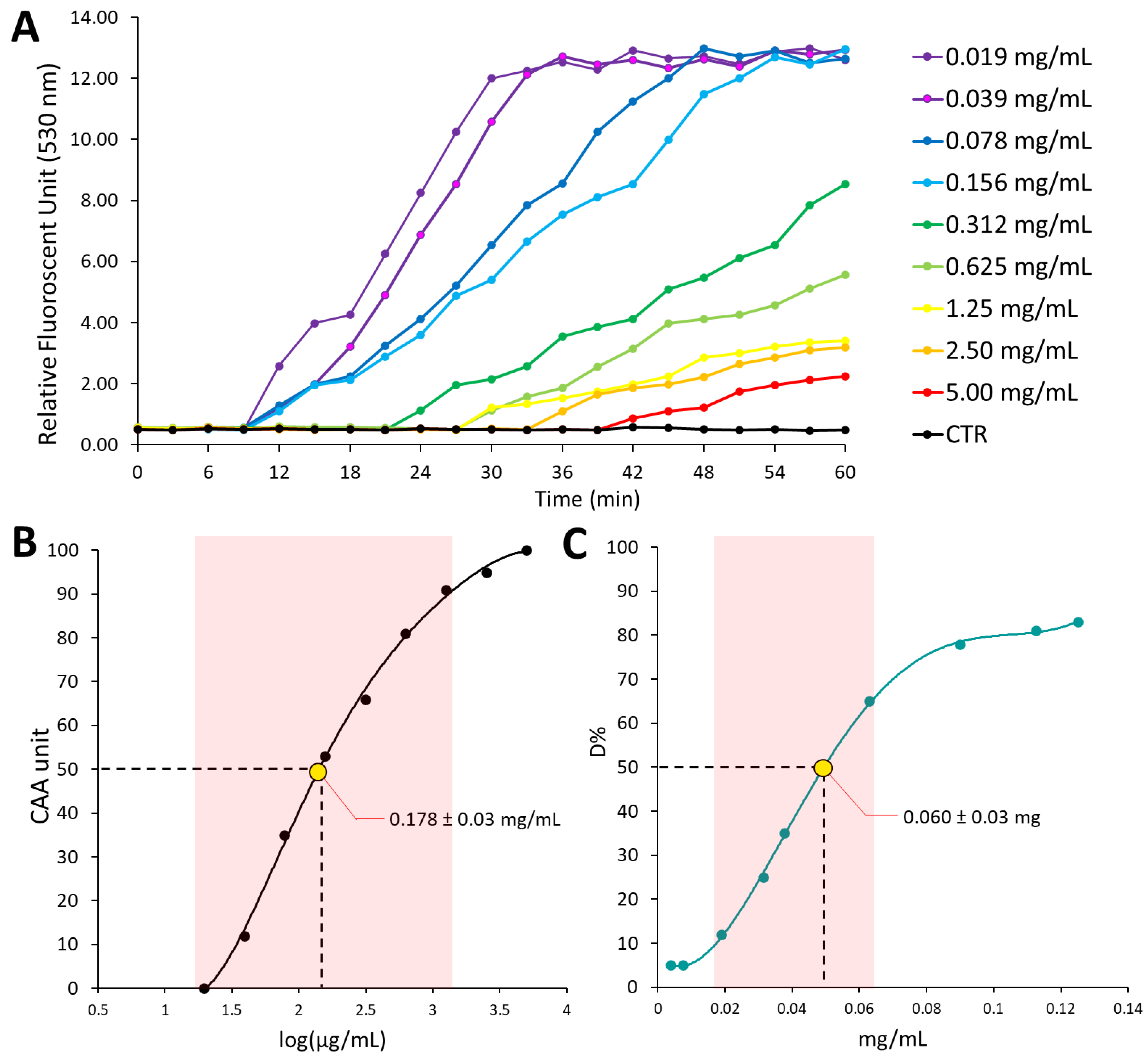
2.5. Cellular Antioxidant Activity

The cellular antioxidant activity (CAA) assay was accomplished as previously described by Wolfe at al. [25] with some minor changes [26]. Briefly, 96-well plates were seeded with HepG2 cells in complete culture medium at the density of 6.0 × 10⁴ cells/well and incubated for 24 h. Subsequently, the cells were treated with 25 μM DCFH-DA salt (Sigma Aldrich, St. Louis, MO, USA) and the WRSP extract at different concentrations for 2 h. Ethanol concentration never exceeded 0.25% (v/v). Control and blank wells were incubated with 25 μM DCFH-DA in culture medium containing 0.25% ethanol (v/v). After the incubation time, cells were washed with PBS salt (Sigma Aldrich, St. Louis, MO, USA), then 600 μM2,2′-Azobis(2-methylpropionamidine) dihydrochloride (ABAP) (Sigma Aldrich, St. Louis, MO, USA) in Hanks′ Balanced Salt solution (HBSS) (Sigma Aldrich, St. Louis, MO, USA) was added to treated and control wells. HBSS alone was added to blank wells. The fluorescence of treated, control, and blank wells were evaluated every 5 min for 1 h by using a plate-reader at 37 °C. The area under the curve of fluorescence versus time was integrated to calculate the CAA values for treated and control wells using Equation (2):
CAA = 100 × [ 100 ( S A C A ) ]
where ∫ SA is the integrated area of the sample wells and ∫ CA is the integrated area of the control wells. The antioxidant activity was expressed as CAA50, that is the WRSP extract concentration necessary for 50% of DCF formation inhibition and CAA50 was calculated from a concentration-response (CAA) curve using linear regression analysis, and it was expressed as mg of DW per mL of cell medium. The result, expressed as mg/mL of cell medium, is the mean value of three separate experiments.

2.6. Antiproliferative Activity

Exponentially growing cells were used for the 3-(4,5-Dimethyl-2-thiazolyl)-2,5-diphenyl-2H-tetrazolium bromide (MTT) assay. The assay was executed as previously described [21]. Briefly, the cells were seeded into standard 96-well plates at a density depending on the doubling times of each cell line. After 24 h of incubation, the WRSP extract at appropriate concentrations (500-50 μg DW/mL cell culture medium) was added and cells were incubated for another 48 hrs. Ethanol concentration never exceeded 0.25% (v/v). Control cells were incubated with culture medium containing 0.25% ethanol (v/v). After the incubation time, 0.5 mg/mL MTT reagent (Sigma Aldrich, St. Louis, MO, USA) was added and discarded after a 3-h incubation at 37 °C. The blue formazan produced in living cells was dissolved by dimethyl sulfoxide (DMSO). The absorbance was measured in a microplate reader (UV-1900i, Shimadzu®, Milan, Italy) at 570 nm, and the percentage of growth (PG) with respect to control cells (untreated cells) was calculated according to Equation (3):
P G = 100 × ( O D t e s t O D t z e r o ) ( O D c t r O D t z e r o )
where ODtest is the average of optical density after cell exposure to the extract for a chosen period of time; ODtzero is the average of optical density before the extract addition; and ODctr is the average of optical density after the chosen period of time with no exposure of cells to treatment. The concentration needed to induce 50% growth inhibition (GI50) for each cell line was determined from concentration-response (PG) curves using linear regression analysis, and it was expressed as mg of DW per mL of cell medium. The results, expressed as µg/mL of cell medium, is the mean value of three separate experiments.

2.7. Microbiological Characterization of WRSP

Ten grams of WRSP were transferred into a sterile bag (BagFilter® 400, Interscience, Saint Nom, France) diluted with 90 mL of Ringer’s solution (Sigma Aldrich, Milan, Italy), and homogenized by the stomacher apparatus BagMixer® 400 (Interscience, Saint Nom, France) at the highest speed (blending power 4) for 2 min. Homogenized WRSP was serially diluted and then plated on agar media for the enumeration of the main microbial groups belonging to the pro-technological, spoilage, and pathogenic populations following the approach of Messina et al. (2019) [27]. Briefly, the different microorganisms were inoculated as follows: total mesophilic microorganisms (TMM) on plate count agar (PCA); mesophilic lactic acid bacteria (LAB) rods and cocci on de Man-Rogosa-Sharpe (MRS) and Medium 17 (M17) agar, respectively; enterococci on kanamycin aesculin azide (KAA) agar; Pseudomonads on Pseudomonas agar base (PAB); members of the Enterobacteriaceae family on violet red bile glucose agar (VRGBA); coagulase-positive staphylococci (CPS) on Baird Parker (BP) supplemented with rabbit plasma fibrinogen (RPF); Listeria monocytogenes on Listeria selective agar base with SR0140E supplement; Salmonella spp. and Escherichia coli on hektoen enteric agar (HEA); and yeasts and moulds on malt agar (MA) supplemented with chloramphenicol (0.1 g/L). All media and chemicals were purchased from Microbiol Diagnostici (Uta, Italy). Plate counts were performed in triplicate.

2.8. Determination of Antibacterial Activity of WRSP

The ethanolic extract of WRSP was tested against bacteria of food origin. In particular, Enterococcus mundtii, Fructilactobacillus sanfranciscensis, Latilactobacillus sakei, Levilactobacillus brevis, Lactococcus lactis, and Leuconostoc mesenteroides were chosen among pro-technological species, Brochothrix thermosphacta, Pseudomonas endophytica, Pseudomonas fluorescens, Pseudomonas lactis, and Pseudomonas poae among spoilage bacteria, while Acinetobacter guillouiae, Bacillus cereus, Escherichia coli, Listeria monocytogenes, Pseudomonas aeruginosa, Salmonella Enteritidis, Salmonella Typhimurium, Staphylococcus aureus, and Staphylococcus epidermidis among bacteria responsible for human diseases. All spoilage and pathogenic bacteria were sub-cultured in Brain Heart Infusion (BHI) broth (Condalab, Madrid, Spain) incubated for 24 h at 37 °C, En. munditii, Lc. lactis and Ln. mesenteroides were reactivated in M17 (Microbiol Diagnostici) incubated for 24 h at 30 °C, Lt. sakei and Lv. brevis in MRS (Microbiol Diagnostici) incubated for 24 h at 30 °C, while F. sanfranciscensis in modified MRS [28]. The inhibitory activity of WRSP extract was tested by the well diffusion assay (WDA) [27,29] using sterile water as negative control, while streptomycin (10% w/v) was used as positive control. The inhibitory activity was evaluated after incubation at the optimal temperature for each strain and was considered positive only in case of a definite clear halo surrounding the wells. The strains sensitive to the extract were subjected to the minimum inhibitory concentration (MIC) determination by broth microdilution method in 96-well microplates [30]. Briefly, the extract was serially diluted in the optimal growth media for each strain (1:2) and their concentrations ranged between 100 and 3.125 mg/mL. After inoculation with approximately 106 CFU/mL of each sensitive strain, the bacterial growth was measured using a ScanReady Microplate photometer P-800 (Life Real Biotechnology Co., Ltd., Hangzhou, China). WDA and MIC tests were carried out in triplicate.

2.9. Statistical Analysis

All the statistical analyses were carried out using SPSS ver. 24 software (IBM, Armonk, New York, USA). The results were expressed as mean ± standard deviation (SD).

3. Results and Discussion

3.1. Phytochemical Characterization

In order to determine the phytochemical profile of the WRSP extract, HPLC-DAD-MS/MS analyses were performed. Specifically, 37 different compounds were identified. In particular, fourteen were flavonols (#6, #7, #9, #10, #11, #12, #14, #15, #16, #22, #24, #25, #34, and #35), three were flavanones (#20, #27, and #37), two were flavonones (#18 and #26), three were flavanonol (#1, #2, and #19), and one was an O-methylated flavonol (#33) (Table 1). Interestingly, five flavan-3-ols (#3, #4, #5, #21, and #36), and nine proanthocyanidins (#8, #13, #17, #23, #28, #29, #30, #31, and #32) (PACs), polyphenols composed of subunits of flavan-3-ols [31,32], were also detected (Table 2). The sum of the identified compounds accounted to 1605.54 ± 44.98 mg per 100 g of DW. The major contribution to the quantification was given by flavan-3-ol family, which represented almost half (48%) of the quantified phytochemicals. In particular, #3, #4, #5, and #36 were glycosylated forms of the simplest existing flavan-3-ol, Catechin (#21). Moreover, HPLC analysis allowed the identification and quantification of a large amount of PACs (641.23 mg per 100 g DW) mostly characterized by a single C6-C4 interflavanic binding. The large presence of those polyphenols in the WRSP extract should not be surprising. Indeed, it is well known that PACs play an essential physiological role in seeds from the early stages of germination [31,33,34]. The most abundant PACs in WRSP were B-type (#8, #17, and #29). Regarding the degree of polymerization, most of the PACs were dimers (#8), representing about 68% of total identified PACs.
Concerning the identified flavonols, representing about 45% of the total quantified polyphenols, #12 (quercetin) accounted for almost all of the weight determined for these polyphenols. On the other hand, this is one of the most widespread polyphenols in the plant kingdom, and it is involved in one of the main steps during the synthesis of most polyphenols in plants [35].
Regarding the conjugation of polyphenols with sugar moieties, most of the identified polyphenols were not glycosylated forms (#1, #8, #12, #13, #17, #19, #21, #22, #23, #24, #25, #27, #28, #29, #30, #31, and #32). Specifically, almost 97% of the identified polyphenols were aglycone, accounting for 1548.91 ± 96.47 mg per 100 g of DW. The remaining glycosylated polyphenols were conjugated to galactose (#3, #11, and #20), glucose (#2, #4, #14, #15, and #18), arabinose (#7), diglucoside (#10, #33, #36, and #37), rutinoside (#5, #6, #26, and #34), xylose (#9), sambubiose (#16), or glucuronic acid (#35) moiety. Scientific studies evaluating the potential bioavailability of polyphenols have frequently indicated that aglycones are poorly permeable to biological membranes [31,35,36,37]. Consequently, potential local activity of these phenols after ingestion of foods enriched with these phytochemicals can be presumed [31,36].

3.2. Antioxidant Proprieties

Oxidative species physiologically generated by aerobic metabolism are involved in different processes, such as proliferation, apoptosis, and gene expression. However, an unbalance among endogenous antioxidant defenses and reactive species production causes cellular oxidative damage associated with etiology and progression of several chronic diseases [38,39,40]. Dietary antioxidants intake contributes to preventing this unbalance. In particular, from the literature it is known that phytochemicals, such as polyphenols, are able to increase the endogenous antioxidant defenses thanks to both their redox-active properties and modulating effects on antioxidant gene expression [21]. On the other hand, the bioactivity of phytochemicals has frequently been referred to by their antioxidant properties. Indeed, by influencing cellular redox homeostasis, dietary phytochemicals can induce structural changes in redox-sensitive targets and, consequently, modulate their function [41].
Different in solution methods have been used to estimate redox active proprieties of pure compounds, plant extracts, or biological fluids. These assays generally use radical generating systems and evaluate the ability of antioxidant compounds to scavenge synthetic free radicals. One of the most popular is the ABTS assay. Radical scavenging activity of the WRSP extract was equal to 4.86 ± 0.06 mmol TE/100 g DW (Figure 1, Panel C). With respect to the redox scavenging activity of WRSP of other berries seeds powder, the ABTS value measured for WRSP is comparable to that estimated for black currant and cranberry, meanwhile is it 2.5 and 50 folds higher than that of elder and rose hip, respectively [16,17]. In order to study whether the redox scavenging ability of WRSP extract prevent oxidative damage in a biological system, the antioxidant activity of the extract was also evaluated in a lipid peroxidation cell-based model. CAA assay not only allowed the evaluation of the antioxidant potential against peroxyl radicals but also the ability of antioxidant substances to interact with or cross membranes and their stability to cellular metabolism [42,43,44]. The estimated CAA50 was 0.178 ± 0.03 mg DW/mL cell medium (Figure 1, Panel A and B). This value is indicative of a relevant antioxidant activity. Moreover, this data turns out to be two orders of magnitude lower, thus indicating a greater activity, compared to that determined by Wolfe 2008 for raspberry extracts [25,42] (togliere 43).

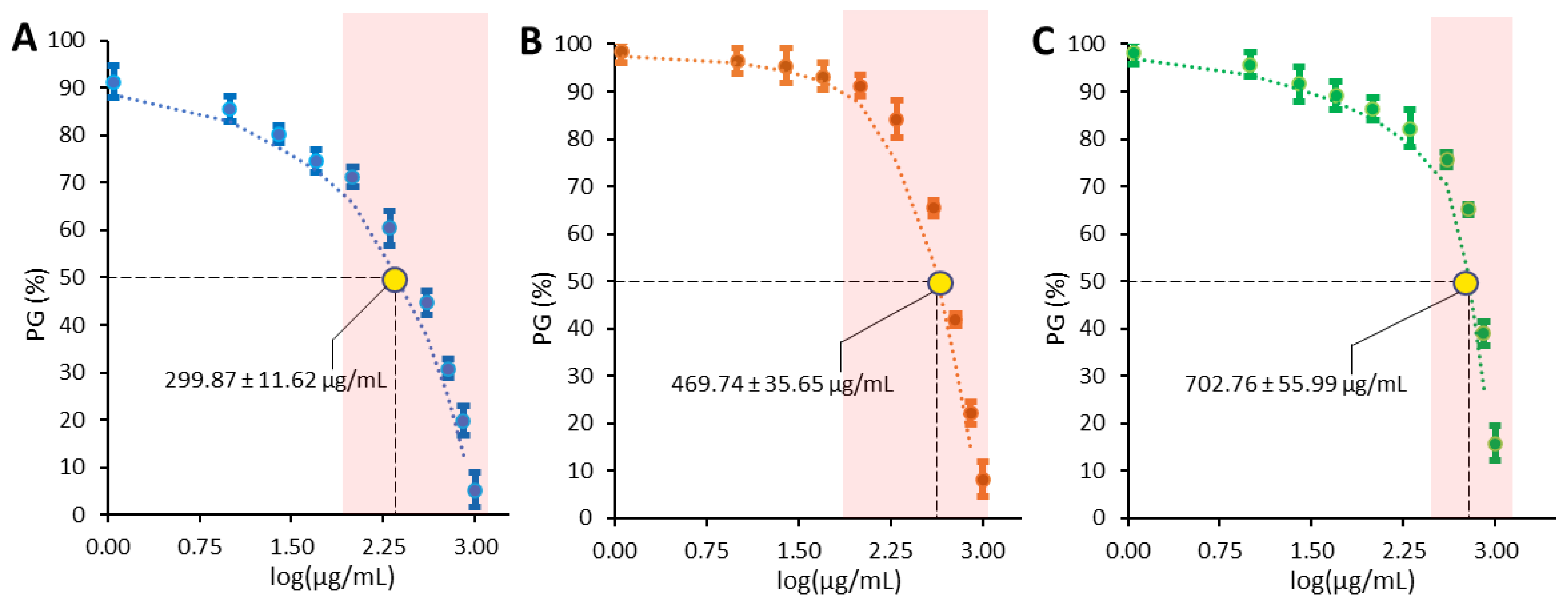
3.3. Antiproliferative Activity

The antiproliferative potential of the WRSP extract was assessed via MTT assay, using three epithelial human tumor cell lines: cervical cancer cells (HeLa), hepatocarcinoma cells (HepG2), and breast cancer cells (MCF-7). The concentration required for 50% inhibition of cell growth (GI50, Growth Inhibitory 50%) was calculated by linear regression, using a concentration/percentage growth (PG) curve (Figure 2). Cells were exposed to concentrations of WRSP extract ranging from 10 to 1000 µg DW/mL of cell culture medium. The results displayed an antiproliferative activity concentration-dependent, with variability of effects in function of the cell line considered. In particular, WRSP extract showed a stronger effect on HepG2 cells (299.866 ± 11.617 µg/mL) (Figure 2, Panel A), while a progressively smaller effects was observed on MCF-7 (469.739 ± 35.646 µg/mL) (Figure 2, Panel B) and HeLa (702.760 ± 55.986) cells (Figure 2, Panel C). This difference could be a consequence of peculiar protein expression patterns in the three cell lines. On the other hand, peculiar phytochemicals in WRSP, acting on differently expressed targets in the cells, could contribute, presumably in synergistic actions, to the observed activity.
Quercetin and catechin are the main dietary polyphenols and, with dimer B-type PAC, represent the most abundant phytochemicals in the WRSP extract. In particular, the quantity of quercetin is equal to over 40% of the total weight of the identified polyphenols. Bioavailability of quercetin and its glycosylated forms has been previously demonstrated [45] and several studies attest the antiproliferative properties of these polyphenols. Moreover, induction of cell cycle arrest and apoptosis by quercetin in cancer cells has been documented [46]. On the other hand, epidemiological studies have reported that intake of quercetin-rich food reduces the risk of different types of cancer [47].
HPLC-DAD-MS/MS analysis revealed that WRSP is also a very rich source of PACs. Along with other health protective effects [33], anti-carcinogenic activity of proanthocyanidins from different sources have been extensively documented and the involved molecular targets identified. Although the very low bioavailability of high-molecular-weight PACs, the literature data showed that smaller oligomeric procyanidins, in particular dimers and trimers, are both stable in digestive conditions and absorbed in the gut. WRSP extract contains mainly low-molecular-weight PACs. In particular, the dimer B-Type represents over 60% of the total identified PACs in the extract [31,45,48].
It must be emphasized that the combined action of multiple polyphenols, as occurs in a complex matrix, may be higher than that of a single polyphenol. A greater cell growth inhibition activity has been demonstrated for various combinations of polyphenols when compared to the activity of the single polyphenol. This evidence may not only be the result of the ability of phytochemical mixtures to affect multiple pathways involved in cancer simultaneously but also of the possible ability of different phytochemicals to reciprocally increase their bioavailability [49]. Anticancer effects of combinations of epigallocatechin gallate (ECGC) and quercetin have been demonstrated in ovarian and prostate cancer [50] and co-adjuvant therapy efficacy of catechin and procyanidin B2 with docetaxel was demonstrated in MCF-7 cells [51].

3.4. Microbiological Characterization and Inhibitory Properties of WRSP

In order to investigate the microbiological safety of WRSP and its potential application as a functional ingredient or natural antimicrobial food additive, microbiological characterization of WRSP was performed along with the evaluation of the inhibitory properties of the WRSP extract against bacteria growth.
The microbiological counts of WRPS did not reveal the presence of bacteria or microscopic fungi (yeasts and molds) (data not shown), highlighting the hygienic suitability of this by-product for food applications. In fact, it is well known that the presence of microorganisms in a by-product besides impairing its stability, when the by-product is used as a food additive, can endanger both consumer safety and stability of the food to which it is added [52].
The search for new antimicrobial food additives is a topic that has been attracting increasing attention over the past few years, since foodborne resistant diseases are one of the most important public health problems associated with the risk of emergence of multidrug-resistant bacterial strains in the food chain [53]. As a result, there is a raising demand for natural plant-derived preservatives because they are perceived as more natural and safe [54,55,56]. To evaluate a potential use of WRSP as food preservative, we evaluated its ability to inhibit the growth of undesirable microorganisms generally associated with foods [57].
The antibacterial activity of WRSP extract against spoilage and pathogenic bacteria is shown in Table 3. A considerable inhibition activity has been observed against both Br. thermosphacta, which is commonly associated with meat spoilage [58], and all Pseudomonas strains, commonly involved in the physicochemical change and generation of off-flavours in a wide range of foods of both plant and animal origin [59,60]. Regarding pathogenic bacteria, the WRSP extract was highly effective against four out of the nine tested strains, recording a diameter of the inhibition area around the wells of 13.5 mm for the B. cereus and of about 18 mm for A. guillouiae, St. aureus, and St. epidermidis. These microorganisms are relevant pathogens commonly associated with food consumption [61,62]. The antimicrobial activity of WRSP was also characterized in terms of MIC (Table 3). Only a MIC of 100 mg/mL was reached for the strain B. cereus ICE70, while a MIC of 25 mg/mL was observed for all the other strains. These results suggest that this extract has great potential applications as natural food preservative.
Interestingly, the development of pro-technological bacteria was not inhibited by WRSP (Table 3), indicating it is harmless against the lactic acid bacteria (LAB) used in food fermentation processes [63]. These results are very relevant because, in consideration of the previously demonstrated antioxidant and antimicrobial properties of WRSP, they indicate the suitability of WRSP as a functional or natural antimicrobial ingredient also in fermented foods.
The obtained inhibition profiles, resulting in no effects on LAB growth, are rather unexpected considering that usually antibacterial agents show greater activity against Gram-positive rather than Gram-negative bacteria. The mechanism underlying the observed activity needs further investigation. Here we can just assume that the strain-dependent resistance to WRSP components can justify the observed inhibition profiles.

4. Conclusions

This work demonstrates that the waste generated by the cold pressing of red raspberry seeds can be an interesting plant raw material for the extraction of bioactive compounds with different biological properties.
In particular, WRSP is shown to be really rich in several phytochemicals, including quercetin, catechin, and low weight proanthocyanidins. Further studies to elucidate the bioavailability of phytochemicals in WRSP are needed. However, considering the concentration of polyphenols in WRSP, the intake of even small quantities (5–10 g) of this plant matrix in a nutraceutical formulation or as a functionalizing ingredient can provide significant quantities of bioactive molecules with potential protective effects on human health. Therefore, antioxidant, anticancer, and antimicrobial properties evaluated and discussed in this article suggest a potential use of this agro-food waste in both food and non-food applications.

Author Contributions

Conceptualization, C.G.; methodology, G.M., G.S:, R.G., L.S. and C.G.; software, G.M., G.S. and A.L.; validation, G.M., G.S., R.G. and A.L. and C.G.; formal analysis, G.M., G.S., R.G. and A.L.; investigation, G.M., G.S., R.G. and G.B. and L.L.R.; resources, G.M., A.L., L.S. and C.G.; data curation, G.S., G.M. and R.G.; writing—original draft preparation, G.M., G.S. and C.G.; writing—review and editing, all authors; supervision, C.G. and L.S.; project administration, C.G.; funding acquisition, C.G. and L.S. All authors have read and agreed to the published version of the manuscript.

Funding

This research was partially funded by the University of Palermo, FFR2021 Carla Gentile.

Data Availability Statement

Data is contained within the article.

Acknowledgments

The authors acknowledge Abel Nutraceuticals S.R.L and Andrea Capuzzo for providing WRSP for this study free of charge. Moreover, the authors would like to express gratitude to Ylenia Intravaia for their technical support.

Conflicts of Interest

The authors declare no conflict of interest.

References

  1. Gustavsson, J.; Cederberg, C.; Sonesson, U.; Otterdijk, R.; Meybeck, A. Global Food Losses and Food Waste-Extent, Causes and Prevention; FAO: Rome, Italy, 2011. [Google Scholar]
  2. Ojha, S.; Bußler, S.; Schlüter, O.K. Food waste valorisation and circular economy concepts in insect production and processing. Waste Manag. 2020, 118, 600–609. [Google Scholar] [CrossRef] [PubMed]
  3. Bertocci, F.; Mannino, G. Can Agri-Food Waste Be a Sustainable Alternative in Aquaculture? A Bibliometric and Meta-Analytic Study on Growth Performance, Innate Immune System, and Antioxidant Defenses. Foods 2022, 11, 1861. [Google Scholar] [CrossRef] [PubMed]
  4. Socaci, S.A.; Farcas, A.C.; Vodnar, D.C.; Tofana, M. Food Wastes as Valuable Sources of Bioactive Molecules; IntechOpen: London, UK, 2017; pp. 75–93. [Google Scholar]
  5. Ben-Othman, S.; Jõudu, I.; Bhat, R. Bioactives from agri-food wastes: Present insights and future challenges. Molecules 2020, 25, 510. [Google Scholar] [CrossRef] [PubMed]
  6. Campobenedetto, C.; Mannino, G.; Agliassa, C.; Acquadro, A.; Contartese, V.; Garabello, C.; Bertea, C.M. Transcriptome Analyses and Antioxidant Activity Profiling Reveal the Role of a Lignin-Derived Biostimulant Seed Treatment in Enhancing Heat Stress Tolerance in Soybean. Plants 2020, 9, 1308. [Google Scholar] [CrossRef]
  7. Ispiryan, A.; Viškelis, J.; Viškelis, P. Red raspberry (Rubus idaeus L.) seed oil: A review. Plants 2021, 10, 944. [Google Scholar] [CrossRef]
  8. FAOSTAT. Available online: www.fao.org/faostat (accessed on 24 August 2022).
  9. George, B.P.; Abrahamse, H. Traditional Uses and Bioactivities of Common Rubus Species With Reference to Cancer: A Mini-Review. In Phytopharmaceuticals; John Wiley & Sons: Hoboken, NJ, USA, 2021; pp. 259–270. [Google Scholar]
  10. Mihailović, N.R.; Mihailović, V.B.; Ćirić, A.R.; Srećković, N.Z.; Cvijović, M.R.; Joksović, L.G. Analysis of Wild Raspberries (Rubus idaeus L.): Optimization of the Ultrasonic-Assisted Extraction of Phenolics and a New Insight in Phenolics Bioaccessibility. Plant Foods Hum. Nutr. 2019, 74, 399–404. [Google Scholar] [CrossRef]
  11. Kähkönen, M.P.; Hopia, A.I.; Heinonen, M. Berry phenolics and their antioxidant activity. J. Agric. Food Chem. 2001, 49, 4076–4082. [Google Scholar] [CrossRef]
  12. Derrick, S.A.; Kristo, A.S.; Reaves, S.K.; Sikalidis, A.K. Effects of Dietary Red Raspberry Consumption on Pre-Diabetes and Type 2 Diabetes Mellitus Parameters. Int. J. Environ. Res. Public Health 2021, 18, 9364. [Google Scholar] [CrossRef]
  13. Veljkovic, B.; Djordjevic, N.; Dolicanin, Z.; Licina, B.; Topuzovic, M.; Stankovic, M.; Zlatic, N.; Dajic-Stevanovic, Z. Antioxidant and anticancer properties of leaf and fruit extracts of the wild raspberry (Rubus idaeus L.). Not. Bot. Horti Agrobot. Cluj-Napoca 2019, 47, 359–367. [Google Scholar] [CrossRef]
  14. Skrovankova, S.; Sumczynski, D.; Mlcek, J.; Jurikova, T.; Sochor, J. Bioactive compounds and antioxidant activity in different types of berries. Int. J. Mol. Sci. 2015, 16, 24673–24706. [Google Scholar] [CrossRef] [Green Version]
  15. Naji-Tabasi, S.; Emadzadeh, B.; Shahidi-Noghabi, M.; Abbaspour, M.; Akbari, E. Physico-chemical and antioxidant properties of barberry juice powder and its effervescent tablets. Chem. Biol. Technol. Agric. 2021, 8, 23. [Google Scholar] [CrossRef]
  16. Bakowska-Barczak, A.M.; Schieber, A.; Kolodziejczyk, P. Characterization of Canadian black currant (Ribes nigrum L.) seed oils and residues. J. Agric. Food Chem. 2009, 57, 11528–11536. [Google Scholar] [CrossRef] [PubMed]
  17. Helbig, D.; Böhm, V.; Wagner, A.; Schubert, R.; Jahreis, G. Berry seed press residues and their valuable ingredients with special regard to black currant seed press residues. Food Chem. 2008, 111, 1043–1049. [Google Scholar] [CrossRef]
  18. Kosmala, M.; Zdunczyk, Z.; Juskiewicz, J.; Jurgonski, A.; Karlinska, E.; Macierzynski, J.; Jańczak, R.; Roj, E. Chemical composition of defatted strawberry and raspberry seeds and the effect of these dietary ingredients on polyphenol metabolites, intestinal function, and selected serum parameters in rats. J. Agric. Food Chem. 2015, 63, 2989–2996. [Google Scholar] [CrossRef]
  19. Parry, J.; Su, L.; Moore, J.; Cheng, Z.; Luther, M.; Rao, J.N.; Wang, J.Y.; Yu, L.L. Chemical compositions, antioxidant capacities, and antiproliferative activities of selected fruit seed flours. J. Agric. Food Chem. 2006, 54, 3773–3778. [Google Scholar] [CrossRef]
  20. Kang, I.; Espín, J.C.; Carr, T.P.; Tomás-Barberán, F.A.; Chung, S. Raspberry seed flour attenuates high-sucrose diet-mediated hepatic stress and adipose tissue inflammation. J. Nutr. Biochem. 2016, 32, 64–72. [Google Scholar] [CrossRef]
  21. Mannino, G.; Serio, G.; Bertea, C.M.; Chiarelli, R.; Lauria, A.; Gentile, C. Phytochemical profile and antioxidant properties of the edible and non-edible portions of black sapote (Diospyros digyna Jacq.). Food Chem. 2022, 380, 132137. [Google Scholar] [CrossRef]
  22. Vigliante, I.; Mannino, G.; Maffei, M.E. Chemical Characterization and DNA Fingerprinting of Griffonia simplicifolia Baill. Molecules 2019, 24, 1032. [Google Scholar] [CrossRef]
  23. Campobenedetto, C.; Agliassa, C.; Mannino, G.; Vigliante, I.; Contartese, V.; Secchi, F.; Bertea, C.M. A biostimulant based on seaweed (Ascophyllum nodosum and Laminaria digitata) and yeast extracts mitigates water stress effects on tomato (Solanum lycopersicum L.). Agriculture 2021, 11, 557. [Google Scholar] [CrossRef]
  24. Lauria, A.; Mannino, S.; Gentile, C.; Mannino, G.; Martorana, A.; Peri, D. DRUDIT: Web-based DRUgs DIscovery Tools to design small molecules as modulators of biological targets. Bioinformatics 2020, 36, 1562–1569. [Google Scholar] [CrossRef]
  25. Wolfe, K.L.; Kang, X.; He, X.; Dong, M.; Zhang, Q.; Liu, R.H. Cellular antioxidant activity of common fruits. J. Agric. Food Chem. 2008, 56, 8418–8426. [Google Scholar] [CrossRef] [PubMed]
  26. Vigliante, I.; Mannino, G.; Maffei, M.E. OxiCyan®, a phytocomplex of bilberry (Vaccinium myrtillus) and spirulina (Spirulina platensis), exerts both direct antioxidant activity and modulation of ARE/Nrf2 pathway in HepG2 cells. J. Funct. Foods 2019, 61, 103508. [Google Scholar] [CrossRef]
  27. Messina, C.M.; Gaglio, R.; Morghese, M.; Tolone, M.; Arena, R.; Moschetti, G.; Santulli, A.; Francesca, N.; Settanni, L. Microbiological profile and bioactive properties of insect powders used in food and feed formulations. Foods 2019, 8, 400. [Google Scholar] [CrossRef] [PubMed]
  28. Corsetti, A.; Settanni, L.; Braga, T.M.; de Fatima Silva Lopes, M.; Suzzi, G. An investigation of the bacteriocinogenic potential of lactic acid bacteria associated with wheat (Triticum durum) kernels and non-conventional flours. LWT-Food Sci. Technol. 2008, 41, 1173–1182. [Google Scholar] [CrossRef]
  29. Gaglio, R.; Guarcello, R.; Venturella, G.; Palazzolo, E.; Francesca, N.; Moschetti, G.; Settanni, L.; Saporita, P.; Gargano, M.L. Microbiological, chemical and sensory aspects of bread supplemented with different percentages of the culinary mushroom Pleurotus eryngii in powder form. Int. J. Food Sci. Technol. 2019, 54, 1197–1205. [Google Scholar] [CrossRef]
  30. Barbaccia, P.; Busetta, G.; Barbera, M.; Alfonzo, A.; Garofalo, G.; Francesca, N.; Moscarelli, A.; Moschetti, G.; Settanni, L.; Gaglio, R. Effect of grape pomace from red cultivar’Nero d’Avola’on the microbiological, physicochemical, phenolic profile and sensory aspects of ovine Vastedda-like stretched cheese. J. Appl. Microbiol. 2021, 133, 130–144. [Google Scholar] [CrossRef]
  31. Mannino, G.; Chinigò, G.; Serio, G.; Genova, T.; Gentile, C.; Munaron, L.; Bertea, C.M. Proanthocyanidins and Where to Find Them: A Meta-Analytic Approach to Investigate Their Chemistry, Biosynthesis, Distribution and Effect on Human Health. Antioxidants 2021, 10, 1229. [Google Scholar] [CrossRef]
  32. Gentile, C.; Mannino, G.; Palazzolo, E.; Gianguzzi, G.; Perrone, A.; Serio, G.; Farina, V. Pomological, Sensorial, Nutritional and Nutraceutical Profile of Seven Cultivars of Cherimoya (Annona cherimola Mill). Foods 2021, 10, 35. [Google Scholar] [CrossRef]
  33. Rauf, A.; Imran, M.; Abu-Izneid, T.; Patel, S.; Pan, X.; Naz, S.; Silva, A.S.; Saeed, F.; Suleria, H.A.R. Proanthocyanidins: A comprehensive review. Biomed. Pharmacother. 2019, 116, 108999. [Google Scholar] [CrossRef]
  34. Lee, Y.A.; Cho, E.J.; Yokozawa, T. Effects of proanthocyanidin preparations on hyperlipidemia and other biomarkers in mouse model of type 2 diabetes. J. Agric. Food Chem. 2008, 56, 7781–7789. [Google Scholar] [CrossRef]
  35. Mannino, G.; Gentile, C.; Ertani, A.; Serio, G.; Bertea, C.M. Anthocyanins: Biosynthesis, Distribution, Ecological Role, and Use of Biostimulants to Increase Their Content in Plant Foods—A Review. Agriculture 2021, 11, 212. [Google Scholar] [CrossRef]
  36. Kaur, K.; Asthir, B.; Verma, D.K. Biosynthesis, bioavailability, and metabolism of plant polyphenols: Biological activities and their potential benefits in human health. In Phytochemicals in Food and Health; Apple Academic Press: Cambridge, MA, USA, 2021; pp. 231–255. ISBN 1003082122. [Google Scholar]
  37. Aneklaphakij, C.; Saigo, T.; Watanabe, M.; Naake, T.; Fernie, A.R.; Bunsupa, S.; Satitpatipan, V.; Tohge, T. Diversity of chemical structures and biosynthesis of polyphenols in Nut-bearing species. Front. Plant Sci. 2021, 12, 642581. [Google Scholar] [CrossRef] [PubMed]
  38. Stahl, W. Oxidative Stress—Antioxidants in Foods. Ernahrungs Umschau 2011, 58, 536–541. [Google Scholar]
  39. Kruk, J.; Aboul-Enein, H.Y.; Kładna, A.; Bowser, J.E. Oxidative stress in biological systems and its relation with pathophysiological functions: The effect of physical activity on cellular redox homeostasis. Free Radic. Res. 2019, 53, 497–521. [Google Scholar] [CrossRef]
  40. Guan, R.; Van Le, Q.; Yang, H.; Zhang, D.; Gu, H.; Yang, Y.; Sonne, C.; Lam, S.S.; Zhong, J.; Jianguang, Z. A review of dietary phytochemicals and their relation to oxidative stress and human diseases. Chemosphere 2021, 271, 129499. [Google Scholar] [CrossRef]
  41. Caradonna, F.; Consiglio, O.; Luparello, C.; Gentile, C. Science and healthy meals in the world: Nutritional epigenomics and nutrigenetics of the mediterranean diet. Nutrients 2020, 12, 1748. [Google Scholar] [CrossRef]
  42. Wolfe, K.L.; Liu, R.H. Cellular antioxidant activity (CAA) assay for assessing antioxidants, foods, and dietary supplements. J. Agric. Food Chem. 2007, 55, 8896–8907. [Google Scholar] [CrossRef]
  43. Allegra, M.; Gentile, C.; Tesoriere, L.; Livrea, M.A. Protective effect of melatonin against cytotoxic actions of malondialdehyde: An in vitro study on human erythrocytes. J. Pineal Res. 2002, 32, 187–193. [Google Scholar] [CrossRef]
  44. Mannino, G.; Pernici, C.; Serio, G.; Gentile, C.; Bertea, C.M. Melatonin and phytomelatonin: Chemistry, biosynthesis, metabolism, distribution and bioactivity in plants and animals—An overview. Int. J. Mol. Sci. 2021, 22, 9996. [Google Scholar] [CrossRef]
  45. Almanza-Aguilera, E.; Ceballos-Sánchez, D.; Achaintre, D.; Rothwell, J.A.; Laouali, N.; Severi, G.; Katzke, V.; Johnson, T.; Schulze, M.B.; Palli, D. Urinary Concentrations of (+)-Catechin and (-)-Epicatechin as Biomarkers of Dietary Intake of Flavan-3-ols in the European Prospective Investigation into Cancer and Nutrition (EPIC) Study. Nutrients 2021, 13, 4157. [Google Scholar] [CrossRef]
  46. Cossarizza, A.; Gibellini, L.; Pinti, M.; Nasi, M.; Montagna, J.P.; De Biasi, S.; Roat, E.; Bertoncelli, L.; Cooper, E.L. Quercetin and cancer chemoprevention. Evid. Based Complement. Altern. Med. 2011, 2011, 591356. [Google Scholar]
  47. Ekström, A.M.; Serafini, M.; Nyren, O.; Wolk, A.; Bosetti, C.; Bellocco, R. Dietary quercetin intake and risk of gastric cancer: Results from a population-based study in Sweden. Ann. Oncol. 2011, 22, 438–443. [Google Scholar] [CrossRef] [PubMed]
  48. Nandakumar, V.; Singh, T.; Katiyar, S.K. Multi-targeted prevention and therapy of cancer by proanthocyanidins. Cancer Lett. 2008, 269, 378–387. [Google Scholar] [CrossRef] [PubMed]
  49. Niedzwiecki, A.; Roomi, M.W.; Kalinovsky, T.; Rath, M. Anticancer efficacy of polyphenols and their combinations. Nutrients 2016, 8, 552. [Google Scholar] [CrossRef]
  50. Wang, P.; Vadgama, J.V.; Said, J.W.; Magyar, C.E.; Doan, N.; Heber, D.; Henning, S.M. Enhanced inhibition of prostate cancer xenograft tumor growth by combining quercetin and green tea. J. Nutr. Biochem. 2014, 25, 73–80. [Google Scholar] [CrossRef] [PubMed]
  51. Núñez-Iglesias, M.J.; Novio, S.; García, C.; Pérez-Muñuzuri, M.E.; Martínez, M.-C.; Santiago, J.-L.; Boso, S.; Gago, P.; Freire-Garabal, M. Co-Adjuvant Therapy Efficacy of Catechin and Procyanidin B2 with Docetaxel on Hormone-Related Cancers In Vitro. Int. J. Mol. Sci. 2021, 22, 7178. [Google Scholar] [CrossRef]
  52. Dlugaszewska, J.; Ratajczak, M.; Kamińska, D.; Gajecka, M. Are dietary supplements containing plant-derived ingredients safe microbiologically? Saudi Pharm. J. 2019, 27, 240–245. [Google Scholar] [CrossRef]
  53. Caniça, M.; Manageiro, V.; Abriouel, H.; Moran-Gilad, J.; Franz, C.M.A.P. Antibiotic resistance in foodborne bacteria. Trends Food Sci. Technol. 2019, 84, 41–44. [Google Scholar] [CrossRef]
  54. Can-Cauich, C.A.; Sauri-Duch, E.; Betancur-Ancona, D.; Chel-Guerrero, L.; González-Aguilar, G.A.; Cuevas-Glory, L.F.; Pérez-Pacheco, E.; Moo-Huchin, V.M. Tropical fruit peel powders as functional ingredients: Evaluation of their bioactive compounds and antioxidant activity. J. Funct. Foods 2017, 37, 501–506. [Google Scholar] [CrossRef]
  55. Ofosu, F.K.; Daliri, E.B.-M.; Elahi, F.; Chelliah, R.; Lee, B.-H.; Oh, D.-H. New insights on the use of polyphenols as natural preservatives and their emerging safety concerns. Front. Sustain. Food Syst. 2020, 4, 525810. [Google Scholar] [CrossRef]
  56. Al-Maqtari, Q.A.; Rehman, A.; Mahdi, A.A.; Al-Ansi, W.; Wei, M.; Yanyu, Z.; Phyo, H.M.; Galeboe, O.; Yao, W. Application of essential oils as preservatives in food systems: Challenges and future prospectives—A review. Phytochem. Rev. 2021, 21, 1209–1246. [Google Scholar] [CrossRef]
  57. Jay, J.M.; Loessner, M.J.; Golden, D.A. Microbiologia Degli Alimenti; Springer Science & Business Media: Berlin/Heidelberg, Germany, 2009; ISBN 8847007860. [Google Scholar]
  58. Gribble, A.; Mills, J.; Brightwell, G. The spoilage characteristics of Brochothrix thermosphacta and two psychrotolerant Enterobacteriacae in vacuum packed lamb and the comparison between high and low pH cuts. Meat Sci. 2014, 97, 83–92. [Google Scholar] [CrossRef]
  59. Caldera, L.; Franzetti, L.; Van Coillie, E.; De Vos, P.; Stragier, P.; De Block, J.; Heyndrickx, M. Identification, enzymatic spoilage characterization and proteolytic activity quantification of Pseudomonas spp. isolated from different foods. Food Microbiol. 2016, 54, 142–153. [Google Scholar] [CrossRef]
  60. Miceli, A.; Gaglio, R.; Francesca, N.; Ciminata, A.; Moschetti, G.; Settanni, L. Evolution of shelf life parameters of ready-to-eat escarole (Cichorium endivia var. latifolium) subjected to different cutting operations. Sci. Hortic. 2019, 247, 175–183. [Google Scholar] [CrossRef]
  61. Settanni, L.; Gaglio, R.; Stucchi, C.; De Martino, S.; Francesca, N.; Moschetti, G. Presence of pathogenic bacteria in ice cubes and evaluation of their survival in different systems. Ann. Microbiol. 2017, 67, 827–835. [Google Scholar] [CrossRef]
  62. Podkowik, M.; Seo, K.S.; Schubert, J.; Tolo, I.; Robinson, D.A.; Bania, J.; Bystroń, J. Genotype and enterotoxigenicity of Staphylococcus epidermidis isolate from ready to eat meat products. Int. J. Food Microbiol. 2016, 229, 52–59. [Google Scholar] [CrossRef]
  63. Bintsis, T. Lactic acid bacteria as starter cultures: An update in their metabolism and genetics. AIMS Microbiol. 2018, 4, 665. [Google Scholar] [CrossRef]
Figure 1. Antioxidant properties of the WRSP extract. Panel (A) shows the relative fluorescence of treated (mg/mL per cell medium) or untreated (control) HepG2 cells evaluated each 5 min for 1 h, after exposure to 600 µM ABA, as reported in materials and methods. Panel (B) shows the dose-response (CAA) curve, used to calculate CAA50 value, reported with the yellow dot. Panel (C) shows dose-response (D%) curve obtained from the decolorization of radical ABTS solution, after adding different concentrations of the WRSP extract, as reported in materials and methods. The yellow dot in Panel (C) shows the WRSP concentration (mg/per mL of reaction mix) necessary to 50% decolorization of the radical ABTS solution (D50). The red box represents the linear portion of the curve within which the CAA50 and D50 values were determined.
Foods 11 02605 g001
Figure 2. Antiproliferative activity exerted by the WRSP extract on (A) HepG2, (B) MCF-7, and (C) HeLa cell line. Dose/response curves are shown as percent growth (PG%) versus concentration expressed as Log(µg DW/mL cell medium). The bars reported near the coloured dots represent the standard deviation of each measurement at the relative concentration. The yellow dot reports the GI50 (µg DW/mL cell medium) of WRSP on each cell line. The red box represents the linear portion of inhibition curve within which the GI50 value was determined.
Foods 11 02605 g002
Table 1. Qualitative and quantitative phytochemical characterization of flavanonols, flavonols, flavonones, flavanones, and o-methylated flavonols in the WRSP extract. Results (mg per 100 g of FW) are expressed as mean ± SD of three different experiments.
#RT [Min] m/zMS/MSCAS-IDChemical
Formula
Compound(s)mg/100 g
Flavanonols
110.7287 480-20-6C15H12O6Dihydrokaempferol12.74 ± 0.73
213.6481319 n.a.C21H22O13Dihydromyricetin-3-O-glucoside4.38 ± 0.11
1928.2319 27,200-12-0C15H12O8Dihydromyricetin64.15 ± 3.96
Flavonols
615.762547946331741,093-68-9C27H30O17Myricetin-rutinoside0.95 ± 0.06
722.2433301 572-30-5C20H18O11Quercetin-3-O-arabinoside5.25 ± 0.07
923.2433 549-32-6C20H18O11Quercetin-3-O-xyloside3.83 ± 0.23
1024.2625463301 6892-74-6C27H30O17Quercetin-3,7-O-diglucoside3.11 ± 0.09
1124.4463301 482-36-0C21H20O12Quercetin-3-O-galactoside0.44 ± 0.01
1224.8301 117-39-5C15H10O7Quercetin697.84 ± 16.81
1425447285 482-35-9C21H20O12Kaempferol-3-O-glucoside0.46 ± 0.02
1525463301 482-35-9C21H20O12Quercetin-3-O-glucoside0.71 ± 0.02
1626.4477176 22,688-79-5C21H18O13Quercetin-3-O-glucoronide2.44 ± 0.13
2231.1317 529-44-2C15H10O8Myricetin18.23 ± 0.75
2432.7285 520-18-3C15H10O6Kaempferol5.05 ± 0.27
2533.9609463447301153-18-4C27H30O16Quercetin-3-O-rutinoside0.48 ± 0.03
3445.659344743128517,650-84-9C27H30O15Kaempherol-3-O-rutinoside4.82 ± 0.28
3545.6595294 83,048-35-5C26H28O16Quercetin-3-O-sambunioside2.42 ± 0.05
Flavonones
1827.7447285 20,344-46-1C21H20O11Luteolin-3-O-glucoside6.32 ± 0.31
2635.2285 491-70-3C15H10O6Luteolin2.32 ± 0.14
Flavanones
2029.4451289 20,344-46-1C21H22O10Naringenin-3-O-galactoside2.17 ± 0.11
2736.4287 552-58-9C21H22O11Eriodictyol5.14 ± 0.26
3757.8595433271 n.a.C27H32O15Naringenin-3,7-O-diglucoside5.66 ± 0.12
O-methylated flavonol
3344.6639477315 n.a.C28H32O17Isohermentin-3,7-O-diglucoside1.33 ± 0.07
RT: Retention Time; m/z: mass-to-charge ratio; MS/MS: detected fragmentations; CAS-ID: Chemical Abstracts Service Identification Number; n.a.: not available.
Table 2. Qualitative and quantitative phytochemical characterization of flavan-3-ols and proanthocyanidins in WRSP extract. Results (mg per 100 g of FW) are expressed as mean ± SD of three different experiments.
#RT [Min]M-HMS/MSCAS-IDChemical
Formula
Compound(s)mg/100 g
Flavan-3-ols
314.2451289 n.a.C21H24O11Catechin-3-O-galactoside0.75 ± 0.03
414.9451289 n.a.C21H24O11Catechin-3-O-glucoside1.32 ± 0.03
515.6597451435289n.a.C27H34O15Catechin-3-O-rutinoside7.34 ± 0.18
2129.7289 154-23-4C15H14O6Catechin122 ± 3.48
3651.2613451289 n.a.C27H34O16Catechin-3,7-O-diglucoside2.46 ± 0.14
Proanthocyanidins
822.8577289 29,106-49-8C30H26O12Dimer B-Type PAC399.61 ± 5.95
1324.9865575289 65,085-09-8C45H38O18Trimer A-Type PAC14.26 ± 0.17
1727.51153867577 n.a.C60H52O24Tetramer B-Type PAC12.63 ± 0.41
2331.8143914371151575n.a.C75H60O30Pentamer A-Type PAC56.16 ± 3.83
2837.220121151863289n.a.C90H72O36Esamer A-Type PAC12.05 ± 0.5
2937.220141153861577n.a.C90H78O36Esamer B-Type PAC5.16 ± 0.17
3037.514371151863575n.a.C75H60O30Pentamer A-Type PAC30.88 ± 1.58
3138.81151863575289n.a.C60H48O24Tetramer A-Type PAC13.35 ± 0.64
3242.514411155865289n.a.C75H65O30Pentamer B-Type PAC 77.33 ± 3.16
RT: retention time; m/z: mass-to-charge ratio; MS/MS: detected fragmentations; CAS-ID: Chemical Abstracts Service Identification Number; n.a.: not available.
Table 3. Antibacterial activity of the ethanolic extract of WRSP.
SpeciesStrainsSource of IsolationInhibition (mm)MIC (mg/mL)
Pro-technological
En. mundtiiWFE3Wheat flours-n.d.
F. sanfranciscensisSD22Sourdough-n.d.
Lt. sakeiSP255Salami-n.d.
Lv. brevisSD70Sourdough-n.d.
Lc. lactisCAG4Curd-n.d.
Ln. mesenteroidesMISE643Raw ewe’s milk-n.d.
Spoilage
Br. thermosphactaSP10Pork meat23.0 ± 0.425
P. fluorescens4G628Ready to eat salad19.3 ± 0.225
P. lactisSP198Salami17.8 ± 0.225
P. poae4G558Ready to eat salad19.0 ± 0.425
Pathogenic
A. guillouiaeICE24Food ice cubes18.2 ± 0.325
B. cereusICE70Food ice cubes13.5 ± 0.2100
E. coliPSL52PDO Pecorino Siciliano cheese-n.d.
L. monocytogenes13BOGorgonzola cheese-n.d.
P. aeruginosaPSA68Animal tissue-n.d.
S. EnteritidisATCC13076Unknown-n.d.
S. Typhimurium50432Molluscs-n.d.
St. aureusATCC33862Unknown17.8 ± 0.225
St. epidermidisICE244Food ice cubes18.6 ± 0.125
Results indicate the mean value of three independent assays. Abbreviations: MIC, minimum inhibitory concentration; En., Enterococcus; F., Fructilactobacillus Lt., Latilactobacillus; Lv., Levilactobacillus; Lc., Lactococcus; Ln., Leuconostoc; Br., Brochothrix; P., Pseudomonas; A., Acinetobacter; B., Bacillus; E., Escherichia; L., Listeria; S., Salmonella; St., Staphylococcus; n.d., not determinated. Symbols:—no inhibition found.
Publisher’s Note: MDPI stays neutral with regard to jurisdictional claims in published maps and institutional affiliations.

