Article

Ethanol at Subinhibitory Concentrations Enhances Biofilm Formation in Salmonella Enteritidis

1
MOST-USDA Joint Research Center for Food Safety, School of Agriculture and Biology, State Key Laboratory of Microbial Metabolism, Shanghai Jiao Tong University, Shanghai 200240, China
2
Food, Nutrition and Health, Faculty of Land and Food Systems, The University of British Columbia, Vancouver, BC V6T 1Z4, Canada
*
Author to whom correspondence should be addressed.
Foods 2022, 11(15), 2237; https://doi.org/10.3390/foods11152237
Submission received: 6 July 2022 / Revised: 21 July 2022 / Accepted: 25 July 2022 / Published: 27 July 2022
(This article belongs to the Special Issue Foodborne Pathogenic Bacteria: Prevalence and Control)

Abstract

:
The survival of Salmonella Enteritidis in the food chain is relevant to its biofilm formation capacity, which is influenced by suboptimal environmental conditions. Here, biofilm formation pattern of this bacterium was assessed in the presence of ethanol at sub-minimal inhibitory concentrations (sub-MICs) by microtiter plate assays, cell characteristic analyses, and gene expression tests. It was observed that ethanol at subinhibitory concentrations (1/4 MIC, 2.5%; 1/2 MIC, 5.0%) was able to stimulate biofilm formation in S. Enteritidis. The OD595 value (optical density at 595 nm) used to quantify biofilm production was increased from 0.14 in control groups to 0.36 and 0.63 under 2.5% and 5.0% ethanol stresses, respectively. Ethanol was also shown to reduce bacterial swimming motility and enhance cell auto-aggregation ability. However, other cell characteristics such as swarming activity, initial attachment and cell surface hydrophobicity were not remarkedly impacted by ethanol. Reverse transcription quantitative real-time PCR (RT-qPCR) analysis further revealed that the luxS gene belonging to a quorum-sensing system was upregulated by 2.49- and 10.08-fold in the presence of 2.5% and 5.0% ethanol, respectively. The relative expression level of other biofilm-related genes (adrA, csgB, csgD, and sdiA) and sRNAs (ArcZ, CsrB, OxyS, and SroC) did not obviously change. Taken together, these findings suggest that decrease in swimming motility and increase in cell auto-aggregation and quorum sensing may result in the enhancement of biofilm formation by S. Enteritidis under sublethal ethanol stress.

1. Introduction

Ethanol is a common component present in a variety of foods such as alcoholic beverages, fruit products, and baked products [1,2,3]. For a long time, ethanol has been utilized as a chemical disinfectant for food contact surfaces, conveyor belts, as well as food processing tools [4,5,6]. Moreover, ethanol is effective for controlling postharvest decay and extending shelf life in the fruit industry [7]. Occasionally, ethanol can be present in food-related environments at subinhibitory concentrations due to its easy evaporation, inappropriate application, or dilution in the environment. The subinhibitory concentrations of ethanol are able to exert an inhibitory activity but are not lethal to bacterial pathogens [6].
Ethanol at subinhibitory concentrations has been recognized as a crucial environmental factor influencing biofilm formation of bacterial pathogens. Tango et al. [8] found that 2.5–3.5% ethanol enabled Staphylococcus aureus ATCC 13150 to form more biofilms. Similarly, biofilm formation in Pseudomonas aeruginosa PAO1 was also induced by ethanol at low levels (1% and 2%) [9]. Bacterial pathogens in biofilms are generally resistant to cleaning and sanitation operations and are extremely difficult to eradicate in food industries [10,11]. In this context, ethanol-triggered biofilm formation can contribute to the survival and persistence of bacterial pathogens on industrial surfaces and equipment, thus posing a threat to food safety. It is thus essential to reveal biofilm formation patterns of other important bacterial pathogens such as Salmonella spp. under ethanol stress.
Biofilm formation mechanisms of Salmonella spp. have been well characterized in laboratory conditions that are optimal for bacterial growth [12]. In terms of physiological strategies, cell surface characteristics (e.g., motility, hydrophobicity, and auto-aggregation) have been demonstrated to participate in biofilm production of this bacterium [13]. At the molecular level, attachment genes (e.g., adrA, csgB, and csgD) and quorum-sensing genes (e.g., luxS and sdiA) have been recognized as key genetic elements for biofilm formation [14]. More recently, a couple of small RNAs (sRNAs) (e.g., ArcZ, CsrB, OxyS, and SroC) have also been revealed to modulate biofilm formation of Salmonella spp. [15]. Nevertheless, it remains unknown whether the aforementioned mechanisms function in Salmonella spp. biofilm formation under ethanol stress.
Practically, the mode of action of biofilm formation by Salmonella spp. under several environmental stress conditions has been characterized in previous studies [16,17]. For instance, Salmonella Typhimurium displayed reduced biofilm formation when exposed to sub-MIC levels of lactobionic acid, which was probably due to repressed synthesis of extracellular polymeric substances, reduced ability of cell motilities, and decreased expression of genes (e.g., adrA, flhD, and fljB) and sRNAs (e.g., ArcZ, CsrB, and SroC) [16]. In addition, the expression of quorum-sensing genes (e.g., luxS) and stress response genes (e.g., rpoS) was involved in biofilm formation of S. Enteritidis and S. Typhimurium in response to quercetin [17]. Thus, these kinds of physiological and gene expression analyses will be helpful in uncovering mechanisms of bacterial biofilm formation under other stress conditions such as exposure to alcoholic disinfectants.
The current work was carried out to evaluate the influence of ethanol at subinhibitory concentrations on biofilm formation in S. Enteritidis, a leading serotype of Salmonella spp. responsible for foodborne diseases. The underlying mechanisms on biofilm formation of this pathogen under ethanol stress will also be investigated via analysis of cell surface characteristics and gene expression levels.

2. Materials and Methods

2.1. Target Bacteria

S. Enteritidis ATCC 13076 was kept in Luria-Bertani (LB) broth containing 50% glycerol at −80 °C. This strain was resuscitated by inoculating stock culture onto LB agar and incubating, at 37 °C, for 24 h. A single bacterial colony was then transferred into LB broth, followed by overnight incubation, at 37 °C, before each test.

2.2. Antibacterial Activity Test

The two-fold dilution method was employed to assess the inhibitory activity of ethanol on S. Enteritidis [18]. An aliquot (5 μL) of an overnight culture (approximately 109 CFU/mL) was added into 5 mL TSB-YE (tryptic soy broth supplemented with 0.6% yeast extract) containing ethanol ranging from 2.5% to 40% (v/v). Negative (5 mL TSB-YE) and positive (5 mL TSB-YE containing 5 μL bacterial suspensions) controls were also included. The samples were stored at 25 °C (room temperature) on a rotator (200 rpm) for 24 h. This temperature was selected because it might be encountered by bacterial pathogens in actual conditions during food processing [19] and temperature abuse scenarios during food storage [20]. Optical density at 600 nm was monitored to measure bacterial growth in different concentrations of ethanol. The minimum inhibitory concentration (MIC) was recorded as the lowest ethanol concentration that completely retarded bacterial growth.

2.3. Biofilm Formation Assay

Biofilm production of S. Enteritidis in the presence of sub-MIC levels (1/4 MIC and 1/2 MIC) of ethanol was quantified using the 96-well microtiter plate assay [21]. The overnight bacteria culture was diluted to an inoculum level of approximately 106 CFU/mL with TSB-YE, and ethanol was later added to achieve a final concentration corresponding to 0, 1/4, and 1/2 of the MIC, respectively. The prepared samples were dispensed into 96-well plates with a volume of 200 μL, followed by static incubation at 25 °C for 72 h. After the biofilm formation, the inoculum was aspirated from 96-well plates, and sterile distilled water was then utilized to wash each well three times. The plates were air-dried at 25 °C for 15 min, and 0.1% (w/v) crystal violet solution (200 μL) was transferred into each well. After dyeing for 45 min, crystal violet was discarded from each well. The plates were allowed to air dry after washing with sterile distilled water, and 95% (v/v) ethanol (200 μL) was then added to each well. After gentle vortex mixing for 45 min, biofilm formation in each well was quantified by optical density at 595 nm (OD595).

2.4. Bacterial Attachment Assay

Bacterial adhesion to polystyrene surfaces was measured as previously detailed by Nilsson et al. [22] and do Valle Gomes et al. [23]. The 24-well microtiter plates (Falcon, Franklin Lakes, NJ, USA) were utilized to increase surface binding areas. The overnight culture of S. Enteritidis was diluted to approximately 106 CFU/mL with TSB-YE and supplemented with 0, 1/4, and 1/2 MIC of ethanol, respectively. An aliquot (3 mL) of the resulting suspensions was then transferred to 24-well plates. After static incubation at 25 °C for 5 h, cell suspensions were discarded from each well, followed by washing with sterile distilled water. Subsequently, the wells were stained with crystal violet (0.1%, w/v), and added with ethanol (95%, v/v). The attachment ability of S. Enteritidis was estimated by reading OD595 values (Tecan Sunrise, Tecan Group Ltd., Mannedorf, Switzerland).

2.5. Cell Motility Test

Cell motility test was conducted based on the method detailed by Roy et al. [24]. The medium was prepared by adding 0.3% and 0.6% agar to TSB-YE for the swimming and swarming assays, respectively, followed by autoclaving. Ethanol was then added to each medium before it hardened and mixed thoroughly. The final concentration of ethanol was 0, 1/4, and 1/2 MIC, respectively. In the swimming assay, 2 μL of pre-diluted bacterial suspension (approximately 106 CFU/mL) was inoculated by passing through a fresh 0.3% agar plate. An aliquot of 5 μL pre-diluted bacterial suspension (approximately 106 CFU/mL) was spotted onto the center of a fresh 0.6% agar plate in the swarming assay. The plates were then stored at 25 °C for 10 and 24 h, respectively. The motility zone diameter was recorded as the distance between the center of the plate and the leading edge of bacterial growth.

2.6. Cell Surface Hydrophobicity Study

Bacterial cell surface hydrophobicity was estimated by a microbial adhesion to solvents test [25,26]. S. Enteritidis cells cultured as described in Section 2.3 were spun, washed, and redissolved in phosphate-buffered saline. The optical density of the resulting bacterial suspensions was recorded at 600 nm (OD600 pre-vortex). Afterwards, 3 mL of hexadecane was added to an equal volume of cell suspensions. Each bacterial suspension was vortexed and incubated for 15 min at 25 °C. The aqueous phase was then subjected to optical density quantification at 600 nm (OD600 post-vortex). Bacterial cell surface hydrophobicity was estimated by the following formula: hydrophobicity (%) = (1 − OD600 post-vortex/OD600 pre-vortex) × 100.

2.7. Cell Auto-Aggregation Assay

The cell auto-aggregation property was estimated as previously detailed by Lee et al. [27] with slight modifications. S. Enteritidis cells cultured as described in Section 2.3 were centrifuged and resuspended in an equal volume of TSB-YE. An aliquot (200 μL) of bacterial suspensions was mixed with 4.8 mL fresh TSB-YE. After overnight incubation at 25 °C statically, the optical density at 600 nm (OD600 pre-vortex) of the upper layer was checked. The samples were then vortexed carefully and evaluated at 600 nm (OD600 post-vortex). Bacterial cell auto-aggregation was calculated using the following formula: auto-aggregation (%) = (1 − OD600 pre-vortex/OD600 post-vortex) × 100.

2.8. Gene Expression Analysis

The RT-qPCR test was carried out to quantify gene expression levels [28,29]. The TRIzol™ reagent (Invitrogen, Carlsbad, CA, USA) was utilized to extract total RNA from S. Enteritidis cells cultured as described in Section 2.3. The cDNA was then synthesized using a HiScript RT SuperMix for qPCR (+gDNA wiper) Kit (Vazyme Biotech Co., Ltd., Nanjing, China). Primers in Table 1 were referenced from published data [14,15]. PCR amplification was initiated at 95 °C for 5 min, followed by 40 cycles at 95 °C for 5 s, 55 °C for 15 s, and 68 °C for 30 s. Change in gene expression levels of S. Enteritidis under ethanol stress was compared with that in pure TSB-YE with 16S rRNA as the reference gene by the 2−ΔΔCt method [30].

2.9. Statistical Analysis

The results from at least three biological experiments were presented as means ± standard deviations. Data were submitted to a one-way Duncan′s ANOVA analysis in the SAS program at the level of p < 0.05.

3. Results and Discussion

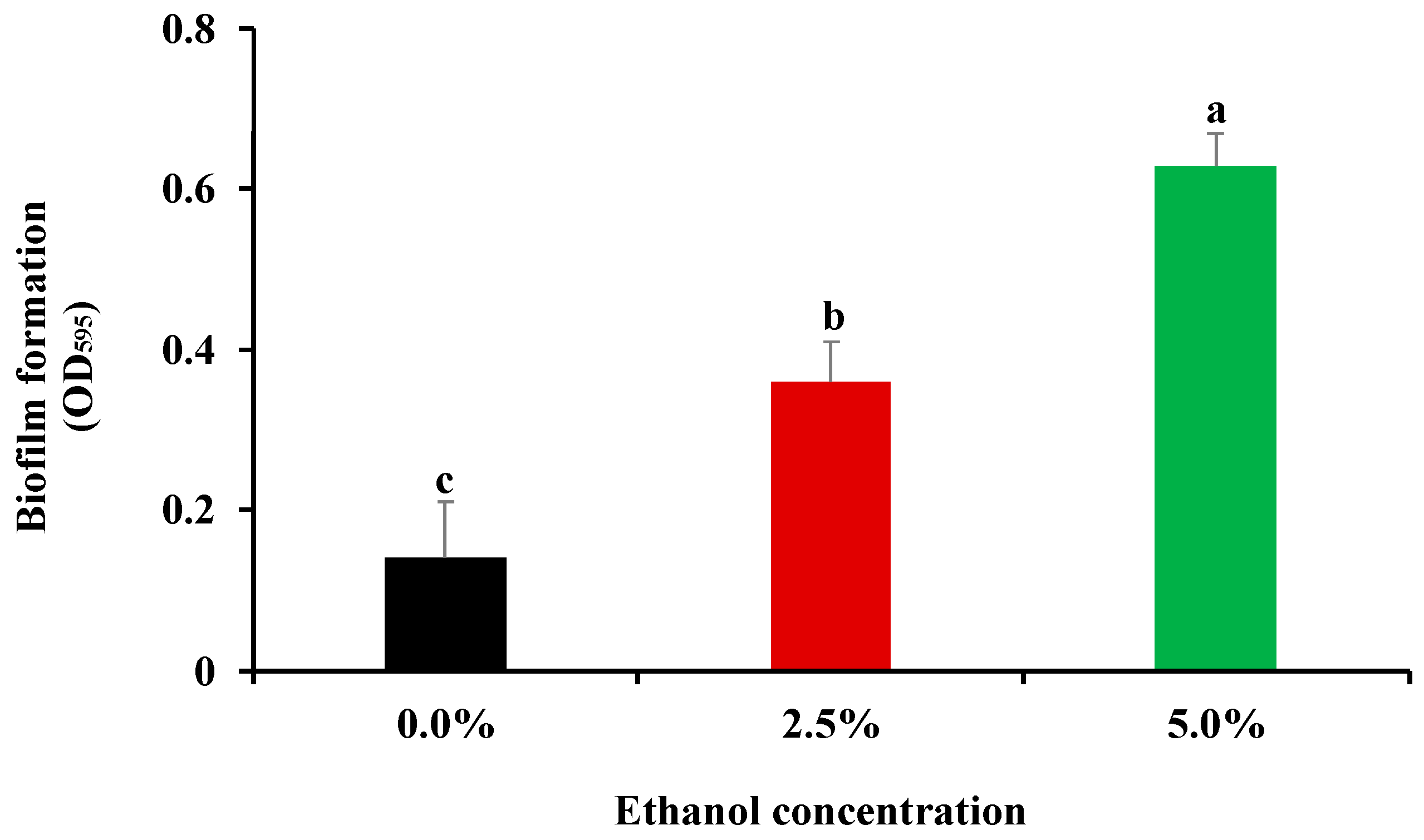
3.1. Influence of Ethanol at Subinhibitory Concentrations on Bacterial Biofilm Formation

Biofilm formation of Salmonella spp. under suboptimal environmental conditions such as acidic pH and high/low temperature has been revealed in previous studies [31,32]. Nevertheless, the capacity of this bacterial pathogen to form biofilms in response to alcoholic disinfectants remains largely unknown. Therefore, the inhibitory activity of ethanol on S. Enteritidis was initially evaluated to screen appropriate concentrations of ethanol for subsequent biofilm formation assay in the current work. The MIC value of ethanol was found to be 10% against S. Enteritidis. Sub-MIC levels of antimicrobials have been considered to exert an inhibitory but not lethal activity against bacterial pathogens [18,33]. Hence, 2.5% (1/4 MIC) and 5.0% (1/2 MIC) were selected as subinhibitory ethanol concentrations for subsequent biofilm formation assay. As shown in Figure 1, exposure to ethanol at subinhibitory levels (2.5% and 5.0%) significantly (p < 0.05) increased biofilm production ability of S. Enteritidis. Moreover, a higher amount of biofilm was produced when ethanol concentration was increased from 2.5% to 5.0%. These results suggest that sublethal ethanol stress stimulated biofilm formation in S. Enteritidis.
Ethanol-mediated biofilm formation has also been assessed in other bacterial pathogens. Tashiro et al. [9] reported that ethanol at low concentrations (1% and 2%) triggered biofilm production by P. aeruginosa. Moreover, biofilm formation ability of S. aureus in tryptic soy broth (TSB) containing 2–8% ethanol was also greater than that in TSB alone [34]. Biofilm growth can provide pathogens with an evaluated degree of resistance to physical and chemical agents commonly applied during food processing, thus serving as a vital source of bacterial contamination in food industries [8]. Therefore, ethanol at low concentrations poses a potential threat to food safety considering its ability to stimulate bacterial biofilm formation.

3.2. Influence of Ethanol at Subinhibitory Concentrations on Cell Attachment Ability

The influence of ethanol at subinhibitory concentrations on bacterial attachment is presented in Figure 2. No significant (p > 0.05) difference in the initial attachment ability was observed among S. Enteritidis cells grown in pure TSB-YE or in TSB-YE supplemented with 2.5% and 5.0% ethanol. Actually, cell adherence to the surface of polystyrene and other substances is the initial step for bacterial pathogens to produce biofilms [35,36]. It was thus indicative that ethanol at subinhibitory levels might not contribute to biofilm formation in S. Enteritidis by affecting its initial attachment in the current work.

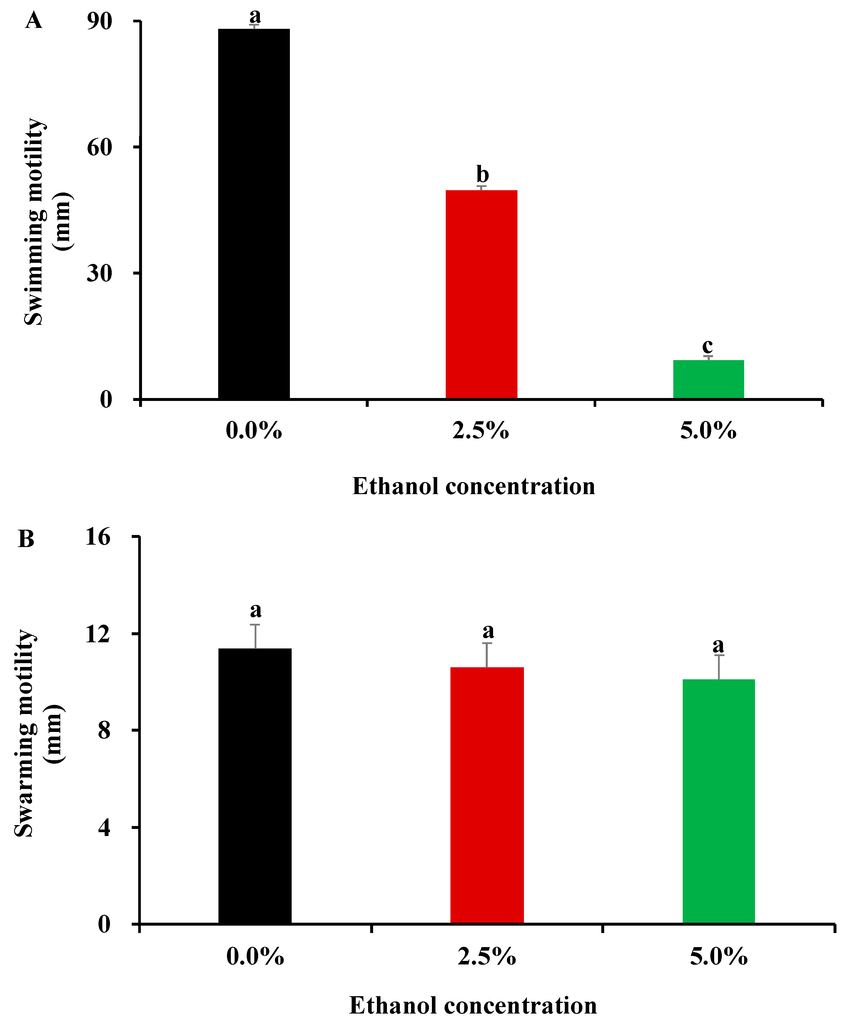
3.3. Influence of Ethanol at Subinhibitory Concentrations on Bacterial Motility Capacity

Bacterial mobility in the presence of ethanol was determined by swimming and swarming assays in the current work. In the swimming assay, S. Enteritidis motility on 0.3% soft-agar plates was partially and completely inhibited by 2.5% and 5.0% ethanol, respectively (Figure 3A). Nevertheless, S. Enteritidis did not alter its swarming ability on 0.6% soft-agar plates under ethanol stress (Figure 3B). Therefore, ethanol at subinhibitory concentrations exerted an inhibitory effect on swimming motility, but not on swarming ability in S. Enteritidis.
Swimming motility is a type of bacterial behavior on semisolid agar media propelled by flagella, which is proven instrumental in biofilm formation [37]. The reduced expression of flagella synthesis genes has been demonstrated to result in marked swimming defect and robust biofilm production in Salmonella spp. [38]. Interestingly, ethanol at subinhibitory concentrations was found to repress the expression of a flagellar basal body protein (FlgF) and a flagellar assembly protein (FliH) in S. Enteritidis in a previous proteomics study [29]. Downregulation of FlgF and FliH might explain the reduced swimming motility and subsequent enhanced biofilm production by S. Enteritidis in the presence of ethanol in the current work.

3.4. Influence of Ethanol at Subinhibitory Concentrations on Bacterial Surface Characteristics

Bacterial surface characteristics (e.g., hydrophobicity and auto-aggregation) were tested to explore their relationship with biofilm formation in S. Enteritidis. As shown in Table 2, ethanol at subinhibitory concentrations did not significantly (p > 0.05) alter bacterial hydrophobicity. On the contrary, S. Enteritidis treated with ethanol displayed significantly (p < 0.05) greater capacity to auto-aggregate than the control group. Moreover, cell auto-aggregation was more pronounced as the concentration of ethanol increased from 2.5% to 5.0% (Table 2). These results indicate a positive correlation between biofilm formation and cell auto-aggregation in S. Enteritidis under ethanol stress.
Auto-aggregation is defined as cell-to-cell interactions wherein bacterial pathogens spontaneously aggregate together [39]. A higher auto-aggregation rate is an indicator of a stronger cell-to-cell interaction, which is beneficial for bacterial pathogens to produce biofilms. Xu et al. [26] observed that auto-aggregation enhanced biofilm formation of several foodborne pathogens (e.g., S. Typhimurium) under NaCl treatment. Therefore, cell-to-cell interactions might be a factor to favor S. Enteritidis biofilm formation under subinhibitory ethanol concentrations in the current work.

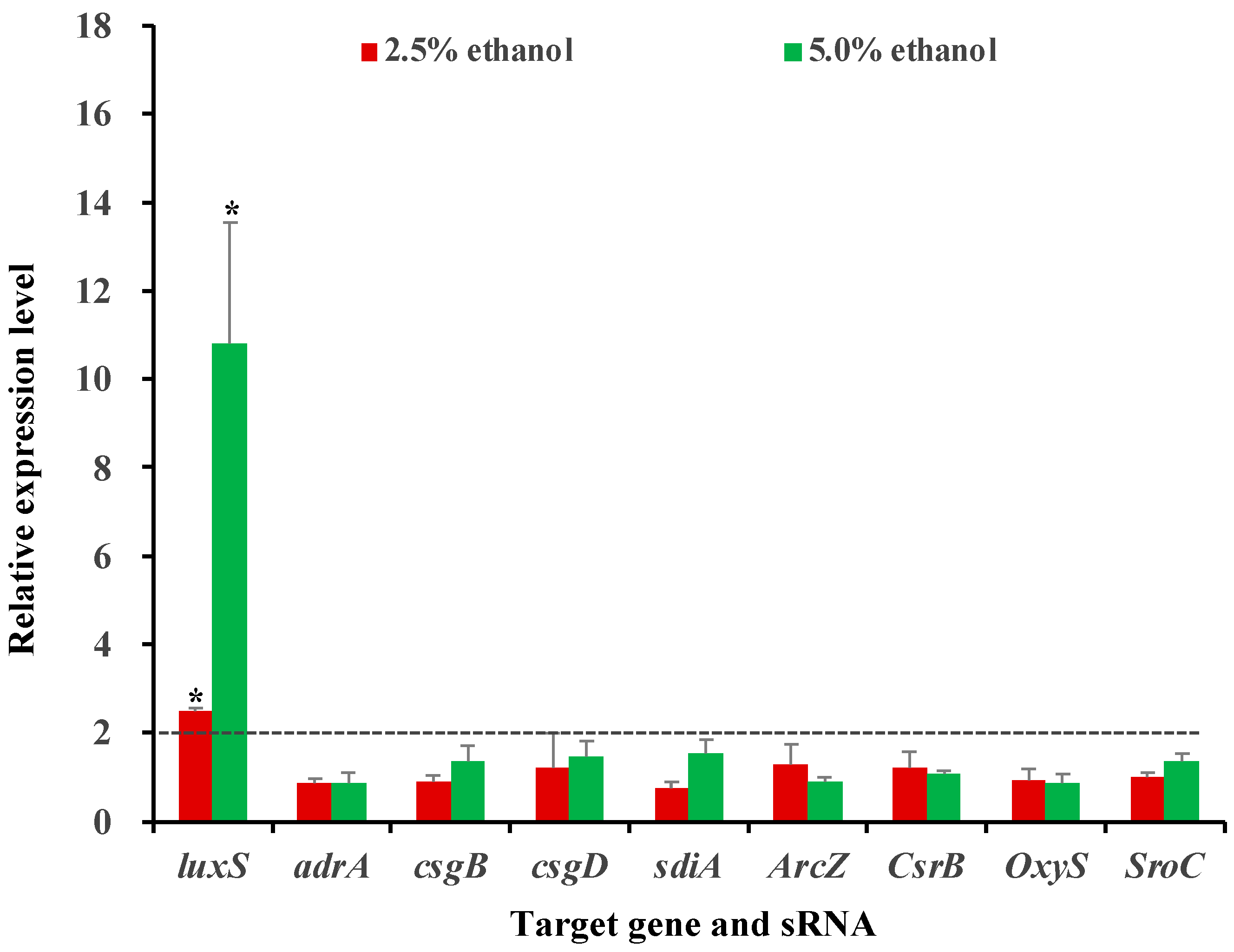
3.5. Influence of Ethanol at Subinhibitory Concentrations on the Expression of Selected Genes and sRNAs

The expression levels of three attachment genes (adrA, csgB, and csgD), two quorum-sensing genes (luxS and sdiA) and four sRNAs (ArcZ, CsrB, OxyS, and SroC) under ethanol stress were determined by RT-qPCR analysis in the current work. Out of these genetic elements, only the luxS gene was significantly (p < 0.05) differentially expressed after exposure to ethanol; this gene was upregulated by 2.49- and 10.08-fold under 2.5% and 5.0% ethanol stresses, respectively. On the contrary, all other tested genes and sRNAs did not show altered expression patterns under ethanol stress in S. Enteritidis (Figure 4).
The aforementioned genetic elements were selected for RT-qPCR test due to their key role in biofilm formation of Salmonella spp. under well-defined laboratory conditions favorable for bacterial growth [14,32]. However, their expression patterns may vary in response to different environmental stimuli [14,32], and it remains largely unknown which elements are involved in bacterial biofilm formation under sub-MIC levels of ethanol. In the current work, three attachment genes (adrA, csgB, and csgD) were not significantly (p > 0.05) differentially expressed in S. Enteritidis, which could be a reason for the remained cell attachment ability under ethanol stress. Similarly, it seemed that four sRNAs (ArcZ, CsrB, OxyS, and SroC) also did not participate in biofilm formation of this pathogen stimulated by ethanol, as revealed by RT-qPCR analysis. However, exposure to the alcohol resulted in the upregulation of the luxS gene, indicating the involvement of this gene in ethanol-induced biofilm formation in S. Enteritidis. In line with this observation, sub-MIC levels of plant-derived quercetin could retard biofilm formation in S. Enteritidis and S. Typhimurium, accompanied by a reduced expression of the luxS gene [17]. It thus seemed that the luxS gene might play a vital role in biofilm formation of Salmonella spp. under some sublethal environmental stresses.
The luxS gene participates in the synthesis of autoinducer-2, which acts as a universal quorum-sensing molecule in bacterial pathogens [24,40]. The LuxS quorum-sensing system offers a means for cell-to-cell communications, which is critical for bacterial biofilm production [41,42]. Deletion of the luxS gene has been demonstrated to significantly (p < 0.05) reduce biofilm production by S. Typhimurium in LB medium [43]. Interestingly, in the current work, ethanol at subinhibitory concentrations induced the expression of the luxS gene, along with enhanced biofilm production in S. Enteritidis. Therefore, the luxS gene might contribute to bacterial biofilm formation under ethanol stress by regulating quorum-sensing processes.
It is noteworthy that the stimulation of biofilm formation by ethanol has been observed in a model organism of S. Enteritidis in the current work. The broader impact of this phenomenon can be addressed by a population-wide survey incorporating more S. Enteritidis isolates in future studies. Moreover, bacterial pathogens are able to exist in multispecies biofilms in food-related environments [44]. Thus, it would also be of importance to further assess ethanol-mediated biofilm formation using a multispecies bacterial cocktail.

4. Conclusions

Ethanol at subinhibitory concentrations could accelerate biofilm production in S. Enteritidis, accompanied by reduced swimming motility and enhanced auto-aggregation ability. Moreover, the luxS gene located on a quorum-sensing system was upregulated in response to sub-MIC levels of ethanol, which suggested the involvement of bacterial quorum-sensing in ethanol-induced biofilm formation. These results demonstrate that alcohols should be used with caution to avoid the presence of subinhibitory ethanol concentrations which may pose a risk to food safety by triggering bacterial biofilm formation.

Author Contributions

Conceptualization, X.S.; methodology, S.H. and Z.Z.; investigation, S.H. and Z.Z.; data curation, S.H. and C.S.; writing—original draft preparation, S.H.; writing—review and editing, C.S., S.W. and X.S.; supervision, X.S.; funding acquisition, S.H. and X.S. All authors have read and agreed to the published version of the manuscript.

Funding

This research was funded by the National Key Research & Development Program of China (No. 2019YFE0119700), the National Natural Science Foundation of China (No. 32001797), the Science and Technology Innovation Agricultural Project of Shanghai Science and Technology Commission (No. 19391902100), the Natural Science Foundation of Shanghai (No. 22ZR1429900), and the Startup Fund for Young Faculty at SJTU (No. 22X010500276).

Institutional Review Board Statement

Not applicable.

Informed Consent Statement

Not applicable.

Data Availability Statement

All data are available in the article.

Conflicts of Interest

The authors declare no conflict of interest.

References

  1. Alzeer, J.; Abou Hadeed, K. Ethanol and its Halal status in food industries. Trends Food Sci. Tech. 2016, 58, 14–20. [Google Scholar] [CrossRef]
  2. Chiou, R.Y.; Phillips, R.D.; Zhao, P.; Doyle, M.P.; Beuchat, L.R. Ethanol-mediated variations in cellular fatty acid composition and protein profiles of two genotypically different strains of Escherichia coli O157: H7. Appl. Environ. Microbiol. 2004, 70, 2204–2210. [Google Scholar] [CrossRef] [Green Version]
  3. Logan, B.K.; Distefano, S. Ethanol content of various foods and soft drinks and their potential for interference with a breath-alcohol test. J. Anal. Toxicol. 1998, 22, 181–183. [Google Scholar] [CrossRef] [Green Version]
  4. Dev Kumar, G.; Mishra, A.; Dunn, L.; Townsend, A.; Oguadinma, I.C.; Bright, K.R.; Gerba, C.P. Biocides and novel antimicrobial agents for the mitigation of coronaviruses. Front. Microbiol. 2020, 11, 1351. [Google Scholar] [CrossRef]
  5. Fagerlund, A.; Møretrø, T.; Heir, E.; Briandet, R.; Langsrud, S. Cleaning and disinfection of biofilms composed of Listeria monocytogenes and background microbiota from meat processing surfaces. Appl. Environ. Microbiol. 2017, 83, e01046-17. [Google Scholar] [CrossRef] [Green Version]
  6. He, S.; Fong, K.; Wang, S.; Shi, X. Ethanol adaptation in foodborne bacterial pathogens. Crit. Rev. Food Sci. Nutr. 2021, 61, 777–787. [Google Scholar] [CrossRef]
  7. Dao, T.; Dantigny, P. Control of food spoilage fungi by ethanol. Food Control 2011, 22, 360–368. [Google Scholar] [CrossRef]
  8. Tango, C.N.; Akkermans, S.; Hussain, M.S.; Khan, I.; Van Impe, J.; Jin, Y.G.; Oh, D.H. Modeling the effect of pH, water activity, and ethanol concentration on biofilm formation of Staphylococcus aureus. Food Microbiol. 2018, 76, 287–295. [Google Scholar] [CrossRef] [PubMed]
  9. Tashiro, Y.; Inagaki, A.; Ono, K.; Inaba, T.; Yawata, Y.; Uchiyama, H.; Nomura, N. Low concentrations of ethanol stimulate biofilm and pellicle formation in Pseudomonas aeruginosa. Biosci. Biotech. Bioch. 2014, 78, 178–181. [Google Scholar] [CrossRef]
  10. Dhivya, R.; Rajakrishnapriya, V.C.; Sruthi, K.; Chidanand, D.V.; Sunil, C.K.; Rawson, A. Biofilm combating in the food industry: Overview, non-thermal approaches, and mechanisms. J. Food Process. Pres. 2022, 00, e16282. [Google Scholar] [CrossRef]
  11. Díez-García, M.; Capita, R.; Alonso-Calleja, C. Influence of serotype on the growth kinetics and the ability to form biofilms of Salmonella isolates from poultry. Food Microbiol. 2012, 31, 173–180. [Google Scholar] [CrossRef]
  12. Wang, H.H.; Ye, K.P.; Zhang, Q.Q.; Dong, Y.; Xu, X.L.; Zhou, G.H. Biofilm formation of meat-borne Salmonella enterica and inhibition by the cell-free supernatant from Pseudomonas aeruginosa. Food Control 2013, 32, 650–658. [Google Scholar] [CrossRef]
  13. Wang, H.; Ding, S.; Dong, Y.; Ye, K.; Xu, X.; Zhou, G. Biofilm formation of Salmonella serotypes in simulated meat processing environments and its relationship to cell characteristics. J. Food Protect. 2013, 76, 1784–1789. [Google Scholar] [CrossRef]
  14. Wang, H.; Dong, Y.; Wang, G.; Xu, X.; Zhou, G. Effect of growth media on gene expression levels in Salmonella Typhimurium biofilm formed on stainless steel surface. Food Control 2016, 59, 546–552. [Google Scholar] [CrossRef]
  15. Lamas, A.; Paz-Mendez, A.M.; Regal, P.; Vazquez, B.; Miranda, J.M.; Cepeda, A.; Franco, C.M. Food preservatives influence biofilm formation, gene expression and small RNAs in Salmonella enterica. LWT-Food Sci. Technol. 2018, 97, 1–8. [Google Scholar] [CrossRef]
  16. Fan, Q.; He, Q.; Zhang, T.; Song, W.; Sheng, Q.; Yuan, Y.; Yue, T. Antibiofilm potential of lactobionic acid against Salmonella Typhimurium. LWT-Food Sci. Technol. 2022, 162, 113461. [Google Scholar] [CrossRef]
  17. Kim, Y.K.; Roy, P.K.; Ashrafudoulla, M.; Nahar, S.; Toushik, S.H.; Hossain, M.I.; Mizan, M.F.R.; Park, S.H.; Ha, S.D. Antibiofilm effects of quercetin against Salmonella enterica biofilm formation and virulence, stress response, and quorum-sensing gene expression. Food Control 2022, 137, 108964. [Google Scholar] [CrossRef]
  18. Song, X.; Sun, Y.; Zhang, Q.; Yang, X.; Zheng, F.; He, S.; Wang, Y. Failure of Staphylococcus aureus to acquire direct and cross tolerance after habituation to cinnamon essential oil. Microorganisms 2019, 7, 18. [Google Scholar] [CrossRef] [Green Version]
  19. Von Hertwig, A.M.; Prestes, F.S.; Nascimento, M.S. Biofilm formation and resistance to sanitizers by Salmonella spp. isolated from the peanut supply chain. Food Res. Int. 2022, 152, 110882. [Google Scholar] [CrossRef]
  20. Dhakal, J.; Sharma, C.S.; Nannapaneni, R.; McDANIEL, C.D.; Kim, T.; Kiess, A. Effect of chlorine-induced sublethal oxidative stress on the biofilm-forming ability of Salmonella at different temperatures, nutrient conditions, and substrates. J. Food Protect. 2019, 82, 78–92. [Google Scholar] [CrossRef]
  21. Obe, T.; Nannapaneni, R.; Sharma, C.S.; Kiess, A. Homologous stress adaptation, antibiotic resistance, and biofilm forming ability of Salmonella enterica serovar Heidelberg ATCC8326 on different food-contact surfaces following exposure to sublethal chlorine concentrations. Poultry Sci. 2018, 97, 951–961. [Google Scholar] [CrossRef] [PubMed]
  22. Nilsson, R.E.; Ross, T.; Bowman, J.P. Variability in biofilm production by Listeria monocytogenes correlated to strain origin and growth conditions. Int. J. Food Microbiol. 2011, 150, 14–24. [Google Scholar] [CrossRef] [PubMed]
  23. Do Valle Gomes, M.Z.; Nitschke, M. Evaluation of rhamnolipid and surfactin to reduce the adhesion and remove biofilms of individual and mixed cultures of food pathogenic bacteria. Food Control 2012, 25, 441–447. [Google Scholar] [CrossRef]
  24. Roy, P.K.; Song, M.G.; Park, S.Y. Impact of quercetin against Salmonella Typhimurium biofilm formation on food-contact surfaces and molecular mechanism pattern. Foods 2022, 11, 977. [Google Scholar] [CrossRef] [PubMed]
  25. Prabhu, Y.; Phale, P.S. Biodegradation of phenanthrene by Pseudomonas sp. strain PP2: Novel metabolic pathway, role of biosurfactant and cell surface hydrophobicity in hydrocarbon assimilation. Appl. Microbiol. Biot. 2003, 61, 342–351. [Google Scholar] [CrossRef]
  26. Xu, H.; Zou, Y.; Lee, H.Y.; Ahn, J. Effect of NaCl on the biofilm formation by foodborne pathogens. J. Food Sci. 2010, 75, M580–M585. [Google Scholar] [CrossRef]
  27. Lee, D.U.; Park, Y.J.; Yu, H.H.; Jung, S.C.; Park, J.H.; Lee, D.H.; Lee, N.K.; Paik, H.D. Antimicrobial and antibiofilm effect of ε-polylysine against Salmonella Enteritidis, Listeria monocytogenes, and Escherichia coli in tryptic soy broth and chicken juice. Foods 2021, 10, 2211. [Google Scholar] [CrossRef]
  28. He, S.; Cui, Y.; Qin, X.; Zhang, F.; Shi, C.; Paoli, G.C.; Shi, X. Influence of ethanol adaptation on Salmonella enterica serovar Enteritidis survival in acidic environments and expression of acid tolerance-related genes. Food Microbiol. 2018, 72, 193–198. [Google Scholar] [CrossRef]
  29. He, S.; Qin, X.; Wong, C.W.; Shi, C.; Wang, S.; Shi, X. Ethanol adaptation strategies in Salmonella enterica serovar Enteritidis revealed by global proteomic and mutagenic analyses. Appl. Environ. Microbiol. 2019, 85, e01107-19. [Google Scholar] [CrossRef] [Green Version]
  30. Livak, K.J.; Schmittgen, T.D. Analysis of relative gene expression data using real-time quantitative PCR and the 2−ΔΔCT method. Methods 2001, 25, 402–408. [Google Scholar] [CrossRef]
  31. Roy, P.K.; Ha, A.J.W.; Mizan, M.F.R.; Hossain, M.I.; Ashrafudoulla, M.; Toushik, S.H.; Nahar, S.; Kim, Y.K.; Ha, S.D. Effects of environmental conditions (temperature, pH, and glucose) on biofilm formation of Salmonella enterica serotype Kentucky and virulence gene expression. Poultry Sci. 2021, 100, 101209. [Google Scholar] [CrossRef] [PubMed]
  32. Lamas, A.; Regal, P.; Vázquez, B.; Miranda, J.M.; Cepeda, A.; Franco, C.M. Influence of milk, chicken residues and oxygen levels on biofilm formation on stainless steel, gene expression and small RNAs in Salmonella enterica. Food Control 2018, 90, 1–9. [Google Scholar] [CrossRef]
  33. Ranieri, M.R.; Whitchurch, C.B.; Burrows, L.L. Mechanisms of biofilm stimulation by subinhibitory concentrations of antimicrobials. Curr. Opin. Microbiol. 2018, 45, 164–169. [Google Scholar] [CrossRef] [PubMed]
  34. Kim, B.R.; Bae, Y.M.; Lee, S.Y. Effect of environmental conditions on biofilm formation and related characteristics of Staphylococcus aureus. J. Food Saf. 2016, 36, 412–422. [Google Scholar] [CrossRef]
  35. Chang, Y.; Gu, W.; McLandsborough, L. Low concentration of ethylenediaminetetraacetic acid (EDTA) affects biofilm formation of Listeria monocytogenes by inhibiting its initial adherence. Food Microbiol. 2012, 29, 10–17. [Google Scholar] [CrossRef]
  36. Chathoth, K.; Fostier, L.; Martin, B.; Baysse, C.; Mahé, F. A multi-skilled mathematical model of bacterial attachment in initiation of biofilms. Microorganisms 2022, 10, 686. [Google Scholar] [CrossRef]
  37. Li, Y.; Bai, F.; Xia, H.; Zhuang, L.; Xu, H.; Jin, Y.; Zhang, X.; Bai, Y.; Qiao, M. A novel regulator PA5022 (aefA) is involved in swimming motility, biofilm formation and elastase activity of Pseudomonas aeruginosa. Microbiol. Res. 2015, 176, 14–20. [Google Scholar] [CrossRef]
  38. Zhou, X.; Liu, B.; Shi, C.; Shi, X. Mutation of a Salmonella serogroup-C1-specific gene abrogates O7-antigen biosynthesis and triggers NaCl-dependent motility deficiency. PLoS ONE 2014, 9, e106708. [Google Scholar] [CrossRef] [Green Version]
  39. Wu, D.; Ding, X.; Zhao, B.; An, Q.; Guo, J. The essential role of hydrophobic interaction within extracellular polymeric substances in auto-aggregation of P. stutzeri strain XL-2. Int. Biodeter. Biodegr. 2022, 171, 105404. [Google Scholar] [CrossRef]
  40. Zhang, Y.; Yu, H.; Xie, Y.; Guo, Y.; Cheng, Y.; Yao, W. Quorum sensing inhibitory effect of hexanal on Autoinducer-2 (AI-2) and corresponding impacts on biofilm formation and enzyme activity in Erwinia carotovora and Pseudomonas fluorescens isolated from vegetables. J. Food Process. Pres. 2022, 46, e16293. [Google Scholar] [CrossRef]
  41. Ju, X.; Li, J.; Zhu, M.; Lu, Z.; Lv, F.; Zhu, X.; Bie, X. Effect of the luxS gene on biofilm formation and antibiotic resistance by Salmonella serovar Dublin. Food Res. Int. 2018, 107, 385–393. [Google Scholar] [CrossRef] [PubMed]
  42. Yu, T.; Ma, M.; Sun, Y.; Xu, X.; Qiu, S.; Yin, J.; Chen, L. The effect of sublethal concentrations of benzalkonium chloride on the LuxS/AI-2 quorum sensing system, biofilm formation and motility of Escherichia coli. Int. J. Food Microbiol. 2021, 353, 109313. [Google Scholar] [CrossRef]
  43. Jesudhasan, P.R.; Cepeda, M.L.; Widmer, K.; Dowd, S.E.; Soni, K.A.; Hume, M.E.; Zhu, J.; Pillai, S.D. Transcriptome analysis of genes controlled by luxS/autoinducer-2 in Salmonella enterica serovar Typhimurium. Foodborne Pathog. Dis. 2010, 7, 399–410. [Google Scholar] [CrossRef] [PubMed] [Green Version]
  44. Carrascosa, C.; Raheem, D.; Ramos, F.; Saraiva, A.; Raposo, A. Microbial biofilms in the food industry-a comprehensive review. Int. J. Env. Res. Pub. He. 2021, 18, 2014. [Google Scholar] [CrossRef] [PubMed]
Figure 1. Influence of subinhibitory concentrations of ethanol (1/4 MIC, 2.5%; 1/2 MIC, 5.0%) on biofilm formation of S. Enteritidis. Different letters above vertical bars represent a significant difference (p < 0.05).
Foods 11 02237 g001
Figure 2. Influence of subinhibitory concentrations of ethanol (1/4 MIC, 2.5%; 1/2 MIC, 5.0%) on cell attachment of S. Enteritidis. The same letters above vertical bars represent no significant difference (p > 0.05).
Foods 11 02237 g002
Figure 3. Influence of subinhibitory concentrations of ethanol (1/4 MIC, 2.5%; 1/2 MIC, 5.0%) on cell motility of S. Enteritidis. (A) Swimming motility; (B) Swarming motility. Different letters above vertical bars represent a significant difference (p < 0.05).
Foods 11 02237 g003
Figure 4. Relative expression levels of selected genes and sRNAs under subinhibitory ethanol concentrations (1/4 MIC, 2.5%; 1/2 MIC, 5.0%). Stars (*) above vertical bars represent a significant difference (p < 0.05) in gene expression.
Foods 11 02237 g004
Table 1. Primer sequences of biofilm-related genes and sRNAs.
Gene/sRNANCBI Accession No. or Gene IDSequence (5′ to 3′)Reference
luxSCAR34242.1F: ATGCCATTATTAGATAGCTT[14]
R: GAGATGGTCGCGCATAAAGCCAGC
adrACAR31954.1F: GAAGCTCGTCGCTGGAAGTC[15]
R: TTCCGCTTAATTTAATGGCCG
csgBCAR33485.1F: TCCTGGTCTTCAGTAGCGTAA[14]
R: TATGATGGAAGCGGATAAGAA
csgDCAR33486.1F: TCCTGGTCTTCAGTAGCGTAA[15]
R: TATGATGGAAGCGGATAAGAA
sdiACAR32642.1F: AATATCGCTTCGTACCAC[15]
R: GTAGGTAAACGAGGAGCAG
ArcZ2847690F: ACTGCGCCTTTGACATCATC[15]
R: CGAATACTGCGCCAACACCA
CsrB1254489F: CAAAGTGGAAAGCGCAGGAT[15]
R: TGACCTTACGGCCTGTTCAT
OxyS6797054F: TAACCCTTGAAGACACCGCC[15]
R: ACCAGAGGTCCGCAAAAGTT
SroC6793706F: GGGACTCCTGTCCTCTCGAT[15]
R: CAGCGCTACCCTCGAAGATT
16S rRNAX80676.1F: AGGCCTTCGGGTTGTAAAGT[15]
R: GTTAGCCGGTGCTTCTTCTG
Table 2. Influence of subinhibitory concentrations of ethanol (1/4 MIC, 2.5%; 1/2 MIC, 5.0%) on cell surface characteristics of S. Enteritidis.
Properties0.0% Ethanol2.5% Ethanol5.0% Ethanol
Hydrophobicity (%)3.92 ± 1.33 a4.98 ± 0.65 a3.60 ± 1.16 a
Auto-aggregation (%)40.53 ± 1.35 c58.92 ± 1.06 b72.32 ± 0.45 a
Note: Different letters in a row represent a significant difference (p < 0.05).
Publisher’s Note: MDPI stays neutral with regard to jurisdictional claims in published maps and institutional affiliations.

