Article

Efficacy of 405 nm Light-Emitting Diode Illumination and Citral Used Alone and in Combination for Inactivation of Vibrio parahaemolyticus on Shrimp

1
College of Food Science and Engineering, Northwest A&F University, Xianyang 712100, China
2
School of Food Science and Technology, National Engineering Research Center of Seafood, Dalian Polytechnic University, Dalian 116034, China
*
Author to whom correspondence should be addressed.
Foods 2022, 11(14), 2008; https://doi.org/10.3390/foods11142008
Submission received: 16 June 2022 / Revised: 29 June 2022 / Accepted: 30 June 2022 / Published: 7 July 2022

Abstract

:
Vibrio parahaemolyticus is a widely distributed pathogen, which is frequently the lead cause of infections related to seafood consumption. The objective of the present study was to investigate the antimicrobial effect of the combination of 405 nm light-emitting diode (LED) and citral on V. parahaemolyticus. The antimicrobial effect of LED illumination and citral was evaluated on V. parahaemolyticus not only in phosphate-buffered saline (PBS) but also on shrimp. Quality changes of shrimp were determined by sensory evaluation. Changes in bacteria cell membrane morphology, cell membrane permeability, cell lipid oxidation level, and DNA degradation were examined to provide insights into the antimicrobial mechanism. The combination of LED treatments and citral had better antimicrobial effects than either treatment alone. LED combined with 0.1 mg/mL of citral effectively reduced V. parahaemolyticus from 6.5 log CFU/mL to below the detection limit in PBS. Combined treatment caused a 3.5 log reduction of the pathogen on shrimp within 20 min and a 6 log reduction within 2 h without significant changes in the sensory score. Furthermore, combined LED and citral treatment affected V. parahaemolyticus cellular morphology and outer membrane integrity. The profile of the comet assay and DNA fragmentation analysis revealed that combination treatment did not cause a breakdown of bacterial genomic DNA. In conclusion, LED may act synergistically with citral. They have the potential to be developed as novel microbial intervention strategies.

1. Introduction

The Gram-negative bacterium Vibrio parahaemolyticus is a natural constituent of fresh water, estuarine, and marine environments [1]. It is the primary cause of bacterial food poisoning in coastal areas, mostly related to contaminated seafood consumption [2]. As a widely consumed seafood, shrimps are often contaminated with V. parahaemolyticus. Previous studies have isolated and characterized various V. parahaemolyticus strains from commonly consumed fresh shrimps in domestic markets in China [3,4]. These studies provided data in support of the high risk of V. parahaemolyticus contamination in shrimp. After eating uncooked seafood contaminated by V. parahaemolyticus, people could develop acute gastroenteritis within 24 h. In addition, V. parahaemolyticus also causes nausea, vomiting, diarrhea, fever, and other symptoms [2].
Various methods have been used for microbial control of shrimp, such as the use of chlorine, modified atmosphere packaging, and irradiation [5]. As a very common strategy to control pathogens in seafood, the effect of chlorine to inactivate Vibrio cells on shrimp has been reported [6]. However, a long contact time and high concentration of chlorine are required to destroy pathogens completely, which may cause health problems in workers, such as severe respiratory tract damage and permanently reduced lung function [7]. Furthermore, the seafoods treated with chlorine often have an undesirable smell with low consumer acceptance. Therefore, an environmentally friendly and safe method should be developed as an alternative to chlorine in the seafood industry.
Recently, many research studies have been focused on the antibacterial effect of artificial light treatments with different wavelengths in agriculture and the food industry. As an alternative energy-saving light source, light-emitting diodes (LED) have attracted attraction in the microbiological safety field [8]. As a conventional approach, UV light (wavelength < 400 nm) emitted by mercury vapor lamps is becoming a worldwide recognized strategy to against potential pathogens such as bacteria, yeasts, viruses, and fungi [9]. However, UV light has shortcomings, such as its decolorization effects on food products, and its potential health risk for the operator [10,11]. Compared to traditional visible light sources, LEDs with wavelengths in the range of 400–420 nm (blue light) have several advantages, such as low cost, high durability, low energy consumption, and a significant antibacterial effect [9]. Recent studies have proven the efficacy of 405 nm LED against various foodborne pathogens, such as Escherichia coli O157:H7, Salmonella Typhimurium, L. monocytogenes, S. aureus, and V. parahaemolyticus [12,13,14]. In addition, 405 nm LED has exhibited promising antimicrobial effectiveness in food such as fresh-cut papaya, fresh-cut mango, and ready-to-eat fresh salmon [15,16,17]. These results have been correlated with the photodynamic inactivation of bacteria (PDI) [18]. Some intracellular molecules known as photosensitizers will absorb oxygen and produce reactive oxygen species (ROS) under 400–420 nm LED irradiation [18,19]. These in turn react with cell membranes’ lipids, enzymes, proteins, or DNA, leading to bacterial death [20].
Although the antimicrobial activity of 405 nm LED illumination has been widely studied, the limitations on the application of LED systems have also been found. In food production environments, 405 nm LED takes a long time to completely sterilize food matrices, causing food sensory and nutritional quality loss. Recently, Josewin et al. [21] demonstrated that the synergistic effects of an LED and riboflavin (100 μM) produced a 1.2 log reduction of L. monocytogenes on smoked salmon at 4 °C, which was more effective than either treatment alone. Thus, an additional hurdle combined with 405 nm LED illumination should be developed for the improvement of sterilization efficiency with less impact on food quality. Plant-derived compounds have been extensively used as flavoring agents and many exhibit a wide spectrum of antimicrobial activity [22]. The challenge for the practical application of plant origin antimicrobial agents is to exploit optimized low-dose combined processes to minimize toxicological effects and sensory changes [23]. Citral (C10H16O) is one of the essential oil compounds originating from herbal plants such as lemon myrtle, lemongrass, orange, lime, and bergamot [24]. It has been recognized as a safe food additive according to the National Food Safety Standard for Uses of Food Additives (GB 2760-2011) [25] and approved by the Food and Drug Administration of the United States (FDA) as a safe ingredient [26]. It has been reported to exert antimicrobial effects on Campylobacter jejuni, E. coli O157, L. monocytogenes, Bacillus cereus, S. aureus, and Cronobacter sakazakii in food matrices [27,28,29,30]. Additionally, there is increasing evidence that citral has potential anti-inflammatory and anti-corrosive effects [31,32].
These findings indicate that the use of LEDs or citral is a promising alternative to traditional preservation technologies for reducing the potential risk of food safety. However, little is known about the inactivating effect of the combination of LEDs and citral. The objective of this study was to examine the antimicrobial efficiency of the combination of 405 nm LED irradiation and citral against V. parahaemolyticus on fresh shrimp, as a novel application for seafood preservation. Moreover, it’s possible antimicrobial mechanism was also elucidated by investigating the cell morphology, cell membrane permeability, as well as DNA degradation.

2. Materials and Methods

2.1. Reagents

Citral (CAS 5392-40-5) was obtained from LGC Labor GmbH (Germany) at a HPLC purity of at least 99%. Citral was dissolved in dimethyl sulfoxide (DMSO; Tianjin Kemiou Chemical Reagent Co., Ltd., Tianjin, China) before being used. The final concentration of DMSO in all treated and control sample solutions was 0.5% (v/v). Fresh shrimp (Litopenaeus vannamei) was purchased from a local retail market in Yangling, China.

2.2. Bacterial Strains and Culture Conditions

V. parahaemolyticus ATCC 17802 was purchased from the American Type Culture Collection (ATCC, Manassas, VA, USA). V. parahaemolyticus 240 and V. parahaemolyticus 245 were seafood isolates obtained from the Food Safety and Technology Research Centre in The Hong Kong Polytechnic University. Frozen cultures were recovered from the cryovial by surface spreading on plates of Tryptone soya agar (TSA; Beijing Land Bridge Technology Co., Ltd., Beijing, China), supplemented with 3% (w/v) NaCl, and were incubated for 24 h at 37 °C. To ensure strain purity, a single colony on TSA was cultured into Tryptone Soya Broth (TSB; Beijing Land Bridge Technology Co., Ltd., China) supplemented with 3% NaCl, incubated with shaking at 130 rpm overnight at 37 °C.

2.3. Light-Emitting Diode (LED) Illumination System

A slightly modified version of the illumination system comprising 405 nm LED was confirmed with the method of Zheng et al. [33], with slight modifications. Initially, the top of a heat sink with a cooling fan attached with 405 nm LED was used for minimizing the thermal production of the LED chip and preventing its thermal damage. To avoid the entry of extraneous light, an ABS (acrylonitrile butadiene styrene) housing was utilized to accommodate the system assembly. During LED illumination, the bacterial suspension below the system was subjected to temperature surveillance once every 3 min with a thermocouple thermometer (Everett, WA, USA).
The light dosage was calculated based on Equation (1) [14]. Derivation of the equation was based on the fact that an inverse-square law is obeyed by the intensity of point source-irradiated light or other linear waves:
P = 2 π I 0 h 2 ( 1 1 1 + ( r h ) 2 ) ,
where P (mW) refers to the quantity of energy falling on the plate every second, I0 denotes the intensity reading at the plate center from a mobile LED radiometer (Linshang, Shenzhen, China), r (cm) represents the radius of the plate, and h (cm) refers to the LED (point source) height from the plate center. In this study, I0 = 3.23 mW/cm2, r = h = 4.5 cm, and P = 18.94 ± 0.05 mW/cm2
The computational formula for dose, D (energy density) (J/cm2), is shown below:
D = P A t ,
where A represents the overall area of the plate (cm2), and t refers to the time (seconds).

2.4. Temperature Control

The temperature of the PBS bacterial suspension treated with LED increased due to the thermal effect of LED irradiation [14]. To monitor the temperature conditions, activated V. parahaemolyticus ATCC 17802, and isolates 240 and 245 were mixed equally. The ten milliliters of mixed bacterial liquid were placed in a sterile dish (d = 90 mm) and measured with a thermoelectric thermometer. The temperature changes of the fresh shrimp surface were also monitored with the thermometer (Hebei Shuangqiao Instrument Factory, Hengshui, China). The LED assembly was placed in a refrigerator at 4 °C and the temperature of bacterial suspension and fresh shrimp was recorded every 3 min for 120 min. The 405 nm LED elevated the temperature rapidly from the set temperature of 4.0 °C to 9.4 and 8.9 °C of the bacterial suspension and fresh shrimp, respectively. For this reason, non-illuminated control experiments of PBS and shrimp were carried out at 9 °C to eliminate the effect of temperature rise during the irradiation.

2.5. Antimicrobial Activity of LED Combined with Citral against V. parahaemolyticus in PBS

The three working cultures of V. parahaemolyticus (ATCC 17802, isolate 240, isolate 245) were centrifuged (Eppendorf, Hamburg, Germany) at 8000× g for 5 min following washing twice with PBS (pH 7.2). The resultant pellets were re-suspended in PBS to make up an initial population of approximately 106 CFU/mL. Three working cultures were mixed to obtain the final bacterial suspension.
In the LED and the LED plus 0.1 mg/mL citral groups, 10 mL of the bacterial suspension was placed in a glass petri dish (d = 60 mm) and then illuminated for 1 h at 4 °C. Experiments of control and citral groups were performed with the same amount of bacterial suspension but placed in an incubator at 9 °C. An aliquot of 100 μL was withdrawn at 0, 2, 5, 10, 20, 30, and 60 min, and these aliquots were plated onto TSA supplemented with 3% NaCl, followed by incubation at 37 °C for 24 h and counting.

2.6. Antibacterial Effect of LED Combined with Citral against V. parahaemolyticus on Shrimp

One hundred fresh shrimps were collected from a retail market, frozen, and stored in a −20 °C refrigerator. Individual shrimp had an average weight of 15.0 g and a length of 13.0 cm. The difference of shrimp length and weight of all experimental samples did not exceed ±10%. Briefly, shrimp with head and shell were washed with sterile water and immersed in 0.02% (v/v) NaClO disinfectant for 10 min under aseptic conditions. Then, residual NaClO was removed from shrimp by washing twice with sterile water and the surface of the samples was wiped with a sterilized paper towel. Bacterial suspension of V. parahaemolyticus strains ATCC 17802, 240, and 245 was prepared according to the method in Section 2.5. Citral was dissolved in DMSO, then 50 μL of citral solution was added into the bacterial solution of the LED plus citral group and the citral group. The concentration of citral in the bacterial solution of both groups was 0.1 mg/mL. The same dose of DMSO was added to the control group and the LED treatment. The shrimp samples were then immersed into bacterial suspensions and mixed, followed by being placed in sterile petri dishes. The dishes were placed under LED illumination at 4 °C, while the treatments without illumination were set at 9 °C to offset the rise in temperature generated by LED irradiation. Samples were withdrawn at 0, 5, 10, 20, 60, and 120 min post-inoculation. After vortexing for 2 min in PBS solution, aliquots of 100 μL of the dilution series were plated on TSA supplemented with 3% NaCl medium and incubated at 37 °C for 24 h.

2.7. Sensory Evaluation for Fresh Shrimp by Trained Panel

Fresh shrimp samples were prepared as described in Section 2.6. Shrimps of each group were treated with 405 nm LED, 0.1 mg/mL of citral, and 405 nm LED plus 0.1 mg/mL of citral for 120 min, respectively. Then, the sensory quality of shrimp samples was evaluated as described by Jeyasekaran et al. [34] with some modifications, as shown in Table S1. The 10 trained sensory assessors carried out sensory evaluation in terms of the smell, appearance, and texture of the samples, respectively. Then, the 3 parameters were added together as the overall acceptance. The total scores were between 18 points (extremely fresh) and 3 points (totally corrupted).

2.8. Bacterial Morphology

The cell morphology of V. parahaemolyticus ATCC 17802 was observed and recorded by FESEM (S-4800; Hitachi, Tokyo, Japan). The bacterial suspension of the four groups (control, LED-illuminated, 0.1 mg/mL citral, and LED-illuminated + 0.1 mg/mL citral) was washed twice with PBS buffer and added with 2.5% (v/v) glutaraldehyde solution (prepared with PBS buffer), sealed with plastic film, and fixed for 10 h at 4 °C. Suspension was washed with PBS solution and sterile water, then added with 1% osmium acid, followed by fixing for 5 h. The bacteria were gradient eluted with 30%, 50%, 70%, 80%, and 90% ethanol, followed by dissolving in ethanol. The droplets of the final suspension were added on the sterilized round cover slides and transferred into a centrifugal tube for air-drying. The slides were dried in a high vacuum, coated with gold, and observed at 20 kV.

2.9. Determination of Bacterial Outer Membrane Integrity

To elucidate the antimicrobial mechanism of LED illumination and citral against V. parahaemolyticus, bacterial outer membrane integrity was performed in accordance with the method of Shi et al. [29], with minor modifications. LIVE/DEAD® BacLight Viability Kit L-7007 (Molecular Probes, Eugene, OR, USA) was used, which encompassed SYTO®9 (green fluorescent dye) and PI (propidium iodide, red fluorescent dye). When bacterial suspension was incubated with these nucleic acid dyes, green fluorescence was produced from the viable bacteria having intact cellular membranes, whereas red fluorescence was produced from the dead bacteria with compromised membranes. Briefly, the overnight culture of V. parahaemolyticus ATCC 17802 was collected and resuspended using 0.85% NaCl. For harvesting of live and dead cells, 1 mL of bacteria suspension was added separately into 20 mL of 0.85% NaCl and 20 mL of 70% isopropyl alcohol. After incubating at 25 °C for 1 h, the optical density of both samples was adjusted at 600 nm to 0.5. Bacterial suspensions were formulated by mixing varying percentages of viable cells (0, 10%, 50%, 90%, and 100%) with non-viable cells, and the standard curve was plotted. Cells treated with 405 nm LED and 0.1 mg/mL of citral for 30 min were centrifuged, followed by pipetting 100 μL of samples into 96-well black, opaque microtiter plates. Each well was pipetted with 100 μL aliquots of the 2× working staining solution, and thorough mixing proceeded. The mixture was incubated at 25 °C for 10 min in the dark. Dyes trapped inside the cells were immediately measured using InfiniteTM M200 PRO (TECAN, Mannedorf, Switzerland), and set of fluorochrome filters: excitation wavelength (485 nm), emission wavelength SYTO®9 (542 nm), and PI (610 nm).

2.10. Comet Assay

Bacterial suspensions of V. parahaemolyticus ATCC 17802 were prepared according to the method of Kim et al. [12] with a slight modification, using the Comet Assay Kit (Abcam ab238544, Shanghai, China). Here, 75 μL of the aliquot was pipetted on comet slides, then slides were placed at 4 °C for 15 min to fix the agarose. Thereafter, a 1 h immersion of the comet slides was accomplished in pH 10 Lysis Buffer under dark conditions at 4 °C, and then a further 30 min immersion proceeded in alkaline solution for the slides under the same conditions. Electrophoresis was performed for 20 min (12 V, 100 mA) using alkaline electrophoresis buffer. The slides were then subjected sequentially to three washings with distilled water, each for 2 min, a 10 min dehydration using 70% (v/v) chilled ethanol, drying in air, and subsequent staining with 100 μL of Vista Green DNA Dye staining solution for each well. A Leica DM6 B epifluorescent microscope (Wetzlar, Germany) was utilized in conjunction with Vista Green DNA Dye (WB, 450–480 nm) for acquisition of micrographs at a magnification of 1000×.

2.11. DNA Fragmentation Analysis

V. parahaemolyticus ATCC 17802 DNA fragmentation following the combination of 405 nm LED treatment and 0.1 mg/mL of citral was analyzed according to the method of Kim et al. [12] with a slight modification, using the TaKaRa MiniBEST Bacteria Genomic DNA Extraction Kit (TAKARA BIO INC). The purified DNA was dissolved in 150 µL of Elution Buffer. The 40 mL 1% (w/v) agarose gel solution was boiled 3 times, cooled for a moment, and added with 4 µL of GelRed nucleic acid dye. Samples with loading buffer (5:1, v/v) were electrophoresed at 120 V for 30 min. The gel was visualized with GELDOC XR+ (Bio-Rad Laboratories, Co., Ltd., Shanghai, China).

2.12. Statistical Analysis

Significant differences in the mean value were calculated at the 95% confidence interval (p < 0.05) using one-way analysis of variance (ANOVA). The Kruskal–Wallis test was used for a comparison of sensory scores. All statistical analysis was performed using the IBM SPSS statistical software (version 19.0; SPSS Inc., Chicago, IL, USA). Data were expressed as mean ± standard deviation (SD) (n = 6).

3. Results

3.1. Bacterial Inactivation by Combination of LED Illumination and Citral in PBS

The inactivation effects of 405 nm LED and 0.1 mg/mL of citral against V. parahaemolyticus in PBS were investigated and the results are shown in Table 1. The population of non-illuminated cells in the control group remained stable within 1 h, at 3.16 × 10⁶ CFU/mL. The population of citral-treated cells showed a 1.5 log (CFU/mL) reduction after 1 h. The inactivation of V. parahaemolyticus treated with the LED illumination and 0.1 mg/mL of citral was most effective compared to the other treatments, producing a severe reduction of 6.5 log after 5 min. The number of viable cells was lower than the detectable limit after 10 and 5 min of LED treatment and LED combined with citral treatment, respectively.

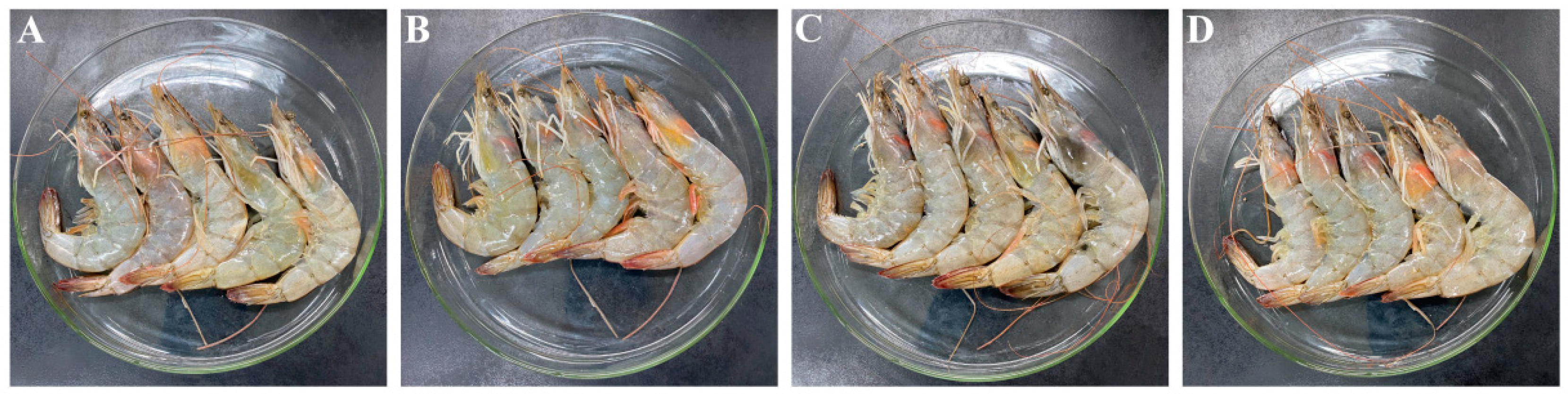
3.2. Bacterial Inactivation by Combination of LED Illumination and Citral on Shrimp

Shrimp were inoculated with bacterial suspension and observed for 2 h, as shown in Table 2. After 2 h of LED illumination, the bacterial population reduced about 3 log (CFU/mL). Additionally, the presence of 0.1 mg/mL of citral led to a 2 log reduction in the population of V. parahaemolyticus on shrimp. The inactivation of V. parahaemolyticus through LED illumination treatment in combination with 0.1 mg/mL of citral was most effective, and the number of bacteria decreased to 3.5 log (CFU/mL) within 20 min and fell below the detection limit (10 CFU/mL) after 2 h.

3.3. Sensory Evaluation for Shrimp

The effect of 405 nm LED and 0.1 mg/mL of citral on shrimp is shown in Figure 1. The overall sensory scores of shrimp with difference treatments are shown in Table 3. The statistical analysis of the overall acceptance showed no significant difference (p > 0.05) between samples. Although the combination of LED and the citral treatment sample had obtained lower scores for the odor attribute (5.0 ± 0.4), the overall acceptance of all shrimp samples was higher than 15 points with all treatments, indicating that the three treatments did not affect the overall sensory quality of the raw shrimp.

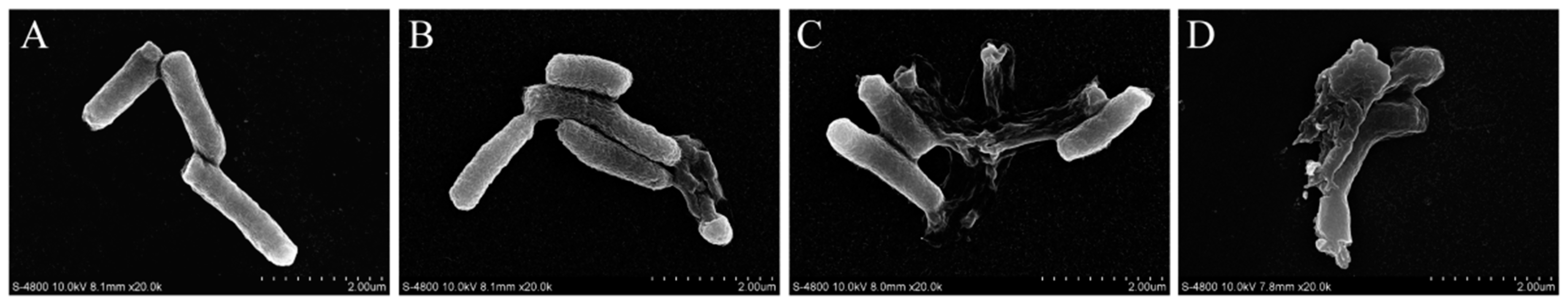
3.4. FESEM Observations

As shown in Figure 2, the shape of untreated cells was rod-shaped and smooth, while LED-treated cells were ruptured after 10 min of exposure (Figure 2B). Cells showed shrinkage with the presence of 0.1 mg/mL of citral (Figure 2C). When cells were treated with LED and citral, cells showed significant shrinkage, with some cells breaking into pieces (Figure 2D).

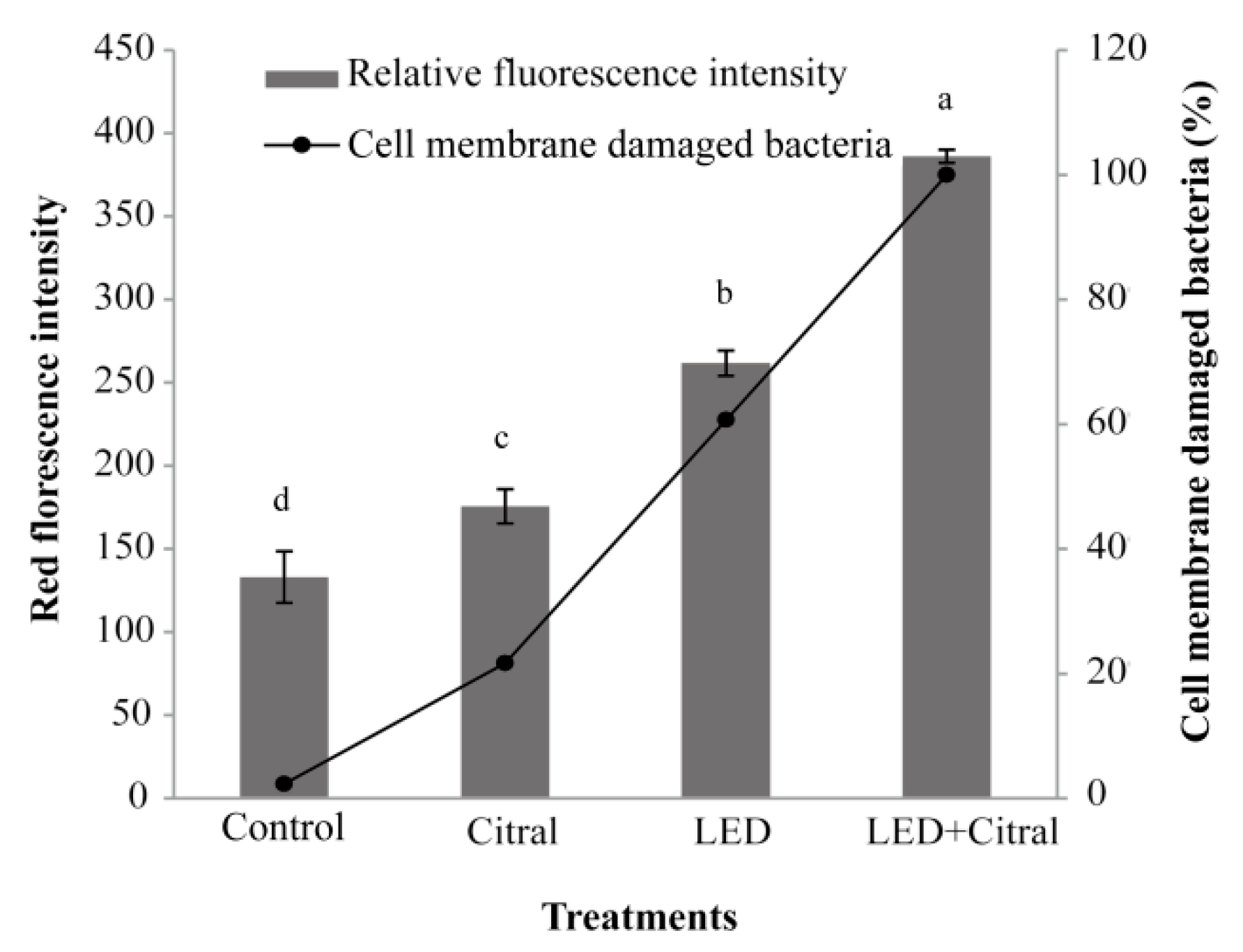
3.5. Effect of 405 nm LED Illumination and Citral on Outer Membrane Integrity
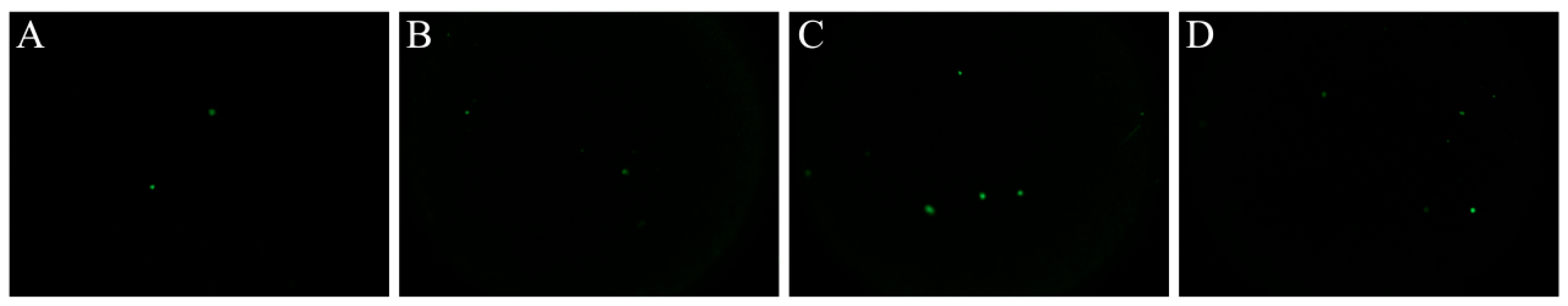
The bacterial suspensions were prepared by mixing 0%, 10%, 50%, 90%, and 100% of the volume of live bacteria with the dead bacteria, respectively. The standard curve was set properly. As shown in Figure 3, LED illumination and 0.1 mg/mL of citral treatment had certain effects on the cell membrane permeability of V. parahaemolyticus. The relative intensity of red fluorescence of the citral group was 175 ± 9.6 (A.U), while the relative intensity of red fluorescence of the LED group was 261 ± 7.3 (A.U), and the relative red fluorescence intensity of LED and 0.1 mg/mL of citral was 385 ± 4.3 (A.U). After being treated with LED illumination combined with citral, the proportion of dead bacteria reached 100%.

3.6. Comet Assay

The comet assay was adopted for determining whether 405 nm LED irradiation and 0.1 mg/mL of citral addition would lead to DNA degradation. As shown in Figure 4, only clear heads were found in both LED-treated and untreated cells, suggesting that LED irradiation did not cause DNA breakage. Similarly, no tails (comets) were observed in citral and the LED combined with citral treatments. All single-cell electrophoresis photos observed by microscopy presented clear zones of the nucleus in cells without DNA tails.

3.7. DNA Fragmentation Analysis

There was only one positive band present at 2000 bp in all DNA ladder profiles (Figure 5). No DNA fragments were observed, and the DNA migration bands of all samples were the same, indicating that no differences were observed in total genomic DNA among untreated, LED illumination, 0.1 mg/mL citral treatment, and the combination of LED and citral treatment cells. These results indicated that the three sterilization treatments did not induce DNA breakage in V. parahaemolyticus.

4. Discussion

The present study detected the effectiveness of 405 nm LED combined with 0.1 mg/mL of citral in inactivating V. parahaemolyticus on the shrimp surface to see if the LED technology has a potential to be applied to seafood preservation. Moreover, the bacterial membrane damage and DNA breakage of V. parahaemolyticus were determined to reveal the mechanism of the inactivation by 405 nm LED and citral.
LED with a specific wavelength (405 nm) has been proven to have a microbial inactivation effect [10,35,36]. The optimized LED sterilizing device in this study has a power of 20 W, a wavelength of 405 ± 5 nm, and was sterilized at 4 °C to simulate a practical food storage condition. Citral is a kind of edible plant-derived bacteriostatic agent whose antimicrobial action against some common pathogens has been demonstrated. Somolinos et al. [37] showed that citral at 0.2 μL/mL caused more than a 5 log CFU/mL E. coli cells’ reduction at pH 4.0 for 24 h. Citral also showed antimicrobial activity against L. monocytogenes, C. sakazakii, and V. parahaemolyticus at relatively low concentrations. The MICs of citral against L. monocytogenes strains ranged from 60 to 300 μg/mL [38]. The MIC of citral against C. sakazakii strains was 0.6 μL/mL [37]. In Guo’s study, the MICs of citral against V. parahaemolyticus ATCC 17802 was 0.1 mg/mL [38]. For this reason, we chose 0.1 mg/mL as an effective concentration of citral in this study. In the present study, 405 nm LED illumination combined with 0.1 mg/mL of citral inactivated 6 log CFU/mL of the populations of V. parahaemolyticus at 4 °C for 5 min, which could improve the efficiency of sterilization and significantly shorten the sterilization time. Additionally, the LED illumination treatment produced a severe reduction of 6 log in the population of bacteria. Similarly, Maclean et al. [10] reported that a 5 log reduction of S. aureus was conducted by 405 nm LED irradiation at a total dose of 36 J/cm2. Endarko et al. [39] also showed that L. monocytogenes was decreased by about 5 log during the 405 nm LED irradiation at a dose of 185 J/cm2. However, Endarko et al. [39] demonstrated that only slight reductions were observed in the population of S. Enteritidis by 1.36 log at a total dose of 739 J/cm2, indicating that the antibacterial efficacy of 405 nm LED might be strain-dependent.
In the present study, 405 nm LED inactivated about 3 log CFU/mL of the population of V. parahaemolyticus for 2 h on shrimp. It was noticed that the population of V. parahaemolyticus was reduced to below the detection limit from 106 CFU/mL in LED, synergistic with 0.1 mg/mL of citral treatment for 2 h on the shrimp sample. It was more efficient to inactivate the bacteria on the shrimp surface than that of LED or citral alone. The effectiveness of 460 nm LED was also shown by Zheng et al. [33], where C. sakazakii was inactivated in powder infant milk with the concentration decreased from 8 to 1 log CFU/g. However, 460 nm LED illumination only led to significant bacterial inactivation in PBS, and did not decrease L. monocytogenes populations surviving on salmon [18]. The differences of temperature, acidity, and polysaccharides’ content in food matrices are probably the factors affecting the sterilization efficiency of LED and plant-derived natural components [16]. For sensory evaluation, LED illumination combined with 0.1 mg/mL of citral treatment did not affect the appearance, texture, odor, and acceptance of the shrimp sample. In terms of food quality parameters, whether LED combined with citral can be directly applied in other foods needs to be further studied.
As a selective permeation barrier around bacteria, the cell outer membrane protects bacteria from harmful substances, but allows nutrients to enter to maintain growth [40]. The results of SEM showed that V. parahaemolyticus cells shrink and rupture severely after 10 min treatment by LED illumination and 0.1 mg/mL of citral (Figure 2). In the LIVE/DEAD® BacLight™ assay, LED combined with 0.1 mg/mL of citral treatment made the cell grinding rupture ratio 100%, eventually (Figure 3). In the present study, some cells treated with citral alone showed obvious shrinkage without cell disintegration, suggesting that citral may bind to the cell surface [41]. Consistent with a previous investigation, Shi et al. showed that citral changes the bacteria cell morphology, and affects the bacteria cell membrane by decreased intracellular ATP concentration and reduced intracellular pH and cell membrane hyperpolarization. Compared with citral treatment, LED illumination caused more severe outer membrane damage in V. parahaemolyticus. Maclean et al. [10] found that the molecular conformation of partial proteins and lipids in the outer cellular membranes was altered by 405 nm light, which could gradually disrupt such membranes, progressively. The membrane lipids, being one of the chief ROS targets in the oxidative stress context, can be used to explain the LED illumination-elicited disruption of cellular membranes [42]. In our postulation, the ROS generated by LED-illuminated bacteria probably interact with the unsaturated fatty acids in the bacterial membranes directly to alter the membrane components, which facilitates better penetration of the outer cellular membranes by citral, ultimately to attain a synergistic bactericidal effect.
Genomic DNA is a critical ROS target generated upon oxidative stress, such as ultraviolet light irradiation and ionizing radiation. Through generation of oxidized derivatives (e.g., 8-hydroxy-deoxyguanosine) and damage of guanine bases, the ROS may result in the DNA breakage [43]. The comet assay and DNA ladder analysis were performed to investigate whether ROS generated by 405 nm LED irradiation would lead to DNA degradation, as well as determine the effect of 0.1 mg/mL of citral on DNA breakage. In this work, no DNA tailing was observed in the comet assay, and the overall assessment of thge genomic DNA ladder revealed no difference among untreated, LED-illuminated, and 0.1 mg/mL of citral treatment cells, implying that the 405 nm LED or 0.1 mg/mL of citral probably do not lead to rupture of bacterial DNA. Similarly, Nitzan and Ashkenazi [43] also found no breakage of DNA in E. coli after visible light irradiation with different wavelengths (400–450, 480–550, and 600–700 nm), while cytoplasmic membrane damage was observed. Kim et al. [12,20] also reported that the illumination with 405 ± 5 nm LED did not lead to DNA breakage in B. cereus, E. coli O157:H7, L. monocytogenes, S. aureus, S. Typhimurium, and S. sonnei. Most likely, the low concentration of ROS generated by V. parahaemolyticus treated with LED irradiation was not sufficient to induce breakage of bacterial DNA. It might only oxidize other cellular components, such as proteins and lipids. Furthermore, the effects of LED illumination and citral on plasmid DNA in V. parahaemolyticus need to be confirmed in the future.

5. Conclusions

In conclusion, the present study evidenced the antibacterial effect of 405 nm LED combined with citral on V. parahaemolyticus under refrigerated conditions. The results reveal that a 405 nm LED illumination combined with 0.1 mg/mL of citral could effectively reduce the number of V. parahaemolyticus in both PBS and fresh shrimp than either treatment individually. Besides, the findings indicate that 405 nm LED and 0.1 mg/mL of citral treatment had no significant effect on the sensory quality of the food sample, destroyed the cell outer membrane morphology of V. parahaemolyticus, and damaged the outer membrane integrity. In addition, neither LED nor citral induced genomic DNA fragmentation in V. parahaemolyticus. Thus, 405 nm LED in combination with 0.1 mg/mL of citral might be a promising technology in eliminating V. parahaemolyticus in stored shrimp. The present study suggests the potential for using 405 nm LED combined with citral as a non-thermal and green technology for the control of pathogenic bacteria in the food matrix. In the future, studies are expected to better simulate a practical scenario with LED illumination and citral treatment in different food matrices.

Supplementary Materials

The following are available online at https://www.mdpi.com/article/10.3390/foods11142008/s1, Figure S1: Light-emitting diode (LED) illumination system. Table S1: The grading standard for fresh shrimp sensory evaluation.

Author Contributions

Conceptualization, X.X. and Y.Z.; methodology, S.W.; software, D.G.; validation, Z.L. and J.G.; formal analysis, X.Z.; investigation, S.W.; resources, Y.W., C.S. and X.X.; data curation, S.W.; writing—original draft preparation, Y.Z.; writing—review and editing, Y.Z.; supervision, X.X.; project administration, X.X.; funding acquisition, C.S. and X.X. All authors have read and agreed to the published version of the manuscript.

Funding

This work was supported by the National Natural Science Foundation of China (31801659), the class General Financial Grant from the Shaanxi Postdoctoral Science Foundation (2018BSHEDZZ150), the Key Research and Development Program of Shaanxi Province, China (2021NY-125), and the Science and Technology Program of Shaanxi Province, China (2022JM-111).

Institutional Review Board Statement

Not applicable.

Informed Consent Statement

Not applicable.

Data Availability Statement

The data presented in this study are available upon request from the corresponding author.

Conflicts of Interest

The authors declare no conflict of interest.

References

  1. Baker-Austin, C.; Trinanes, J.; Gonzalez-Escalona, N.; Martinez-Urtaza, J. Non-Cholera Vibrios: The Microbial Barometer of Climate Change. Trends Microbiol. 2017, 25, 76–84. [Google Scholar] [CrossRef] [Green Version]
  2. Baker-Austin, C.; Oliver, J.; Alam, M.; Ali, A.; Waldor, M.; Qadri, F.; Martinez-Urtaza, J. Vibrio spp. infections. Nat. Rev. Dis. Primers 2018, 4, 8. [Google Scholar] [CrossRef]
  3. Kang, C.-H.; Shin, Y.; Yu, H.; Kim, S.; So, J.-S. Antibiotic and heavy-metal resistance of Vibrio parahaemolyticus isolated from oysters in Korea. Mar. Pollut. Bull. 2018, 135, 69–74. [Google Scholar] [CrossRef] [PubMed]
  4. Yang, Y.; Xie, J.; Li, H.; Tan, S.; Chen, Y.; Yu, H. Prevalence, Antibiotic Susceptibility and Diversity of Vibrio parahaemolyticus Isolates in Seafood from South China. Front. Microbiol. 2017, 8, 2566. [Google Scholar] [CrossRef] [PubMed] [Green Version]
  5. Wan Norhana, M.N.; Poole, S.E.; Deeth, H.C.; Dykes, G.A. Prevalence, persistence and control of Salmonella and Listeria in shrimp and shrimp products: A review. Food Control 2010, 21, 343–361. [Google Scholar] [CrossRef]
  6. Chaiyakosa, S.; Charernjiratragul, W.; Umsakul, K.; Vuddhakul, V. Comparing the efficiency of chitosan with chlorine for reducing Vibrio parahaemolyticus in shrimp. Food Control 2007, 18, 1031–1035. [Google Scholar] [CrossRef]
  7. Henneberger, P.K.; Olin, A.-C.; Andersson, E.; Hagberg, S.; Torén, K. The Incidence of Respiratory Symptoms and Diseases Among Pulp Mill Workers with Peak Exposures to Ozone and Other Irritant Gases. Chest 2005, 128, 3028–3037. [Google Scholar] [CrossRef]
  8. Kumar, A.; Ghate, V.; Kim, M.J.; Zhou, W.; Khoo, G.H.; Yuk, H.G. Inactivation and changes in metabolic profile of selected foodborne bacteria by 460 nm LED illumination. Food Microbiol. 2017, 63, 12–21. [Google Scholar] [CrossRef]
  9. Prasad, A.; Du, L.; Zubair, M.; Subedi, S.; Ullah, A.; Roopesh, M.S. Applications of Light-Emitting Diodes (LEDs) in Food Processing and Water Treatment. Food Eng. Rev. 2020, 12, 268–289. [Google Scholar] [CrossRef]
  10. Maclean, M.; MacGregor, S.J.; Anderson, J.; Woolsey, G. Inactivation of Bacterial Pathogens following Exposure to Light from a 405-Nanometer Light-Emitting Diode Array. Appl. Environ. Microbiol. 2009, 75, 1932–1937. [Google Scholar] [CrossRef] [Green Version]
  11. Murdoch, L.E.; Maclean, M.; MacGregor, S.J.; Anderson, J.G. Inactivation of Campylobacter jejuni by exposure to high-intensity 405-nm visible light. Foodborne Pathog. Dis. 2010, 7, 1211–1216. [Google Scholar] [CrossRef] [PubMed] [Green Version]
  12. Kim, M.-J.; Mikš-Krajnik, M.; Kumar, A.; Yuk, H.-G. Inactivation by 405 ± 5 nm light emitting diode on Escherichia coli O157:H7, Salmonella Typhimurium, and Shigella sonnei under refrigerated condition might be due to the loss of membrane integrity. Food Control 2016, 59, 99–107. [Google Scholar] [CrossRef]
  13. Ghate, V.S.; Ng, K.S.; Zhou, W.; Yang, H.; Khoo, G.H.; Yoon, W.-B.; Yuk, H.-G. Antibacterial effect of light emitting diodes of visible wavelengths on selected foodborne pathogens at different illumination temperatures. Int. J. Food Microbiol. 2013, 166, 399–406. [Google Scholar] [CrossRef] [PubMed]
  14. Kumar, A.; Ghate, V.; Kim, M.J.; Zhou, W.; Khoo, G.H.; Yuk, H.G. Antibacterial efficacy of 405, 460 and 520 nm light emitting diodes on Lactobacillus plantarum, Staphylococcus aureus and Vibrio parahaemolyticus. J. Appl. Microbiol. 2016, 120, 49–56. [Google Scholar] [CrossRef] [PubMed] [Green Version]
  15. Kim, M.-J.; Bang, W.S.; Yuk, H.-G. 405 ± 5 nm light emitting diode illumination causes photodynamic inactivation of Salmonella spp. on fresh-cut papaya without deterioration. Food Microbiol. 2017, 62, 124–132. [Google Scholar] [CrossRef]
  16. Kim, M.J.; Tang, C.H.; Bang, W.S.; Yuk, H.G. Antibacterial effect of 405 ± 5 nm light emitting diode illumination against Escherichia coli O157:H7, Listeria monocytogenes, and Salmonella on the surface of fresh-cut mango and its influence on fruit quality. Int. J. Food Microbiol. 2017, 244, 82–89. [Google Scholar] [CrossRef] [Green Version]
  17. Li, X.; Kim, M.-J.; Yuk, H.-G. Influence of 405 nm light-emitting diode illumination on the inactivation of Listeria monocytogenes and Salmonella spp. on ready-to-eat fresh salmon surface at chilling storage for 8 h and their susceptibility to simulated gastric fluid. Food Control 2018, 88, 61–68. [Google Scholar] [CrossRef]
  18. Kim, M.J.; Yuk, H.G. Antibacterial Mechanism of 405-Nanometer Light-Emitting Diode against Salmonella at Refrigeration Temperature. Appl. Env. Microbiol. 2017, 83, e02582-16. [Google Scholar] [CrossRef] [Green Version]
  19. Luksienė, Z.; Zukauskas, A. Prospects of photosensitization in control of pathogenic and harmful micro-organisms. J. Appl. Microbiol. 2009, 107, 1415–1424. [Google Scholar] [CrossRef]
  20. Kim, M.-J.; Mikš-Krajnik, M.; Kumar, A.; Ghate, V.; Yuk, H.-G. Antibacterial effect and mechanism of high-intensity 405 ± 5 nm light emitting diode on Bacillus cereus, Listeria monocytogenes, and Staphylococcus aureus under refrigerated condition. J. Photochem. Photobiol. B Biol. 2015, 153, 33–39. [Google Scholar] [CrossRef]
  21. Josewin, S.W.; Ghate, V.; Kim, M.-J.; Yuk, H.-G. Antibacterial effect of 460 nm light-emitting diode in combination with riboflavin against Listeria monocytogenes on smoked salmon. Food Control 2018, 84, 354–361. [Google Scholar] [CrossRef]
  22. Tajkarimi, M.M.; Ibrahim, S.A.; Cliver, D.O. Antimicrobial herb and spice compounds in food. Food Control 2010, 21, 1199–1218. [Google Scholar] [CrossRef]
  23. Tiwari, B.K.; Valdramidis, V.P.; O’Donnell, C.P.; Muthukumarappan, K.; Bourke, P.; Cullen, P.J. Application of natural antimicrobials for food preservation. J. Agric. Food Chem. 2009, 57, 5987–6000. [Google Scholar] [CrossRef] [PubMed] [Green Version]
  24. Hyldgaard, M.; Mygind, T.; Meyer, R.L. Essential oils in food preservation: Mode of action, synergies, and interactions with food matrix components. Front. Microbiol. 2012, 3, 12. [Google Scholar] [CrossRef] [PubMed] [Green Version]
  25. National Food Safety Standard for Uses of Food Additives (GB 2760-2011). Available online: www.nhc.gov.cn (accessed on 9 December 2020).
  26. Food and Drug Administration (2005). GRAS Notifications. Available online: www.fda.gov (accessed on 5 December 2020).
  27. Fisher, K.; Phillips, C.A. The effect of lemon, orange and bergamot essential oils and their components on the survival of Campylobacter jejuni, Escherichia coli O157, Listeria monocytogenes, Bacillus cereus and Staphylococcus aureus In Vitro and in food systems. J. Appl. Microbiol. 2006, 101, 1232–1240. [Google Scholar] [CrossRef]
  28. Kawacka, I.; Olejnik-Schmidt, A.; Schmidt, M.; Sip, A. Natural Plant-Derived Chemical Compounds as Listeria monocytogenes Inhibitors In Vitro and in Food Model Systems. Pathogens 2021, 10, 12. [Google Scholar] [CrossRef]
  29. Shi, C.; Song, K.; Zhang, X.; Sun, Y.; Sui, Y.; Chen, Y.; Jia, Z.; Sun, H.; Sun, Z.; Xia, X. Antimicrobial Activity and Possible Mechanism of Action of Citral against Cronobacter sakazakii. PLoS ONE 2016, 11, e0159006. [Google Scholar] [CrossRef] [Green Version]
  30. Somolinos, M.; García, D.; Condón, S.; Mackey, B.; Pagán, R. Inactivation of Escherichia coli by citral. J. Appl. Microbiol. 2010, 108, 1928–1939. [Google Scholar] [CrossRef]
  31. Korenblum, E.; Regina de Vasconcelos Goulart, F.; de Almeida Rodrigues, I.; Abreu, F.; Lins, U.; Alves, P.B.; Blank, A.F.; Valoni, É.; Sebastián, G.V.; Alviano, D.S.; et al. Antimicrobial action and anti-corrosion effect against sulfate reducing bacteria by lemongrass (Cymbopogon citratus) essential oil and its major component, the citral. AMB Express 2013, 3, 44. [Google Scholar] [CrossRef] [Green Version]
  32. Ortiz, M.I.; González-García, M.P.; Ponce-Monter, H.A.; Castañeda-Hernández, G.; Aguilar-Robles, P. Synergistic effect of the interaction between naproxen and citral on inflammation in rats. Phytomedicine Int. J. Phytother. Phytopharm. 2010, 18, 74–79. [Google Scholar] [CrossRef]
  33. Zheng, Z.; Xie, Y.; Ma, S.; Tu, J.; Li, J.; Liang, S.; Xu, Y.; Shi, C. Effect of 405-nm light-emitting diode on environmental tolerance of Cronobacter sakazakii in powdered infant formula. Food Res. Int. 2021, 144, 110343. [Google Scholar] [CrossRef] [PubMed]
  34. Jeyasekaran, G.; Ganesan, P.; Anandaraj, R.; Jeya Shakila, R.; Sukumar, D. Quantitative and qualitative studies on the bacteriological quality of Indian white shrimp (Penaeus indicus) stored in dry ice. Food Microbiol. 2006, 23, 526–533. [Google Scholar] [CrossRef] [PubMed]
  35. Maclean, M.; MacGregor, S.J.; Anderson, J.; Woolsey, G. High- intensity narrow-spectrum light inactivation and wavelength Sensitivity of Staphylococcus aureus. FEMS Microbiol. Lett. 2008, 285, 227–232. [Google Scholar] [CrossRef] [PubMed] [Green Version]
  36. Murdoch, L.E.; Maclean, M.; Endarko, E.; MacGregor, S.J.; Anderson, J.G. Bactericidal effects of 405 nm light exposure demonstrated by inactivation of Escherichia, Salmonella, Shigella, Listeria, and Mycobacterium species in liquid suspensions and on exposed surfaces. Sci. World J. 2012, 2012, 137805. [Google Scholar] [CrossRef] [PubMed] [Green Version]
  37. Yang, Y.; Ma, S.; Guo, K.; Guo, D.; Li, J.; Wang, M.; Wang, Y.; Zhang, C.; Xia, X.; Shi, C. Efficacy of 405-nm LED illumination and citral used alone and in combination for the inactivation of Cronobacter sakazakii in reconstituted powdered infant formula. Food Res. Int. 2022, 154, 111027. [Google Scholar] [CrossRef]
  38. Guo, D.; Sun, H.; Sun, Z.; Xia, X.; Shi, C. Antimicrobial activity of citral against Vibrio parahaemolyticus. Food Sci. 2019, 40, 113–120. [Google Scholar] [CrossRef]
  39. Endarko, E.; Maclean, M.; Timoshkin, I.V.; MacGregor, S.J.; Anderson, J.G. High-Intensity 405 nm Light Inactivation of Listeria monocytogenes. Photochem. Photobiol. 2012, 88, 1280–1286. [Google Scholar] [CrossRef]
  40. Guo, D.; Wang, S.; Li, J.; Bai, F.; Yang, Y.; Xu, Y.; Liang, S.; Xia, X.; Wang, X.; Shi, C. The antimicrobial activity of coenzyme Q(0) against planktonic and biofilm forms of Cronobacter sakazakii. Food Microbiol. 2020, 86, 103337. [Google Scholar] [CrossRef]
  41. Bajpai, V.K.; Sharma, A.; Baek, K.-H. Antibacterial mode of action of Cudrania tricuspidata fruit essential oil, affecting membrane permeability and surface characteristics of food-borne pathogens. Food Control 2013, 32, 582–590. [Google Scholar] [CrossRef]
  42. Cabiscol, E.; Tamarit, J.; Ros, J. Oxidative stress in bacteria and protein damage by reactive oxygen species. Int. Microbiol. Off. J. Span. Soc. Microbiol. 2000, 3, 3–8. [Google Scholar]
  43. Nitzan, Y.; Ashkenazi, H. Photoinactivation of Acinetobacter baumannii and Escherichia coli B by a Cationic Hydrophilic Porphyrin at Various Light Wavelengths. Curr. Microbiol. 2001, 42, 408–414. [Google Scholar] [CrossRef] [PubMed]
Figure 1. Effect of combination of 405 nm LED and 0.1 mg/mL of citral on sensory evaluation of shrimp. (A) Untreated, (B) treated with 405 nm LED irradiation for 120 min, (C) treated with 0.1 mg/mL of citral for 120 min, and (D) LED treatment combined with 0.1 mg/mL of citral for 120 min.
Foods 11 02008 g001
Figure 2. Scanning electron micrographs of Vibrio parahaemolyticus. (A) Untreated, (B) treated with 405 nm LED irradiation for 10 min, (C) treated with 0.1 mg/mL of citral for 10 min, and (D) LED treatment combined with 0.1 mg/mL of citral for 10 min.
Foods 11 02008 g002
Figure 3. Effect of 405 nm LED illumination and 0.1 mg/mL of citral on outer membrane integrity of Vibrio parahaemolyticus for 30 min. Means marked with different lower-case letters are statistically different (p < 0.05).
Foods 11 02008 g003
Figure 4. Comet assay of DNA extracted from V. parahaemolyticus ATCC 17802 from different treatments. (A) Untreated, (B) treated with 0.1 mg/mL of citral for 30 min, (C) treated with LED irradiation for 30 min, and (D) LED treatment combined with 0.1 mg/mL of citral for 30 min.
Foods 11 02008 g004
Figure 5. DNA fragmentation profiles of DNA extracted from Vibrio parahaemolyticus. Lane: M, λ/HindIII DNA marker; 1, untreated; 2, treated with 405 nm LED irradiation for 30 min; 3, treated with 0.1 mg/mL of citral for 30 min; 4, LED treatment combined with 0.1 mg/mL of citral for 30 min.
Foods 11 02008 g005
Table 1. Inactivation of Vibrio parahaemolyticus by combination of 405 nm LED illumination and citral in PBS.
Time (min)Vibrio parahemolyticus (log CFU/mL)
ControlLEDCitralLED + Citral
06.56 ± 0.08 a6.56 ± 0.08 a6.56 ± 0.08 a6.56 ± 0.08 a
26.61 ± 0.10 a5.54 ± 0.12 a6.71 ± 0.10 a2.70 ± 0.01 a
56.49 ± 0.23 a4.51 ± 0.19 b6.15 ± 0.33 aND c
106.28 ± 0.27 aND c6.03 ± 0.25 bND c
206.37 ± 0.12 aND c5.58 ± 0.22 bND c
306.52 ± 0.41 aND b5.54 ± 0.27 aND b
606.26 ± 0.22 aND c5.03 ± 0.30 bND c
ND: Not detected. Different letters indicate significant (p < 0.05) differences.
Table 2. Inactivation of Vibrio parahaemolyticus by combination of 405 nm LED illumination and citral on shrimp.
Time (min)Vibrio parahemolyticus (log CFU/mL)
ControlLEDCitralLED + Citral
06.02 ± 0.18 a6.02 ± 0.18 a6.02 ± 0.18 a6.02 ± 0.18 a
56.02 ± 0.18 a4.63 ± 0.25 ab5.83 ± 0.23 b4.65 ± 0.38 b
105.84 ± 0.26 a4.43 ± 0.19 a5.76 ± 0.22 b4.30 ± 0.15 b
205.81 ± 0.19 a3.12 ± 0.09 a5.60 ± 0.24 b2.45 ± 0.10 b
606.07 ± 0.10 a2.75 ± 0.09 a5.28 ± 0.07 b1.78 ± 0.33 c
1205.90 ± 0.18 a2.53 ± 0.02 b4.35 ± 0.21 cND d
ND: Not detected. Different letters indicate significant (p < 0.05) differences.
Table 3. Sensory evaluation of fresh shrimp with LED illumination, 0.1 mg/mL of citral, and LED plus 0.1 mg/mL of citral treatment for 120 min.
TreatmentsOdorAppearanceTextureOverall Acceptance
Control5.3 ± 0.6 a5.3 ± 0.5 a5.3 ± 0.8 a15.9 ± 1.3 a
LED5.5 ± 0.5 a5.3 ± 0.6 a5.2 ± 0.7 a16.0 ± 1.3 a
Citral5.6 ± 0.5 a5.0 ± 0.8 a5.2 ± 0.6 a15.8 ± 1.4 a
LED + Citral5.0 ± 0.4 a5.3 ± 0.6 a5.6 ± 0.5 a15.9 ± 1.0 a
Mean ± standard deviation (n = 10). Same letters indicate no significant differences (p > 0.05).
Publisher’s Note: MDPI stays neutral with regard to jurisdictional claims in published maps and institutional affiliations.

