Article

METTL3 Regulated the Meat Quality of Rex Rabbits by Controlling PCK2 Expression via a YTHDF2–N6-Methyladenosine Axis

College of Animal Science and Technology, Northwest A&F University, Xianyang 712100, China
*
Author to whom correspondence should be addressed.
Foods 2022, 11(11), 1549; https://doi.org/10.3390/foods11111549
Submission received: 22 April 2022 / Revised: 10 May 2022 / Accepted: 15 May 2022 / Published: 25 May 2022
(This article belongs to the Section Meat)

Abstract

:
N6-methyladenosine (m6A) is the most prevalent internal mRNA modification in eukaryotes. The M6A modification plays an important role in transcription and cell function. The mechanism by which m6A modification regulates meat quality remains elusive. In this study, gene knockout and overexpression were used to explore m6A-modified regulation of meat quality. The content of PCK2 in blood increased significantly with the increase of Rex rabbits’ age. PCK2 expression levels in the longissimus lumborum and liver also increased significantly with the increase of Rex rabbits’ age. However, the expression level of PCK2 showed no significant difference in adipose tissue. In cell experiments, we found that METTL3 inhibited adipocyte differentiation by targeting the PCK2 gene via the recognition function of YTHDF2. Finally, the results of correlation analysis showed that PCK2 expression was positively correlated with intramuscular fat, whereas PCK2 expression was negatively correlated with total water loss rate at three different stages. In addition, PCK2 expression was also negatively correlated with reduced pH value at 75 and 165 days. Intramuscular fat content, pH and muscle water holding capacity are the main factors affecting the taste and flavor of muscle. Therefore, N6-methyladenosine regulated muscle quality by targeting the PCK2 gene.

1. Introduction

Rabbit meat has been widely accepted as a healthy food worldwide due to its high nutritional value and easy digestibility, which make it especially suitable for children and elderly people. It contains not only a high content of protein, essential amino acids, mineral and trace element components such as selenium and cobalt but also a low fat and cholesterol level [1]. However, the low intramuscular fat content reduced rabbit meat consumption and hindered the development of the rabbit industry. Appropriately increased IMF (intramuscular fat) content can improve the meat quality, including color, tenderness, flavor and juiciness [2,3,4,5]. In addition, greater intramuscular fat can lead to an increased postmortem pH [6]. Besides, studies on pork showed that the higher the pH value, the smaller the cooking loss [7]; the pH value has a significant effect on the tenderness, color, water retention and moisture distribution of muscle fibers [8]. Thus, the formation of fat has a great impact on the taste, flavor and juiciness of the muscle. Obesity or adipogenesis is characterized by the process of cell proliferation and differentiation at the cellular level. Therefore, since the manipulation of adipocyte differentiation could be a promising strategy for the flavor improvement of rabbit meat, improving our understanding of the molecular mechanism of adipogenesis is of major significance.
N6-methyladenosine (m6A) occurs in the sixth nitrogen atom of the RNA adenylate and is the most abundant internal modification in eukaryotic mRNAs [9]. M6A is installed by a multicomponent methyltransferase complex consisting of methyltransferase-like 3 (METTL3), METTL14 and Wilms tumor 1–associated protein (WTAP) and erased by m6A demethylase fat mass and obesity-associated protein (FTO) [10]. YTHDF2 act as the m6A “readers” that can promote the translation of mRNAs and mediate m6A-dependent RNA degradation [11]. In addition, m6A modification also plays a key role in biological processes such as cellular differentiation, lipid accumulation and energy metabolism [12,13,14]. However, the detailed regulation mechanism of m6A modification is unknown.
Most of what is known about the regulation and function of phosphoenolpyruvate carboxykinase (PEPCK-C) comes from studies in rats and mice, where its exclusive function is gluconeogenesis [15]. PCK2 encodes phosphoenolpyruvate carboxykinase 2 and participates in the metabolic pathway of gluconeogenesis [16,17]. Overexpression of PCK2 increased gluconeogenesis and free fatty acid (FFA) reesterification [16]. The results showed that the expression of PCK2 was significantly correlated with the content of polyunsaturated fatty acids (PUFAs), which affected the oxidative sensitivity of meat [18]. PCK2 has a direct repetitive sequence with the AGGTCA motif and can bind to peroxisome-proliferator-activated receptor gamma (PPARγ) [19]. Peroxisome-proliferator-activated receptor-γ (PPARγ) and members of the CCAAT/enhancer-binding family of proteins (C/EBPα) play a significant role in the regulation of adipogenesis [20,21]. These results suggest that PCK2 may regulate fat deposition. In addition, the expression of PCK2 was positively correlated with the content of IMF [22], which suggested that PCK2 may participate in the regulation of IMF. However, the regulation of PCK2 in adipogenesis is poorly understood.
In the present study, the content of PCK2 in the blood and the expression levels of the PCK2 gene in the longissimus lumborum and liver tissues increased significantly with increasing age in Rex rabbits. However, the expression level of the PCK2 gene remained almost unchanged at different stages. Subsequently, we found that the loss of METTL3 promoted expression of the PCK2 gene via the recognition function of YTHDF2. In addition, PCK2 can promote adipocyte differentiation. Finally, the results of correlation analysis showed that the correlation coefficients between PCK2 expression in the longissimus lumborum and intramuscular fat content in 35-day-old, 75-day-old and 165-day-old rabbits were 0.94 (p < 0.01), 0.85 (p < 0.05) and 0.82 (p < 0.05), respectively. The expression level of the PCK2 gene was negatively correlated with the total water loss rate, and the correlation coefficient were 0.87 (p < 0.05), 0.79 (p < 0.05) and 0.74 (p < 0.05) in 35-day-old, 75-day-old and 165-day-old rabbits, respectively. Reduced pH value was negatively correlated with PCK2 expression only in 75-day-old and 165-day-old rabbits, and the correlation coefficient were 0.85 (p < 0.05) and 0.88 (p < 0.05), respectively. Intramuscular fat content, pH and muscle water holding capacity are the main factors affecting the taste and flavor of muscle [23]. Therefore, N6-methyladenosine affected the flavor of muscle by targeting the PCK2 gene. This study provides a scientific, molecular theoretical basis for cultivating rabbits with good meat quality.

2. Material and Methods

2.1. Animals and Tissue Collection

Three 0-day-old, six 35-day-old, six 75-day-old and six 165-day-old female Rex rabbits were used in this study, which were raised under standard conditions at the Northwest A&F University farm (Yangling, Shanxi, China). All the experimental procedures in this study were approved by the guidelines of Animal Experiment Committee Northwest A&F University, China. Three replicates were selected for all cell experiments, and six replicates were selected for all tissue or blood experiments in this study.

2.2. Sample Collection

Before slaughtering, 5 mL of heart blood was collected from all rabbits except 0-day-old rabbits and placed at room temperature for 2 h. Serum was isolated and stored at −80 °C. After slaughtering, the required amount of the left perirenal fat, the longissimus lumborum and the liver were quickly separated and immediately cooled in liquid nitrogen. All samples were transferred to a −80 °C refrigerator after half an hour. Meat samples required for meat quality determination were separated as required and determined at an appropriate time.

2.3. Cell Culture and Transfection

The perirenal fat of 0-day-old rabbits was isolated immediately after slaughter and placed in PBS containing 4% double antibody. Subsequently, the fat was digested with 0.1% collagenase type I (GIBCO, Carlsbad, CA, USA) in a water bath at 37 °C, and the growth medium (DM/F12, 10% fetal bovine serum, 2% penicillin-streptomycin) was used to culture preadipocytes. The cells were cultured in the incubator (5% CO2, 37 °C) using culture bottles after filtration with 70 and 40 nm cell sieves. Cell transfection was achieved by using Lipofectamine 2000 (Invitrogen, Carlsbad, CA, USA) for siRNA following the manufacturer’s protocols when cells grow to 80–90% in the culture bottle or cell dish. Then, an adipogenic cocktail (0.5 mM 3-isobutyl-1- methylxanthine, 10% fetal bovine serum, 1 µM dexamethasone and 1.7 µM insulin) was added into the growth medium to induce differentiation. After 3 days, the culture medium was changed to maintain differentiation. After another 3 days, the growth medium (DM/F12, 10% fetal bovine serum, 2% penicillin-streptomycin and 1.7 µM insulin) was changed to maintain differentiation.

2.4. RT-qPCR

Total RNA was extracted using TRIzol reagent (Invitrogen, CA, USA) and reverse transcribed into cDNA by the Prime Script RT Reagent Kit with genomic DNA Eraser (Takara Bio, Saint-Germain-en Laye, France). The QRT-PCR experiments were carried out with 2.5 ng/µL cDNA template, SYBR Premix Ex Taq II and Rox Plus and 10 pmol/µL forward/reverse primer on a Quant StudioTM (Applied Biosystems by Thermo Fisher Scientific, Waltham, MA, USA). β-Actin was used as an internal control. All the primers used for qPCR are listed in Table 1.

2.5. Measurement of PCK2 in Blood

Concentrations of PCK2 were determined according to the manufacturer’s instructions using PCK2 ELISA kits (Ruixinbio, Quanzhou, China). The actual sensitivities of PCK2 were typically <0.1 ng/mL. This PCK2 ELISA kits only recognizes PCK2 and is not disturbed by other similar substances.

2.6. Gene-Specific m6A qPCR

Total RNA was extracted using TRIzol regent (Invitrogen, CA, USA). Then, the Magna MeRIP m6A Kit (Millipore) was used to examine m6A modifications on individual genes according to the manufacturer’s instructions. mRNA was purified by using mRNA miniprep kit (SIGMA, Cat. No. MRN10). Briefly, 0.5 μg of mRNA was removed and placed into a new microcentrifuge tube labeled “RNA input”. The input sample was then kept at −80 °C. The sample was later used for comparison in RT-PCR methods. Subsequently, RNase inhibitor and 5 × IP buffer were used to enrich the m6A modification in mRNA. Finally, PCR analysis was performed after elution with eluent.

2.7. Western Blotting

Cells were harvested using RIPA lysis buffer (CWBIO, Jiangsu, China) containing protease inhibitor (CWBIO, Jiangsu, China) and incubated on ice for 30 min. Subsequently, WB was performed using the method used in the previous study [24]. The membranes were incubated with the following primary antibodies: PCK2 (Shenggong, Shanghai, China), METTL3 (Shenggong, Shanghai, China), FTO (Fitzgerald, Acton, USA) and β-actin (Absin, Shanghai, China). Finally, the images were obtained with a Bio-Rad GelDoc system equipped with a Universal Hood III (Bio-Rad, California, USA), and the integrated optical density (IOD) was calculated using Gel-Pro Analyzer 4.0.0.4. Actin was used as an internal control.

2.8. Measurement of Triglyceride Content and Oil Red-O Staining

The contents of triglyceride (TG) in adipocytes were measured using the TG Assay Kit (Applygen, Beijing, China) according to the manufacturer’s protocol. The method of oil red-O staining was the same as that used in previous articles [25]. Finally, oil red-O was eluted from the stained cells with isopropanol and quantified by measuring the optical density values at 510 nm wavelength.

2.9. Determination of Meat Quality Traits

Intramuscular fat was measured by Soxhlet extraction method [26]. Briefly, the meat sample was dehydrated to a constant amount in the oven and then crushed. Then, the sample weight, sample and paper package weight and the total weight after extraction were recorded as M1, M2 and M3, respectively. The formula for intramuscular fat content (w) is as follows: w = (M2 − M3)/M1 × 100%. The longissimus lumborum was used to measure the pH values at 45 min and 24 h after rabbits were sacrificed by an insert electrode pH-star according to Blasco’s method [23]. A meat moisture meter was used to determine the total water content according to the instructions. For the determination methods of the cooked meat rate and the drip loss, refer to the previous study [27].

2.10. Statistical Analysis

All data are presented as the mean ± standard deviation (SD). Differences in the mean values between 2 groups were tested for significance with a Student’s t-test, and between 3 groups, the significance was determined with one-way ANOVA using GraphPad Prism7 (GraphPad Software, La Jolla, CA, USA). p < 0.05 and p < 0.01 were deemed to be significant and highly significant, respectively. Correlation analyses were performed using SAS9.2 (Statistical Analysis System; SAS Institute Inc., Raleigh, NC, USA), the method used was Pearson’s correlation coefficient. Two-tail Student’s t-test was used to analyze the significance of the different levels. p-values of less than 0.05 and 0.01 were considered to be significantly and extremely significantly correlated.

3. Results

3.1. Analysis of the Content of PCK2 in Blood, the Expression of the PCK2 Gene and m6A Modification of the PCK2 Gene in Different Tissues of Rex Rabbits

First, by measuring the content of PCK2 in the blood of Rex rabbits at different stages, it was found that the PCK2 content increased with age (Figure 1A). To investigate whether the expression level of the PCK2 gene is related to fat deposition, the expression levels of PCK2 were detected in the perirenal fat, the longissimus lumborum and the liver of 35-day-old, 75-day-old and 165-day-old Rex rabbits. The results showed that the expression level of PCK2 was not significantly different in the perirenal fat of different stages (Figure 1B), while the expression levels of PCK2 in the longissimus lumborum and liver of old Rex rabbits were significantly higher than that of young Rex rabbits (p < 0.01) (Figure 1C,D). Finally, we detected the methylation of the PCK2 gene in the longissimus lumborum and perirenal fat of newborn rabbits and found that the modification of the PCK2 gene was significantly different between the longissimus lumborum and the perirenal fat (Figure 1E). Together, these results showed that the expression of the PCK2 gene may be modified by m6A and related to fat deposition.

3.2. Knockout of YTHDF2 Gene Partially Restored the Effect of METTL3 Knockout on Adipocyte Differentiation

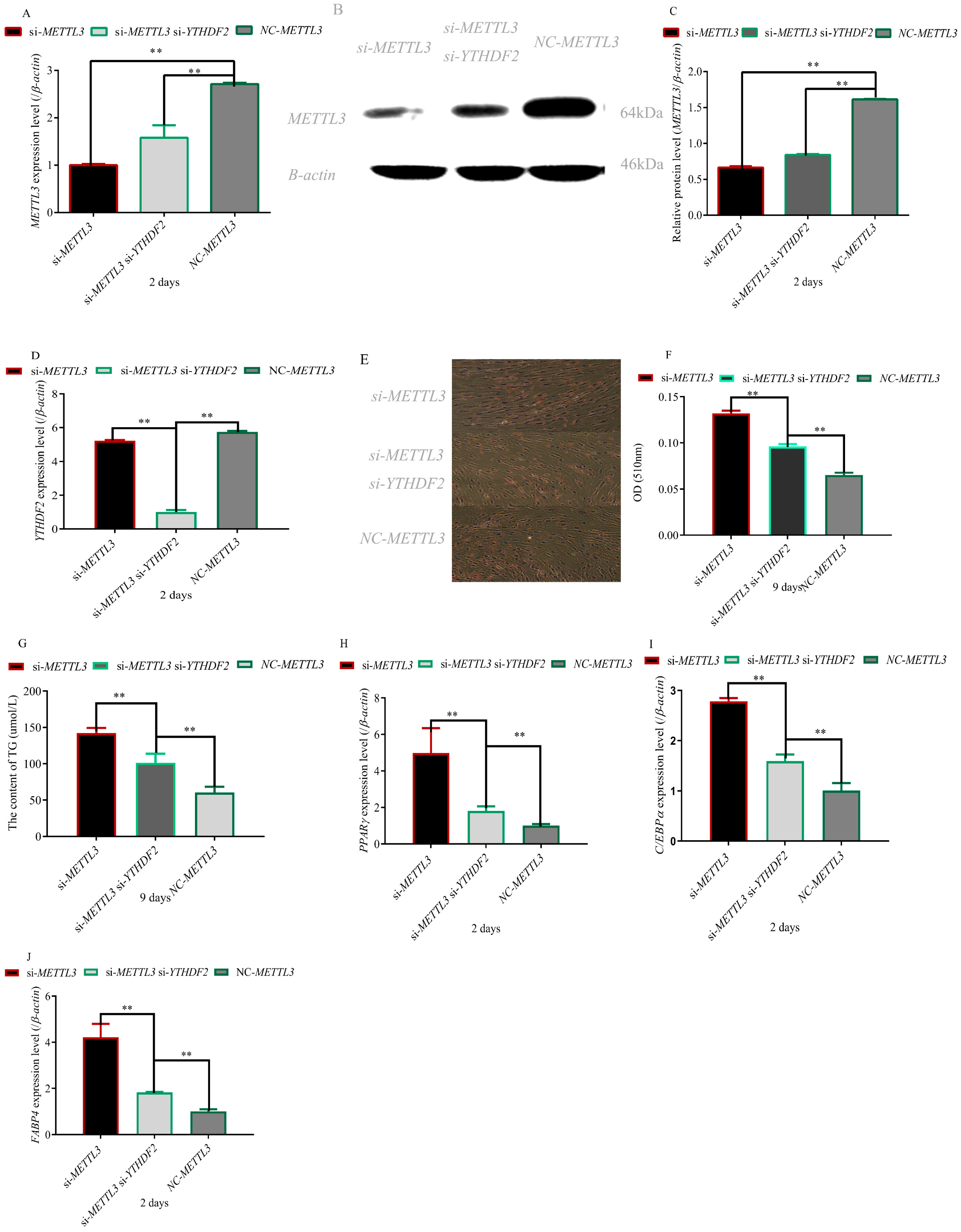
To investigate the regulation of YTHDF2 and METTL3 on adipocyte differentiation, we simultaneously interfered with YTHDF2 and METTL3 in preadipocytes. The results indicated that METTL3 and YTHDF2 were successfully knocked out in the experimental group (Figure 2A–D). Knockout of the METTL3 gene promoted the differentiation of preadipocytes. However, YTHDF2 knockout inhibited the promoting effect of METTL3 knockout on differentiation of preadipocytes. Downexpression of YTHDF2 in METTL3 knockdown preadipocytes significantly reduced the lipid droplets (p < 0.01) (Figure 2E,F), accompanied by significantly decreased TG content (p < 0.01) (Figure 2G). Similarly, knockout of YTHDF2 partly decreased the expression of adipogenic marker PPARγ, C/EBPa and FABP4 genes in METTL3 knockdown preadipocytes (p < 0.01) (Figure 2H–J). Consistently, YTHDF2 partly influenced the regulation of METTL3 on the differentiation of preadipocytes.

3.3. Effect of METTL3 and YTHDF2 on the Expression Level of the PCK2 Gene

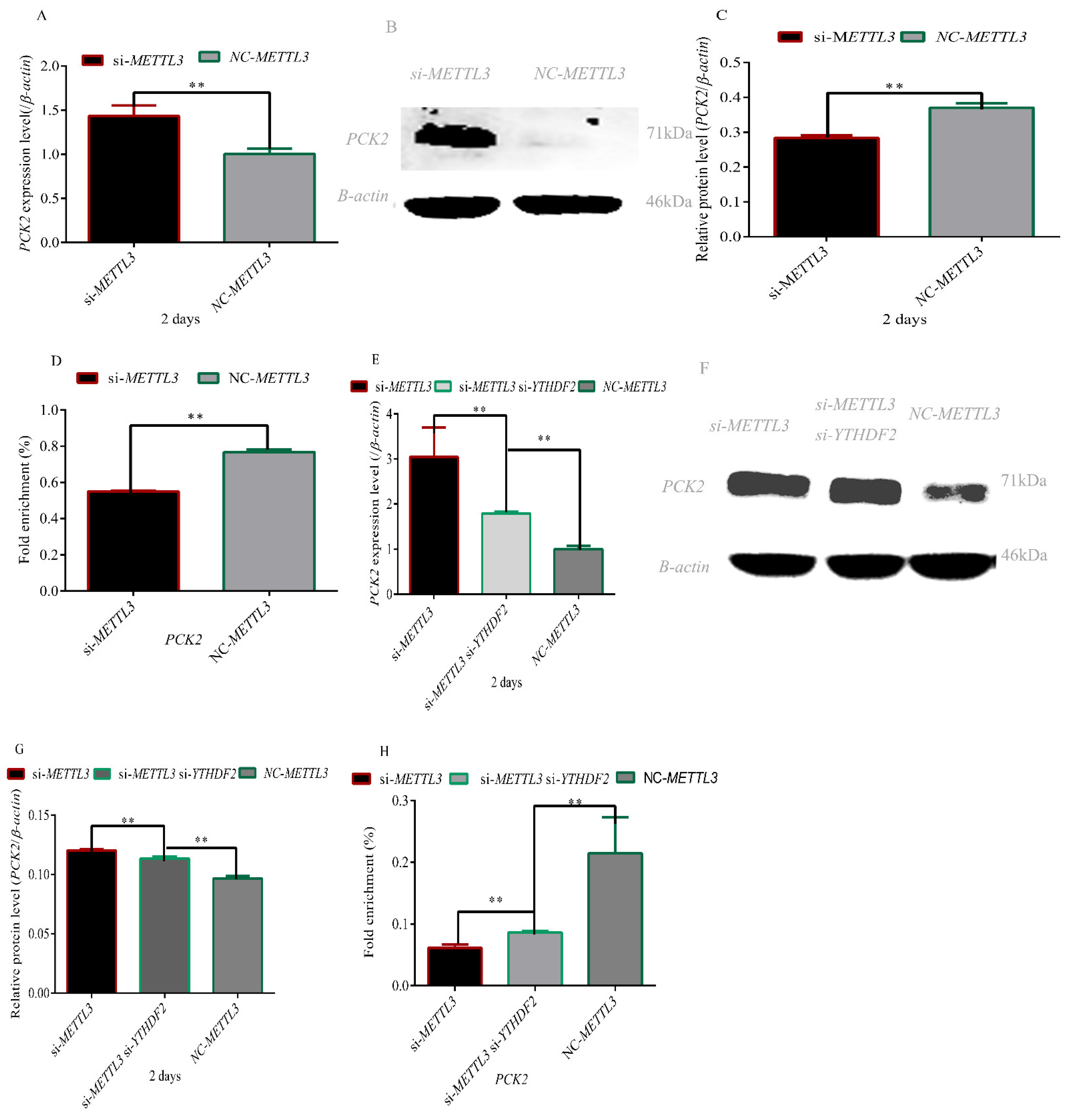
To determine whether METTL3 regulates the PCK2 gene m6A–YTHDF2 dependently, we first detected the expression of PCK2 in METTL3 knockdown adipocytes and found that the expression levels of PCK2 were significantly higher than those of the NC group (p < 0.01) (Figure 3A–C). However, the fold enrichment of PCK2 methylation modification decreased significantly in the METTL3-depleted adipocytes (p < 0.01) (Figure 3D). Next, we assessed the role of YTHDF2 by knocking down METTL3 and YTHDF2. Knockdown of YTHDF2 significantly decreased the mRNA and protein levels of the PCK2 gene in METTL3 knockdown cells (p < 0.01) (Figure 3E–G). In addition, knockdown of YTHDF2 partly recovered the fold enrichment of PCK2 methylation modification (Figure 3H). These results demonstrated that METTL3 m6A–YTHDF2 dependently regulates the PCK2 gene.

3.4. Effect of PCK2 on the Differentiation of Preadipocytes

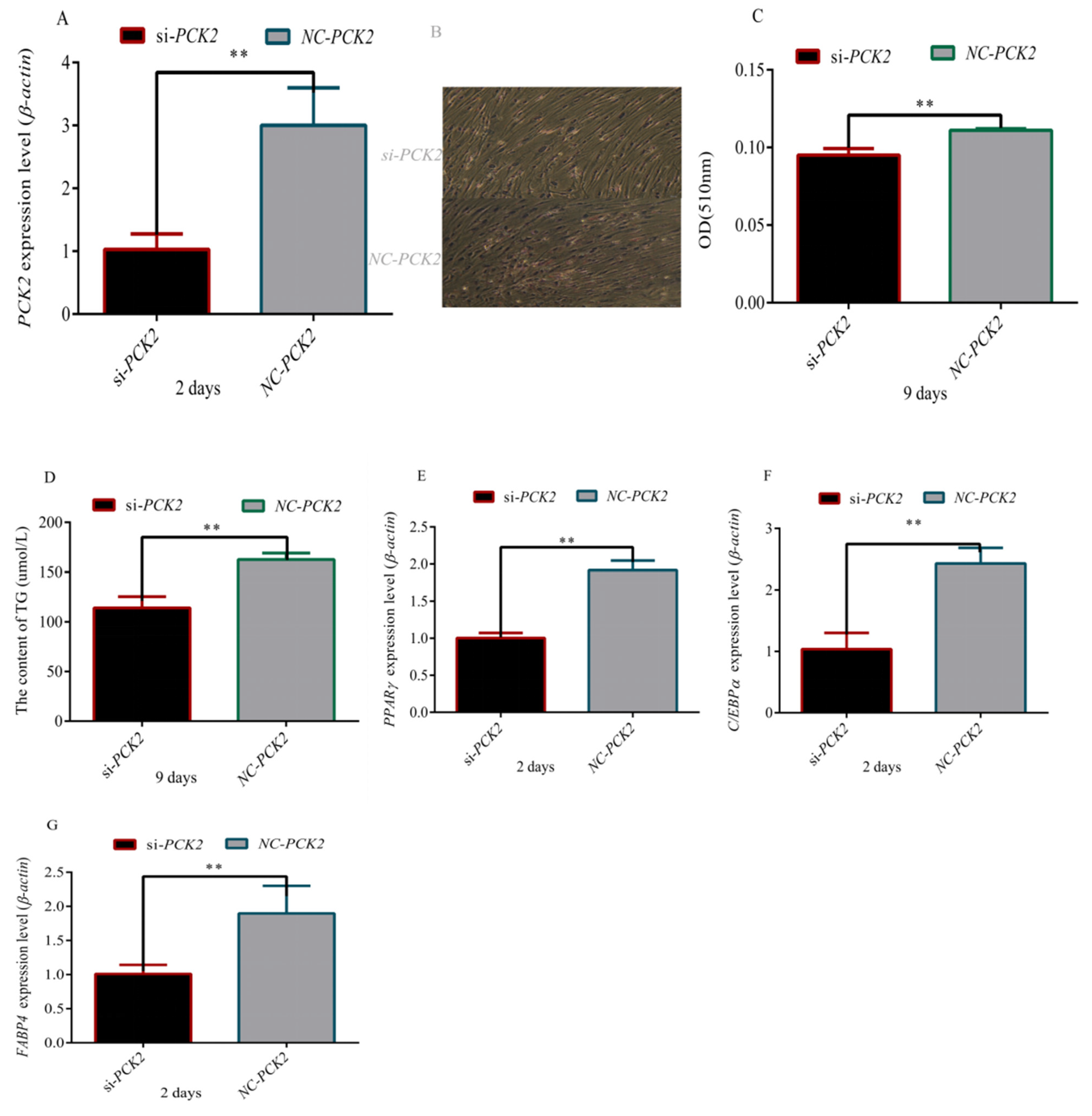
To understand the mechanism by which PCK2 regulates the differentiation of preadipocytes, we first interfered with the PCK2 gene in preadipocytes (Figure 4A–C). In PCK2-knockdown adipocytes, we found that the number and volume of lipid droplets was significantly reduced using oil red-O staining and determination of TG content (Figure 4D–F). At the same time, the expression of adipogenic marker PPARγ, C/EBPa and FABP4 genes was lower in PCK2 knockdown preadipocytes than those in the NC group (p < 0.01) (Figure 4G–I). Overall, PCK2 played an important role in regulating the differentiation of preadipocytes.

3.5. Correlation Analysis between Expression Level of the PCK2 Gene in the Longissimus Lumborum and Meat Quality Traits

The correlation analysis is plotted in Table 2. The correlation coefficients between intramuscular fat content and PCK2 expression in 35-day-old, 75-day-old and 165-day-old rabbits were 0.94 (p < 0.01), 0.85 (p < 0.05) and 0.82 (p < 0.05), respectively. Reduced pH value was negatively correlated with PCK2 expression only in 75-day-old and 165-day-old rabbits, and the correlation coefficients were 0.85 (p < 0.05) and 0.88 (p < 0.05), respectively. The total water loss rate was negatively correlated with the expression level of the PCK2 gene, and the correlation coefficients were 0.87 (p < 0.05), 0.79 (p < 0.05) and 0.74 (p < 0.05) in 35-day-old, 75-day-old and 165-day-old rabbits, respectively.

4. Discussion

In this study, we found that the level of PCK2 in blood also increased with age (Figure 1A). Previous studies showed that there was a positive correlation between fat deposition and age in growing rabbits [27]. It indicated that PCK2 may regulate fat deposition. To investigate whether PCK2 is associated with fat deposition, we detected the expression levels of PCK2 in the perirenal fat, longissimus lumborum and liver of Rex rabbits and found that the expression level of PCK2 increased with age in the longissimus lumborum and liver tissues (Figure 1C,D). However, there was no significant difference in the expression level of PCK2 in the perirenal fat at different stages (Figure 1B). The liver is the main site of fat synthesis [28]. Due to the need of adaptability, many animals deposit a large amount of fat in the liver; the liver returns to its original state after the stored fat is consumed by energy supply, and the whole change process is reversible [29,30]. In addition, the study showed that the volume of adipocytes gradually increased and the rate of fat deposition increased rapidly in lambs entering the fattening stage [30]. These results further indicated that PCK2 played an important role in fat deposition. At the same time, we found that the PCK2 gene was modified by m6A in the longissimus lumborum and perirenal adipose tissues of Rex rabbits.
The profiles and function of METTL3-mediated mRNA m6A are different depending on the tissue and the developmental stage [31,32,33]. Indeed, it has also been reported that the expression of many genes is regulated by either METTL3 or FTO alone [34]. A previous study also suggested the functional importance of the heterodimer formation of METTL3 and METTL14 in adipocyte differentiation [35].
In this study, we found that interference with METTL3 can promote the differentiation of adipocytes (Figure 2) and the expression level of the PCK2 gene (Figure 3A–C). METTL3 inhibits hepatic insulin sensitivity and promotes fatty acid metabolism [36]. A loss of the initial insulin suppression of glucose output in vitro and an associated increased activity of hepatic PCK2 has been reported [37]. It was recently demonstrated that hepatic PCK2 mRNA expression increased following the induction of chronic hypoglycemia in fetal sheep and that this was associated with a decrease in circulating insulin and an increase in plasma cortisol [38]. PPARγ agonists may sensitize the response of peripheral tissues to insulin by inhibiting the expression of 11β-HSD1 in adipocytes [39]. 11β-HSD1-knockout mice lost the ability to convert 11-dehydrocortisone into cortisol and inhibited the activity of PEPCK [40]. Cortisol has been shown to play a key role in the upregulation of hepatic PCK2 activity and expression in the late-gestation fetus [41,42], so the increase in hepatic HSDL1 mRNA expression and intrahepatic cortisol production may stimulate increased PCK2 mRNA expression. RNA N6-methyladenosine methyltransferase METTL3 facilitates colorectal cancer by activating the m6A–GLUT1–mTORC1 axis [43]. There is a positive correlation between the expression of GLUT-1 and PCK2 in laryngeal squamous cell carcinoma [44]. These results indicated METTL3 can regulate the expression of the PCK2 gene. However, the specific regulatory mechanism is still unclear. In this study, we found that mRNA and protein levels of the PCK2 gene increased significantly, whereas m6A methylation of the PCK2 gene decreased significantly after transfection with si-METTL3. Based on the above results, we inferred that METTL3 regulated the expression level of PCK2 by m6A methylation.
METTL3-mediated mRNA m6A modification can be recognized by the individual m6A reader proteins, which play key roles in controlling gene expression. Early studies showed that YTHDF2-binding increases mRNA decay [45]. In addition, other roles for these reader proteins are rapidly emerging in the regulation of specific mRNAs in different types of cells during different stimuli [46,47,48]. Moreover, the study revealed a novel mechanism wherein downregulated spinal cord METTL3 in coordination with YTHDF2 contributes to the modulation of inflammatory pain by stabilizing the upregulation of TET1 in spinal neurons [49]. METTL3 epigenetically repressed YPEL5 in an m6A–YTHDF2-dependent manner by targeting the m6A site in the coding sequence region of the YPEL5 transcript [50]. In this study, we found that YTHDF2 silencing could partially reverse the phenotype induced by METTL3 knockdown, including differentiation of adipocytes and methylation level of the PCK2 gene (Figure 2, Figure 3). These results suggested METTL3 regulates adipocyte differentiation by targeting the PCK2 gene in an m6A–YTHDF2-dependent manner.
In order to explore the mechanism by which PCK2 regulates adipogenesis, we knocked out the PCK2 gene in adipocytes and found that the differentiation of adipocytes was significantly inhibited (Figure 4). It has been found that PCK1 and PCK2 can be used as candidate genes of obesity, which can participate in the synthesis of glycerophosphate and promote the accumulation of fat in human body [51]. In addition, the loss of PCK2 in islets can damage insulin secretion, which suggested that the activity of PCK2 is related to insulin secretion [52]. In the majority of these studies [53,54,55], the degree of obesity and not the direct measurement of insulin resistance was used to identify individuals as insulin resistant vs. insulin sensitive. PCK2 was upregulated by effectors of this pathway by recruiting ATF4 to a consensus AARE site located at the PCK2 proximal promoter [56]. A clue to the mechanism for the integrative role of PCK2 in cancer metabolism arises from its transcription regulation by ATF4 under nutrient stress [57,58]. ATF4 regulates age-related and diet-induced obesity as well as glucose homeostasis in mammals and has conserved metabolic functions in flies [59]. In addition, the expression level of PCK2 was higher in Rex rabbits with morefat deposition. These results indicated that PCK2 can promoted adipocyte differentiation.
Adipocyte differentiation is a necessary process of fat deposition. In order to explore the relationship between the expression level of PCK2 and intramuscular fat, we analyzed the correlation between the expression level of PCK2 and intramuscular fat and found that they are positively correlated (Table 2). Previous study has shown that PCK2 can promote adipogenesis. In summary, PCK2 can regulate the production of intramuscular fat. Studies have shown that intramuscular fat is positively correlated with the pH of muscle [60]. The faster the pH value drops, the faster the meat spoils. Therefore, a reduced pH value is an important indicator of good meat quality. In this study, we found that the expression level of PCK2 was negatively correlated with reduced pH value in rabbits at 75 and 165 days of age. At the same time, there was a strong correlation between intramuscular fat and muscle system hydraulics [61]. Water in muscle also affects meat production. In this study, we found the expression level of PCK2 was negatively correlated with the total water loss rate. Meat with low drip loss is more easily accepted by consumers [62]. A study reported that consumers are attracted to rabbit meat according to its healthiness, sensory properties and hedonistic quality (variability on visual appeal) [63].
Intramuscular fat, pH and muscle water holding capacity are important indicators affecting muscle flavor. At the same time, intramuscular fat is positively correlated with sensory tenderness, juiciness and flavor [64]. Therefore, it can be concluded that PCK2 plays an important role in muscle flavor.

5. Conclusions

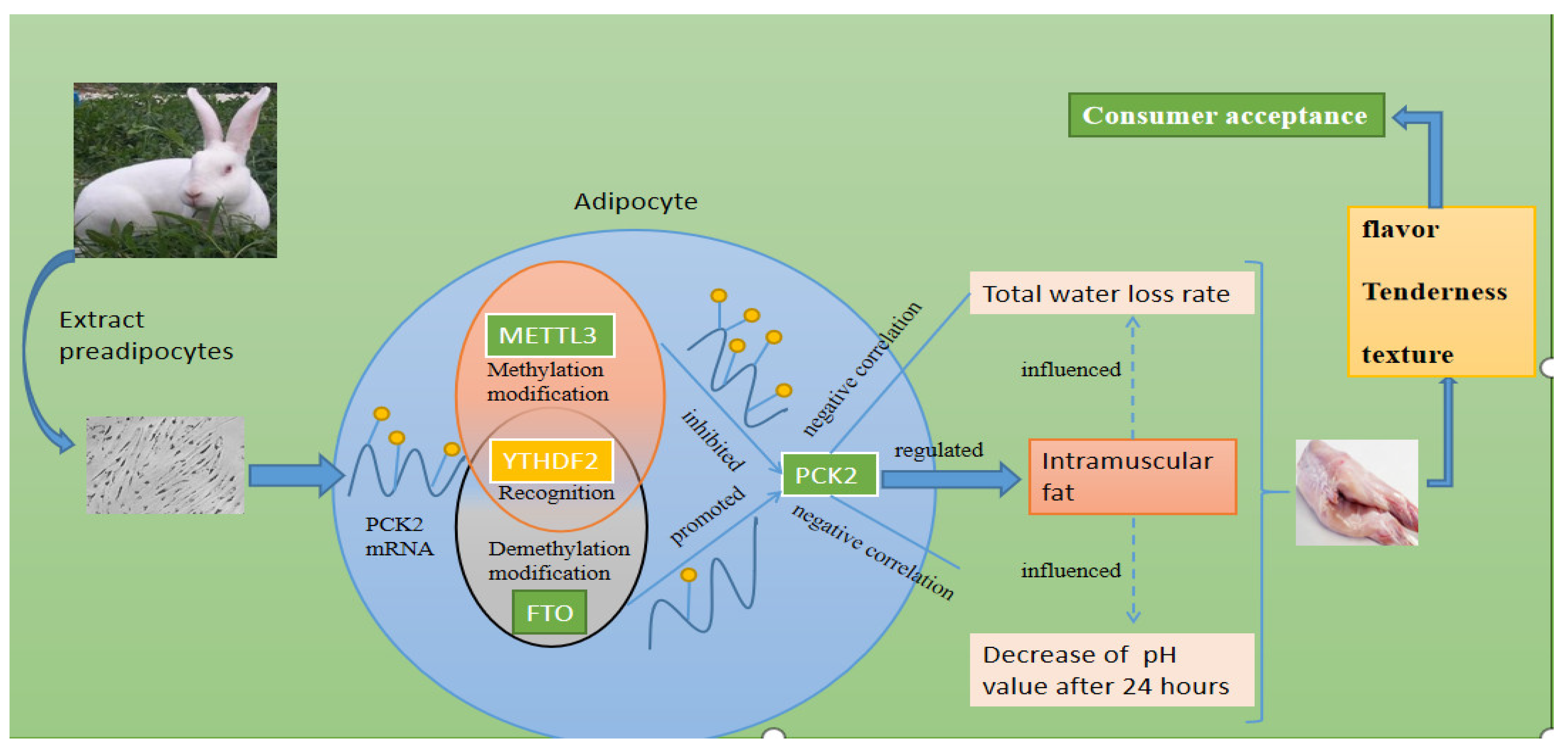
METTL3 decreased the expression level of the PCK2 gene in an m6A–YTHDF2-dependent manner. The expression level of the PCK2 gene was negatively correlated with reduced pH value and total water loss rate. However, intramuscular fat was positively correlated with the expression of the PCK2 gene. As shown in Figure 5, METTL3 affects meat quality by regulating the expression of PCK2. The study provides a scientific, molecular theoretical basis for cultivating rabbits with good meat quality.

Author Contributions

G.L. cultured cells and wrote the manuscript. Z.R. and G.L. conceived and designed the experiments. G.L. and T.Z. performed molecular test operations. All authors have read and agreed to the published version of the manuscript.

Funding

This work was supported by the Yangling Demonstration Area Industry University research and application collaborative innovation major project (1017cxy-15)(Yangling demonstration zone), the agricultural science and technology innovation and tackling key projects in Shaanxi Province (2016NY-108) (Shaanxi Provincial Department of science and technology) and the integration and demonstration of rabbit breeding and factory breeding technology (2018ZDXM-NY-041) (Shaanxi Provincial Department of agriculture and rural).

Institutional Review Board Statement

All experimental procedures were reviewed and approved by the Institutional Animal Care and Use Committee (IACUC) of Northwest A&F University, under the permit No. DK 2022045. All the procedures were performed in accordance with the relevant policies of animal welfare in China.

Informed Consent Statement

Not applicable.

Data Availability Statement

The datasets used and/or analyzed during the current study are available from the corresponding authors upon reasonable request.

Acknowledgments

We thank the staff at our laboratory for their ongoing assistance.

Conflicts of Interest

The authors declare no competing interests.

Abbreviations

m6AN6-methyladenosine
NCDnon-communicable disease
PCK2phosphoenolpyruvate carboxykinase 2
YTHDF2YTH domain family 2
METTL3methyltransferase-like 3
METTL14methyltransferase-like 14
WTAPWilms tumor 1–associated protein
PPARγperoxisome proliferator-activated receptor-γ
C/EBPαCCAAT/enhancer-binding family of proteins
FABP4fatty-acid-binding protein 4
TGtriglyceride content

References

  1. Dalle, Z.A.; Zs, S. The role of rabbit meat as functional food. Meat Sci. 2011, 88, 319–331. [Google Scholar] [CrossRef] [PubMed]
  2. Nishimura, T. The role of intramuscular connective tissue in meat texture. Anim. Sci. J. 2010, 81, 21–27. [Google Scholar] [CrossRef]
  3. Ge, K.; Ye, P.; Yang, L.; Kuang, J.; Chen, X.; Geng, Z. Comparison of slaughter performance, meat traits, serum lipid parameters and fat tissue between Chaohu ducks with high- and low-intramuscular fat content. Anim. Biotechnol. 2020, 31, 245–255. [Google Scholar] [CrossRef] [PubMed]
  4. Frank, D.; Watkins, P.; Ball, A.; Krishnamurthy, R.; Piyasiri, U.; Sewell, J.; OrtuñO, J.; Stark, J.; Warner, R. Impact of Brassica and Lucerne Finishing Feeds and Intramuscular Fat on Lamb Eating Quality and Flavor. A Cross-Cultural Study Using Chinese and Non-Chinese Australian Consumers. J. Agric. Food Chem. 2016, 64, 6856–6868. [Google Scholar] [CrossRef] [PubMed]
  5. Starkey, C.P.; Geesink, G.H.; Collins, D.; Oddy, V.H.; Hopkins, D.L. Do sarcomere length, collagen content, pH, intramuscular fat and desmin degradation explain variation in the tenderness of three ovine muscles? Meat Sci. 2016, 113, 51–58. [Google Scholar] [CrossRef] [PubMed]
  6. Di, S.L.; Brugiapaglia, A.; Galloni, M.; Destefanis, G.; Lisa, C. Effect of the leptin c.73TC mutation on carcass traits in beef cattle. Anim. Genet. 2007, 38, 316–317. [Google Scholar]
  7. Aaslyng, M.D.; Bejerholm, C.; Ertbjerg, P.; Bertram, H.C.; Andersen, H.J. Cooking loss and juiciness of pork in relation to raw meat quality and cooking procedure—ScienceDirect. Food Qual. Prefer. 2003, 14, 277–288. [Google Scholar] [CrossRef]
  8. Mortensen, M.; Andersen, H.J.; Engelsen, S.B.; Bertram, H.C. Effect of freezing temperature, thawing and cooking rate on water distribution in two pork qualities. Meat Sci. 2006, 72, 34–42. [Google Scholar] [CrossRef]
  9. Roundtree, I.A.; Evans, M.E.; Pan, T.; He, C. Dynamic RNA Modifications in Gene Expression Regulation. Cell 2017, 169, 1187–1200. [Google Scholar] [CrossRef] [Green Version]
  10. Yang, Y.; Hsu, P.J.; Chen, Y.S.; Yang, Y.G. Dynamic transcriptomic m6A decoration: Writers, erasers, readers and functions in RNA metabolism. Cell Res. 2018, 28, 616–624. [Google Scholar] [CrossRef] [Green Version]
  11. Zhen, B.; Liu, Y.; Zhao, Y.; Yao, Y.; Wang, X. A dynamic reversible RNA N6-methyladenosine modification: Current status and perspectives. J. Cell. Physiol. 2019, 234, 7948–7956. [Google Scholar]
  12. Wang, Y.; Li, Y.; Toth, J.I.; Petroski, M.; Zhang, Z.; Jing, C.Z. N-methyladenosine modification destabilizes developmental regulators in embryonic stem cells. Nat. Cell Biol. 2014, 16, 191–198. [Google Scholar] [CrossRef] [PubMed]
  13. Wang, X.; Zhu, L.; Chen, J.; Wang, Y. mRNA m6A methylation downregulates adipogenesis in porcine adipocytes. Biochem. Biophys. Res. Commun. 2015, 459, 201–207. [Google Scholar] [CrossRef]
  14. Zhao, X.; Yang, Y.; Sun, B.F.; Shi, Y.; Yang, X.; Xiao, W.; Hao, Y.J.; Ping, X.L.; Chen, Y.S.; Wang, W.J.; et al. FTO-dependent demethylation of N6-methyladenosine regulates mRNA splicing and is required for adipogenesis. Cell Res. 2014, 24, 1403–1419. [Google Scholar] [CrossRef] [PubMed]
  15. Berg, J.; Tymoczko, J.; Stryer, L. Biochemistry; W. H. Freeman and Company: New York, NY, USA, 2002; pp. 93–126. [Google Scholar]
  16. Franckhauser, S.; Munoz, S.; Pujol, A. Increased Fatty Acid Re-esterification by PEPCK Overexpression in Adipose Tissue Leads to Obesity Without Insulin Resistance. Diabetes 2002, 51, 624–630. [Google Scholar] [CrossRef] [PubMed] [Green Version]
  17. Peng, K.Y.; Li, M.; Yu, B.; Fan, M.; Yerle, B.L. Assignment porcine PCK1 and PCK2 genes to SSC17 and SSC7, respectively, by radiation hybrid mapping. Anim. Genet. 2005, 36, 364–365. [Google Scholar] [CrossRef] [PubMed]
  18. Wang, W.; Xue, W.; Jin, B.; Zhang, X.; Ma, F.; Xu, X. Candidate gene expression affects intramuscular fat content and fatty acid composition in pigs. J. Appl. Genet. 2013, 54, 113–118. [Google Scholar] [CrossRef]
  19. Oshel, K.M.; Knight, J.B.; Cao, K.T.; Thai, M.V.; Olson, A.L. Identification of a 30-Base Pair Regulatory Element and Novel DNA Binding Protein That Regulates the Human GLUT4 Promoter in Transgenic Mice. J. Biol. Chem. 2000, 275, 23666–23673. [Google Scholar] [CrossRef] [Green Version]
  20. Hamam, D.; Ali, D.; Kassem, M.; Aldahmash, A.; Alajez, N.M. MicroRNAs as Regulators of Adipogenic Differentiation of Mesenchymal Stem Cells. Stem Cells Dev. 2014, 24, 417–425. [Google Scholar] [CrossRef] [Green Version]
  21. Prayitno, D.S.; Phillips, C.J.C.; Omed, H. The Effects of Color of Lighting on the Behavior and Production of Meat Chickens. Poult. Sci. 1997, 76, 452–457. [Google Scholar] [CrossRef]
  22. Wang, W.; Xue, W.; Xu, X.; Jin, B.; Zhang, X. Correlations of genes expression in PPAR signalling pathway with porcine meat quality traits. Czech J. Anim. Sci. 2016, 61, 333–339. [Google Scholar]
  23. Wood, J.D.; Richardson, R.I.; Nute, G.R.; Fisher, A.V.; Campo, M.M.; Kasapidou, E.; Sheard, P.R.; Enser, M. Effects of fatty acids on meat quality: A review. Meat Sci. 2004, 66, 21–32. [Google Scholar] [CrossRef]
  24. Kang, H.; Zhang, Z.; Yu, L.; Li, Y.; Liang, M.; Zhou, L. FTO reduces mitochondria and promotes hepatic fat accumulation through RNA demethylation. J. Cell. Biochem. 2018, 119, 5676–5685. [Google Scholar] [CrossRef] [PubMed]
  25. Luo, G.; Hu, S.; Lai, T.; Wang, J.; Wang, L.; Lai, S. MiR-9-5p promotes rabbit preadipocyte differentiation by suppressing leptin gene expression. Lipids Health Dis. 2020, 19, 126. [Google Scholar] [CrossRef] [PubMed]
  26. Uezumi, A.; Ito, T.; Morikawa, D.; Shimizu, N.; Yoneda, T.; Segawa, M.; Yamaguchi, M.; Ogawa, R.; Matev, M.M.; Miyagoe-Suzuki, Y. Fibrosis and adipogenesis originate from a common mesenchymal progenitor in skeletal muscle. J. Cell Sci. 2011, 124, 3654–3664. [Google Scholar] [CrossRef] [PubMed] [Green Version]
  27. Luo, G.; Wang, L.; Hu, S.; Du, K.; Lai, S. Association of leptin mRNA expression with meat quality trait in Tianfu black rabbits. Anim. Biotechnol. 2020, 3, 1–7. [Google Scholar] [CrossRef] [PubMed]
  28. Pearce, J. Some differences between avian and mammaeian biochemistry. Int. J. Biochem. 1977, 8, 269–275. [Google Scholar] [CrossRef]
  29. Pilo, B.; George, J.C. Diurnal and seasonal variation in liver glycogen and fat in relation to metabolic status of liver and m. pectoralis in the migratory starling, Sturnus roseus, wintering in India. Comp. Biochem. Physiol. A Comp. Physiol. 1983, 74, 601–604. [Google Scholar] [CrossRef]
  30. Yasuhara, M.; Ohama, T.; Matsuki, N.; Saito, H.; Teramoto, T. Induction of fatty liver by fasting in suncus. J. Lipid Res. 1991, 32, 887–891. [Google Scholar] [CrossRef]
  31. Dominissini, D.; Moshitch-Moshkovitz, S.; Schwartz, S.; Salmon-Divon, M.; Ungar, L.; Osenberg, S.; Cesarkas, K.; Jacob-Hirsch, J.; Amariglio, N.; Kupiec, M. Topology of the human and mouse m6A RNA methylomes revealed by m6A-seq. Nature 2012, 485, 201–206. [Google Scholar] [CrossRef]
  32. Wu, R.; Jiang, D.; Wang, Y.; Wang, X. N (6)-Methyladenosine (m(6)A) Methylation in mRNA with A Dynamic and Reversible Epigenetic Modification. Mol. Biotechnol. 2016, 58, 450–459. [Google Scholar] [CrossRef]
  33. Wu, Y.; Xie, L.; Wang, M.; Xiong, Q.; Guo, Y.; Liang, Y.; Li, J.; Sheng, R.; Deng, P.; Wang, Y.; et al. Mettl3-mediated m6A RNA methylation regulates the fate of bone marrow mesenchymal stem cells and osteoporosis. Nat. Commun. 2018, 9, 1–12. [Google Scholar] [CrossRef] [PubMed]
  34. Liu, J.; Yue, Y.; Han, D.; Wang, X.; Fu, Y.; Zhang, L.; Jia, G.; Yu, M.; Lu, Z.; Deng, X.; et al. A METTL3-METTL14 complex mediates mammalian nuclear RNA N6-adenosine methylation. Nat. Chem. Biol. 2014, 10, 93–95. [Google Scholar] [CrossRef] [PubMed] [Green Version]
  35. Masatoshi, K.; Mitsuru, O.; Takayoshi, S.; Motoharu, A.; Toshihiro, U.; Aya, I.; Naoki, K.; Yukiko, O.; Naoto, K.; Ryo, S. The RNA methyltransferase complex of WTAP, METTL3, and METTL14 regulates mitotic clonal expansion in adipogenesis. Mol. Cell. Biol. 2018, 38, 16–18. [Google Scholar]
  36. Xie, W.; Ma, L.L.; Xu, Y.Q.; Wang, B.H.; Li, S.M. METTL3 inhibits hepatic insulin sensitivity via N6-methyladenosine modification of Fasn mRNA and promoting fatty acid metabolism—ScienceDirect. Biochem. Biophys. Res. Commun. 2019, 518, 120–126. [Google Scholar] [CrossRef] [PubMed]
  37. Desai, M.; Byrne, C.D.; Zhang, J.L.; Petry, C.J.; Hales, C.N. Programming of hepatic insulin-sensitive enzymes in offspring of rat dams fed a protein-restricted diet. Am. J. Physiol. 1997, 272, 1083–1090. [Google Scholar] [CrossRef]
  38. Rozance, P.J.; Limesand, S.W.; Barry, J.S.; Brown, L.D.; Hay, W.W. Chronic late-gestation hypoglycemia upregulates hepatic PEPCK associated with increased PGC1α mRNA and phosphorylated CREB in fetal sheep. Am. J. Physiol. Endocrinol. Metab. 2008, 294, E365–E370. [Google Scholar] [CrossRef] [Green Version]
  39. Berger, J. Peroxisome proliferator-activated receptor-gamma ligands inhibit adipocyte 11beta -hydroxysteroid dehydrogenase type 1 expression and activity. J. Biol. Chem. 2001, 276, 12629–12635. [Google Scholar] [CrossRef] [Green Version]
  40. Kotelevtsev, Y.; Holmes, M.C.; Burchell, A.; Houston, P.M.; Schmoll, D.; Jamieson, P.; Best, R.; Brown, R.; Edwards, C.; Seckl, J.R. 11β-Hydroxysteroid dehydrogenase type 1 knockout mice show attenuated glucocorticoid-inducible responses and resist hyperglycemia on obesity orstress. Proc. Natl. Acad. Sci. USA 1997, 94, 14924–14929. [Google Scholar] [CrossRef] [Green Version]
  41. Forhead, A.J.; Poore, K.R.; Mapstone, J.; Fowden, A.L. Developmental regulation of hepatic and renal gluconeogenic enzymes by thyroid hormones in fetal sheep during late gestation. J. Physiol. 2003, 548, 941–947. [Google Scholar] [CrossRef]
  42. Franko, K.L.; Giussani, D.A.; Forhead, A.J.; Fowden, A.L. Effects of dexamethasone on the glucogenic capacity of fetal, pregnant, and non-pregnant adult sheep. J. Endocrinol. 2007, 192, 67–73. [Google Scholar] [CrossRef]
  43. Chen, H.; Gao, S.; Liu, W.; Wong, C.C.; Wu, J.; Wu, J.; Liu, D.; Gou, H.; Kang, W.; Zhai, J. RNA N6-Methyladenosine Methyltransferase METTL3 Facilitates Colorectal Cancer by Activating the m6A-GLUT1-mTORC1 Axis and Is a Therapeutic Target—ScienceDirect. Gastroenterology 2020, 160, 1284–1300. [Google Scholar] [CrossRef] [PubMed]
  44. Hu, Y.; Deng, K.; Pan, M.; Liu, S.; Zuo, J. Down-regulation of PCK2 inhibits the invasion and metastasis of laryngeal carcinoma cells. Am. J. Transl. Res. 2020, 12, 3842–3857. [Google Scholar]
  45. Wang, X.; Lu, Z.; Gomez, A.; Hon, G.C.; Yue, Y.; Han, D.; Fu, Y.; Parisien, M.; Dai, Q.; Jia, G. N6-methyladenosine-dependent regulation of messenger RNA stability. Nature 2014, 505, 117–120. [Google Scholar] [CrossRef]
  46. Xiang, Y.; Laurent, B.; Hsu, C.H.; Nachtergaele, S.; Lu, Z.; Sheng, W.; Xu, C.; Hen, H.C.; Jian, O.; Wang, S. RNA m6A methylation regulates the ultraviolet-induced DNA damage response. Nature 2017, 552, 573–576. [Google Scholar] [CrossRef] [PubMed]
  47. Meyer, K.; Patil, D.; Zhou, J.; Zinoviev, A.; Jaffrey, S. 5’ UTR m6A Promotes Cap-Independent Translation. Cell 2015, 163, 999–1010. [Google Scholar] [CrossRef] [Green Version]
  48. Yu, J.; Li, Y.; Tian, W.; Xiang, Z.; Gabriele, M. Modification of N6-methyladenosine RNA methylation on heat shock protein expression. PLoS ONE 2018, 13, e0198604. [Google Scholar] [CrossRef] [PubMed]
  49. Pan, Z.; Zhang, Q.; Liu, X.; Zhou, H.; Cao, J.L. Mettl3 contributes to inflammatory pain by targeting TET1 in YTHDF2-dependent manner. Pain 2021, 7. Publish ahead of printing. [Google Scholar]
  50. Zhou, D.; Tang, W.; Xu, Y.; Xu, Y.; Wang, G. METTL3/YTHDF2 m6A axis accelerates colorectal carcinogenesis through epigenetically suppressing YPEL5. Mol. Oncol. 2021, 15, 2172–2184. [Google Scholar] [CrossRef] [PubMed]
  51. Hanson, R.W.; Reshef, L. Glyceroneogenesis revisited. Biochimie 2003, 85, 1199–1205. [Google Scholar] [CrossRef]
  52. Stark, R.; Pasquel, F.; Turcu, A.; Pongratz, R.L.; Roden, M.; Cline, G.W.; Shulman, G.I.; Kibbey, R.G. Phosphoenolpyruvate Cycling via Mitochondrial Phosphoenolpyruvate Carboxykinase Links Anaplerosis and Mitochondrial GTP with Insulin Secretion. J. Biol. Chem. 2009, 284, 26578–26590. [Google Scholar] [CrossRef] [PubMed] [Green Version]
  53. Polonsky, K.S.; Given, B.; Hirsch, L.; Shapiro, E.T.; Cauter, E.V. Quantitative study of insulin secretion and clearance in normal and obese subjects. J. Clin. Investig. 1988, 81, 435–441. [Google Scholar] [CrossRef] [PubMed]
  54. Bonora, E.; Zavaroni, I.; Bruschi, F.; Alpi, O.; Pezzarossa, A.; Guerra, L.; Dall’Aglio, E.; Coscelli, C.; Butturini, U. Peripheral Hyperinsulinemia of Simple Obesity: Pancreatic Hypersecretion or Impaired Insulin Metabolism? J. Clin. Endocrinol. Metab. 1984, 59, 1121–1127. [Google Scholar] [CrossRef] [PubMed]
  55. Meistas, M.T.; Margolis, S.; Kowarski, A.A. Hyperinsulinemia of obesity is due to decreased clearance of insulin. Am. J. Physiol. 1983, 245, 155–159. [Google Scholar] [CrossRef] [PubMed]
  56. Méndez-Lucas, A.; Duarte, J.; Sunny, N.E.; Satapati, S.; He, T.T.; Fu, X.; Bermúdez, J.; Burgess, S.C.; Perales, J.C. PEPCK-M expression in mouse liver potentiates, not replaces, PEPCK-C mediated gluconeogenesis. J. Hepatol. 2013, 59, 105–113. [Google Scholar] [CrossRef] [Green Version]
  57. Ye, J.; Kumanova, M.; Hart, L.S.; Sloane, K.; Zhang, H.; Panis, D.D.; Bobrovnikova-Marjon, E.; Diehl, J.A.; Ron, D.; Koumenis, C. The GCN2-ATF4 pathway is critical for tumour cell survival and proliferation in response to nutrient deprivation. Embo J. 2014, 29, 2082–2096. [Google Scholar] [CrossRef] [Green Version]
  58. Ding, B.; Parmigiani, A.; Divakaruni, A.S.; Archer, K.; Murphy, A.N.; Budanov, A.V. Sestrin2 is induced by glucose starvation via the unfolded protein response and protects cells from non-canonical necroptotic cell death. Sci. Rep. 2016, 6, 22538. [Google Scholar] [CrossRef] [Green Version]
  59. Seo, J.; Fortuno, E.S.; Suh, J.M.; Stenesen, D.; Tang, W.; Parks, E.J.; Adams, C.M.; Townes, T.; Graff, J.M. Atf4 Regulates Obesity, Glucose Homeostasis, and Energy Expenditure. Diabetes 2009, 58, 2565–2573. [Google Scholar] [CrossRef] [Green Version]
  60. Okeudo, N.J.; Moss, B.W. Interrelationships amongst carcass and meat quality characteristics of sheep. Meat Sci. 2005, 69, 1–8. [Google Scholar] [CrossRef]
  61. Wang, Y.H.; Byrne, K.A.; Reverter, A.; Harper, G.S.; Taniguchi, M.; Mcwilliam, S.M.; Mannen, H.; Oyama, K.; Lehnert, S.A. Transcriptional profiling of skeletal muscle tissue from two breeds of cattle. Mamm. Genome 2005, 16, 201–210. [Google Scholar] [CrossRef]
  62. Schäfer, A.; Rosenvold, K.; Purslow, P.P.; Andersen, H.J.; Henckel, P. Physiological and structural events post mortem of importance for drip loss in pork. Meat Sci. 2002, 61, 355–366. [Google Scholar] [CrossRef]
  63. Zotte, A.D. Perception of rabbit meat quality and major factors influencing the rabbit carcass and meat quality. Livest. Prod. Sci. 2002, 75, 11–32. [Google Scholar] [CrossRef]
  64. Anne, L.; Bénédicte, L.; Isabelle, L.; Thierry, A.; Muriel, B.; Louis, L.; Brigitte, P.; JérMe, B. How muscle structure and composition influence meat and flesh quality. Sci. World J. 2016, 2016, 1–14. [Google Scholar]
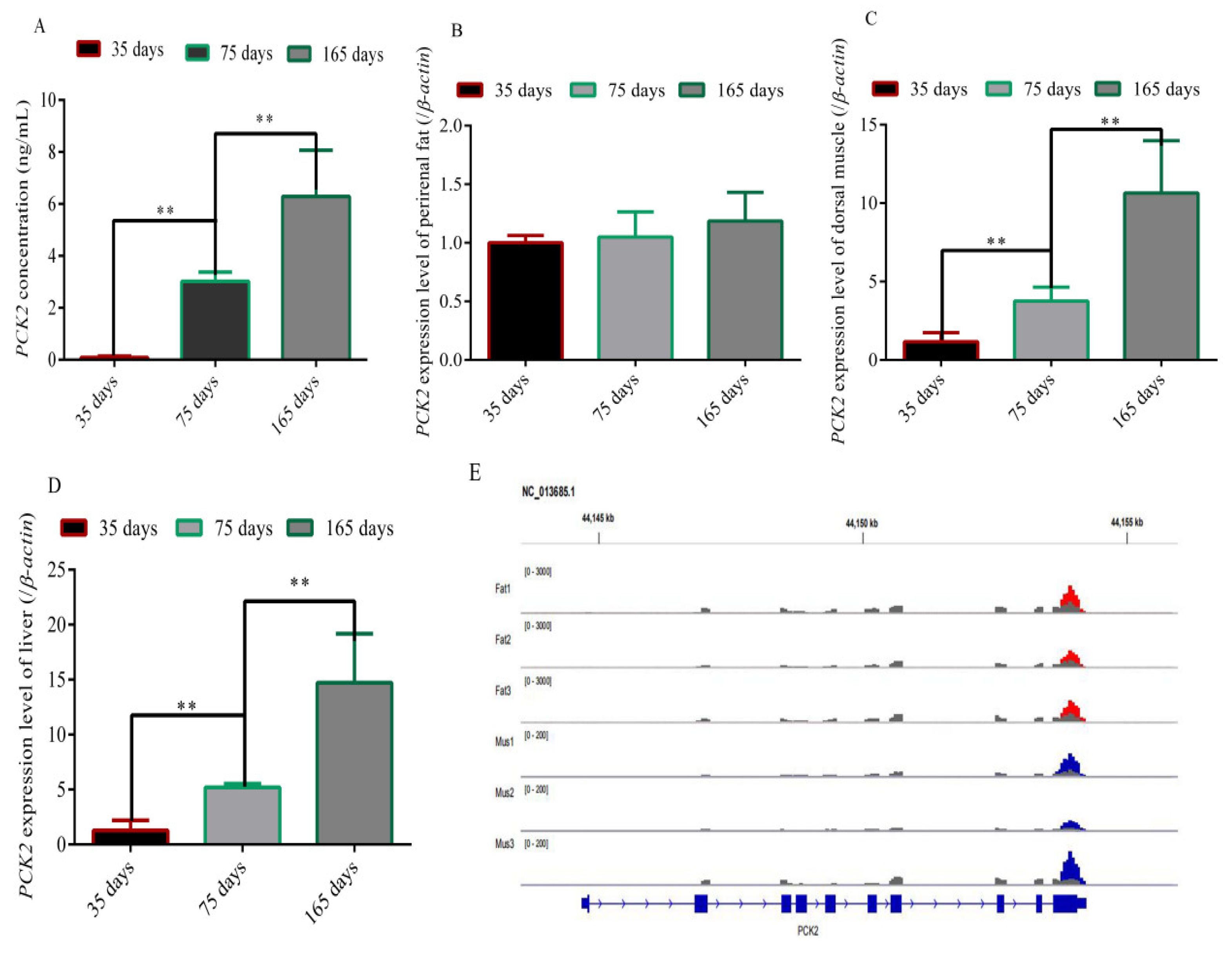
Figure 1. The content of PCK2 in blood and the expression and methylation of the PCK2 gene in various tissues. (A) Content of PCK2 in Rex rabbit blood; (B) expression levels of PCK2 in perirenal fat; (C) expression levels of PCK2 in dorsal muscle; (D) expression levels of PCK2 in the liver; (E) examples of dynamic methylation with m6A peaks in the PCK2 gene. (“**”, p ≤ 0.01).
Foods 11 01549 g001
Figure 2. METTL3-regulated preadipocyte differentiation in an m6A–YTHDF2-dependent manner. (AC) METTL3 expression level during preadipocyte differentiation after transfecting with si-METTL3, si-YTHDF2 and NC; (D) YTHDF2 expression level during preadipocyte differentiation after transfecting with si-METTL3, si-YTHDF2 and NC; (E,F) results of oil red-O staining on day 9 after transfecting with si-METTL3, si-YTHDF2 and NC; (G) triglyceride content on day 9 after transfecting with si-METTL3, si-YTHDF2 and NC; (HJ) expression level of marker genes PPARγ, CEBPα and FABP4 during preadipocyte differentiation after transfecting with si-METTL3, si-YTHDF2 and NC (“**”, p ≤ 0.01).
Foods 11 01549 g002
Figure 3. METTL3 regulated the expression level of the PCK2 gene in an m6A–YTHDF2-dependent manner. (AC) PCK2 expression level during preadipocyte differentiation after transfecting with si-METTL3 and NC; (D) fold enrichment of the PCK2 gene after transfecting with si-METTL3 and NC; (EG) PCK2 expression level during preadipocyte differentiation after transfecting with si-METTL3, si-YTHDF2 and NC; (H) fold enrichment of the PCK2 gene after transfecting with si-METTL3, si-YTHDF2 and NC; (“**”, p ≤ 0.01).
Foods 11 01549 g003
Figure 4. Inhibition of PCK2 inhibited rabbit preadipocyte differentiation. (A) PCK2 expression level during preadipocyte differentiation after transfecting with si-PCK2 and NC; (B,C) results of oil red-O staining on day 9 after transfecting with si-PCK2 and NC; (D) triglyceride content on day 9 after transfecting with si-PCK2 and NC; (EG) expression level of marker genes PPARγ, CEBPα and FABP4 during preadipocyte differentiation after transfecting with si-PCK2 and NC (“**”, p ≤ 0.01).
Foods 11 01549 g004
Figure 5. N6-methyladenosine affected the flavor and taste of muscle by targeting the PCK2 gene.
Foods 11 01549 g005
Table 1. Primers used in this study.
Gene NamePrimer Sequence (5′-3′)Tm (°C)Product Size (bp)
YTHDF2CAGACACAGCCATTGCCTCCAC60122
CCGTTATGACCGAACCCACTGC
METTL3CCCACCTCAGTGGATCTGTT60189
ACCCAGAGGAAGAGAAAGCC
PCK2AACAGGAGGTGCGTGACATT60250
GGGACAGGGAGTGTGAGAAG
PPARγGAGGACATCCAGGACAACC61168
GTCCGTCTCCGTCTTCTTT
β-actinGGAGATCGTGCGGGACAT61.4318
GTTGAAGGTGGTCTCGTGGAT
C/EBPαGCGGGAACGAACAACAT64172
GGCGGTCATTGTCACTGGTC
FABP4GGCCAGGAATTTGATGAAGTC61.4140
AGTTTATCGCCCTCCCGTT
si-YTHDF2CAUGAAUACUAUAGACCAATT
UUGGUCUAUAGUAUUCAUGTT
si-METTL3UCAAGGAACAACAGAGCAATT
UUGCUCUGUUGUUCCUUAGTT
si-PCK2GGGAACAGGAGGUGCGUGATT
UCACGCACCUCCUGUUCCCTT
Negative ControlUUCUCCGAACGUGUCACGUTT
ACGUGACACGUUCGGAGAATT
Table 2. Correlation analysis of expression level of the PCK2 gene with meat quality traits in Rex rabbits.
Expression Level/Meat Quality TraitsIntramuscuLAR Fat ContentCooked Meat RateDrip LossReduced pH ValueTotal Water Loss Rate
Expression level of PCK2Age of 35 days0.94 **0.450.580.39−0.87 *
Age of 75 days0.85 *−0.460.21−0.85 *−0.79 *
Age of 165 days0.82 *−0.04−0.28−0.88 *−0.74 *
Note: Single asterisk (*) and double asterisks (**) indicate significant and highly significant correlations at the 0.05 and 0.01 probability levels, respectively.
Publisher’s Note: MDPI stays neutral with regard to jurisdictional claims in published maps and institutional affiliations.