Article

Combination of Direct Viable Count and Fluorescent In Situ Hybridization (DVC-FISH) as a Potential Method for Identifying Viable Vibrio parahaemolyticus in Oysters and Mussels

by
Jorge García-Hernández
1,*,
Manuel Hernández
1 and
Yolanda Moreno
2
1
Advanced Center for Food Microbiology, Biotechnology Department, Universitat Politècnica de València, 46022 Valencia, Spain
2
Research Institute of Water and Environmental Ingeneering (IIAMA), Universitat Politècnica de València, 46022 Valencia, Spain
*
Author to whom correspondence should be addressed.
Foods 2021, 10(7), 1502; https://doi.org/10.3390/foods10071502
Submission received: 12 May 2021 / Revised: 18 June 2021 / Accepted: 25 June 2021 / Published: 29 June 2021
(This article belongs to the Topic Future Food Analysis and Detection)

Abstract

:
Vibrio parahaemolyticus is a human food-borne pathogen with the ability to enter the food chain. It is able to acquire a viable, non-cultivable state (VBNC), which is not detected by traditional methods. The combination of the direct viable count method and a fluorescent in situ hybridization technique (DVC-FISH) makes it possible to detect microorganisms that can present VBNC forms in complex samples The optimization of the in vitro DVC-FISH technique for V. parahaemolyticus was carried out. The selected antibiotic was ciprofloxacin at a concentration of 0.75 μg/mL with an incubation time in DVC broth of 5 h. The DVC-FISH technique and the traditional plate culture were applied to detect and quantify the viable cells of the affected pathogen in artificially contaminated food matrices at different temperatures. The results obtained showed that low temperatures produced an important logarithmic decrease of V. parahaemolyticus, while at 22 °C, it proliferated rapidly. The DVC-FISH technique proved to be a useful tool for the detection and quantification of V. parahaemolyticus in the two seafood matrices of oysters and mussels. This is the first study in which this technique has been developed to detect viable cells for this microorganism.

1. Introduction

Composed of more than 100 species, the Vibrio genus is a Gram-negative curved rod bacterium. It is widely distributed in estuarine, marine and freshwater systems worldwide. A large number of Vibrio spp. are in commensal or pathogenic relations with marine organisms such as fish, mollusks and crustaceans [1]. Vibrio parahaemolyticus, Vibrio vulnificus and Vibrio cholerae are the most important human pathogens of this genus [2,3,4,5]. V. vulnificus can cause gastroenteritis or severe wound infections due to exposure to highly contaminated water [6,7,8] and, in high-risk groups with their immune systems compromised, it can also cause septicemia [9]. It has been isolated from shellfish, fish, water and sediments of different temperatures and salinities [10]. V. parahaemolyticus is recognized as the leading cause of diarrheal disease linked to the consumption of raw, inadequately cooked or contaminated-after-cooking seafood, particularly oysters [6,7,8,11]. The infection with this species is less serious than the one with Vibrio vulnificus, and it is generally not associated with death. However, illness is not limited to a particular high-risk group [9]. Vibrio spp. occurrence increases to growing areas as water warms up. Therefore, the risk of illness rises during the summer months [12]. In this sense, Garrido-Maestu et al. [3] observed a seasonal distribution of fresh mussels harvested in the southern Rias of Galici having higher densities of V. parahaemolyticus in warmer months. Although there are just a few annual cases of infectious diseases related to Vibrio spp. in European countries, this number is expected to increase soon due to the consumption of raw seafood, the increase of high-risk patients, the impact of anthropogenic activities and the warming of the marine environment owing to the climate change [8,13,14]. Furthermore, Vibrio related outbreaks are more likely to presently occur in geographical areas where these bacteria were once considered absent such as the Pacific Northwest and Northeast coasts of the United States. [15,16,17]. Recently, the Committee for Standardization (CEN) of the European Commission (EC) undertook a work to validate a method designed from the merging of two previous ISO methods for the detection of V. parahaemolyticus, V. cholerae and other potentially pathogenic vibrio [18]. However, these ISO standard methods did not provide accurate quantification because the VBNC forms were not detected.
The VBNC concept was first discovered in 1982 in E. coli and V. cholerae environmental samples [19]. Under stress conditions, some microorganisms are capable of entering a viable but non-culturable (VBNC) state as a means of survival, in which they are living cells but unable to grow in their normal environment [20,21]. Thus, it is not possible to detect this state using traditional plating techniques, which lead to the wrong number of the total viable cells in food or environmental and clinical samples due to underestimation [20]. There are many factors that force the VBNC state such as lower nutrient availability and changes in temperature, pH, osmotic pressure and oxygen concentrations [22].
Some authors have demonstrated the VBNC state of Vibrio spp. by inducing stress–response genes as well as genes involved in the regulation of cold protection [21,23,24]. Moreover, Nowakowska and Oliver [25] described the ability of V. vulnificus cells to enter into a VBNC state when water temperatures were below 13 °C. V. parahaemolyticus is sometimes found entering the VBNC state in unfavorable conditions such as low temperature in aquatic environments [26]. It can still express some metabolic activity, so it can continue to cause infectious diseases in humans [27,28].
According to the recent ISO 21872-1:2017, which describes a method for the determination of potentially enteropathogenic V. parahaemolyticus, V. cholerae and V. vulnificus, the use of traditional culture techniques for detecting V. parahaemolyticus may require about 4–5 days. The methodology consists of four steps: a primary enrichment, a secondary enrichment, isolation in selective culture media and a final confirmation by biochemical assays or PCR. Molecular techniques such as fluorescent in situ hybridization (FISH) and PCR can be used as alternatives to conventional detection methods, although they do not allow differentiation between non-viable and viable cells [29], which can result in false positives [30]. Fluorescent in situ hybridization (FISH) is a method that allows the detection and identification of microbial organisms (bacteria, protozoa and yeasts) at the species or genus level using fluorescence-labeled, rRNA-targeted oligonucleotide probes, followed by an analysis with a fluorescence microscope [31,32].
The direct viable count (DVC) procedure involves exposing bacterial cells to a revival medium that contains antibiotics that prevent cellular division [33]; elongated cells are then enumerated as viable cells [34]. DVC is useful for the detection of specific bacterial viable cells in combination with a microscopic technique such as FISH [31]. DVC assay is based on the incubation of bacterial cells in a medium containing antibiotics that inhibit DNA gyrase and prevent cell division. The resulting cells can continue to metabolize nutrients and elongate and/or increase their size during incubation [31,35]. These viable cells are then counted and distinguished from non-viable cells by size [36]. The combination of the DVC procedure, which increases intracellular rRNA levels, with FISH performed on rRNA-targeted sequences, could prove useful in detecting and identifying viable cells in mixed microbial communities [37]. Armisen and Servais (2003) [38] combined for the first time direct viable count (DVC) and fluorescent in situ hybridization (FISH) to enumerate viable E-coli in rivers and wastewaters. A combination of the DVC procedure and FISH facilitates the detection and identification of viable cells in mixed microbial populations [39]. Several antibiotic inhibitors of DNA gyrase have been used in previous trials of DVC-FISH with other microorganisms [40,41]. Ciprofloxacin is a fluoroquinolone that has been shown to be a good inhibitor of DNA gyrase for both Gram-positive and Gram-negative bacteria [42].
The aim of this work was to optimize DVC-FISH as a rapid, sensitive and specific method for V. parahaemolyticus detection, particularly in bivalves such as oysters and mussels. In addition, some molecular techniques were compared to determine the viability and evolution of such pathogens in bivalve samples submitted to different temperatures.

2. Materials and Methods

2.1. Bacterial Strains and Culture Conditions

The Vibrio parahaemolyticus strain CECT511 (Spanish Type Culture Collection, Valencia, Spain) was used for all assays. This bacterium was grown in TSA (Trypic Soy Agar, Merck, Darmstadt, Germany) modified with 2% NaCl (mTSA), and it was shown to be more efficient to recover V. parahaemolyticus than inTSA agar [43]. Plates were incubated at 37 °C for 24 h in aerobic conditions as CECT-recommended.

2.2. DVC Assays

Experimental assays were carried out to determine the optimal conditions of the DVC technique. An exponential phase culture (16 h) of V. parahaemolyticus was inoculated in TSB medium (Trypic Soy Broth, Merck, Darmstadt, Germany) with 2% NaCl (mTSB) at four different ciprofloxacin concentrations (0.5, 0.75, 1 and 1.5 μg/mL). They were then incubated for different times (5, 6, 7, 8 and 16 hours) at 37 °C. Following that, 1 mL aliquots were fixed for subsequent analysis by FISH, which is described below. A count of the initial inoculum was made by serial decimal dilutions in 9 mL of PBS 1X (phosphate-buffered saline), of which 100 μL were plated in mTSA plates with 2% of NaCl and incubated at 37 °C for 24 h. A control tube of mTSB broth containing no antibiotic was inoculated with V. parahaemolyticus. Thus, the original cellular size of V. parahaemolyticus was determined by FISH. All assays were performed in duplicate.

2.3. Fluorescent In Situ Hybridization (FISH) Analysis

FISH analysis was performed using the V. parahaemolyticus 16S rRNA oligonucleotide-specific probe (VP612: 5′-TGCAATTCCGAGGTTGAGCCCCGG-3′) [44,45]. This probe was labelled with CY3 fluorophore. The EUB338 universal probe mixture, complementary to a region of the 16S rRNA of the domain bacteria [44] was used as a positive control. A mixture of three EUB338 universal probes, complementary to a 16S rRNA region of eubacteria, was used as a positive control [46]. These probes were labeled with 5 (6)-carboxyfluorescein-N-hydroxysuccinimide ester (FLUOS).
For FISH analysis, 1 mL of DVC broth was centrifuged (8000× g for 8 min). The resulting pellet was resuspended in 1 mL PBS 1X buffer to wash the cells and then centrifuged (8000 rpm for 8 min). Samples were fixed with paraformaldehyde (PFA) 4%, and 10 μL of fixed sample was placed on a gelatin-coated slide, air dried, dehydrated (50, 80, 100% ethanol) and hybridized with VP612 and EUB338 16S rRNA probes [46]. To provide a specific hybridization, a final concentration of 30% formamide was included in the hybridization buffer.
Hybridized samples were visualized with a fluorescence Olympus microscope BX50 equipped with U-MWB, U-MWIB and U-MWIG exciter filters. Color micrographs were taken with an Olympus DP-10 digital camera (Olympus Optical Co., Hamburg, Germany). Cells that were elongated to at least twice their original size compared with the exponential culture control were estimated to be viable cells, as described by other authors [40,47]. Bacterial size elongation was evaluated by an ocular micrometer.

2.4. DVC-FISH Sensitivity

A V. parahaemolyticus overnight culture grown in mTSA at 37 °C for 24 h was 10-fold serially diluted in PBS buffer to obtain cultures between 1 and 107 CFU/mL. An aliquot of 1 mL of each dilution was inoculated in DVC medium (mTSB with 2% NaCl and 0.75 μg/mL of ciprofloxacin) and incubated at 37 °C. After 5 h of incubation, 1 mL of each flask medium was taken, and the cells were fixed and processed for in situ hybridization as described above. Counts of viable cells were determined as the mean value obtained from 20 microscopic fields from two different slides. We estimated viable cells as those which were elongated to at least twice their original size. The number of viable cells/mL were obtained by multiplying the average of the counts by the microscope factor and the dilution applied. The initial concentration of each dilution was determined by a duplicate plate count in TSA medium with 2% NaCl.

2.5. Detection of Viable V. parahaemolyticus in Inoculated Samples

Oysters and mussels were bought in the Central Market of Valencia, Spain. The weight of the oyster and mussel meat was between 7 and 11 g for oysters and between 12 and 16 g for mussels. The meat was extracted and treated under UV light (260 nm) for 45 min. The absence of Vibrio spp. cells was verified by culture on TCBS medium (agar-thiosulfate-citrate-bile salts-saccharose) and mTSA. It was also verified by FISH analysis that there was no presence of Vibrio parahaemolyticus cells. Briefly, a portion of the sample was homogenized in PBS buffer (1:10) using a Stomacker® homogenizer (VWR, Barcelona, Spain), and subsequently, 100 μL were spread on the plates and incubated at 37 °C for 24 h in aerobic conditions. FISH analysis was performed as described in Section 2.3.
To determine the usefulness of the DVC-FISH method in Vibrio survival studies, an assay was performed in order to simulate contaminated samples from their purchase in the supermarket until their refrigeration at home. Three replications of this experiment were performed, and the results shown are an average of these. Firstly, the transport from supermarket to home was simulated at 20 °C. In short, 5 mL of an overnight culture of V. parahaemolyticus was added to 5 g of oysters and mussels both treated under UV, respectively, and incubated at 20 °C. At 0, 10 and 20 min, volumes of 1 mL of the inoculated samples were introduced in different tubes containing 9 mL of PBS 1X. Product refrigeration carried out at home under different conditions was also imitated (4 °C and 8 °C). For this purpose, 1 mL of an overnight culture of V. parahaemolyticus was added to 12 tubes, each containing 1 g of oysters and mussels treated under UV. Six of them were incubated at 4 °C, and the other 6 at 8 °C. At 0, 1, 5, 24, 48 and 96 h of incubation, the entire contents of a tube from each temperature were introduced into different flasks containing 9 mL of PBS 1X. Finally, 4 mL were taken from each PBS suspension: 1 mL for FISH analysis, 1 mL for analysis by DVC-FISH, 1 mL for DNA extraction and 1 mL was destined to serial decimal dilution in physiological solution to determine the CFU/mL (colony-forming units). 100 μL of each decimal dilution were plated on mTSA (NaCl 2%) medium supplemented with 0.1% sodium pyruvate for the resuscitation of viable but non-culturable Vibrio parahaemolyticus cells with conditions induced at low temperatures [48]. Plates were incubated under optimal conditions as described above.

2.6. Viability Staining

To determinate live and dead cells, 500 μL of each V. parahaemolyticus suspension were inoculated with 0.8 μL of LIVE/DEAD® BacLight Kit (Molecular Probes, Eugene, OR, USA) and incubated at room temperature in darkness under agitation for 5–10 min following the manufacturer´s recommendations. An aliquot of 10 μL was placed on a diagnostic slide coated with poly-L-lysine (0.01%) (Sigma), and live and dead cells were counted under an epifluorescence microscope using a double band-filter cube (XF 53, Omega, Brattleboro, USA).

2.7. Nucleic Acid Extraction

The stability of the RNA and DNA was also studied as a viability marker. Inoculated samples of oysters and mussels were immediately processed for RNA extraction. They were centrifuged at 14,000× g for 5 min, the supernatant was removed and RNA was extracted using a commercial kit (Hight Pure RNA Isolation Kit, Roche, Madrid, Spain), following the manufacturer’s directions. Next, the RNA was reverse-transcribed to cDNA, using a commercial reaction kit (Transcriptor First Strand cDNA Synthesis Kit, Roche) by following the manufacturer’s protocol. For DNA isolation, aliquots were also centrifuged at 14,000× g for 5 min, the supernatant was removed and DNA was extracted by the commercial system “Real Pure Spin Kit” (REAL™, Durviz, Spain). DNA was stored at −20 °C until PCR was done.

2.8. Detection and Testing of Nucleic Acid Stability of V. parahaemolyticus

A PCR was carried out to detect and check the RNA and DNA stability in V. parahaemolyticus cells, using L-tl and R-tl primers [49], which amplified a specific, 450 pb fragment of the tl gene sequence (located between nucleotides 781 and 1230) that encoded the thermolabile hemolysin. Sensitivity and specificity of the primers, as well as the amplification cycle conditions, were tested in a previous study [49].

3. Results

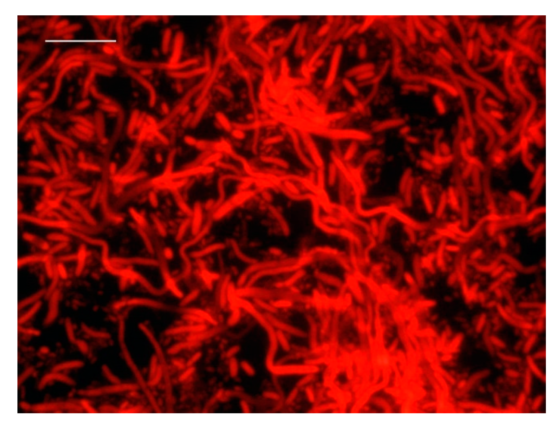
3.1. DVC-FISH Assay Optimization

An optimized protocol was established for the specific detection of viable V. parahaemolyticus cells. Figure 1 shows the control of the original size of the V. parahaemolyticus cells without incubation with DVC broth.
Four concentrations of ciprofloxacin (0.5, 0.75, 1 and 1.5 μg/mL) and five incubation times (5, 6, 7, 8 and 16 h) were tested.
No cells were observed after 16 h of incubation at any concentration.
A concentration of 0.5 μg/mL did not produce enough cell elongation at any of the incubation times. Although the cells were elongated after 7 and 8 h of incubation, at this concentration, they did not increase their size to more than twice the original.
After 7 or more hours of incubation and a high ciprofloxacin concentration (0.75, 1 and 1.5 μg/mL), cells appeared degraded in all cases (Figure 2).
In the subsequent assays, the concentrations of 0.75, 1 and 1.5 μg/mL of ciprofloxacin were tested after 5 and 6 h of incubation. Elongation of viable cells occurred with the three concentrations of ciprofloxacin (0.75, 1 and 1.5 μg/mL), although at 0.75 μg/mL, greater elongation was detected. Moreover, a drop in the number of cells was observed by LIVE/DEAD fluorescent assay at concentrations of 1 and 1.5 μg/mL. In addition, 0.75 μg/mL and 5 and 6 h of incubation yielded similar results, as it was possible to observe viable elongated cells without any alteration associated with the presence of an antibiotic.
Results showed that long incubation periods in the presence of an antibiotic caused cell degradation. Therefore, the optimal conditions for DVC analysis turned out to be 0.75 μg/mL of ciprofloxacin and 5 h of incubation (Figure 3).
Once the optimal conditions for the DVC procedure were selected for V. parahaemolyticus, the number of viable cells before and after the DVC incubation was also tested by both culture on mTSA as described in Materials and Methods and by using the LIVE/DEAD BacLight kit. A DVC broth without an antibiotic, inoculated with the same V. parahaemolyticus inoculum and incubated for 5 h, was used as the control. As shown in Table 1, the results obtained by culture were similar to those obtained by DVC-FISH assay, meaning that that almost all cells were both viable and cultivable for these conditions. Furthermore, in the DVC medium, the number of cells was not affected by 5 h of incubation, and the antibiotic did not seem to affect bacteria viability. Therefore, the selected antibiotic concentration and incubation time results were optimal, taking into consideration that cell elongation was achieved without degradation or replication of the cells. It was observed that in the control culture (DVC broth without antibiotic), viable cells were not elongated, and cells counts were higher than the ones obtained at time 0 h due to their ability to replicate.
Starting from an initial culture of 2.5 × 10⁸ cfu/mL of V. parahaemolyticus, serial decimal dilutions were made in order to determine the detection limit of the DVC-FISH technique. According to the presence and appearance of the cells in each dilution, the detection limit was established at 102 cells/mL (Figure 4).

3.2. Detection of Viable V. parahaemolyticus in Inoculated Samples

The DVC-FISH technique was applied in real samples inoculated with V. parahaemolyticus to assess its usefulness in detecting viable cells of the pathogen in two bivalve mollusks, oysters and mussels. The results were compared with the ones obtained when applying the culture to mTSA agar plates to detect viable bacteria. Different conditions were simulated to ascertain the growth kinetics of V. parahaemolyticus in contaminated oysters and mussels until their arrival to the consumer. The process called “transport”, defined as the time passed from the purchase of a food until its storage in the refrigerator, was simulated for oysters and mussels at 22 °C for 10, 20 and 30 min. As shown in Table 2, the initial inoculum of V. parahaemolyticus in oysters and mussels contained 1.7 × 10⁷ cfu/mL and 1.3 × 10⁶ cfu/mL, respectively. The bacterial plate counts increased in both samples throughout the time. The same tendency was detected in the FISH and DVC-FISH counts, which were slightly higher than those obtained by culture. FISH counts were the highest due to the count of VBNC, viable and non-viable cells.
Subsequently, the microbiological processes that occur when food is stored in refrigeration were simulated at 4 and 8 °C for different periods of time. In the simulation at 4 °C, the initial inocula for the samples of oysters and mussels were around 1.8 × 10⁷ cfu/mL, respectively (Table 3). Results showed that the concentration of the culture and the total cells went down progressively. Viable cells were identified by the DVC-FISH technique throughout the whole refrigeration process at 4 °C (96 h). The counts obtained by cultivation and FISH decreased by between three and five orders of magnitude for both oysters and mussels. However, after 48 h in oysters and 96 h in mussels, V. parahaemolyticus was not detected by culture, even though the cells were elongated after the DVC. For the simulation at 8 °C, the oysters’ and mussels’ initial inocula were around 1.8 × 10⁷ cfu/mL, respectively (Table 4). A reduction was also reported, but more gradually than at 4 °C. A decrease of 3–4 orders of magnitude was observed by traditional cultivation. DVC yielded counts which were one log unit higher than the culture counts. FISH analysis, as resulting from the simulation at 4 °C, generated the highest counts.

3.3. Detection and Testing of NA Stability of V. parahaemolyticus

Bacterial DNA and RNA were isolated from all the samples of oysters and mussels. PCR and later, electrophoresis, were performed to verify the stability of the V. parahaemolyticus nucleic acids. The specific amplifications for the tlh gene from both the DNA and RNA of V. parahaemolyticus were detected in all the assays and times analyzed. Therefore, although the temperature affected the growth of the culture, DNA and RNA remained stable without degrading.

4. Discussion

In this study, a combined DVC-FISH method was optimized for the specific detection and quantification of viable pathogenic V. parahaemolyticus in two bivalve mollusks, oysters and mussels. The developed method reduced the total detection and identification time of V. parahaemolyticus in these seafoods by up to 4 h, which could be a good complement to detection by traditional cultivation methods such as the official ISO 21872-1:2017 method, which requires a total analysis time of 4 days.
The FISH method is a useful tool for the initial identification of microbial pathogens, and it is considered quicker and associated with less hands-on time than traditional culture methods [32,50]. However, this technique on its own cannot ensure that bacteria remain alive.
In this regard, the DVC technique has been widely applied to the study of aquatic samples, including seawater [51,52], groundwater [53], wastewater [54], fresh water [36,37,55] and drinking water [39,56,57]. It has also been used for the study of cell viability in food samples [58]. In this method, bacteria are exposed to a medium that contains a sublethal concentration of an antibiotic substance, which prevents cell division by inhibiting DNA gyrase without affecting other metabolic activities such that cell growth is not impeded [35]. Therefore, when cells are cultivated under these conditions, the amount of ribosomal RNA in active cells increases, resulting in elongated cells [35], which are then enumerated as viable cells because dead cells remain unchanged [59]. Cells are considered viable when their length is at least twice the original one [58]. It is essential that no cell division occurs in order to ensure that the DVC analysis is accurate, which is generally confirmed by comparing cell presence before and after incubation [52]. However, in the DVC medium, bacterial cell division can be underestimated due, among other factors, to cell lysis caused by high concentrations of antibiotics, which can counterbalance the increase in the number of bacteria because of cell division. On the other hand, cell division can be overestimated in some strains that are resistant to antibiotics when there is an insignificant increase in their cell number compared with the total bacteria count but that is significant for their own population [31]. Therefore, in order to obtain reliable and accurate results, it is fundamental to select the antibiotic and its dose properly.
Due to the lack of success with some culture methods [60], and as PCR only indicates the presence of DNA, a combination of molecular techniques such as DVC-FISH seems to be the most efficient strategy for the detection and the quantification of viable cells, including those in a VBNC state. In fact, it has been reported regarding the detection of viable cells of E. coli in various types of samples [61], H. pylori in wastewater and drinking water [62], L. monocytogenes in complex mixed communities such as wastewater samples [30], L. delbrueckii subsp. bulgaricus and S. thermophilus in inoculated feces samples [36] and Yersinia spp. in minced pork meat [50].
To ensure the inhibition of cell division and the maximum elongation of bacteria in aquatic samples, different combinations of several antibiotics (mainly nalidixic, piromidic and pipemidic acid as well as ciprofloxacin) have been proposed [42,51,53,57]. It has been demonstrated in many studies that V. parahaemolyticus exhibit multiple-antibiotic resistance due to the misuse of antibiotics to control infections in aquaculture production [63]. In a research on antimicrobial resistance, Yano et al. [64] revealed the occurrence of resistance to nalidixic acid and ampicillin in V. parahaemolyticus strains derived from shrimp. The selected antibiotic for the current research, ciprofloxacin, is an effective inhibitor of cell division in Gram-positive and Gram-negative bacteria, and its effectiveness has been proven in V. parahaemolyticus strains derived from water and marine samples [65,66] and from oysters [67]. The DVC medium with 0.75 μg/mL of ciprofloxacin turned out to be effective in preventing cell division of V. parahaemolyticus and promoting the elongation of viable cells after 5 h of incubation. Once the DVC-FISH procedure was optimized, the detection limit of the technique was established at 102 cells/mL in inoculated samples. This limit could be improved by centrifuging the whole amount of DVC broth instead of just 1 mL.
However, the DVC-FISH technique has certain limitations. Thus, as indicated by Huber et al. [68], despite the high specificity of FISH and the possibility of direct quantitative imaging, some of its key constraints are that the time of the assay and the probe consumption prevent its regular use in diagnostics. The influence of the fixation/permeabilization step [69] and the design of specific probes which need to be specific, sensitive, and relatively short-chained to make cell penetration easier [70] have also been pointed out. The selection of the appropriate DNA gyrase-inhibitor antibiotic, its concentration and the necessary incubation time are also factors that must be taken into account. Finally, background noise in the sample sometimes limits the ability to observe and determine cell size (36, 41, 58).
In order to determine the suitability of the optimized technique to detect viable cells of V. parahaemolyticus in seafood samples, DVC-FISH was applied to track the pathogen viability through a survival study. Vibrio parahaemolyticus is a Gram-negative bacteria related to seafood-associated illness, which not only causes symptoms of gastroenteritis, but also wound infections and septicemia [71]. Numerous cases of gastroenteritis caused by V. parahaemolyticus have been detected and isolated in Spain, Greece, Britain, Denmark and other countries in Europe over the years [72,73]. Love et al. [74] demonstrated that the cold chain was effective in reducing the risk of V. parahaemolyticus in seafood by modeling the effects of temperature on the growth of this bacterium in oysters. Moderate bacterial growth was detected in oysters that had not entered the cold chain, while bacterial growth stopped once they were under refrigeration. This information is in line with the results of the present study, where it was observed that both bacterial counts and viability of V. parahaemolyticus in samples of oysters and mussels increased throughout the time of transport at 20 °C, from the purchase to the storage of the food in the refrigerator. However, a rapid decrease in viable and, therefore, potentially virulent cells was detected when the samples were under refrigeration at 4 °C and 8 °C. The same trend was detected in the study carried out by López-Joven [75], where the bacterial load of V. parahaemolyticus in Manila clam samples was lower at 4 °C and 15 °C than at 28 °C in all treatments. Therefore, the control of temperatures and times in the production and transport chains as well as optimal manipulation, preparation and storage conditions are crucial to ensure food safety. The risk of Vibrio spp. can be attenuated with shorter times of storage and the rapid cooling of seafood [76]. The wrong storage temperature at above 20 °C, even with short storage times, could increase the risk of V. parahaemolyticus infection through contaminated seafood if the necessary bacterial load was reached because cells of this pathogen remain metabolically active at this temperature, as reported in our work.
Our results showed that a higher number of samples were recorded as positive for V. parahaemolyticus by DVC-FISH analysis than by culture, since the first included the VBNC cells. Furthermore, this technique allowed us to specifically identify and quantify the viable cells directly in a few hours. Similar results have been reported for the detection and enumeration of other pathogens such as L. monocytogenes in wastewater samples [30], where the DVC-FISH technique was more sensitive than PCR and culture methods. Furthermore, an advantage of this technique is that in samples with an initial contamination of the pathogen above 102 viable cells/g, prior culture enrichment is not required due to the detection limit obtained. mTSA medium was also applied to compare the results with the ones obtained by molecular techniques. The presence of V. parahaemolyticus has been commonly detected by different culture-based methods [77]. However, our results revealed that traditional culture methods can be limited in detecting V. parahaemolyticus in food matrices because the cells can acquire a VBNC state, in which they are metabolically active but unable to grow on a synthetic media [77].
As demonstrated in this study, DNA remains stable under stress conditions that cause cell death, so PCR is not a useful technique because it is unable to distinguish between viable and non-viable cells in food samples [29], which leads to false positive results [78]. Moreover, RNA was detected with the same intensity throughout the survival assays. Hence, in this case, the expression of the tlh gene was not a good viability marker to detect viable V. parahaemolyticus. Other authors reported the inefficiency of RNA detection as bacterial viability identification [79].
The combination of traditional culture methods with the DVC-FISH could turn into the most efficient strategy for the detection and quantification of V. parahaemolyticus in bivalve matrices. Our results show that the DVC–FISH method is a useful and fast tool for the identification and quantification of viable forms of V. parahaemolyticus in oysters and mussels and could be used for both survival studies and food quality control in naturally contaminated samples.

5. Conclusions

The DVC–FISH method is a useful and fast tool for the detection and quantification of viable forms of V. parahaemolyticus in oysters and mussels and could be used for both survival studies and food control. The optimal conditions were 0.75 μg/mL of ciprofloxacin in DVC broth and 5 h of incubation. The developed method allowed us to detect and identify viable V. parahaemolyticus in inoculated seafood in just 4 h, which was an excellent complement to the ISO standard, which requires a time of several days.
In the process of simulating transport conditions of mussels and oysters at 22 °C, bacterial counts by culture, FISH and DVC-FISH increased significantly over time. However, the viable cell counts obtained by DVC-FISH were more accurate than the ones obtained by culture because both cultivable and VBNC cells were detected by this technique. In the simulation process of refrigeration conditions at 4 and 8 °C for oysters and mussels, V. parahaemolyticus counts decreased by between three and four orders of magnitude by cultivation and by FISH. After 48 h in refrigeration, not only did Vibrio parahaemolyticus lose the ability to grow, but the cells also did not elongate after DVC. Finally, the V. parahaemolyticus nucleic acid stability test through specific amplification of the tlh gene determined that the DNA and RNA remained stable, without degrading during the process.

Author Contributions

All authors contributed equally to this work. All authors have read and agreed to the published version of the manuscript.

Funding

This research received no external funding.

Conflicts of Interest

The authors declare no conflict of interest.

References

  1. Romalde, J.; Dieguez, A.; Lasa, A.; Balboa, S. New Vibrio species associated to molluscan microbiota: A review. Front. Microbiol. 2014, 4, 413. [Google Scholar] [CrossRef]
  2. Oliver, J. The Biology of Vibrio vulnificus. Microbiol. Spectr. 2015, 3. [Google Scholar] [CrossRef] [PubMed] [Green Version]
  3. Garrido-Maestu, A.; Lozano-Leon, A.; Rodríguez-Souto, R.R.; Vieites-Maneiro, R.; Chapela, M.J.; Ana, G.; Cabado, A.G. Presence of pathogenic Vibrio species in fresh mussels harvested in the southern Rias of Galicia (NW Spain). Food Control 2016, 59, 759–765. [Google Scholar] [CrossRef]
  4. Drake, S.L.; DePaola, A.; Jaykus, L.A. An overview of Vibrio vulnificus and Vibrio parahaemolyticus. Compr. Rev. Food Sci. Food Saf. 2007, 6, 120–144. [Google Scholar] [CrossRef]
  5. Kokashvili, T.; Whitehouse, C.A.; Tskhvediani, A.; Grim, C.J.; Elbakidze, T.; Mitaishvili, N.; Janelidze, N.; Janelidze, E.; Haley, E.J.; Lashkhi, E.; et al. Occurrence and Diversity of Clinically Important Vibrio Species in the Aquatic Environment of Georgia. Front. Public Health 2015, 3, 232. [Google Scholar] [CrossRef]
  6. Cantet, F.; Hervio-Heath, D.; Caro, A.; Le Mennec, C.; Monteil, C.; Quemere, C. Quantification of Vibrio parahaemolyticus, V. vulnificus and V. cholerae in French Mediterranean coastal lagoons. Res. Microbiol. 2013, 164, 867–874. [Google Scholar] [CrossRef] [Green Version]
  7. Johnson, C.N.; Flowers, A.R.; Noriea, N.F.; Zimmerman, A.M.; Bowers, J.C.; DePaola, A. Relationships between environmental factors and pathogenic Vibrios in the northern Gulf of Mexico. Appl. Environ. Microbiol. 2010, 76, 7076–7084. [Google Scholar] [CrossRef] [PubMed] [Green Version]
  8. Tall, A.; Teillon, A.; Boisset, C.; Delesmont, R.; Touron-Bodilis, A.; Hervio-Heath, D. Real-time PCR optimization to identify environmental Vibrio spp. strains. J. Appl. Microbiol. 2012, 113, 361–372. [Google Scholar] [CrossRef] [Green Version]
  9. European Commission. DG (SANTE) 2015-7486—MR—Final Report of an Audit Carried Out in the United States from 17 March 2015 to 27 March 2015 in Order to Evaluate the Control Systems in Place Governing the Production of Bivalve Molluscs and Fishery Products Derived Therefrom Intended for Export to the European Union; European Commission: Brussels, Belgium, 2015. [Google Scholar]
  10. Harwood, V.J.; Gandhi, J.P.; Wright, A.C. Methods for isolation and confirmation of Vibrio vulnificus from oysters and environmental sources: A review. J. Microbiol. Methods 2004, 59, 301–316. [Google Scholar] [CrossRef]
  11. Zhao, F.; Zhao, D.; Cao, H.; Ma, L.; Jiang, Y. Distribution, serological and molecular characterization of Vibrio parahaemolyticus from shellfish in the eastern coast in China. Food Control 2011, 22, 1095–1100. [Google Scholar] [CrossRef]
  12. Abdullah Sani, N.; Ariyawansa, S.; Salam Babji, A.; Khair Hashim, J. The risk assessment of Vibrio parahaemolyticus in cooked black tiger shrimps (Penaeus monodon) in Malaysia. Food Control 2013, 31, 546–552. [Google Scholar] [CrossRef]
  13. Baker-Austin, C.; Stockley, L.; Rangdale, R.; Martinez-Urtaza, J. Environmental occurrence and clinical impact of Vibrio vulnificus and Vibrio parahaemolyticus: A European perspective. Environ. Microbiol. Rep. 2010, 2, 7–18. [Google Scholar] [CrossRef] [PubMed]
  14. Baker-Austin, C.; Trinanes, J.A.; Taylor, N.G.H.; Hartnell, R.; Siitonen, A.; Martinez-Urtaza, J. Emerging Vibrio risk at high latitudes in response to ocean warming. Nat. Clim. Chang. 2012, 3, 73–77. [Google Scholar] [CrossRef]
  15. Baker-Austin, C.; Trinanes, J.; Gonzalez-Escalona, N.; Martinez-Urtaza, J. Noncholera vibrios: The microbial barometer of climate change. Trends Microbiol. 2016, 25, 76–84. [Google Scholar] [CrossRef] [PubMed] [Green Version]
  16. Martinez-Urtaza, J.; Baker-Austin, C.; Jones, J.; Newton, A.; Gonzalez-Aviles, G.; DePaola, A. Spread of Pacific Northwest Vibrio parahaemolyticus Strain. N. Engl. J. Med. 2013, 369, 1573–1574. [Google Scholar] [CrossRef] [PubMed] [Green Version]
  17. Scallan, E.; Hoekstra, R.; Angulo, F.; Tauxe, R.; Widdowson, M.; Roy, S.; Jones, J.; Griffin, P. Foodborne Illness Acquired in the United States—Major Pathogens. Emerg. Infect. Dis. 2011, 17, 7–15. [Google Scholar] [CrossRef]
  18. Hartnell, R.E.; Stockley, L.; Keay, W.; Rosec, J.P.; Hervio-Heath, D.; Van den Berg, H.; Leoni, F.; Ottaviani, D.; Henigman, U.; Denayer, S.; et al. A pan-European ring trial to validate an International Standard for detection of Vibrio cholerae, Vibrio parahaemolyticus and Vibrio vulnificus in seafoods. Int. J. Food Microbiol. 2019, 288, 58–65. [Google Scholar] [CrossRef] [Green Version]
  19. Xu, H.; Roberts, N.; Singleton, F.L.; Attwell, R.W.; Grimes, D.J.; Colwell, R.R. Survival and viability of non culturable Escherichia coli and Vibrio cholerae in the estuarine and marine environment. Microb. Ecol. 1982, 8, 313–323. [Google Scholar] [CrossRef]
  20. Li, L.; Mendis, N.; Trigui, H.; Oliver, J.D.; Faucher, S.P. The importance of the viable but non-culturable state in human bacterial pathogens. Front. Microbiol. 2014, 5, 258. [Google Scholar] [CrossRef] [Green Version]
  21. Meng, L.; Alter, T.; Aho, T.; Huehn, S. Gene expression profiles of Vibrio parahaemolyticus in viable but non-culturable state. FEMS Microbiol. Ecol. 2015, 91, 91. [Google Scholar] [CrossRef] [Green Version]
  22. Oliver, J. Recent findings on the viable but nonculturable state in pathogenic bacteria. FEMS Microbiol. Rev. 2010, 34, 415–425. [Google Scholar] [CrossRef] [Green Version]
  23. Datta, P.P.; Bhadra, R.K. Cold shock response and major cold shock proteins of Vibrio cholerae. Appl. Environ. Microbiol. 2003, 69, 6361–6369. [Google Scholar] [CrossRef] [PubMed] [Green Version]
  24. Limthammahisorn, S.; Brady, Y.; Arias, C. In vivo gene expression of cold shock and other stress-related genes in Vibrio vulnificus during shellstock temperature control conditions in oysters. J. Appl. Microbiol. 2009, 106, 642–650. [Google Scholar] [CrossRef] [PubMed]
  25. Nowakowska, J.; Oliver, J.D. Resistance to environmental stresses by Vibrio vulnificus in the viable but nonculturable state. FEMS Microbiol. Ecol. 2013, 84, 213–222. [Google Scholar] [CrossRef] [Green Version]
  26. Colwell, R.; Grimes, D. Nonculturable Microrganisms in the Environment; American Society for Microbiology Press: Washington, DC, USA, 2000; pp. 325–342. [Google Scholar]
  27. Baffone, W.; Citterio, B.; Vittoria, E.; Casaroli, A.; Campana, R.; Falzano, L.; Donelli, G. Retention of virulence in viable but non-culturable halophilic Vibrio spp. Int. J. Food Microbiol. 2003, 89, 31–39. [Google Scholar] [CrossRef]
  28. Falcioni, T.; Papa, S.; Campana, R.; Manti, A.; Battistelli, M.; Baffone, W. State transitions of Vibrio parahaemolyticus VBNC cells evaluated by flow cytometry. Cytom. B Clin. Cytom. 2008, 74, 272–281. [Google Scholar] [CrossRef] [PubMed]
  29. Okoh, A.I.; Odjadjare, E.E.; Igbinosa, E.O.; Osode, A.N. Wastewater treatment plants as a source of microbial pathogens in receiving watersheds. Afr. J. Biotechnol. 2007, 6, 2932–2944. [Google Scholar]
  30. Moreno, Y.; Ballesteros, L.; García-Hernández, J.; Santiago, P.; González, A.; Ferrús, M. Specific detection of viable Listeria monocytogenes in Spanish wastewater treatment plants by Fluorescent in situ Hybridization and PCR. Water Res. 2011, 45, 4634–4640. [Google Scholar] [CrossRef] [Green Version]
  31. Wu, S.; Ueno, D.; Inoue, K.; Someya, T. Direct Viable Count Combined with Fluorescence in situ Hybridization (DVC-FISH) for Specific Enumeration of Viable Escherichia coli in Cow Manure. Microbes Environ. 2009, 24, 33–38. [Google Scholar] [CrossRef] [Green Version]
  32. Frickmann, H.; Zautner, A.; Moter, A.; Kikhney, J.; Hagen, R.; Stender, H.; Poppert, S. Fluorescence in situ hybridization (FISH) in the microbiological diagnostic routine laboratory: A review. Crit. Rev. Microbiol. 2017, 43, 263–293. [Google Scholar] [CrossRef]
  33. Kogure, K.; Simidu, U.; Taga, N. A tentative direct microscopic method for counting living marine bacteria. Canad. J. Microb. 1979, 25, 415–420. [Google Scholar] [CrossRef]
  34. Servais, P.; Prats, J.; Passerat, J.; Garcia-Armisen, T. Abundance of culturable versus viable Escherichia coli in freshwater. Canad. J. Microb. 2009, 55, 905–909. [Google Scholar] [CrossRef]
  35. Duodu, S.; Colquhoun, D. Monitoring the survival of fish-pathogenic Francisella in water microcosms. FEMS Microbiol. Ecol. 2010, 74, 534–541. [Google Scholar] [CrossRef] [Green Version]
  36. Piqueres, P.; Moreno, Y.; Alonso, J.; Ferrús, M. A combination of direct viable count and fluorescent in situ hybridization for estimating Helicobacter pylori cell viability. Res. Microbiol. 2006, 157, 345–349. [Google Scholar] [CrossRef] [PubMed]
  37. Kalmbach, S.; Manz, W.; Szewzyk, U. Isolation of new bacterial species from drinking water biofilms and proof of their in situ dominance with highly specific 16S rRNA probes. Appl. Environ. Microbiol. 1997, 63, 4164–4170. [Google Scholar] [CrossRef] [Green Version]
  38. Armisen, T.G.; Servais, P. Combining direct viable count (DVC) and fluorescent in situ hybridisation (FISH) to enumerate viable E-coli in rivers and wastewaters. Water Sci. Technol. 2004, 50, 271–275. [Google Scholar] [CrossRef]
  39. Baudart, J.; Coallier, J.; Laurent, P.; Prevost, M. Rapid and Sensitive Enumeration of Viable Diluted Cells of Members of the Family Enterobacteriaceae in Freshwater and Drinking Water. Appl. Environ. Microbiol. 2002, 68, 5057–5063. [Google Scholar] [CrossRef] [PubMed] [Green Version]
  40. García-Hernández, J.; Moreno, Y.; Amorocho, C.M.; Hernández, M. A combination of direct viable count and fluorescence in situ hybridization for specific enumeration of viable Lactobacillus delbrueckii subsp. bulgaricus and Streptococcus thermophilus. Lett. Appl. Microbiol. 2011, 54, 247–254. [Google Scholar] [CrossRef] [PubMed]
  41. Moreno-Mesonero, L.; Moreno, Y.; Alonso, J.L.; Ferrús, M.A. DVC-FISH and PMA-qPCR techniques to assess the survival of Helicobacter pylori inside Acanthamoeba castellanii. Res. Microbiol. 2016, 167, 29–34. [Google Scholar] [CrossRef] [PubMed]
  42. Barcina, I.; Arana, I.; Santoram, P.; Iriberri, J.; Egca, L. Direct viable count of Gram positive and Gram negative bacteria using ciprofloxacin as inhibitor of cellular division. J. Microbiol. Met. 1995, 22, 139–150. [Google Scholar] [CrossRef]
  43. Anupama, K.P.; Deeksh, A.K.; Deeksha, A.; Karunasagar, I.; Karunasagar, I.; Maiti, B. Comparative performance of TCBS and TSA for the enumeration of trh+ Vibrio parahaemolyticus by direct colony hybridization. J. Microbiol. Methods 2019, 157, 37–42. [Google Scholar] [CrossRef] [PubMed]
  44. Sawabe, T.; Yoshizawa, A.; Kawanishi, Y.; Komatsu-Takeda, E.; Nakagawa, S.; Sawabe, T.; Ootubo, M.; Satomi, M.; Yano, Y.; Yamazaki, K. Multi-Probe-Fluorescence in situ Hybridization for the Rapid Enumeration of Viable Vibrio parahaemolyticus. Microbes Environ. 2009, 24, 259–264. [Google Scholar] [CrossRef] [Green Version]
  45. Bisha, B.; Simonson, J.; Janes, M.; Bauman, K.; Goodridge, L. A review of the current status of cultural and rapid detection of Vibrio parahaemolyticus. Int. J. Food Sci. Technol. 2012, 47, 885–899. [Google Scholar] [CrossRef]
  46. Amann, R.I.; ludwig, W.; Schleifer, K.H. Phylogenetic identification and in situ detection of individual microbial cells without cultivation. Microbiol. Mol. Biol. Rev. 1995, 59, 143–169. [Google Scholar] [CrossRef] [Green Version]
  47. Besnard, V.; Federighi, M.; Cappelier, J.M. Development of a direct viable count procedure for the investigation of VBNC state in Listeria monocytogenes. Lett. Appl. Microbiol. 2000, 31, 77–81. [Google Scholar] [CrossRef]
  48. Yoshimitsu, M.; Wai, S.N.; Ishikawa, T.; Takade, A.; Yoshida, S.-I. Resuscitation of viable but nonculturable cells of Vibrio parahaemolyticus induced at low temperature under starvation. FEMS Microbiol. Lett. 2000, 186, 115–120. [Google Scholar]
  49. Bej, A.K.; Patterson, D.P.; Brasher, C.W.; Vickery, M.; Jones, D.D.; Kaysner, C.A. Detection of total and haemolysin-producing Vibrio parahaemolyticus in shellfish using multiplex PCR amplification of tl, tdh and trh. J. Microbiol. Methods 1999, 36, 215–225. [Google Scholar] [CrossRef]
  50. Rohde, A.; Hammerl, J.A.; Appel, B.; Dieckmann, R.; Al Dahouk, S. Differential detection of pathogenic Yersinia spp. by fluorescence in situ hybridization. Food Microbiol. 2017, 62, 39–45. [Google Scholar] [CrossRef] [PubMed]
  51. Joux, F.; Lebaron, P. Ecological implications of an improved direct viable count method for aquatic bacteria. Appl. Environ. Microbiol. 1997, 63, 3643–3647. [Google Scholar] [CrossRef] [Green Version]
  52. Kogure, K.; Simidu, U.; Taga, N. An improved direct viable count method for aquatic bacteria. Arch. Hydrobiol. 1984, 102, 117–122. [Google Scholar]
  53. Bakermans, C.; Madsen, E.L. Use of substrate responsive-direct viable counts to visualize naphthalene degrading bacteria in a coal tar-contaminated groundwater microbial community. J. Microbiol. Methods 2000, 43, 81–90. [Google Scholar] [CrossRef]
  54. Garcia-Armisen, T.; Servais, P. Enumeration of viable E. coli in rivers and wastewaters by fluorescent in situ hybridization. J. Microbiol. Methods 2004, 58, 269–279. [Google Scholar] [CrossRef]
  55. Kenzaka, T.; Yamaguchi, N.; Prapagdee, B.; Mikami, E.; Nasu, M. Bacterial community composition and activity in urban rivers in Thailand and Malaysia. J. Health Sci. 2001, 47, 353–361. [Google Scholar] [CrossRef] [Green Version]
  56. Bjergbaek, L.; Roslev, P. Formation of nonculturable Escherichia coli in drinking water. J. Appl. Microbiol. 2005, 99, 1090–1098. [Google Scholar] [CrossRef] [PubMed]
  57. Guyard, S.; Mary, P.; Defives, C.; Hornez, J.P. Enumeration and characterization of bacteria in mineral water by improved direct viable count method. J. Appl. Microbiol. 1999, 86, 841–850. [Google Scholar] [CrossRef] [PubMed] [Green Version]
  58. Moreno, Y.; Sanchez-Contreras, J.; Montes, R.M.; García-Hernández, J.; Ballesteros, L.; Ferrús, M.A. Detection and enumeration of viable Listeria monocytogenes cells from ready-to-eat and processed vegetable foods by culture and DVC-FISH. Food Control 2012, 27, 374–379. [Google Scholar] [CrossRef]
  59. García Hernández, J.; Moreno Trigos, Y.; Hernández Pérez, M. In Vivo Study of the Survival of Lactobacillus delbruecki subsp. bulgaricus CECT 4005T and Streptococcus thermophilus CECT 801 by DVC-FISH after Consumption of Fermented Milk. J. Food Sci. 2012, 77, 593–597. [Google Scholar] [CrossRef] [PubMed]
  60. Laupland, K.; Valiquette, L. The changing culture of the microbiology laboratory. Can. J. Infect. Dis Med. Microbiol. 2013, 24, 125–128. [Google Scholar] [CrossRef]
  61. Regnault, B.; Martin-Delautre, S.; Lejay-Collin, M.; Lefèvre, M.; Grimont, P.A.D. Oligonucleotide probe for the visualization of Escherichia coli/Escherichia fergusonii cells by in situ hybridation: Specificity and potential applications. Res. Microbiol. 2000, 151, 521–533. [Google Scholar] [CrossRef]
  62. Moreno, Y.; Ferrús, M.A. Specific detection of cultivable Helicobacter pylori cells from wastewater treatment plants. Helicobacter 2012, 17, 327–332. [Google Scholar] [CrossRef]
  63. Elmahdi, S.; DaSilva, L.V.; Parveen, S. Antibiotic resistance of Vibrio parahaemolyticus and Vibrio vulnificus in various countries: A review. Food Microbiol. 2016, 57, 128–134. [Google Scholar] [CrossRef] [PubMed] [Green Version]
  64. Yano, Y.; Hamano, K.; Satomi, M.; Tsutsui, I.; Aue-umneoy, D. Diversity and characterization of oxytetracycline-resistant bacteria associated with non-native species, white-leg shrimp (Litopenaeus vannamei), and native species, black tiger shrimp (Penaeus monodon), intensively cultured in Thailand. J. Appl. Microbiol. 2014, 110, 713–722. [Google Scholar] [CrossRef] [PubMed]
  65. Akinbowale, O.L.; Peng, H.; Barton, M.D. Antimicrobial resistance in bacteria isolated from aquaculture sources in Australia. J. Appl. Microbiol. 2006, 100, 1103–1113. [Google Scholar] [CrossRef] [PubMed]
  66. Baker-Austin, C.; McArthur, J.V.; Lindell, A.H.; Wright, M.F.; Tuckfield, R.C.; Gooch, J.; Warner, L.; Oliver, J.; Stepanauskas, R. Multi-site Analysis Reveals Widespread Antibiotic Resistance in the Marine Pathogen Vibrio vulnificus. Microb. Ecol. 2009, 57, 151–159. [Google Scholar] [CrossRef] [PubMed]
  67. Han, F.; Walker, R.D.; Janes, M.E.; Prinyawiwatkul, W.; Ge, B. Antimicrobial Susceptibilities of Vibrio parahaemolyticus and Vibrio vulnificus Isolates from Louisiana Gulf and Retail Raw Oysters. Appl. Environ. Microbiol. 2007, 73, 7096–7098. [Google Scholar] [CrossRef] [Green Version]
  68. Huber, D.; von Voithenberg, V.L.; Kaigala, G.V. Fluorescence in situ hybridization (FISH): History, limitations and what to expect from micro-scale FISH? Micro Nano Eng. 2018, 1, 15–24. [Google Scholar] [CrossRef]
  69. Rocha, R.; Almeida, C.; Azevedo, N.F. Influence of the fixation/permeabilization step on peptide nucleic acid fluorescence in situ hybridization (PNA-FISH) for the detection of bacteria. PLoS ONE 2018, 13, e0196522. [Google Scholar]
  70. Locke, A.; Fitzgerald, S.; Mahadevan-Jansen, A. Advances in Optical Detection of Human-Associated Pathogenic Bacteria. Molecules 2020, 25, 5256. [Google Scholar] [CrossRef]
  71. García, E.; Hernández, A.; Herrero, J.A.; Gómez, J. Cholera and other infections of the Vibrio genus. Medicine 2014, 11, 3317–3321. [Google Scholar]
  72. Qadri, F.; Chowdhury, N.R.; Takeda, Y.; Nair, G.B. Vibrio parahaemolyticus—seafood safety and associations with higher organisms. In Oceans and Health: Pathogens in the Marine Environment; Springer: Boston, MA, USA, 2005; pp. 277–295. [Google Scholar]
  73. Letchumanan, V.; Chan, K.G.; Lee, L.H. Vibrio parahaemolyticus: A review on the pathogenesis, prevalence, and advance molecular identification techniques. Front. Microbiol. 2014, 5, 705. [Google Scholar] [CrossRef] [Green Version]
  74. Love, D.; Lane, R.; Davis, B.; Clancy, K.; Fry, J.; Harding, J.; Hudson, B. Performance of Cold Chains for Chesapeake Bay Farmed Oysters and Modeled Growth of Vibrio parahaemolyticus. J. Food Prot. 2019, 82, 168–178. [Google Scholar] [CrossRef] [PubMed]
  75. López-Joven, C.; de Blas, I.; Roque, A. Temperature effects on the growth and survival of tdh positive Vibrio parahaemolyticus in tissues of postharvest Manila clam (Ruditapes philippinarum). Food Microbiol. 2017, 75, 61–64. [Google Scholar] [CrossRef] [PubMed] [Green Version]
  76. Jones, J.L.; Lydon, K.A.; Kinsey, T.P.; Friedman, B.; Curtis, R.; Schuster, R.; Bowers, J.C. Vibrio parahaemolyticus abundances in oysters. Int. J. Food Microbiol. 2017, 253, 54–58. [Google Scholar] [CrossRef]
  77. Ndraha, N.; Wong, H.; Hsiao, H. Managing the risk of Vibrio parahaemolyticus infections associated with oyster consumption: A review. Compr. Rev. Food Sci. Food Saf. 2020, 19, 1187–1217. [Google Scholar] [CrossRef] [PubMed] [Green Version]
  78. Xu, W. The Identification and Detection Technology of Research in Microorganisms Including Living or Dead Bacteria. In Functional Nucleic Acids Detection in Food Safety; Springer: Singapore, 2016; pp. 343–364. [Google Scholar]
  79. Ju, W.; Moyne, A.; Marco, M.L. RNA-based detection does not accurately enumerate living Escherichia coli O157:H7 cells on plants. Front. Microbiol. 2016, 7, 1–9. [Google Scholar] [CrossRef] [Green Version]
Figure 1. FISH micrographs showing the hybridization of V. parahaemolyticus cells, showing the usual cell size as the control. (bar: 10 μm).
Foods 10 01502 g001
Figure 2. Viable but degraded V. parahaemolyticus cells after incubation in DVC broth (0.75 μg/mL—7 h) (bar: 10 μm).
Foods 10 01502 g002
Figure 3. Detection by FISH of live and dead cells of V. parahaemolyticus after 5 h incubation with ciprofloxacin (0.75 μg/mL). (bar: 10 μm).
Foods 10 01502 g003
Figure 4. Detection of viable V. parahaemolyticus cells by DVC-FISH at a concentration of 102 cells/mL. (bar: 10 μm).
Foods 10 01502 g004
Table 1. Results of culture viability using the LIVE/DEAD BacLight kit.
TimeViable (SYTO) (cells/mL)Not Viable (PI) (cells/mL)mTSA Plate Count (cfu/mL)FISH (cells/mL)
Initial culture (0 h)9.6 × 1063.4 × 1061.4 × 1071.5 × 107
DVC (5 h)2.4 × 1073.3 × 1071.7 × 1073.2 × 107
Control (5 h)6.6 × 1072.6 × 1077.3 × 1071.3 × 108
Table 2. Counts of oyster and mussel samples in transport simulation.
Time (Minutes)mTSA Plate Count (cfu/mL)FISH (cells/mL)DVC-FISH (cells/mL)
OystersMusselsOystersMusselsOystersMussels
01.70 × 1071.30 × 1061.80 × 1079.70 × 1061.70 × 1074.90 × 106
107.00 × 1066.90 × 1068.70 × 1062.10 × 1078.10 × 1069.70 × 106
208.80 × 1069.70 × 1061.40 × 1074.00 × 1079.40 × 1062.80 × 107
Table 3. Counts of oyster and mussel samples in refrigeration simulation at 4 °C.
Time (Hours)mTSA Plate Count (cfu/mL)FISH (cells/mL)DVC-FISH (cells/mL)
OystersMusselsOystersMusselsOystersMussels
09.50 × 1061.65 × 1071.80 × 1071.97 × 1087.04 × 1062.23 × 107
16.24 × 1061.02 × 1076.09 × 1061.57 × 1083.93 × 1061.74 × 107
52.00 × 1058.10 × 1051.78 × 1068.54 × 1071.36 × 1068.54 × 106
241.57 × 1042.06 × 1058.80 × 1059.76 × 1061.40 × 1051.44 × 106
48<104.50 × 1031.68 × 1043.50 × 1056.40 × 1037.80 × 104
96<10<101.30 × 1036.50 × 1046.20 × 1027.30 × 103
Table 4. Counts of oyster and mussel samples in refrigeration simulation at 8 °C.
Time (Hours)mTSA Plate Count (cfu/mL)FISH (cells/mL)DVC-FISH (cells/mL)
OystersMusselsOystersMusselsOystersMussels
01.65 × 1081.78 × 1071.90 × 1087.80 × 1082.00 × 1081.83 × 107
11.03 × 1081.50 × 1061.41 × 1087.19 × 1071.61 × 1087.32 × 106
58.50 × 1061.70 × 1054.80 × 1076.34 × 1071.31 × 1071.44 × 106
242.26 × 1067.70 × 1046.30 × 1063.53 × 1072.40 × 1061.22 × 106
483.50 × 1054.70 × 1041.30 × 1067.32 × 1068.30 × 1051.30 × 105
961.98 × 1041.10 × 1042.11 × 1051.22 × 1067.60 × 1048.20 × 104
Publisher’s Note: MDPI stays neutral with regard to jurisdictional claims in published maps and institutional affiliations.

