
The Bioactivity Prediction of Peptides from Tuna Skin Collagen Using Integrated Method Combining In Vitro and In Silico


Abstract
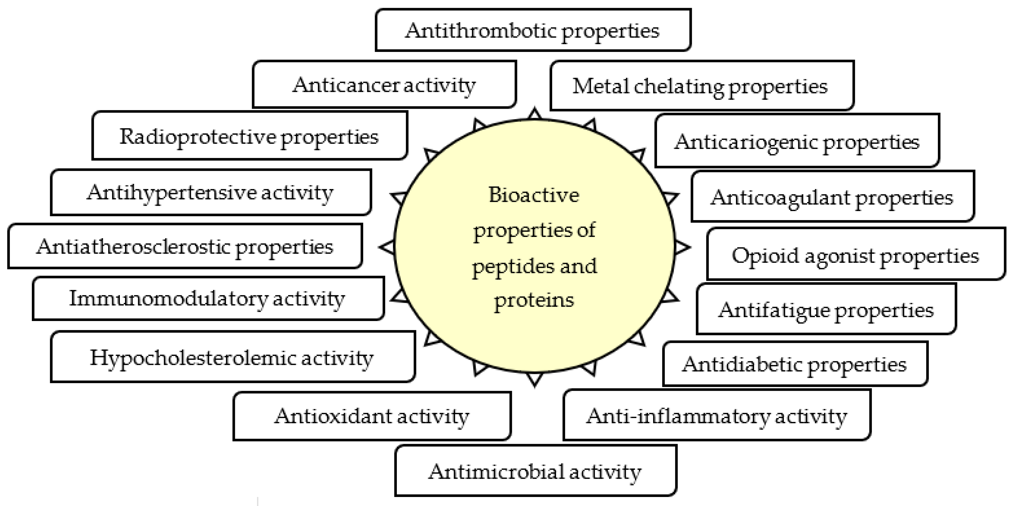
:1. Introduction
2. Materials and Methods
2.1. Materials
2.2. Skin Preparation and Collagen Extraction
2.3. Preparation of Hydrolysates and Peptide Fractions
2.4. Protein Concentration Measurement of Hydrolysates and Peptide Fractions
2.5. Antioxidant Activity Test
2.6. Statistical Study of the Effects of Collagen Types, Enzymes, and Molecular Sizes on Antioxidant Activities
2.7. Amino Acid Sequence Analysis Using LC-MS-MS
2.8. In Silico Analysis for Bioactivity Prediction and Characteristics Study
2.8.1. Bioactivity Prediction Using BIOPEP
2.8.2. Characteristics of Peptides Using PepDraw
2.9. Statistical Analysis
3. Results
3.1. Collagen Extraction
3.2. Hydrolysates and Peptide Fractions
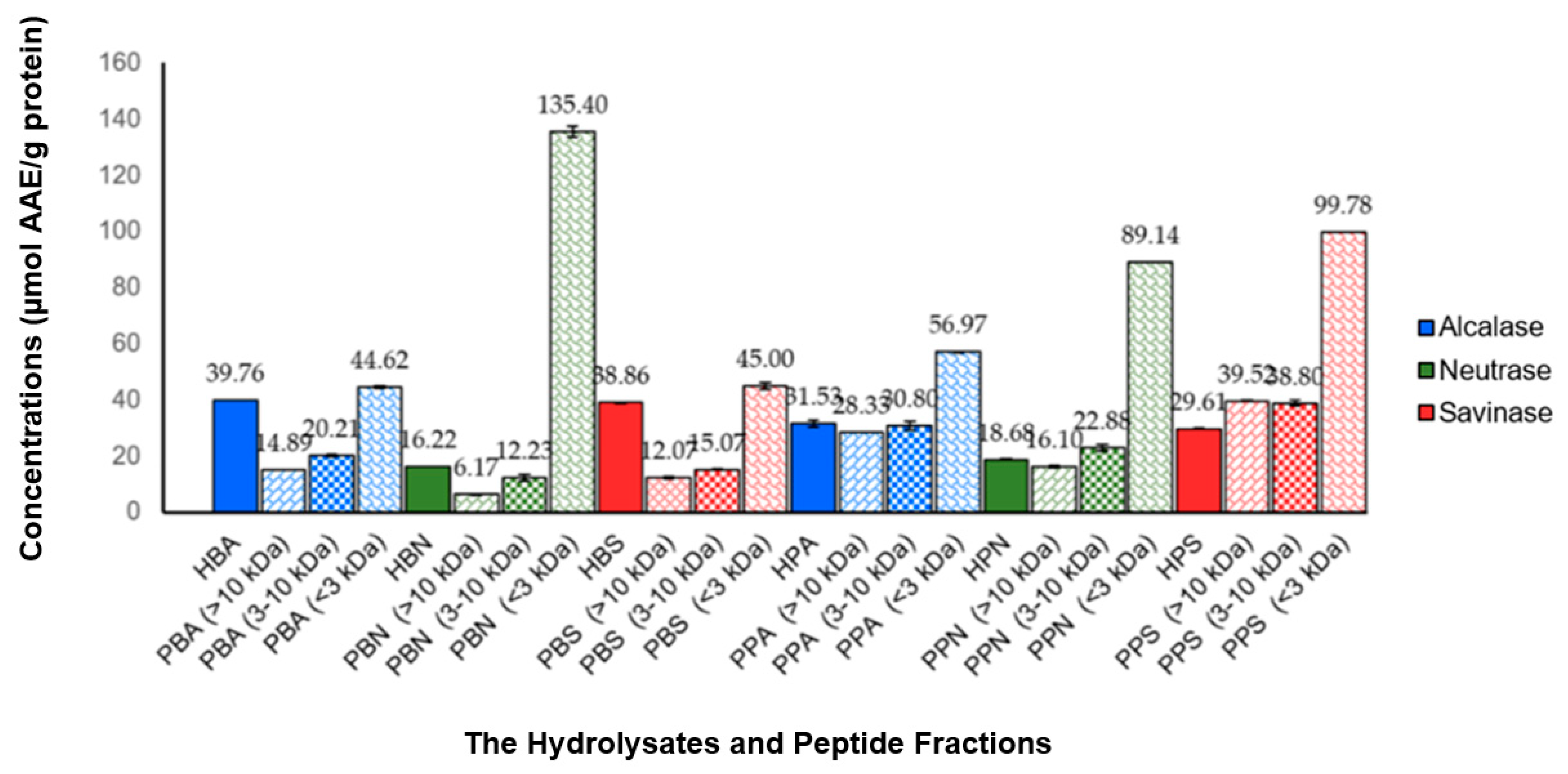
3.3. Concentration of Hydrolysates and Peptide Fractions
3.4. Antioxidant Activity
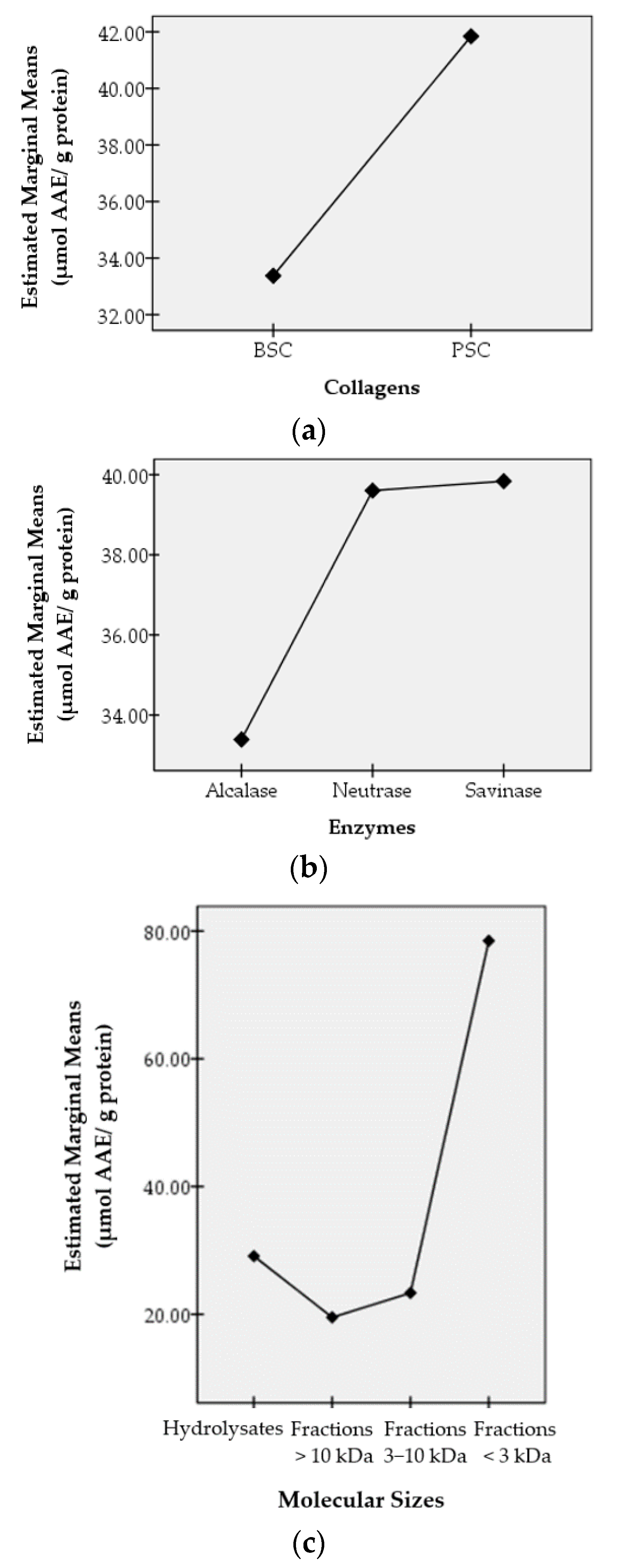
3.5. Effects of Collagen Types, Enzymes, and Molecular Sizes on Antioxidant Activities
3.6. Amino Acid Sequence
3.7. Prediction of Bioactive Properties and Characteristics of Peptides in the PBN and PPS Fractions of <3 kDa
4. Discussion
5. Conclusions
Author Contributions
Funding
Institutional Review Board Statement
Informed Consent Statement
Data Availability Statement
Acknowledgments
Conflicts of Interest
Abbreviations of Sample
| PSC | pepsin soluble collagen |
| BSC | bromelain soluble collagen |
| HPA | hydrolysates from PSC that hydrolyzed using alcalase |
| HPN | hydrolysates from PSC that hydrolyzed using neutrase |
| HPS | hydrolysates from PSC that hydrolyzed using savinase |
| HBA | hydrolysates from BSC that hydrolyzed using alcalase |
| HBN | hydrolysates from BSC that hydrolyzed using neutrase |
| HBS | hydrolysates from BSC that hydrolyzed using savinase |
| PPA | peptide fractions from PSC that hydrolyzed using alcalase |
| PPN | peptide fractions from PSC that hydrolyzed using neutrase |
| PPS | peptide fractions from PSC that hydrolyzed using savinase |
| PBA | peptide fractions from BSC that hydrolyzed using alcalase |
| PBN | peptide fractions from BSC that hydrolyzed using neutrase |
| PBS | peptide fractions from BSC that hydrolyzed using savinase |
References
- Bhat, Z.F.; Kumar, S.; Bhat, H.F. Bioactive peptides of animal origin: A review. J. Food Sci. Technol. 2015, 52, 5377–5392. [Google Scholar] [CrossRef] [Green Version]
- Wu, R.; Chen, L.; Liu, D.; Huang, J.; Zhang, J.; Xiao, X.; Lei, M.; Chen, Y.; He, H. Preparation of antioxidant peptides from salmon by-products with bacterial extracellular proteases. Mar. Drugs 2017, 15, 4. [Google Scholar] [CrossRef] [Green Version]
- Bahar, A.A.; Ren, D. Antimicrobial peptides. Pharmaceuticals 2013, 6, 1543–1575. [Google Scholar] [CrossRef] [Green Version]
- Okamoto, K.; Kawamura, S.; Tagawa, M.; Mizuta, T.; Zahid, H.M.; Nabika, T. Production of an antihypertensive peptide from milk by the brown rot fungus Neolentinus lepideus. Eur. Food Res. Technol. 2020, 246, 1773–1782. [Google Scholar] [CrossRef]
- Korhonen, H.; Pihlanto, A. Bioactive peptides: Production and functionality. Int. Dairy J. 2006, 16, 945–960. [Google Scholar] [CrossRef]
- Carrasco-Castilla, J.; Hernández-Álvarez, A.J.; Jiménez-Martínez, C.; Jacinto-Hernández, C.; Alaiz, M.; Girón-Calle, J.; Vioque, J.; Dávila-Ortiz, G. Antioxidant and metal chelating activities of Phaseolus vulgaris L. var. Jamapa protein isolates, phaseolin and lectin hydrolysates. Food Chem. 2012, 131, 1157–1164. [Google Scholar] [CrossRef]
- Di Bernardini, R.; Mullen, A.M.; Bolton, D.; Kerry, J.; O’Neill, E.; Hayes, M. Assessment of the angiotensin-I-converting enzyme (ACE-I) inhibitory and antioxidant activities of hydrolysates of bovine brisket sarcoplasmic proteins produced by papain and characterisation of associated bioactive peptidic fractions. Meat Sci. 2012, 90, 226–235. [Google Scholar] [CrossRef] [PubMed]
- Yu, Z.; Yin, Y.; Zhao, W.; Wang, F.; Yu, Y.; Liu, B.; Liu, J.; Chen, F. Characterization of ACE-inhibitory peptide associated with antioxidant and anticoagulation properties. J. Food Sci. 2011, 76, 1149–1155. [Google Scholar] [CrossRef] [PubMed]
- Agyei, D.; Pan, S.; Acquah, C.; Danquah, M.K. Bioactivity profiling of peptides from food proteins. In Soft Chemistry and Food Fermentation; Academic Press: Cambridge, MA, USA; Elsevier Inc.: New York, NY, USA, 2017; pp. 49–77. ISBN 9780128114124. [Google Scholar]
- Guaní-Guerra, E.; Santos-Mendoza, T.; Lugo-Reyes, S.O.; Terán, L.M. Antimicrobial peptides: General overview and clinical implications in human health and disease. Clin. Immunol. 2010, 135, 1–11. [Google Scholar] [CrossRef] [PubMed]
- Liu, X.; Cao, X.; Wang, S.; Ji, G.; Zhang, S.; Li, H. Identification of Ly2 members as antimicrobial peptides from zebrafish Danio rerio. Biosci. Rep. 2017, 37, BSR20160265. [Google Scholar] [CrossRef] [PubMed] [Green Version]
- Masso-Silva, J.A.; Diamond, G. Antimicrobial peptides from fish. Pharmaceuticals 2014, 7, 265–310. [Google Scholar] [CrossRef] [PubMed] [Green Version]
- Kim, S.K.; Ngo, D.H.; Vo, T.S. Marine fish-derived bioactive peptides as potential antihypertensive agents. In Advances in Food and Nutrition Research, 1st ed.; Elsevier Inc.: New York, NY, USA, 2012; Volume 65, pp. 249–260. ISBN 9780124160033. [Google Scholar]
- Beaulieu, L.; Bondu, S.; Doiron, K.; Rioux, L.E.; Turgeon, S.L. Characterization of antibacterial activity from protein hydrolysates of the macroalga Saccharina longicruris and identification of peptides implied in bioactivity. J. Funct. Foods 2015, 17, 685–697. [Google Scholar] [CrossRef]
- Qian, B.; Xing, M.; Cui, L.; Deng, Y.; Xu, Y.; Huang, M.; Zhang, S. Antioxidant, antihypertensive, and immunomodulatory activities of peptide fractions from fermented skim milk with Lactobacillus delbrueckii ssp. bulgaricus LB340. J. Dairy Res. 2011, 78, 72–79. [Google Scholar] [CrossRef] [PubMed]
- Chotphruethipong, L.; Aluko, R.E.; Benjakul, S. Hydrolyzed collagen from porcine lipase-defatted seabass skin: Antioxidant, fibroblast cell proliferation, and collagen production activities. J. Food Biochem. 2019, 43, e12825. [Google Scholar] [CrossRef] [PubMed]
- Barzideh, Z.; Latiff, A.A.; Gan, C.Y.; Abedin, M.Z.; Alias, A.K. ACE inhibitory and antioxidant activities of collagen hydrolysates from the ribbon jellyfish (Chrysaora sp.). Food Technol. Biotechnol. 2014, 52, 495–504. [Google Scholar] [CrossRef]
- Sheih, I.C.; Wu, T.K.; Fang, T.J. Antioxidant properties of a new antioxidative peptide from algae protein waste hydrolysate in different oxidation systems. Bioresour. Technol. 2009, 100, 3419–3425. [Google Scholar] [CrossRef]
- Najafian, L.; Babji, A.S. Isolation, purification and identification of three novel antioxidative peptides from patin (Pangasius sutchi) myofibrillar protein hydrolysates. LWT-Food Sci. Technol. 2015, 60, 452–461. [Google Scholar] [CrossRef]
- Gao, D.; Cao, Y.; Li, H. Antioxidant activity of peptide fractions derived from cottonseed protein hydrolysate. J. Sci. Food Agric. 2010, 90, 1855–1860. [Google Scholar] [CrossRef]
- Stadnik, J.; Keska, P. Meat and fermented meat products as a source of bioactive peptides. Acta Sci. Pol. Technol. Aliment. 2015, 14, 181–190. [Google Scholar] [CrossRef]
- Sae-leaw, T.; Benjakul, S. Antioxidant activities of hydrolysed collagen from salmon scale ossein prepared with the aid of ultrasound. Int. J. Food Sci. Technol. 2018, 53, 2786–2795. [Google Scholar] [CrossRef]
- Ding, D.D.; Du, B.; Zhang, C.; Zaman, F.; Huang, Y. Isolation and identification of an antioxidant collagen peptide from skipjack tuna (Katsuwonus pelamis) bone. RSC Adv. 2019, 9, 27032–27041. [Google Scholar] [CrossRef] [Green Version]
- Benjakul, S.; Karnjanapratum, S.; Visessanguan, W. Production and characterization of odorless antioxidative hydrolyzed collagen from seabass (Lates calcarifer) skin without descaling. Waste Biomass Valorization 2018, 9, 549–559. [Google Scholar] [CrossRef]
- Chi, C.F.; Cao, Z.H.; Wang, B.; Hu, F.Y.; Li, Z.R.; Zhang, B. Antioxidant and functional properties of collagen hydrolysates from Spanish mackerel skin as influenced by average molecular weight. Molecules 2014, 19, 11211–11230. [Google Scholar] [CrossRef] [PubMed] [Green Version]
- Zhang, J.B.; Zhao, Y.Q.; Wang, Y.M.; Chi, C.F.; Wang, B. Eight collagen peptides from hydrolysate fraction of Spanish mackerel skins: Isolation, identification, and in vitro antioxidant activity evaluation. Mar. Drugs 2019, 17, 224. [Google Scholar] [CrossRef] [PubMed] [Green Version]
- Chen, Y.P.; Liang, C.H.; Wu, H.T.; Pang, H.Y.; Chen, C.; Wang, G.H.; Chan, L.P. Antioxidant and anti-inflammatory capacities of collagen peptides from milkfish (Chanos chanos) scales. J. Food Sci. Technol. 2018, 55, 2310–2317. [Google Scholar] [CrossRef] [PubMed]
- Xia, G.; Zhang, X.; Dong, Z.; Shen, X. Comparative study on the antioxidant activity of peptides from pearl oyster (Pinctada martensii) mantle type V collagen and tilapia (Oreochromis niloticus) scale type I collagen. J. Ocean Univ. China 2017, 16, 1175–1182. [Google Scholar] [CrossRef]
- Zhang, R.; Chen, J.; Jiang, X.; Yin, L.; Zhang, X. Antioxidant and hypoglycaemic effects of tilapia skin collagen peptide in mice. Int. J. Food Sci. Technol. 2016, 51, 2157–2163. [Google Scholar] [CrossRef]
- Park, S.H.; Jo, Y.J. Static hydrothermal processing and fractionation for production of a collagen peptide with anti-oxidative and anti-aging properties. Process Biochem. 2019, 83, 176–182. [Google Scholar] [CrossRef]
- Wang, B.; Wang, Y.M.; Chi, C.F.; Luo, H.Y.; Deng, S.G.; Ma, J.Y. Isolation and characterization of collagen and antioxidant collagen peptides from scales of croceine croaker (Pseudosciaena crocea). Mar. Drugs 2013, 11, 4641–4661. [Google Scholar] [CrossRef]
- Nurilmala, M.; Pertiwi, R.M.; Nurhayati, T.; Fauzi, S.; Batubara, I.; Ochiai, Y. Characterization of collagen and its hydrolysate from yellowfin tuna Thunnus albacares skin and their potencies as antioxidant and antiglycation agents. Fish. Sci. 2019, 85, 591–599. [Google Scholar] [CrossRef]
- Nurilmala, M.; Hizbullah, H.H.; Karnia, E.; Kusumaningtyas, E.; Ochiai, Y. Characterization and antioxidant activity of collagen, gelatin, and the derived peptides from yellowfin tuna (Thunnus albacares) skin. Mar. Drugs 2020, 18, 98. [Google Scholar] [CrossRef] [PubMed] [Green Version]
- Mooney, C.; Haslam, N.J.; Holton, T.A.; Pollastri, G.; Shields, D.C. Peptidelocator: Prediction of bioactive peptides in protein sequences. Bioinformatics 2013, 29, 1120–1126. [Google Scholar] [CrossRef] [PubMed] [Green Version]
- Agirbasli, Z.; Cavas, L. In silico evaluation of bioactive peptides from the green algae Caulerpa. J. Appl. Phycol. 2017, 29, 1635–1646. [Google Scholar] [CrossRef]
- Udenigwe, C.C. Bioinformatics approaches, prospects and challenges of food bioactive peptide research. Trends Food Sci. Technol. 2014, 36, 137–143. [Google Scholar] [CrossRef]
- Minkiewicz, P.; Iwaniak, A.; Darewicz, M. BIOPEP-UWM database of bioactive peptides: Current opportunities. Int. J. Mol. Sci. 2019, 20, 5978. [Google Scholar] [CrossRef] [Green Version]
- Ciociola, T.; Pertinhez, T.A.; De Simone, T.; Magliani, W.; Ferrari, E.; Belletti, S.; D’Adda, T.; Conti, S.; Giovati, L. In vitro and in vivo anti-Candida activity and structural analysis of killer peptide (KP)-derivatives. J. Fungi 2021, 7, 129. [Google Scholar] [CrossRef]
- Amigo, L.; Martinez-Maqueda, D.; Hernandez-Ledessma, B. In silico and in vitro analysis of multifunctionality of animal food-derived peptides. Foods 2020, 9, 991. [Google Scholar] [CrossRef] [PubMed]
- Available online: https://indonesiantuna.com/2019/03/05/facts-and-figures-of-indonesian-tuna-exports/ (accessed on 12 August 2019).
- Available online: https://kkp.go.id/djpdspkp/artikel/2746-ekspor-tuna-cakalang-tongkol-indonesia-6-tahun-terakhir-2012-2017-kondisi-dan-harapan (accessed on 12 August 2019).
- Available online: https://kkp.go.id/artikel/4409-pesona-tuna-sebagai-penggerak-bisnis-perikanan-indonesia (accessed on 12 August 2019).
- Ahmed, R.; Haq, M.; Chun, B.S. Characterization of marine derived collagen extracted from the by-products of bigeye tuna (Thunnus obesus). Int. J. Biol. Macromol. 2019, 135, 668–676. [Google Scholar] [CrossRef]
- Lin, X.; Chen, Y.; Jin, H.; Zhao, Q.; Liu, C.; Li, R.; Yu, F.; Chen, Y.; Huang, F.; Yang, Z.; et al. Collagen extracted from bigeye tuna (Thunnus obesus) skin by isoelectric precipitation: Physicochemical properties, proliferation, and migration activities. Mar. Drugs 2019, 17, 261. [Google Scholar] [CrossRef] [Green Version]
- Available online: https://danadesa.id/usaha-kerupuk-kulit-ikan/ (accessed on 30 October 2021).
- Devita, L.; Nurilmala, M.; Lioe, H.N.; Suhartono, M.T. Chemical and antioxidant characteristics of skin-derived collagen obtained by acid-enzymatic hydrolysis of bigeye tuna (Thunnus obesus). Mar. Drugs 2021, 19, 222. [Google Scholar] [CrossRef]
- Liu, D.; Liang, L.; Regenstein, J.M.; Zhou, P. Extraction and characterisation of pepsin-solubilised collagen from fins, scales, skins, bones and swim bladders of bighead carp (Hypophthalmichthys nobilis). Food Chem. 2012, 133, 1441–1448. [Google Scholar] [CrossRef]
- Wang, B.; Li, L.; Chi, C.F.; Ma, J.H.; Luo, H.Y.; Xu, Y.F. Purification and characterisation of a novel antioxidant peptide derived from blue mussel (Mytilus edulis) protein hydrolysate. Food Chem. 2013, 138, 1713–1719. [Google Scholar] [CrossRef] [PubMed]
- Lowry, O.H.; Randall, R.J. Protein measurement by the Folin reagent. J. Biol. Chem. 1951, 193, 265–275. [Google Scholar] [CrossRef]
- Waksman De Torres, N.; Salazar-Aranda, R.; Pérez-López, L.A.; López-Arroyo, J.; Alanís-Garza, B.A. Antimicrobial and antioxidant activities of plants from northeast of Mexico. Evidence-Based Complement. Altern. Med. 2011, 2011, 536139. [Google Scholar] [CrossRef] [Green Version]
- Sun, L.; Chang, W.; Ma, Q.; Zhuang, Y. Purification of antioxidant peptides by high resolution mass spectrometry from simulated gastrointestinal digestion hydrolysates of Alaska pollock (Theragra chalcogramma) skin collagen. Mar. Drugs 2016, 14, 186. [Google Scholar] [CrossRef] [PubMed] [Green Version]
- BIOPEP Website. Available online: http://www.uwm.edu.pl/biochemia/index.php/pl/biopep (accessed on 23 January 2021).
- PepDraw Website. Available online: http://www.tulane.edu/~biochem/WW/PepDraw/ (accessed on 23 January 2021).
- Coscueta, E.R.; Brassesco, M.E.; Pintado, M. Collagen-based bioactive bromelain hydrolysate from salt-cured cod skin. Appl. Sci. 2021, 11, 8538. [Google Scholar] [CrossRef]
- Jeong, H.S.; Venkatesan, J.; Kim, S.K. Isolation and characterization of collagen from marine fish (Thunnus obesus). Biotechnol. Bioprocess Eng. 2013, 18, 1185–1191. [Google Scholar] [CrossRef]
- Nagai, T.; Araki, Y.; Suzuki, N. Collagen of the skin of ocellate puffer fish (Takifugu rubripes). Food Chem. 2002, 78, 173–177. [Google Scholar] [CrossRef]
- Liu, H.Y.; Li, D.; Guo, S.D. Studies on collagen from the skin of channel catfish (Ictalurus punctaus). Food Chem. 2007, 101, 621–625. [Google Scholar] [CrossRef]
- Nagai, T.; Suzuki, N. Preparation and partial characterization of collagen from paper nautilus (Argonauta argo, Linnaeus) outer skin. Food Chem. 2002, 76, 149–153. [Google Scholar] [CrossRef]
- Rowan, A.D. Stem bromelain. In Handbook of Proteolytic Enzymes, 3rd ed.; Academic Press: New York, NY, USA; Elsevier Ltd.: Amsterdam, The Netherlands, 2013; pp. 1871–1873. ISBN 9780123822192. [Google Scholar]
- Rowan, A.D.; Buttle, D.J.; Barrett, A.J. Ananain: A novel cysteine proteinase found in pineapple stem. Arch. Biochem. Biophys. 1988, 267, 262–270. [Google Scholar] [CrossRef]
- Martinez-Gonzalez, A.I.; Díaz-Sánchez, Á.G.; De La Rosa, L.A.; Vargas-Requena, C.L.; Bustos-Jaimes, I.; Alvarez-Parrilla, E. Polyphenolic compounds and digestive enzymes: In vitro non-covalent interactions. Molecules 2017, 22, 669. [Google Scholar] [CrossRef] [PubMed] [Green Version]
- Chen, Y.; Jin, H.; Yang, F.; Jin, S.; Liu, C.; Zhang, L.; Huang, J.; Wang, S.; Yan, Z.; Cai, X.; et al. Physicochemical, antioxidant properties of giant croaker (Nibea japonica) swim bladders collagen and wound healing evaluation. Int. J. Biol. Macromol. 2019, 138, 483–491. [Google Scholar] [CrossRef] [PubMed]
- Ren, J.; Zhao, M.; Shi, J.; Wang, J.; Jiang, Y.; Cui, C.; Kakuda, Y.; Xue, S.J. Purification and identification of antioxidant peptides from grass carp muscle hydrolysates by consecutive chromatography and electrospray ionization-mass spectrometry. Food Chem. 2008, 108, 727–736. [Google Scholar] [CrossRef] [PubMed]
- Skibsted, L.H. Understanding oxidation processes in Foods. In Oxidation in Foods and Beverages and Antioxidant Applications; Woodhead Publishing: Cambridge, UK, 2010; ISBN 9781845699833. [Google Scholar]
- Chi, C.F.; Wang, B.; Wang, Y.M.; Zhang, B.; Deng, S.G. Isolation and characterization of three antioxidant peptides from protein hydrolysate of bluefin leatherjacket (Navodon septentrionalis) heads. J. Funct. Foods 2015, 12, 1–10. [Google Scholar] [CrossRef]
- Najafian, L.; Babji, A.S. A review of fish-derived antioxidant and antimicrobial peptides: Their production, assessment, and applications. Peptides 2012, 33, 178–185. [Google Scholar] [CrossRef]
- Guo, Y.; Zhang, T.; Jiang, B.; Miao, M.; Mu, W. The effects of an antioxidative pentapeptide derived from chickpea protein hydrolysates on oxidative stress in Caco-2 and HT-29 cell lines. J. Funct. Foods 2014, 7, 719–726. [Google Scholar] [CrossRef]
- Shahidi, F. Antioxidants: Principles and applications. In Handbook of Antioxidants for Food Preservation, 1st ed.; Woodhead Publishing: Cambridge, UK, 2015; pp. 1–14. ISBN 9781782420897. [Google Scholar]
- Jang, H.L.; Liceaga, A.M.; Yoon, K.Y. Purification, characterisation and stability of an antioxidant peptide derived from sandfish (Arctoscopus japonicus) protein hydrolysates. J. Funct. Foods 2016, 20, 433–442. [Google Scholar] [CrossRef]
- Wang, B.; Li, Z.R.; Chi, C.F.; Zhang, Q.H.; Luo, H.Y. Preparation and evaluation of antioxidant peptides from ethanol-soluble proteins hydrolysate of Sphyrna lewini muscle. Peptides 2012, 36, 240–250. [Google Scholar] [CrossRef]
- Byun, H.G.; Lee, J.K.; Park, H.G.; Jeon, J.K.; Kim, S.K. Antioxidant peptides isolated from the marine rotifer, Brachionus rotundiformis. Process Biochem. 2009, 44, 842–846. [Google Scholar] [CrossRef]
- Chen, H.M.; Muramoto, K.; Yamauchi, F.; Nokihara, K. Antioxidant activity of designed peptides based on the antioxidative peptide isolated from digests of a soybean protein. J. Agric. Food Chem. 1996, 44, 2619–2623. [Google Scholar] [CrossRef]
- Ryan, J.T.; Ross, R.P.; Bolton, D.; Fitzgerald, G.F.; Stanton, C. Bioactive peptides from muscle sources: Meat and fish. Nutrients 2011, 3, 765–791. [Google Scholar] [CrossRef] [PubMed] [Green Version]


| Sample | Content | Sample | Content | Sample | Content |
|---|---|---|---|---|---|
| HBA | 1.03 ± 0.02 | HBN | 1.99 ± 0.00 | HBS | 1.11 ± 0.09 |
| PBA > 10 kDa | 2.12 ± 0.00 | PBN > 10 kDa | 4.24 ± 0.08 | PBS > 10 kDa | 1.75 ± 0.06 |
| PBA 3–10 kDa | 1.71 ± 0.04 | PBN 3–10 kDa | 1.95 ± 0.04 | PBS 3–10 kDa | 1.69 ± 0.03 |
| PBA < 3 kDa | 0.75 ± 0.00 | PBN < 3 kDa | 0.11 ± 0.00 | PBS < 3 kDa | 0.50 ± 0.00 |
| HPA | 1.23 ± 0.03 | HPN | 2.04 ± 0.07 | HPS | 1.34 ± 0.05 |
| PPA > 10 kDa | 0.97 ± 0.02 | PPN > 10 kDa | 2.52 ± 0.00 | PPS > 10 kDa | 1.14 ± 0.00 |
| PPA 3–10 kDa | 0.93 ± 0.04 | PPN 3–10 kDa | 1.25 ± 0.00 | PPS 3–10 kDa | 1.02 ± 0.04 |
| PPA < 3 kDa | 0.44 ± 0.00 | PPN < 3 kDa | 0.22 ± 0.00 | PPS < 3 kDa | 0.23 ± 0.02 |
| Peptide Fraction | Amino Acid Sequence | Acc. Number | Length | Mass |
|---|---|---|---|---|
| PBN < 3 kDa | HFDcTVHGSLFPWcSLDADYVGR | P02784 | 23 | 2624.1539 |
| PPS < 3 kDa | IGSmWmSWcSLSPNYDKDR | P02784 | 19 | 2274.9827 |
| cVFPFIYGNKK | P04557 | 11 | 1314.6773 | |
| KGEPGTPGNPGPRGPPGSPS | H2M082 | 20 | 1841.8992 | |
| DEQLSEDNVILPK | P04557 | 13 | 1498.7489 | |
| PGEPGTPGEPGAP | A0A1G7X1M5 | 13 | 1161.5284 | |
| cVFPFIYGNK | P04557 | 10 | 1186.5826 | |
| GPRGPPGEPGPPGSPG | A0A3P6HM70 | 16 | 1411.6823 | |
| EVGPQGLKGDMGPQ | A0A3B3BHI3 | 14 | 1411.6743 | |
| GGADTGEVGPRDTGEAG | A0A498LDH1 | 17 | 1544.6680 | |
| PGEPGPPGPNGEDGRP | A0A0M3JYC0 | 16 | 1528.6884 | |
| GPAGPGGAAGAQGP | A0A536PFB4 | 14 | 1063.5031 | |
| PGAGPGGAGPGVGGG | A0A368EWB0 | 15 | 1063.5031 |
| Peptide Fractions | PBN < 3 kDa | PPS < 3 kDa | |||||||||||
|---|---|---|---|---|---|---|---|---|---|---|---|---|---|
| Amino acid sequence | HFDcTVHGSLFPWcSLDADYVGR | IGSmWmSWcSLSPNYDKDR | cVFPFIYGNKK | KGEPGTPGNPGPRGPPGSPS | DEQLSEDNVILPK | PGEPGTPGEPGAP | cVFPFIYGNK | GPRGPPGEPGPPGSPG | EVGPQGLKGDMGPQ | GGADTGEVGPRDTGEAG | PGEPGPPGPNGEDGRP | GPAGPGGAAGAQGP | PGAGPGGAGPGVGGG |
| Accession number | P02784 | P02784 | P04557 | H2M082 | P04557 | A0A1G7X1M5 | P04557 | A0A3P6HM70 | A0A3B3BHI3 | A0A498LDH1 | A0A0M3JYC0 | A0A536PFB4 | A0A368EWB0 |
| Antioxidant | PW | KD | IY | GPP | IY | GPP | LK | GPP | GAA | ||||
| ACE inhibitors | FP, LF, VG, GR, DA, GS, HG, DY, YV | LSP, IG, GS, NY, WM, MW | IY, VF, FP, YG, NK | PR, GP, GEP, KG, GS, GT, GE, PG, GPP, PP, TP, RG | ILP, IL | GEP, AP, GA, GT, GE, PG, TP | IY, VF, FP, YG, NK | PR, GP, GEP, GS, GE, PG, GPP, PP, RG | GP, VG, GL, KG, MG, QG, GD, EV, PQ, VGP, DM, MGP | PR, GP, VG, GA, AG, GE, GG, TG, EA, EV, VGP | GRP, GP, GEP, RP, GR, GE, NG, PG, DG, GPP, PP | GPA, GP, AA, GA, AG, GG, QG, PG, AGP, QGP | GP, VG, GA, AG, GV, GG, PG, AGP, GAGP |
| Inhibitor | PGP | PGP | PGP, PPGP | ||||||||||
| Chemotactic | PGP | PGP | PGP | ||||||||||
| Alpha-glucosidase inhibitor | AD | PP | PP | EA, AD | PP | ||||||||
| Dipeptidyl peptidase III inhibitor | DA, HF | WM, SM | PF, YG | GE, PR | GE | PF, YG | GE, PR | GE, PR | GE | ||||
| Dipeptidyl peptidase IV inhibitor | FP, SL, WC, AD, HF, PW, TV, VG, VH, YV | SP, SL, WM, WC, MW, DR, NY, PN, SW, YD | FP, KK, PF, VF, YG | GP, PP, TP, SP, EP, NP, PPG, GE, KG, PG, PS, RG | LP, DN, IL, NV, PK, QL, VI | AP, TP, GA, EP, GE, PG | FP, PF, VF, YG | GP, PP, SP, EP, PPG, GE, PG, RG | GP, GL, EV, KG, MG, PQ, QG, VG | GP, GA, AD, AG, EV, GE, GG, TG, VG | GP, PP, RP, EP, PPG, GE, NG, PG, PN | GP, PA, GPA, GA, AA, AG, GG, PG, QG, GPAG | GP, GA, AG, GG, GV, PG, VG |
| Antithrombotic | GPRG, GPRGP, GPRGPP, GPR, GP, PGP, PG | PG | GPRG, GPRGP, GPRGPP, GPR, GP, PGP, PG | GP | GPR, GP | GP, PGP, PG | GP, PG, GPGG | GP, PG, GPGG | |||||
| Antiamnestic | PGP, PG, GP | PG | PGP, PG, GP | GP | GP | PGP, PG, GP | GPGG, PG, GP | GPGG, PG, GP | |||||
| Characteristic | Statistics | Antioxidant | ACE Inhibitor | Chemotactic | Alpha-Glucosidase Inhibitor |
|---|---|---|---|---|---|
| Amino acid sequence | - | PW | FP, LF, VG, GR, DA, GS, HG, DY, YV | AD | DA, HF |
| Percentage of bioactivity | - | 6.25 | 56.25 | 6.25 | 12.50 |
| Sequence length | Max | 2.00 | 2.00 | 2.00 | 2.00 |
| Min | 2.00 | 2.00 | 2.00 | 2.00 | |
| Q1 | 2.00 | 2.00 | 2.00 | 2.00 | |
| Q2 | 2.00 | 2.00 | 2.00 | 2.00 | |
| Q3 | 2.00 | 2.00 | 2.00 | 2.00 | |
| Molecular weight | Max | 301.14 | 296.10 | 204.07 | 302.14 |
| Min | 301.14 | 162.06 | 204.07 | 204.07 | |
| Q1 | 301.14 | 196.58 | 204.07 | 228.59 | |
| Q2 | 301.14 | 246.63 | 204.07 | 253.11 | |
| Q3 | 301.14 | 278.66 | 204.07 | 277.62 | |
| Isoelectric point | Max | 5.21 | 10.73 | 3.13 | 7.68 |
| Min | 5.21 | 2.95 | 3.13 | 2.95 | |
| Q1 | 5.21 | 5.45 | 3.13 | 4.13 | |
| Q2 | 5.21 | 5.51 | 3.13 | 5.32 | |
| Q3 | 5.21 | 5.64 | 3.13 | 6.50 | |
| Net charge | Max | 0.00 | 1.00 | −1.00 | 0.00 |
| Min | 0.00 | −1.00 | −1.00 | −1.00 | |
| Q1 | 0.00 | 0.00 | −1.00 | −0.75 | |
| Q2 | 0.00 | 0.00 | −1.00 | −0.50 | |
| Q3 | 0.00 | 0.00 | −1.00 | −0.25 | |
| Hydrophobicity (Kcal/mol) | Max | 5.95 | 12.04 | 12.04 | 12.04 |
| Min | 5.95 | 4.94 | 12.04 | 8.52 | |
| Q1 | 5.95 | 6.73 | 12.04 | 9.40 | |
| Q2 | 5.95 | 9.51 | 12.04 | 10.28 | |
| Q3 | 5.95 | 10.86 | 12.04 | 11.16 |
| Characteristic | Statistics | Antioxidant | ACE Inhibitor | Chemotactic | Alpha-Glucosidase Inhibitor |
|---|---|---|---|---|---|
| Amino acid sequence | - | KD, IY, GPP, LK, GAA | LSP, IG, GS, NY, WM, MW, IY, VF, FP, YG, NK, PR, GP, GEP, KG, GT, GE, PG, GPP, PP, TP, RG, ILP, IL, AP, GA, VG, GL, MG, QG, GD, EV, PQ, VGP, DM, MGP, AG, GG, TG, EA, GRP, RP, GR, NG, DG, GPA, AA, AGP, QGP, GV, GAGP | PGP | PP, EA, AD |
| Percentage of bioactivity | - | 6.02 | 61.45 | 1.20 | 3.61 |
| Sequence length | Max | 3.00 | 4.00 | 3.00 | 2.00 |
| Min | 2.00 | 2.00 | 3.00 | 2.00 | |
| Q1 | 2.00 | 2.00 | 3.00 | 2.00 | |
| Q2 | 2.00 | 2.00 | 3.00 | 2.00 | |
| Q3 | 3.00 | 2.00 | 3.00 | 2.00 | |
| Molecular weight | Max | 294.16 | 341.23 | 269.14 | 204.07 |
| Min | 217.11 | 146.07 | 269.14 | 204.07 | |
| Q1 | 259.19 | 196.59 | 269.14 | 204.07 | |
| Q2 | 261.13 | 244.18 | 269.14 | 204.07 | |
| Q3 | 269.14 | 282.66 | 269.14 | 204.07 | |
| Isoelectric point | Max | 9.80 | 11.56 | 5.25 | 5.25 |
| Min | 5.48 | 2.95 | 5.25 | 3.09 | |
| Q1 | 5.61 | 5.38 | 5.25 | 3.11 | |
| Q2 | 5.65 | 5.57 | 5.25 | 3.13 | |
| Q3 | 6.77 | 5.65 | 5.25 | 4.19 | |
| Net charge | Max | 1.00 | 1.00 | 0.00 | 0.00 |
| Min | 0.00 | −1.00 | 0.00 | −1.00 | |
| Q1 | 0.00 | 0.00 | 0.00 | −1.00 | |
| Q2 | 0.00 | 0.00 | 0.00 | −1.00 | |
| Q3 | 0.00 | 0.00 | 0.00 | −0.50 | |
| Hydrophobicity (Kcal/mol) | Max | 14.34 | 12.82 | 9.33 | 12.04 |
| Min | 6.07 | 5.14 | 9.33 | 8.18 | |
| Q1 | 9.33 | 8.32 | 9.33 | 10.11 | |
| Q2 | 9.45 | 9.30 | 9.33 | 12.03 | |
| Q3 | 10.05 | 10.52 | 9.33 | 12.04 |
| Characteristic | Statistics | Dipeptidyl Peptidase III Inhibitor | Dipeptidyl Peptidase IV Inhibitor | Antithrombotic | Antiamnestic |
|---|---|---|---|---|---|
| Amino acid sequence | - | WM, SM, PF, YG, GE, PR | SP, SL, WM, WC, MW, DR, NY, PN, SW, YD, FP, KK, PF, VF, YG, GP, PP, TP, EP, NP, PPG, GE, KG, PG, PS, RG, LP, DN, IL, NV, PK, QL, VI, AP, GA, GL, EV, MG, PQ, QG, VG, AD, AG, GG, TG, RP, NG, PA, GPA, AA, GPAG, GV | GPRG, GPRGP, GPRGPP, GPR, GP, PGP, PG, GPGG | PGP, PG, GP, GPGG |
| Percentage of bioactivity | - | 7.23 | 62.65 | 9.64 | 4.82 |
| Sequence length | Max | 2.00 | 4.00 | 6.00 | 4.00 |
| Min | 2.00 | 2.00 | 2.00 | 2.00 | |
| Q1 | 2.00 | 2.00 | 2.75 | 2.00 | |
| Q2 | 2.00 | 2.00 | 3.50 | 2.50 | |
| Q3 | 2.00 | 2.00 | 4.25 | 3.25 | |
| Molecular weight | Max | 271.16 | 335.13 | 579.31 | 286.13 |
| Min | 204.07 | 132.05 | 172.08 | 172.08 | |
| Q1 | 220.08 | 216.11 | 211.11 | 172.08 | |
| Q2 | 236.08 | 244.11 | 356.70 | 220.61 | |
| Q3 | 253.62 | 267.66 | 458.00 | 273.38 | |
| Isoelectric point | Max | 10.73 | 11.56 | 11.56 | 5.65 |
| Min | 3.21 | 2.95 | 5.23 | 5.23 | |
| Q1 | 5.24 | 5.25 | 5.51 | 5.25 | |
| Q2 | 5.43 | 5.45 | 8.19 | 5.43 | |
| Q3 | 5.57 | 5.61 | 11.24 | 5.61 | |
| Net charge | Max | 1.00 | 2.00 | 1.00 | 0.00 |
| Min | −1.00 | −1.00 | 0.00 | 0.00 | |
| Q1 | 0.00 | 0.00 | 0.00 | 0.00 | |
| Q2 | 0.00 | 0.00 | 0.50 | 0.00 | |
| Q3 | 0.00 | 0.00 | 1.00 | 0.00 | |
| Hydrophobicity (Kcal/mol) | Max | 12.68 | 13.50 | 12.43 | 11.49 |
| Min | 5.14 | 5.14 | 9.19 | 9.19 | |
| Q1 | 6.67 | 7.98 | 9.30 | 9.19 | |
| Q2 | 8.02 | 8.85 | 11.25 | 9.26 | |
| Q3 | 9.47 | 9.98 | 12.19 | 9.87 |
| Peptide Fractions | Antidiabetic Peptides | Antihypertensive Peptides | Antioxidant Peptides | Other Bioactive Peptides | ||||
|---|---|---|---|---|---|---|---|---|
| C-Terminal | N-Terminal | C-Terminal | N-Terminal | C-Terminal | N-Terminal | C-Terminal | N-Terminal | |
| PBN < 3 kDa | 6 non polar (hydrophobic), 1 positively charged, 1 negatively charged, 3 polar uncharged | 7 non polar (hydrophobic), 1 positively charged, 1 negatively charged, 2 polar uncharged | 3 non polar (hydrophobic), 1 positively charged, 2 negatively charged, 3 polar uncharged | 4 non polar (hydrophobic), 1 positively charged, 4 polar uncharged | 1 non polar (hydrophobic) | 1 non polar (hydrophobic) | - | - |
| PPS < 3 kDa | 24 non polar (hydrophobic), 4 positively charged, 5 negatively charged, 22 polar uncharged | 29 non polar (hydrophobic), 4 positively charged, 3 negatively charged, 19 polar uncharged | 21 non polar (hydrophobic), 3 positively charged, 4 negatively charged, 23 polar uncharged | 28 non polar (hydrophobic), 3 positively charged, 2 negatively charged, 18 polar uncharged | 2 non polar (hydrophobic), 1 positively charged, 2 polar uncharged | 2 non polar (hydrophobic), 1 positively charged, 1 negatively charged, 1 polar uncharged | 2 non polar (hydrophobic), 6 polar uncharged | 4 non polar (hydrophobic), 1 positively charged, 3 polar uncharged |
Publisher’s Note: MDPI stays neutral with regard to jurisdictional claims in published maps and institutional affiliations. |
© 2021 by the authors. Licensee MDPI, Basel, Switzerland. This article is an open access article distributed under the terms and conditions of the Creative Commons Attribution (CC BY) license (https://creativecommons.org/licenses/by/4.0/).
Share and Cite
Devita, L.; Lioe, H.N.; Nurilmala, M.; Suhartono, M.T. The Bioactivity Prediction of Peptides from Tuna Skin Collagen Using Integrated Method Combining In Vitro and In Silico. Foods 2021, 10, 2739. https://doi.org/10.3390/foods10112739
Devita L, Lioe HN, Nurilmala M, Suhartono MT. The Bioactivity Prediction of Peptides from Tuna Skin Collagen Using Integrated Method Combining In Vitro and In Silico. Foods. 2021; 10(11):2739. https://doi.org/10.3390/foods10112739
Chicago/Turabian StyleDevita, Liza, Hanifah Nuryani Lioe, Mala Nurilmala, and Maggy T. Suhartono. 2021. "The Bioactivity Prediction of Peptides from Tuna Skin Collagen Using Integrated Method Combining In Vitro and In Silico" Foods 10, no. 11: 2739. https://doi.org/10.3390/foods10112739
APA StyleDevita, L., Lioe, H. N., Nurilmala, M., & Suhartono, M. T. (2021). The Bioactivity Prediction of Peptides from Tuna Skin Collagen Using Integrated Method Combining In Vitro and In Silico. Foods, 10(11), 2739. https://doi.org/10.3390/foods10112739