Article

An Integrated Bile Acids Profile Determination by UHPLC-MS/MS to Identify the Effect of Bile Acids Supplement in High Plant Protein Diet on Common Carp (Cyprinus carpio)

1
National Aquafeed Safety Assessment Center, Institute of Feed Research of CAAS, Beijing 100081, China
2
Beijing Institute of Feed Control, Beijing 100107, China
*
Author to whom correspondence should be addressed.
Foods 2021, 10(10), 2465; https://doi.org/10.3390/foods10102465
Submission received: 27 August 2021 / Revised: 5 October 2021 / Accepted: 13 October 2021 / Published: 15 October 2021
(This article belongs to the Section Foods of Marine Origin)

Abstract

:
Bile acids (BAs) have considerable importance in the metabolism of glycolipid and cholesterol. The purpose of the present study is to clarify the effects of bile acids supplementary in a high plant protein diet for the common carp BA profiles and hepatopancreas and intestine health. An 11-week feeding trial was conducted with high plant protein diet (18% soybean meal and 18% cottonseed protein concentrated) (HP) and HP added 600 mg/kg BAs (HP+BAs) for common carp, and then, the UHPLC-MS/MS technology was used to analyze the BAs in the bile and plasma of two groups. HP could induce vacuolation of hepatocytes and accumulation of glycogen in the common carp, while these phenotypes were significantly improved in the HP+BAs group. In addition, the BA profile of the HP group and HP+BAs group are described in detail, for the common carp bile with treatment by exogenous BAs, TCA, CA, TβMCA, and TωMCA were the main components. Furthermore, in the HP+BAs group plasma, CDCA, CA, LCA, and GCDCA increased significantly; they could activate TGR5, and the activation of hepatopancreas TGR5 might regulate glucose metabolism to relieve hepatopancreas glycogen accumulation. This study proved that BAs supplemented to plant protein diet could relieve the common carp hepatopancreas glycogen accumulation by changing the BAs’ profile, thereby promoting its healthy growth, which has important guiding significance for the promotion of aquaculture development and makes an important contribution to expanding the strategic space of food security.

Graphical Abstract

1. Introduction

Bile acids (BAs) are a series of amphipathic molecules that are synthesized in the liver from cholesterol and stored in the gallbladder [1]. Most of the BAs are conjugated with taurine or glycine in liver [2], and then hydrolyzed, dehydroxylated, and deconjugated in the gut [3,4]. BAs are secreted into the duodenum to promote lipid digestion and absorption in the small intestine and then are reabsorbed in the ileum by the liver via BAs transporters and the portal vein, which is defined as metabolism enterohepatic circulation [5]. BAs have been known to accelerate the digestion and absorption of lipids in the gut [6], and to regulate cholesterol homeostasis [7]. Moreover, in recent years, scientists have found that BAs also act as various signal receptors to participate in the regulations of homeostasis of glucose and energy metabolism [8], as well as in signaling pathways [9,10]. LCA (Lithocholic acid) and DCA (Deoxycholic acid) activate TGR5 (G protein coupled bile acid receptor 1) to regulate glucose metabolism and anti-inflammatory response [11,12]. TβMCA (Tauro-β-muricholic acid) and CDCA (Chenodeoxycholic acid) act together on FXR (Farnesoid X receptor) to regulate BAs’ synthesis and glycolipid metabolism [13,14]. These new findings of BAs’ functions helped to solve various diseases caused by metabolic disorders.
In order to meet the needs of environmental protection and reduce the cost of feed, plant protein is increasingly widely used in aquatic feed [15]. However, the application of plant protein could induce intestine damage and interfere with BAs’ metabolism, which results in hepatic lesions and disrupts the body’s overall metabolism, and finally, reduces fish growth performance remarkably as well as the efficiency of breeding. [16,17]. Bile acids were widely used in aquaculture of China, and they have a positive effect on fish growth performance, nutrient digestibility, and immunity [18,19]. However, not all BAs have a positive effect; some BAs would bring negative effects; for example, both TCA (Taurocholic acid) and bovine bile salt supplementation in a low fishmeal diet to the Atlantic salmon could cause slight or moderate inflammation of the distal intestine [20]. At present, in mammals, the profile of BAs generated a lot of results and interesting discoveries [21,22], while there were few reports that concentrated on BAs in fish, since most of them focused on the roles of BAs in fish pheromone systems and the identification of some new BAs in saltwater fish [23,24,25,26]. In general, BAs in fish have undergone fewer studies, which are relatively one-sided [27,28,29]. Previous studies by our team suggested that high plant protein induced common carp liver injury, which BA supplements could help to alleviate [30], but the questions of which BA played the leading role or how they affect liver health remain unanswered and BA profiles of fish are still unknown.
Common carp (Cyprinus carpio) [31] is a kind of omnivorous fish, an important economical freshwater fish around the world. It is estimated by the FAO that by 2030, freshwater species such as carp, catfish (including pangasius), and tilapia will account for about 62% of the global aquaculture production. The common carp needs a certain amount of animal protein, and a high proportion of plant protein may cause intestinal and liver diseases, thereby reducing the benefit of breeding. Therefore, the present study combined the UHPLC-MS/MS technology to explore the BA profile of the common carp comprehensively and the effect of BA supplement in high plant protein feed on the common carp’s BA profile and hepatopancreas health.

2. Materials and Methods

During the feeding period, the experimental fishes were maintained in compliance with the Laboratory Animal Welfare Guidelines of China (General Administration of Quality Supervision, Inspection and Quarantine of the People’s Republic of China, Standardization Administration of China, GB/T 35,892–2018).

2.1. Chemicals and Reagents

Reference standards of unconjugated and conjugated BAs (list in Table 1) including cholic acid-d4 (CA-d4), chenodeoxycholic acid-d4 (CDCA-d4), lithocholic acid-d4 (LCA-d4), and glycocholic acid -d4 (GCA-d4) were purchased from Steraloids Inc. (Newport, RI, USA). Taurocholic acid-d5 sodium salt (TCA-d5), tauro-β-muricholic acid-d4 sodium salt (TβMCA-d4), and tauroursodeoxycholic acid-d5 (TUDCA-d5) were obtained from Toronto Research Chemicals (North York, Ontario, Canada). β-muricholic acid-d5 MCA-d5) was bought from IsoSciences (Ambler, PA, USA). Seven deuterium-labeled BAs containing deoxycholic acid-d4 (DCA-d4), glycolithocholic acid-d4 (GLCA-d4), glycoursodeoxycholic acid-d4 (GUDCA-d4), taurodeoxycholic acid-d4 sodium salt (TDCA-d4), glycochenodeoxycholic acid-d4 (GCDCA-d4), glycodeoxycholic acid-d4 (GDCA-d4), and ursodeoxycholic acid-d4 (UDCA-d4) were the products of Cambridge Isotope Laboratories Inc (Tewksbury, MA, USA). LC-MS grade methanol, acetonitrile and formic acid were the products of Fisher Scientific. Other materials were obtained from Shanghai Anpel Laboratory Technologies (Shanghai, China). Bile acids supplementary products were supplied by Shandong Longchang Animal Health Care Co., Ltd., Dezhou, China (8.0% hyocholic acid (HCA), 70.9% hyodeoxycholic acid (HDCA), and 20.2% CDCA).
The ultra-high performance liquid chromatography-tandem mass spectrometry (UHPLC-MS/MS) utilized in the project was an Agilent 1290 Infinity II UHPLC coupled to an Agilent 6470A TripleQQQ (TQQQ) and AB SCIEX TripleTOF6600 (QTOF). The UPLC BEH C18 column (100 mm × 2.1 mm, 1.7 μm) (Waters, Milford, CT, USA) C18-Aq GracePure TM (500 mg/3 mL), was the product of Grace Davison Discovery Sciences TM (Waukegan Rd, IL, USA). The refrigerated centrifuge, Type 5430R, was bought from Eppendorf Inc, Germany. The Tissue Gnostics Fluorescence Imaging System was purchased from TissueGnostics (Vienna, Austria).

2.2. Bile and Plasma Sampling

As described in Yao et al.’s study [30], high plant (Cottonseed concentrate protein) protein diet (18% soybean meal and 18% cottonseed protein concentrated) (HP) and HP added 600 mg/kg BAs (HP+BAs) (HP+BAs) (Table 2) for common carp, respectively, were used. The fishes were fed to apparent satiation four times daily (8: 00, 11:00, 14:00 17:00) for 11 weeks in laboratory conditions to the re-circulating system. Six replicates were assigned to HP and HP+BAs groups, respectively, and every replicate distributed 30 fishes. Subsequently, the plasma, liver, and bile of stochastic twelve fishes from each group (two fishes from each replicate) were collected after obtaining an empty stomach for 24 h and stored at −80 °C for analysis. The body weight, hepatopancreas weight, and hepatopancreas intuitive phenotype of each fish were recorded in detail.

2.3. Plasma Biochemical Parameters

Plasma ALT (alanine aminotransferase), AST (aspartate aminotransferase), glucose, and total cholesterol (TC) were measured by Reagent kits (Nanjing Jiancheng Co., Nanjing, China) following the given protocols.

2.4. Histopathological Detections of Hepatopancreas Tissues

The hepatopancreas tissue fixation, dehydration, embedding, hematoxylin and eosin (H&E) and periodic acid Schiff (PAS) staining procedures were conducted as described by Yu et al. [32]. Then, the pictures were visualized using TissueGnostics Fluorescence Imaging System (TissueGnostics, Vienna, Austria) and the glycogen granules and effective nucleus analyzed by the StrataQuest Analysis Software (TissueGnostics, Vienna, Austria). BAs were extracted and analyzed for the corresponding plasma and bile samples with obvious hepatopancreas damage observed in HP group and no obvious abnormalities in the hepatopancreas observed in HP+BAs group. The graph abstract is shown in Figure 1.

2.5. Bile Acids Quantitative Analysis

Plasma and bile samples were prepared following the previous report [33]. The eluted substances of UHPLC-TQqQ-MS/MS were ionized in an electrospray ionization source in negative mode (ESI). Both temperatures of ESI source drying gas and sheath gas were 300 °C. The flow rate of ESI- source drying gas and sheath gas were 5 and 11 L/min, respectively. The pressure of the nebulizer was 45 psi, and capillary voltage was 4000 V. The dynamic multiple reaction monitoring (dMRM) was used to acquire data in optimized MRM transition (precursor -> product), fragment, and collision energy (CE) as Table 1. The total scan time per cycle was 300 ms. Chromatographic separation was operated on a UPLC BEH C18 column (100 mm × 2.1 mm, 1.7 μm). The column temperature was 40 °C, and the flow rate was 0.45 mL/min. The mobile phase consisted of water in 0.1% formic acid (A) and acetonitrile in 0.1% formic acid (B). The chromatographic separation was conducted by a gradient elution program as follows: 0.5 min, 15% B; 1 min, 25% B; 3min, 25% B; 5 min, 34% B; 8 min, 40% B; 9 min, 52% B; 10.2 min, 58% B; 10.21 min, 100% B; 11.2 min, 100% B; 11.21 min, 15% B; 12.5 min, 15% B. The gradient elution was applied and MS detection proceeded in negative mode. Standards for all BAs were used to identify the different BA metabolites detected by UHPLC-MS/MS. The Agilent Mass Hunter software (version B.08.00) was used to control instruments and acquire data. The raw data were processed by Agilent Mass Hunter Workstation Software (version B.08.00) by using the default parameters and assisting manual inspection to ensure the qualitative and quantitative accuracies of each compound. The peak areas of target compounds were integrated and output for quantitative calculation.

2.6. TβMCA and TωMCA Qualitative Analysis

TβMCA and TωMCA were qualified by UHPLC (Agilent 1290)-Q-TOF (AB SCIEX| 6600)-MS/MS with an ESI source. The main parameters of ESI-MS/MS were as follows: declustering potential (DP): −100 v, collision energy (CE): −60 v, ion source gas1 (GS1): 50 arb, ion source gas2 (GS2): 60 arb, curtain gas (CUR):30 arb, temperature: 600 °C.
Chromatographic separation was operated as 2.4. A mass range of m/z 50 to 1000 was acquired. PeakView 2.1 Software of AB SCIEX was used to analyze the ion fragment information of TβMCA and TωMCA standards and samples.

2.7. Statistical Analyses

Independent-samples t-test of variance by the software SPSS Statistics 20 was used to analyze all data. Homogeneity test of variance (F-test) was also performed for the data between the two groups; log transformation analysis was executed on the data when variance was irregular. Data are presented as mean ± SEM. Statistically significant results are indicated by asterisks (*, p < 0.05; **, p < 0.01; ***, p < 0.001). Graphics were drawn using GraphPad Prism 8.0 (GraphPad Software Inc. USA). Hepatosomatic index (HSI) was calculated by the formula of weight of the hepatopancreas (g)/body weight (g)∗100%. All BAs’ unit conversion was calculated from ng/mL to mM, and we summed individual BA concentration as total BA concentration (TBA). The average concentration of individual BAs and the total BAs’ concentration were normalized to calculate the ratio of individual BAs to the total BAs.

3. Results

3.1. Growth Performance

Compared with the HP group, the growth performance of the HP+BAs group improved significantly (p < 0.01) (Figure 2), the final body weight (BW) increased significantly (p < 0.01), and HSI significantly decreased. The mean of HSI in HP and HP+BAs groups were 7% and 3%, respectively.

3.2. Hepatopancreas Histopathological Examination

Fish hepatopancreas sections were examined after PAS staining and H & E staining, and eight samples were selected to be observed and quantified the glycogen granules and effective nucleus in each group. Two typical phenotypes are shown in Figure 3A. Phenotypes I: Hepatopancreas have obvious glycogen accumulation, vacuolization, blurred cell membrane boundaries, and cell nuclei aggregation. Phenotypes II: No significant accumulation of glycogen, and cell morphology showed no obvious abnormality, and effective nuclei also increased. Glycogen granules in the HP group were significant more than HP+BAs group (p < 0.001), while the HP+BAs group has a more active nucleus (Figure 3B).

3.3. Plasma Biochemical Parameters and Hepatic Glycogen

Plasma biochemical parameters of ALT, AST, and TC are listed in Table 3. ALT and AST in the HP group were apparently higher than HP+BAs (p < 0.05). Supplement BAs to a high plant protein diet not affected the content of TC in plasma. Plasma glucose and liver glycogen in the HP+BAs group were significantly lower (p < 0.05) than those in the HP group (Figure 3D).

3.4. Bile Acids Profile of in Common Carp Bile and Plasma

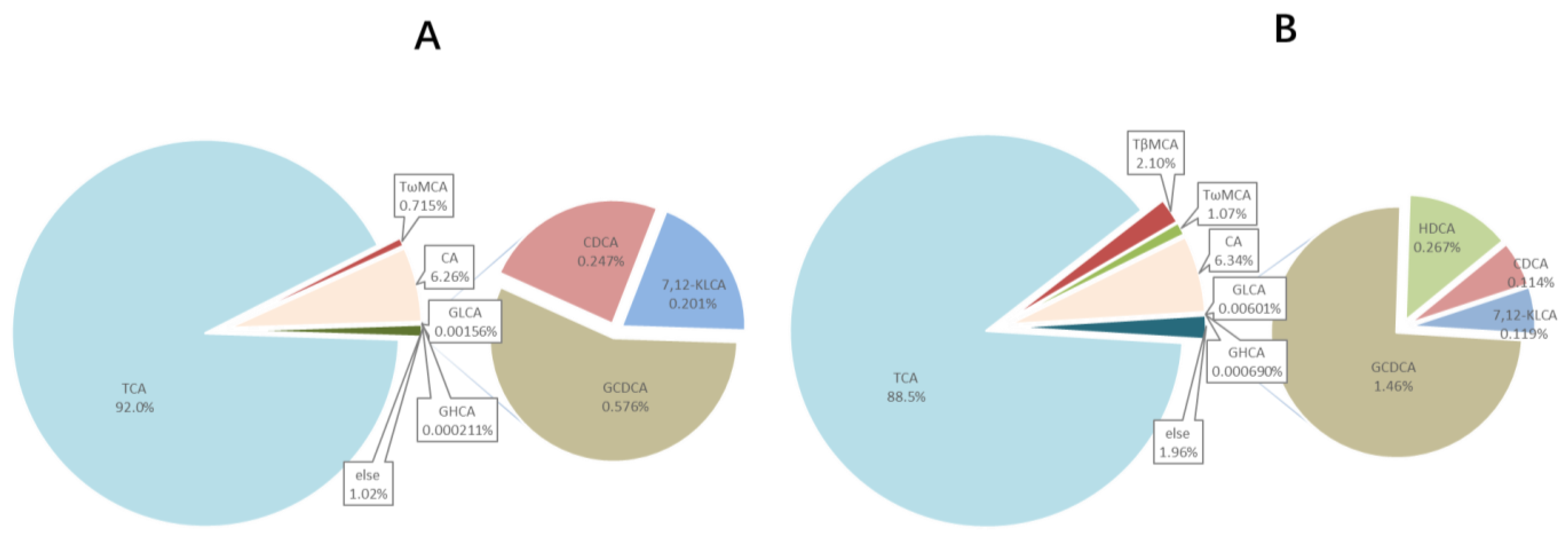
Ten compounds, including TCA, TβMCA, TωMCA, CA, GLCA, GHCA, GCDCA, HDCA, CDCA, and 7,12-KLCA were found quantified in bile samples, whose EIC is shown in Figure 4. BA profiles of common carp bile are summarized in Table 4 and Figure 5, which suggested that TCA was the main bile acids in common carp, followed by CA which accounted for 88–92% and 6–7% respectively; CA ranged from 140–170 μM; however, no TCDCA existed.
Moreover, eight BAs, including TCDCA, CDCA, CA, LCA, HDCA, GLCA, GCDCA, and DCA were detected and quantified from the plasma samples. Table 5 shows the detailed BAs in bile and plasma.
This was the first time that MCA was found to exist in fish. Thus, this was confirmed at this point. The confirmations of MCA detected were verified by the retention time (RT) and the abundance ratio of MS/MS. The RT and abundance ratio of MS/MS of TβMCA and TωMCA standard were 2.79 min and 2.37 min, m/z 124.0017:106.9758:80.9615:79.9523 = 6:3:2:4, and m/z 124.0018:106.9759:80.9611:79.9536 = 40:19:13:40, respectively, which in the sample was consistent (Figure 6).

3.5. Supplement BAs to High Plant Protein Feed Altered the BA profile

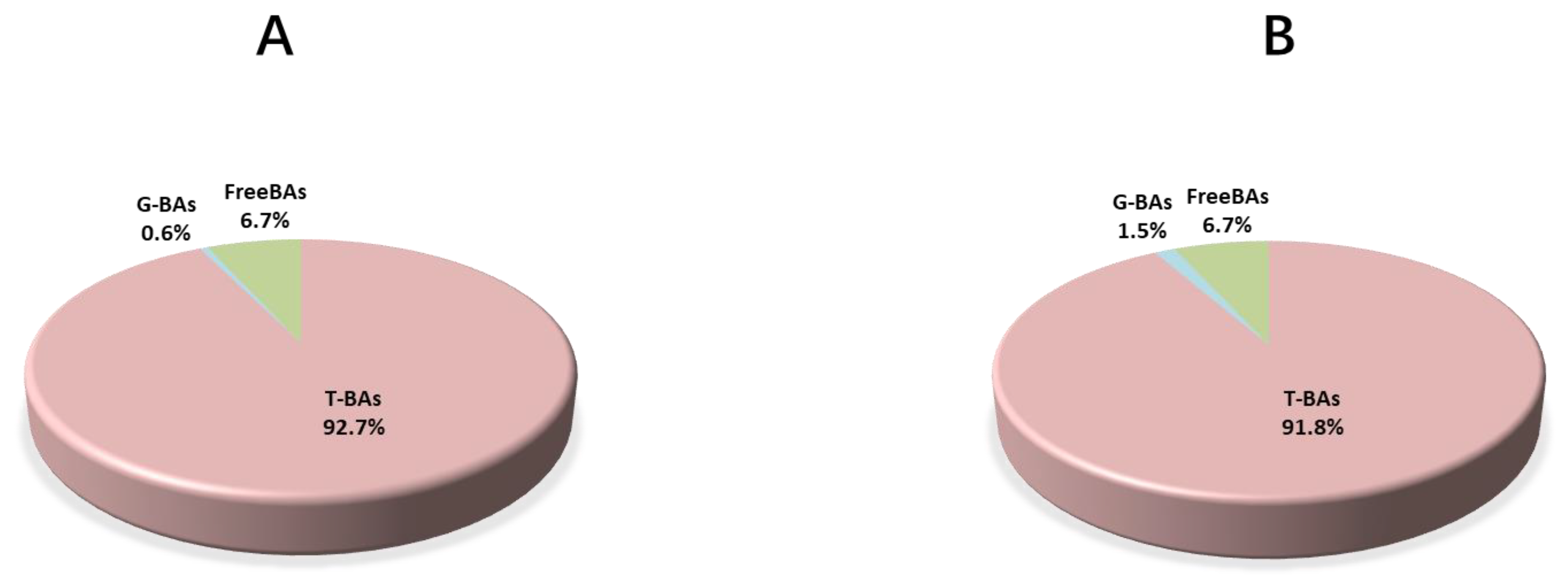
The ratio of GCDCA, TβMCA, GLCA, and HDCA increased significantly in the HP+BAs group, while TCA and CDCA decreased (Figure 6). An increase of BA diversity in the HP+BAs group could be observed in both bile and plasma. Supplementary BAs to a high plant protein diet increased the proportion of G-BAs in bile, which accounted for 0.6% and 1.5% in the HP group and HP+BAs group, respectively (Figure 7).

4. Discussion

4.1. BA Profile Changes Caused by Supplements of BAs

TCA was the main BA in the common carp bile, which was consistent with the results of previously reported studies about the BA profile of fish (angelfish (Pterophyllumeimekei)) bile [24]. In this study, G-BAs such as GCDCA, GLCA, GHCA, and GCA were detected in fish. The BA family in animals is quite complex. Similar to the reports on the human [34], mice [35], and rainbow trout [36], we also found that the common carp can conjugate bile acid with glycine, but not only with taurine. In some early views, it was pointed out that animals, except for the mammals, conjugate their BAs exclusively with taurine [37,38], which led to the fact that less attention was paid to G-BAs when studying fish bile acids. The glycine conjugated bile acid in fish could be partially from fishmeal or other animal ingredients in the feed. We determined the bile acids of fish meal sample that we used in the present study, and the content of G-BAs is about 1.07 × 10⁻⁷ umol/mg. However, the fish meal in the experimental diets of this study only accounted for 10%, which made the bile acids level in the basal diet lower than the detection limit of LC-MS/MS. In the study of Staessen et al. (2021) [36], 27% fishmeal and 1.3% fish oil were used and about 1.1 × 10⁻⁵ umol/mg GBA were detected in the basal diet. In the present study, the dietary bile acid profile was composed with HCA (8.0%), HDCA (70.9%), and CDCA (20.2%), and the two experimental diets were designed with the same level of fishmeal. Hence, we can conclude that the increased GBA in the HP+Bas group should be majorly endogenous for the common carp. This is a report, for the first time, that bile acids can be conjugated with glycine in common carp, although it has been found that bile acids could hardly be combined with glycine in some fish species, such as Sea Lamprey [26] and lake charr [39]. In addition, compared with the HP group, the percentage of G-BA in the HP+BA group increased while the percentage of T-BA decreased; that is consistent with the finding that G-BA and T-BA have a mutual inhibition relationship in previous studies [40,41].
Supplementary BAs (mainly HCA, HDCA, CDCA) increased the contents of TβMCA, GLCA, GCDCA, and HDCA in the common carp bile; we suppose the following could account for this with the help of intestinal microorganisms. HCA transformed into βMCA by 6b-epimerization and further 7b-epimerization [42], then βMCA was reabsorbed back to hepatopancreas and combined with taurine into TβMCA, while HDCA was directly reabsorbed to the hepatopancreas. CDCA is dehydroxylated into LCA (based on KEGG secondary bile acid biosynthesis, map00121), which is partly excreted from the body, and part is reabsorbed to the liver and combined with glycine to form GLCA. The other part of CDCA is reabsorbed to the hepatopancreas to combine with glycine.
The discovery of TβMCA and TωMCA in Common Carp was a breakthough since they were thought to be a rodent specific bile acid [42,43,44,45,46], and indeed, they were not found in birds and monogastric animal BA analysis [47,48], but have also been found in humans according to some reports [21,49]. Rodents branch off from fish in the evolutionary tree, and humans and rodents are on a small branch; we considered that MCA maybe a common species of bile acids that existed in fish, rodents, and primates. More work should be done in the future.

4.2. Supplement BAs Affected Common Carp BA Profile to Reduce Hepatopancreas Glycogen Accumulation and Alleviated Hepatopancreas Damage with a High Plant Protein Diet

Supplement BAs reduced liver glycogen accumulation and alleviated liver damage in common carp with a high plant protein diet. High plant protein could induce fish intestinal and liver damage that has been confirmed in the previous research through our laboratory [16,30,50,51]. It was reported a sturgeon intestinal obvious damage when it was fed a diet with more than 50% of plant protein content [50]. In this study, the proportion of plant protein was as high as 78%, and it has been confirmed by Yoa et al. [30] that this plant protein level causes the intestine of carp serious injury. The intestinal organ is the organ to digest and absorb nutrients, while damage and functional barriers would lead to nutritional metabolism disorders, especially proteins [52,53], thus affecting the synthesis of key enzymes of other nutrients. Therefore, in this study, the reason that a high-plant-protein diet caused hepatopancreas glycogen accumulation and damage is possible because the high-plant-protein diet injures the common carp’s intestines, leading to protein digestion and absorption disorders, and then resulting in a lack of phosphorylase (the key enzymes in liver glycogen decomposition) synthesis. In addition, disorders of glucose metabolism can stimulate cell inflammation and apoptosis, which cause liver damage [32,54]. It has been reported that supplementary BAs to high plant protein feed could alleviate intestinal and liver damage [30], and our results also confirmed that. The added BAs probably play a role as indicated in the following aspects to overcome the negative effects of high plant protein.
Firstly, the soysaponins of soybean meal may be the main cause of common carp intestinal damage [55]. BAs can be combined with non-starch polysaccharides and excreted from the body [56]. Saponins are composed of sapogenins and glycosyl, and a study has shown that soysaponins could increase the excretion of BAs [57]. This shows that soysaponins may have similar binding power to BAs as non-starch polysaccharides. Subsequently, BA supplements could be combined with saponins, thereby reducing the damage of saponins to the intestinal organ, and then improving protein digestion and absorption, reducing liver glycogen accumulation.
Secondly, common carp hepatopancreatic inflammation and glucose metabolism may be regulated by three purposes that were LCA, CDCA, and CA to activate liver TGR, increased liver glycine concentration and TβMCA inhibits intestinal FXR. TGR5 could be activated by some BAs, in which LCA is the most potent agonist for TGR5, DCA and the conjugations, CDCA and the conjugations, and CA and the conjugations activate TGR5 effectively simultaneously [58]. TGR5 plays an important role in anti-inflammatory activities and glucose metabolism [59]. TGR5 restrains the activated B cells (NF-κB) to control the proinflammatory factors secretion by the mediation of the interaction between IκBα and β-arrestin2 and thus exert anti-inflammatory effects [60,61,62]. Activating liver TGR5 could reduce blood glucose in mice with a high-fat diet [58]. That suggested the potential hypoglycemic function of TGR5. In the present study, plasma CDCA, CA, LCA, and GCDCA increased significantly in the HP+BAs group; with the assistance of enterohepatic circulation of Bas [63], they will enter the liver and activate TGR5, especially LCA, that cannot be synthesized directly in the liver while it can only be recovered from intestinal BAs by blood circulation, thus enhancing the anti-inflammatory ability of the body, regulating glucose metabolism and reducing blood glucose, and decreasing the liver inflammation [64]. The expression of the TGR5 gene in the liver increase after supplementation of BAs to HP has been confirmed by Yao et al. [30].
In addition, in the results of IDE et al. (1994), it can be found that the content of G-BAs in bile increases with the increase of liver glycine concentration [65]. Kupffer cells in an activated state could release a variety of inflammatory mediators and play a leading role when the liver is invaded, Glycine inactivates Kupffer cells and can protect the liver from inflammation [66]. TβMCA varied quite distinctly in bile between the HP group and HP+BAs group. TβMCA is a farnesoid X receptor (FXR) nuclear receptor antagonist [14]. FXR is a member of the nuclear receptor superfamily that is primarily expressed in the liver, kidney, and intestine [67]. In the FXR gene knockout mice, intestinal glucose absorption was delayed, together with blood glucose decreased [68]. TβMCA suppressed the enterohepatic FXR-FGF15 signaling and could affect glucose metabolism, reduce blood glucose, and treat diabetes [69]. Therefore, the increase of G-BAs and TβMCA in bile after the addition of BAs also plays a certain role in keeping the hepatopancreas from avoiding histological damage and the reduction of plasma glucose.

5. Conclusions

In summary, HP could induce glycogen accumulation in common carp hepatopancreas while supplemented BAs to HP could mitigate this symptom. BAs supplements in a high plant protein diet could change the BA profile of common carp, among them, plasma LCA, CDCA, and CA increased significantly, TβMCA and the proportion of G-BAs in bile increased significantly, which might play a leading role in it that reduced the accumulation of hepatopancreas glycogen and maintained hepatopancreas health. This study proceeded with an integrated bile acid profile determination by UHPLC-MS/MS to identify the effect of exogenous BAs supplementary on the endogenous BA profile and hepatopancreas health of common carp and discussed how the BAs supplementary is transformed in the body, providing a theoretical basis for the application of BAs products in fish and a data basis for revealing the mystery of fish BAs. In addition, this study has important significance for the development of aquaculture and has a potential contribution to expanding the strategic space of food security.

Author Contributions

X.W., T.Y. and F.N.F. conceived the experiment; X.W. analyzed all data; X.W. and T.Y. wrote the manuscript; X.W., T.Y., F.N.F., X.L., J.W., W.D., X.G. and M.X. reviewed the manuscript. All authors have read and agreed to the published version of the manuscript.

Funding

This study was supported by the National Key R&D Program of China (2018YFD0900400 and 2019YFD0900200), National Natural Science Foundation of China (31902382), Beijing Natural Science Foundation (6204047), The Agricultural Science and Technology Innovation Program of CAAS, China (CAAS-ASTIP-2017-FRI-08) and Beijing Technology System for Sturgeon and Salmonids (BAIC08-2021).

Institutional Review Board Statement

Feed Research Institute, Chinese Academy of Agricultural Sciences Experimental Animal Committee (100081, 31 August 2021).

Informed Consent Statement

Not applicable.

Conflicts of Interest

The authors declare that they have no conflict of interest.

References

  1. Lefebvre, P.; Cariou, B.; Lien, F.; Kuipers, F.; Staels, B. Role of Bile Acids and Bile Acid Receptors in Metabolic Regulation. Physiol. Rev. 2009, 89, 147–191. [Google Scholar] [CrossRef] [Green Version]
  2. Bachmann, V.; Kostiuk, B.; Unterweger, D.; Diaz-Satizabal, L.; Ogg, S.; Pukatzki, S. Bile Salts Modulate the Mucin-Activated Type VI Secretion System of Pandemic Vibrio cholerae. PLoS Negl. Trop. Dis. 2015, 9, e0004031. [Google Scholar] [CrossRef] [PubMed]
  3. Sun, J.; Cao, Z.; Smith, A.D.; Carlson, P.E., Jr.; Coryell, M.; Chen, H.; Beger, R.D. Bile Acid Profile and its Changes in Response to Cefoperazone Treatment in MR1 Deficient Mice. Metabolites 2020, 10, 127. [Google Scholar] [CrossRef] [PubMed] [Green Version]
  4. Ridlon, J.M.; Kang, D.-J.; Hylemon, P.B. Bile salt biotransformations by human intestinal bacteria. J. Lipid Res. 2006, 47, 241–259. [Google Scholar] [CrossRef] [PubMed] [Green Version]
  5. Begley, M.; Gahan, C.G.; Hill, C. The interaction between bacteria and bile. FEMS Microbiol. Rev. 2005, 29, 625–651. [Google Scholar] [CrossRef] [PubMed] [Green Version]
  6. Insull, W., Jr. Clinical utility of bile acid sequestrants in the treatment of dyslipidemia a scientific review. South. Med. J. 2006, 99, 257–273. [Google Scholar] [CrossRef]
  7. Staels, B.; Fonseca, V.A. Bile Acids and Metabolic Regulation: Mechanisms and clinical responses to bile acid sequestration. Diabetes Care 2009, 32, S237–S245. [Google Scholar] [CrossRef] [Green Version]
  8. Ferrell, J.M.; Chiang, J.Y.L. Understanding Bile Acid Signaling in Diabetes: From Pathophysiology to Therapeutic Targets. Diabetes Metab. J. 2019, 43, 257–272. [Google Scholar] [CrossRef]
  9. Lien, F.; Berthier, A.; Bouchaert, E.; Gheeraert, C.; Alexandre, J.; Porez, G.; Prawitt, J.; Dehondt, H.; Ploton, M.; Colin, S.; et al. Metformin interferes with bile acid homeostasis through AMPK-FXR crosstalk. J. Clin. Investig. 2014, 124, 1037–1051. [Google Scholar] [CrossRef] [Green Version]
  10. Chiang, J.Y.L. Bile Acid Metabolism and Signaling. Compr. Physiol. 2013, 3, 1191–1212. [Google Scholar] [CrossRef] [Green Version]
  11. Katsuma, S.; Hirasawa, A.; Tsujimoto, G. Bile acids promote glucagon-like peptide-1 secretion through TGR5 in a murine enteroendocrine cell line STC-1. Biochem. Biophys. Res. Commun. 2005, 329, 386–390. [Google Scholar] [CrossRef]
  12. Pols, T.W.; Nomura, M.; Harach, T.; Sasso, G.L.; Oosterveer, M.H.; Thomas, C.; Rizzo, G.; Gioiello, A.; Adorini, L.; Pellicciari, R.; et al. TGR5 Activation Inhibits Atherosclerosis by Reducing Macrophage Inflammation and Lipid Loading. Cell Metab. 2011, 14, 747–757. [Google Scholar] [CrossRef] [Green Version]
  13. Vallim, T.Q.D.A.; Tarling, E.J.; Edwards, P.A. Pleiotropic Roles of Bile Acids in Metabolism. Cell Metab. 2013, 17, 657–669. [Google Scholar] [CrossRef] [Green Version]
  14. Li, F.; Jiang, C.; Krausz, K.W.; Li, Y.; Albert, I.; Hao, H.; Fabre, K.M.; Mitchell, J.B.; Patterson, A.; Gonzalez, F.J. Microbiome remodelling leads to inhibition of intestinal farnesoid X receptor signalling and decreased obesity. Nat. Commun. 2013, 4, 2384. [Google Scholar] [CrossRef]
  15. Abdel-Latif, H.M.; Abdel-Daim, M.M.; Shukry, M.; Nowosad, J.; Kucharczyk, D. Benefits and applications of Moringa oleifera as a plant protein source in Aquafeed: A review. Aquaculture 2021, 547, 737369. [Google Scholar] [CrossRef]
  16. Wei, H.; Xing, S.; Chen, P.; Wu, X.; Gu, X.; Luo, L.; Liang, X.; Xue, M. Plant protein diet-induced hypoimmunity by affecting the spiral valve intestinal microbiota and bile acid enterohepatic circulation in Amur sturgeon (Acipenser schrenckii). Fish Shellfish Immunol. 2020, 106, 421–430. [Google Scholar] [CrossRef]
  17. Zhang, Y.; Chen, P.; Liang, X.F.; Han, J.; Wu, X.F.; Yang, Y.H.; Xue, M. Metabolic disorder induces fatty liver in Japanese seabass, Lateolabrax japonicas fed a full plant protein diet and regulated by cAMP-JNK/NF-kB-caspase signal pathway. Fish Shellfish Immunol. 2019, 90, 223–234. [Google Scholar] [CrossRef] [PubMed]
  18. Romano, N.; Kumar, V.; Yang, G.; Kajbaf, K.; Rubio, M.B.; Overturf, K.; Brezas, A.; Hardy, R. Bile acid metabolism in fish: Disturbances caused by fishmeal alternatives and some mitigating effects from dietary bile inclusions. Rev. Aquac. 2020, 12, 1792–1817. [Google Scholar] [CrossRef]
  19. Jin, M.; Pan, T.; Cheng, X.; Zhu, T.T.; Sun, P.; Zhou, F.; Ding, X.; Zhou, Q.-C. Effects of supplemental dietary l-carnitine and bile acids on growth performance, antioxidant and immune ability, histopathological changes and inflammatory response in juvenile black seabream (Acanthopagrus schlegelii) fed high-fat diet. Aquaculture 2019, 504, 199–209. [Google Scholar] [CrossRef]
  20. Kortner, T.M.; Penn, M.H.; Björkhem, I.; Måsøval, K.; Krogdahl, Å. Bile components and lecithin supplemented to plant based diets do not diminish diet related intestinal inflammation in Atlantic salmon. BMC Vet. Res. 2016, 12, 190. [Google Scholar] [CrossRef] [PubMed] [Green Version]
  21. Bathena, S.P.R.; Mukherjee, S.; Olivera, M.; Alnouti, Y. The profile of bile acids and their sulfate metabolites in human urine and serum. J. Chromatogr. B 2013, 942–943, 53–62. [Google Scholar] [CrossRef]
  22. Chen, C.; Hu, B.; Wu, T.; Zhang, Y.; Xu, Y.; Feng, Y.; Jiang, H. Bile acid profiles in diabetic (db/db) mice and their wild type littermates. J. Pharm. Biomed. Anal. 2016, 131, 473–481. [Google Scholar] [CrossRef]
  23. Buchinger, T.J.; Li, W.; Johnson, N.S. Bile Salts as Semiochemicals in Fish. Chem. Senses 2014, 39, 647–654. [Google Scholar] [CrossRef]
  24. Satoh, R.; Saito, T.; Ogata, H.; Ohsaki, A.; Iida, T.; Asahina, K.; Mitamura, K.; Ikegawa, S.; Hofmann, A.F.; Hagey, L.R. N-Methyltaurine N-acyl amidated bile acids and deoxycholic acid in the bile of angelfish (Pomacanthidae): A novel bile acid profile in Perciform fish. Steroids 2014, 80, 15–23. [Google Scholar] [CrossRef]
  25. Schmucker, A.K.; Johnson, N.S.; Bussy, U.; Li, K.; Galbraith, H.S.; Chung-Davidson, Y.; Li, W. American eels produce and release bile acid profiles that vary across life stage. J. Fish Biol. 2020, 96, 1024–1033. [Google Scholar] [CrossRef]
  26. Wang, H.; Yeh, C.-Y.; Li, K.; Chung-Davidson, Y.-W.; Li, W. An UPLC–MS/MS method for quantitative profiling of bile acids in sea lamprey plasma and tissues. J. Chromatogr. B 2015, 980, 72–78. [Google Scholar] [CrossRef]
  27. Xu, H.; Zhang, Q.; Kim, S.-K.; Liao, Z.; Wei, Y.; Sun, B.; Jia, L.; Chi, S.; Liang, M. Dietary taurine stimulates the hepatic biosynthesis of both bile acids and cholesterol in the marine teleost, tiger puffer (Takifugu rubripes). Br. J. Nutr. 2020, 123, 1345–1356. [Google Scholar] [CrossRef] [Green Version]
  28. Yao, T.; Wei, X.; Xue, M.; Sun, B.; Gu, X.; Zhang, S. Analysis of Bile Acid Profile in Bile and Plasma of Turbot. Chin. J. Anim. Nutr. 2020, 32, 5816–5826. [Google Scholar]
  29. Zhang, J.; Li, W.; Zhou, H.; Li, M.; Wang, M.; Wu, S. An analysis of Bile Acid composition in different tissues of Grass Carp (Ctenopharyngodon idellus). Acta Hydrobiol. Sin. 2017, 41, 479–482. [Google Scholar]
  30. Yao, T.; Gu, X.; Liang, X.; Fall, F.N.; Cao, A.; Zhang, S.; Guan, Y.; Sun, B.; Xue, M. Tolerance assessment of dietary bile acids in common carp (Cyprinus carpio L.) fed a high plant protein diet. Aquaculture 2021, 543, 737012. [Google Scholar] [CrossRef]
  31. Long, Y.; Li, X.; Li, F.; Ge, G.; Liu, R.; Song, G.; Li, Q.; Qiao, Z.; Cui, Z. Transcriptional Programs Underlying Cold Acclimation of Common Carp (Cyprinus carpio L.). Front. Genet. 2020, 11, 556418. [Google Scholar] [CrossRef] [PubMed]
  32. Yu, H.; Zhang, L.; Chen, P.; Liang, X.; Cao, A.; Han, J.; Wu, X.; Zheng, Y.; Qin, Y.; Xue, M. Dietary Bile Acids Enhance Growth, and Alleviate Hepatic Fibrosis Induced by a High Starch Diet via AKT/FOXO1 and cAMP/AMPK/SREBP1 Pathway in Micropterus salmoides. Front. Physiol. 2019, 10, 1430. [Google Scholar] [CrossRef] [Green Version]
  33. Gu, Y.; Wang, X.; Li, J.; Zhang, Y.; Zhong, H.; Liu, R.; Zhang, D.; Feng, Q.; Xie, X.; Hongmei, Z.; et al. Analyses of gut microbiota and plasma bile acids enable stratification of patients for antidiabetic treatment. Nat. Commun. 2017, 8, 1785. [Google Scholar] [CrossRef]
  34. Humbert, L.; Maubert, M.A.; Wolf, C.; Duboc, H.; Mahé, M.; Farabos, D.; Seksik, P.; Mallet, J.-M.; Trugnan, G.; Masliah, J.; et al. Bile acid profiling in human biological samples: Comparison of extraction procedures and application to normal and cholestatic patients. J. Chromatogr. B 2012, 899, 135–145. [Google Scholar] [CrossRef]
  35. Zheng, X.; Huang, F.; Zhao, A.; Lei, S.; Zhang, Y.; Xie, G.; Chen, T.; Qu, C.; Rajani, C.; Dong, B.; et al. Bile acid is a significant host factor shaping the gut microbiome of diet-induced obese mice. BMC Biol. 2017, 15, 120. [Google Scholar] [CrossRef] [Green Version]
  36. Staessen, T.W.O.; Verdegem, M.C.J.; Nederlof, M.A.J.; Eding, E.H.; Schrama, J.W. Effect of type of dietary non- protein energy source (starch vs. fat) on the body bile acid pool size and composition, faecal bile acid loss and bile acid synthesis in rainbow trout (Oncorhynchus mykiss). Aquac. Nutr. 2021, 27, 865–879. [Google Scholar] [CrossRef]
  37. Vessey, D.A. The biochemical basis for the conjugation of bile acids with either glycine or taurine. Biochem. J. 1978, 174, 621–626. [Google Scholar] [CrossRef] [Green Version]
  38. Hofmann, A.F.; Hagey, L.R.; Krasowski, M. Bile salts of vertebrates: Structural variation and possible evolutionary significance. J. Lipid Res. 2010, 51, 226–246. [Google Scholar] [CrossRef] [Green Version]
  39. Li, K.; Buchinger, T.J.; Bussy, U.; Fissette, S.D.; Johnson, N.S.; Li, W. Quantification of 15 bile acids in lake charr feces by ultra-high performance liquid chromatography–tandem mass spectrometry. J. Chromatogr. B 2015, 1001, 27–34. [Google Scholar] [CrossRef] [PubMed]
  40. Jurkowska, H.; Stipanuk, M.H.; Hirschberger, L.L.; Roman, H.B. Propargylglycine inhibits hypotaurine/taurine synthesis and elevates cystathionine and homocysteine concentrations in primary mouse hepatocytes. Amino Acids 2015, 47, 1215–1223. [Google Scholar] [CrossRef] [PubMed] [Green Version]
  41. Ridlon, J.M.; Wolf, P.G.; Gaskins, H.R. Taurocholic acid metabolism by gut microbes and colon cancer. Gut Microbes 2016, 7, 201–215. [Google Scholar] [CrossRef] [Green Version]
  42. Wahlström, A.; Sayin, S.I.; Marschall, H.-U.; Bäckhed, F. Intestinal Crosstalk between Bile Acids and Microbiota and Its Impact on Host Metabolism. Cell Metab. 2016, 24, 41–50. [Google Scholar] [CrossRef] [Green Version]
  43. DiMarzio, M.; Rusconi, B.; Yennawar, N.H.; Eppinger, M.; Patterson, A.D.; Dudley, E. GIdentification of a mouse Lactobacillus johnsonii strain with deconjugase activity against the FXR an-tagonist T-beta-MCA. PLoS ONE 2017, 12, e0183564. [Google Scholar] [CrossRef] [PubMed]
  44. Sun, L.; Xie, C.; Wang, G.; Wu, Y.; Wu, Q.; Wang, X.; Liu, J.; Deng, Y.; Xia, J.; Chen, B.; et al. Gut microbiota and intestinal FXR mediate the clinical benefits of metformin. Nat. Med. 2018, 24, 1919–1929. [Google Scholar] [CrossRef] [PubMed]
  45. Sayin, S.I.; Wahlström, A.; Felin, J.; Jäntti, S.; Marschall, H.-U.; Bamberg, K.; Angelin, B.; Hyötyläinen, T.; Oresic, M.; Bäckhed, F. Gut Microbiota Regulates Bile Acid Metabolism by Reducing the Levels of Tauro-beta-muricholic Acid, a Naturally Occurring FXR Antagonist. Cell Metab. 2013, 17, 225–235. [Google Scholar] [CrossRef] [PubMed] [Green Version]
  46. Takahashi, S.; Fukami, T.; Masuo, Y.; Brocker, C.N.; Xie, C.; Krausz, K.W.; Wolf, C.R.; Henderson, C.J.; Gonzalez, F.J. Cyp2c70 is responsible for the species difference in bile acid metabolism between mice and humans. J. Lipid Res. 2016, 57, 2130–2137. [Google Scholar] [CrossRef] [Green Version]
  47. Bansal, M.; Fu, Y.; Alrubaye, B.; Abraha, M.; Almansour, A.; Gupta, A.; Liyanage, R.; Wang, H.; Hargis, B.; Sun, X. A secondary bile acid from microbiota metabolism attenuates ileitis and bile acid reduction in subclinical necrotic enteritis in chickens. J. Anim. Sci. Biotechnol. 2020, 11, 37. [Google Scholar] [CrossRef] [Green Version]
  48. Jia, W.; Rajani, C.; Zheng, X.; Jia, W. Hyocholic acid and glycemic regulation: Comments on ‘Hyocholic acid species improve glucose homeostasis through a distinct TGR5 and FXR signaling mechanism’. J. Mol. Cell Biol. 2021, 13, 460–462. [Google Scholar] [CrossRef]
  49. Cui, Y.; Xu, B.; Zhang, X.; He, Y.; Shao, Y.; Ding, M. Diagnostic and therapeutic profiles of serum bile acids in women with intrahepatic cholestasis of pregnancy-a pseudo-targeted metabolomics study. Clin. Chim. Acta 2018, 483, 135–141. [Google Scholar] [CrossRef]
  50. Wei, H.; Chen, P.; Liang, X.; Yu, H.; Wu, X.; Han, J.; Luo, L.; Gu, X.; Xue, M. Plant protein diet suppressed immune function by inhibiting spiral valve intestinal mucosal barrier integrity, anti-oxidation, apoptosis, autophagy and proliferation responses in amur sturgeon (Acipenser schrenckii). Fish Shellfish Immunol. 2019, 94, 711–722. [Google Scholar] [CrossRef]
  51. Urán, P.; Gonçalves, A.; Taverne-Thiele, J.; Schrama, J.; Verreth, J.; Rombout, J. Soybean meal induces intestinal inflammation in common carp (Cyprinus carpio L.). Fish Shellfish Immunol. 2008, 25, 751–760. [Google Scholar] [CrossRef]
  52. Kaushik, S.; Cravedi, J.; Lallès, J.-P.; Sumpter, J.; Fauconneau, B.; Laroche, M. Partial or total replacement of fish meal by soybean protein on growth, protein utilization, potential estrogenic or antigenic effects, cholesterolemia and flesh quality in rainbow trout, Oncorhynchus mykiss. Aquaculture 1995, 133, 257–274. [Google Scholar] [CrossRef]
  53. Escaffre, A.M.; Infante, J.L.Z.; Cahu, C.L.; Mambrini, M.; Bergot, P.; Kaushik, S.J. Nutritional value of soy protein concentrate for larvae of common carp (Cyprinus carpio). Aquaculture 1997, 153, 63–80. [Google Scholar] [CrossRef]
  54. Laubertová, L.; Koňariková, K.; Gbelcová, H.; Ďuračková, Z.; Žitňanová, I. Effect of walnut oil on hyperglycemia-induced oxidative stress and pro-inflammatory cytokines production. Eur. J. Nutr. 2015, 54, 291–299. [Google Scholar] [CrossRef]
  55. Knudsen, D.; Urán, P.; Arnous, A.; Koppe, A.W.; Frøkiær, H. Saponin-Containing Subfractions of Soybean Molasses Induce Enteritis in the Distal Intestine of Atlantic Salmon. J. Agric. Food Chem. 2007, 55, 2261–2267. [Google Scholar] [CrossRef] [PubMed]
  56. Matin, H.H.; Shariatmadari, F.; Torshizi, M.K.; Chiba, L. In vitro bile acid-binding capacity of dietary fibre sources and their effects with bile acid on broiler chicken performance and lipid digestibility. Br. Poult. Sci. 2016, 57, 348–357. [Google Scholar] [CrossRef]
  57. Lee, S.-O.; Simons, A.L.; Murphy, P.A.; Hendrich, S. Soyasaponins Lowered Plasma Cholesterol and Increased Fecal Bile Acids in Female Golden Syrian Hamsters. Exp. Biol. Med. 2005, 230, 472–478. [Google Scholar] [CrossRef]
  58. Sato, H.; Genet, C.; Strehle, A.; Thomas, C.; Lobstein, A.; Wagner, A.; Mioskowski, C.; Auwerx, J.; Saladin, R. Anti-hyperglycemic activity of a TGR5 agonist isolated from Olea europaea. Biochem. Biophys. Res. Commun. 2007, 362, 793–798. [Google Scholar] [CrossRef]
  59. Guo, C.; Chen, W.-D.; Wang, Y.-D. TGR5, Not Only a Metabolic Regulator. Front. Physiol. 2016, 7, 646. [Google Scholar] [CrossRef]
  60. Wang, Y.-D.; Chen, W.-D.; Yu, D.; Forman, B.M.; Huang, W. The G-protein-coupled bile acid receptor, Gpbar1 (TGR5), negatively regulates hepatic inflammatory response through antagonizing nuclear factor kappa light-chain enhancer of activated B cells (NF-kappaB) in mice. Hepatology 2011, 54, 1421–1432. [Google Scholar] [CrossRef] [Green Version]
  61. Keitel, V.; Donner, M.; Winandy, S.; Kubitz, R.; Häussinger, D. Expression and function of the bile acid receptor TGR5 in Kupffer cells. Biochem. Biophys. Res. Commun. 2008, 372, 78–84. [Google Scholar] [CrossRef]
  62. Häussinger, D.; Keitel, V. Role of TGR5 (GPBAR1) in Liver Disease. Semin. Liver Dis. 2018, 38, 333–339. [Google Scholar] [CrossRef]
  63. Li, Y.-F.; Wu, J.-S.; Li, Y.-Y.; Dai, Y.; Zheng, M.; Zeng, J.-K.; Wang, G.-F.; Wang, T.-M.; Li, W.-K.; Zhang, X.-Y.; et al. Chicken bile powder protects against α-naphthylisothiocyanate-induced cholestatic liver injury in mice. Oncotarget 2017, 8, 97137–97152. [Google Scholar] [CrossRef]
  64. Holter, M.M.; Chirikjian, M.K.; Govani, V.N.; Cummings, B.P. TGR5 Signaling in Hepatic Metabolic Health. Nutrients 2020, 12, 2598. [Google Scholar] [CrossRef] [PubMed]
  65. Ide, T.; Kano, S.; Murata, M.; Yanagita, T.; Sugano, M. Dietary modifications of the biliary bile acid glycine:taurine ratio and activity of hepatic bile acid-CoA:amino acid N-acyltransferase (EC 2.3.1) in the rat. Br. J. Nutr. 1994, 72, 93–100. [Google Scholar] [CrossRef] [PubMed] [Green Version]
  66. Rivera, C.A.; Bradford, B.U.; Hunt, K.J.; Adachi, Y.; Schrum, L.W.; Koop, D.R.; Burchardt, E.-R.; Rippe, R.A.; Thurman, R.G. Attenuation of CCl4-induced hepatic fibrosis by GdCl3 treatment or dietary glycine. Am. J. Physiol. Gastrointest. Liver Physiol. 2001, 281, G200–G208. [Google Scholar] [CrossRef] [PubMed] [Green Version]
  67. Ma, K.; Saha, P.K.; Chan, L.; Moore, D.D. Farnesoid X receptor is essential for normal glucose homeostasis. J. Clin. Investig. 2006, 116, 1102–1109. [Google Scholar] [CrossRef] [Green Version]
  68. Van Dijk, T.H.; Grevhorst, A.; Oosterveer, M.H.; Bloks, V.W.; Staels, B.; Reijngoud, D.-J.; Kuipers, F. An increased flux through the glucose 6-phosphate pool in enterocytes delays glucose absorption in Fxr−/− mice. J. Biol. Chem. 2009, 284, 10315–10323. [Google Scholar] [CrossRef] [Green Version]
  69. Hui, S.; Liu, Y.; Chen, M.; Wang, X.; Lang, H.; Zhou, M.; Yi, L.; Mi, M. Capsaicin Improves Glucose Tolerance and Insulin Sensitivity Through Modulation of the Gut Microbiota-Bile Acid-FXR Axis in Type 2 Diabetic db/db Mice. Mol. Nutr. Food Res. 2019, 63, e1900608. [Google Scholar] [CrossRef]
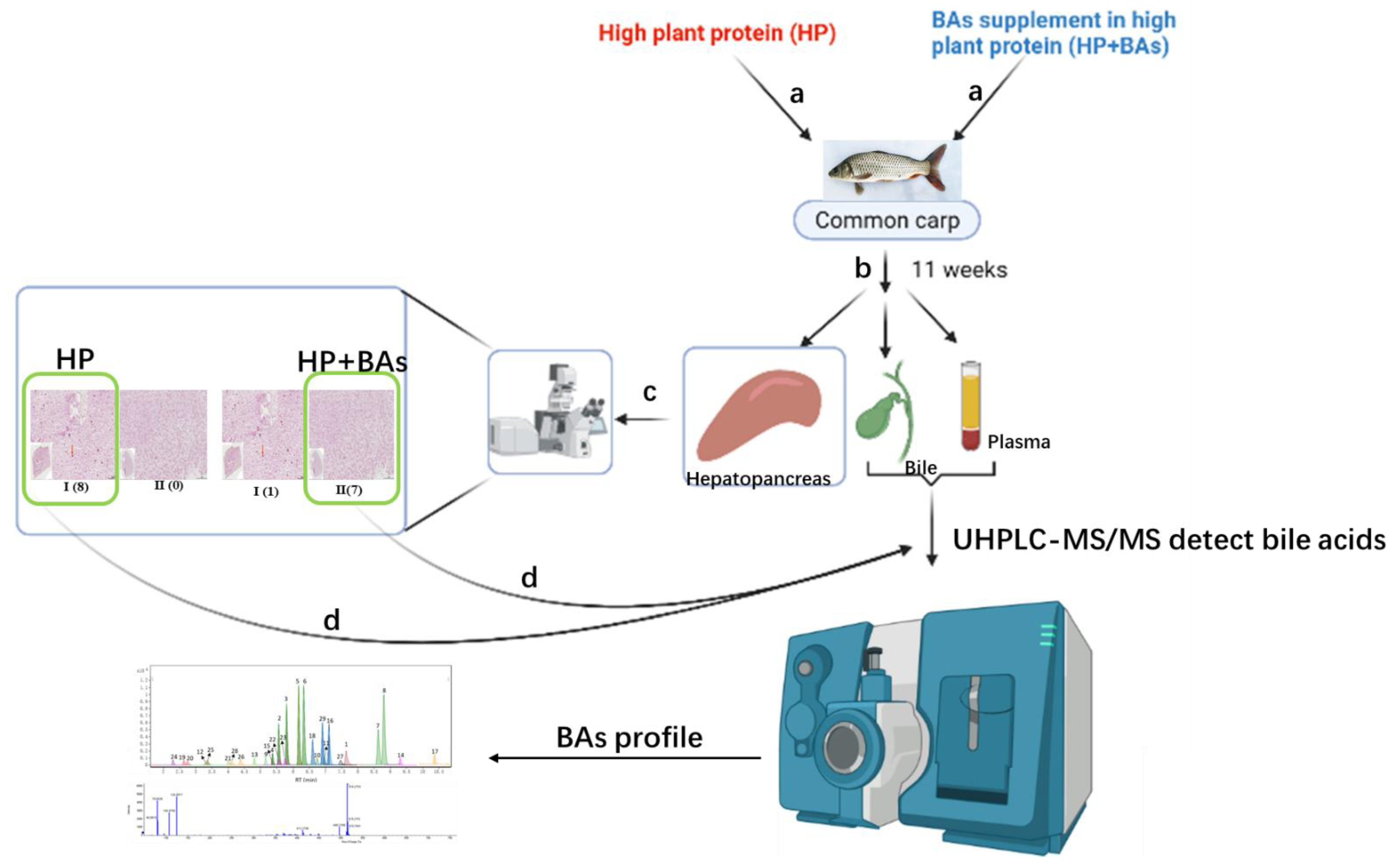
Figure 1. The workflow of the effect in bile acid supplement to high plant protein diet on common carp bile acid profile and hepatopancreas health. Arrows a: Common carp fed with HP and HP+BAs 11 weeks, respectively. Arrows b: Collect hepatopancreas, gallbladder, and plasma. Arrows c: Histopathological detections of hepatopancreas tissues. Arrows d: BAs analysis was performed on the bile and plasma corresponding to phenotype I of the hepatopancreas in the HP group (n = 8), the gallbladder and plasma corresponding to phenotype II of HP+BAs were treated in the same way (n = 7).
Foods 10 02465 g001
Figure 2. Supplement bile acids to high plant protein feed improved the growth performance and reduced the hepatosomatic index (HSI) in common carp (Cyprinus carpio). (Statistically significant results are indicated by asterisks (**, p < 0.01), n = 7.
Foods 10 02465 g002
Figure 3. Supplement bile acids to high plant protein feed reduced carp hepatopancreas histological lesions (Statistically significant results were indicated by asterisks (*, p < 0.05; ***, p < 0.001)): (A) PAS and H&E staining of liver sections with bar = 100 μm, intracellular accumulation of glycogen (marked with yellow arrow), deformed cells (marked with green arrow) and Nuclear gathered (marked with yellow arrow) were clearly observed in the damaged hepatopancreas. (B) Quantification of glycogenosome and active nucleus, the number of hepatopancreas glycogenosome in the HP+BAs group carp was significantly lower than that in the HP group. (C) The phenotype of hepatopancreas histopathological examination in HP group and HP+BAs group. (D) Supplement BAs to high plant protein feed reduced hepatopancreas glycogen and plasma glucose (Statistically significant results are indicated by asterisks (*, p < 0.05; ***, p < 0.001), n = 7).
Foods 10 02465 g003
Figure 4. Bile acids Extracted Ion Chromatogram (EIC) in sample and standard; the peak times between bile acids do not interfere with each other. (A) is standard EIC, (B) is for bile sample, and (C) for plasma sample.
Foods 10 02465 g004
Figure 5. Bile acids species composition in bile, (A,B) for HP and HP+BAs, respectively (n = 7). TCA is the major BA of carp. Compared to HP group carp, the ratio of GCDCA, TβMCA, GLCA, and HDCA increased significantly in HP+BAs group, while TCA and CDCA decreased.
Foods 10 02465 g005
Figure 6. Tandem mass spectrometry (MS/MS) of TβMCA and TωMCA in sample and standard. (A) for TβMCA, (B) for TωMCA. The RT and abundance ratio of MS/MS of TβMCA and TωMCA standard were 2.79 min and 2.37 min, m/z 124.0017:106.9758:80.9615:79.9523 = 6:3:2:4 and m/z 124.0018: 106.9759:80.9611:79.9536 = 40:19:13:40, respectively, which in sample was consistent.
Foods 10 02465 g006
Figure 7. Supplement bile acids to a high plant protein diet increase the proportion of Glycine-conjugation bile acids significantly, while Tauro-conjugation bile acids decreased unremarkably in carp bile, (A,B) for HP and HP+BAs, respectively (n = 7).
Foods 10 02465 g007
Table 1. Parameters for quantification on BAs by UHPLC-MS/MS.
No.CompoundsRetention TimeTransitionFragmentorCELinearity RangeCalibration Curves *R2Internal Standard
(min)(m/z)(V)(eV)(μg/mL)
1CA6.339407.3 -> 407.3240100.0025–5y = 13.798x0.9989CA -d4
2αMCA5.570407.3 -> 407.3240100.001–5y = 100.08x0.9992βMCA -d5
3ωMCA5.384407.3 -> 407.3240100.001–5y = 65.329x0.9982βMCA -d5
4HCA6.178407.3 -> 407.3240100.001–5y = 11.074x0.9994βMCA -d5
5CDCA8.628391.3 -> 391.3230100.001–5y = 39.802x0.9995CDCA-d4
6DCA8.805391.3 -> 391.3230100.001–5y = 8.9922x0.9998DCA-d4
7HDCA7.110391.3 -> 391.3230100.001–5y = 8.6365x0.9993DCA-d4
87,12KLCA7.641389.3 -> 389.3225100.005–0.5y = 52.86x0.9934LCA-d4
9LCA10.367375.3 -> 375.3235100.005–0.5y = 17.63x0.9972LCA-d4
10GCA5.169464.3 -> 74.0230460.001–5y = 44.935x0.9926GCA-d4
11GHCA4.823464.3 -> 74.0230460.001–5y = 118.29x0.9974GCDCA-d4
12GCDCA6.744448.3 -> 74.0220400.001–5y = 27.884x0.9930GCDCA-d4
13GLCA9.304432.3 -> 74.0225360.001–5y = 17.814x0.9996GLCA-d4
14TCA4.059514.3 -> 79.9300770.001–5y = 32.025x + 0.29140.9989TCA-d5
15TβMCA2.787514.3 -> 79.9300770.005–5y = 28.963x − 0.07430.9966TβMCA -d4
16TωMCA2.371514.3 -> 79.9300770.005–5y = 28.963x − 0.07430.9966TβMCA -d4
17TCDCA5.572498.3 -> 79.9280800.001–5y = 51.689x + 0.07820.9999TUDCA-d5
18TDCA5.753498.3 -> 79.9280800.001–5y = 191.74x0.9950TDCA-d5
19THDCA4.412498.3 -> 79.9280800.001–5y = 48.675x − 0.01040.9999TUDCA-d5
20TLCA7.463482.3 -> 79.9290800.001–0.5y = 9.4573x0.9931LCA-d4
21βMCA5.812407.3 -> 407.3240100.001–5y = 83.77x + 0.10570.9995βMCA -d5
22UDCA6.913391.3 -> 391.3230100.001–5y = 61.645x + 1.34250.9991UDCA-d4
23MuroCA6.622391.3 -> 391.3230100.0025–5y = 1.2135x + 0.03620.9993DCA-d4
24GDCA6.976448.3 -> 74.0220400.001–5y = 7.6437x + 0.05410.9997GDCA-d4
25GUDCA5.387448.3 -> 74.0220400.005–5y = 130.8x − 5.53750.9982GCDCA-d4
26GDHCA3.354458.2 -> 74.0205360.001–5y = 88.496x + 4.23690.9973GCDCA-d4
27TαMCA2.657514.3 -> 79.9300770.001–5y = 27.164x − 0.05740.9977TβMCA -d4
28THCA3.405514.3 -> 79.9300770.0025–5y = 23.806x + 0.15180.9994TDCA-d5
29TUDCA4.151498.3 -> 79.9280800.001–5y = 35.379x − 0.10250.9999TUDCA-d5
30TDHCA2.314508.3 -> 79.9285720.001–5y = 15.83x + 0.10230.9997TUDCA-d5
* y, the integral peak area ratio between standard and IS (internal standard); x, concentration in the detected samples or standard curves samples.
Table 2. Experimental diets’ formula and composition of HP and HP+BAs groups (air-dry basis, %) (The content of this table has been published [30]).
Feed FormulationHPHP+BAs
Fish meal a10.0010.00
Soybean meal18.0018.00
Cottonseed protein concentrated18.0018.00
Tapioca flour5.005.00
Wheat flour39.8039.74
Soy oil4.004.00
Vitamin and mineral premix b4.104.10
Lecithin oi1.001.00
DL-me0.100.10
Total100100
Bile Acid (mg/kg) c0600
Analyzed nutrients compositions
Moisture8.249.11
Crude protein30.4729.15
Crude lipid7.537.47
Crude Ash7.257.31
Gross energy (MJ/Kg)18.2818.20
a Fish meal: Shandong Chishan Fishmeal Factory, Shandong, China; Soybean meal: Yihai Kerry Investment Co. Ltd., China; CPC: Xinjiang Jinlan Plant Protein Co. Ltd., China. b Vitamin premix (mg·kg-1 diets): Vitamin A 28; Vitamin B1 12; Vitamin B212; Vitamin B6 16; Vitamin B12 0.2; Vitamin E 300; Vitamin K3 20, Vitamin D 14; Niacinamide 80; Vitamin C 600; Calciumpantothenate 100; Biotin 0.4; Folicacid 3; Corn protein powder 314.4. Mineral premix (mg·kg−1 diets): FeSO4·H2O 300; ZnSO4·H2O 300; MnSO4·H2O 100; Na2SeO3 10; CoCl2·6H2O (10% Co) 2.5; Kl 80; Zeolite 1307.5; MgSO4 500. c Bile acids: supplied by Shandong Longchang Animal Health Care Co. Ltd. (Dezhou, China), with 8.0%HCA, 70.9% HDCA, and 20.2% CDCA. Bile acids were added and well mixed in premix at levels of 0 and 600 mg/kg, respectively.
Table 3. Plasma biochemical parameters. The concentrations of AST and ALT in the HP+BAs group was significantly lower than in the HP group. * Data are shown as mean ± SEM (n = 7). Statistically significant results are indicated by asterisks (*, p < 0.05).
HPHP+BAs
AST(U/L)103 ± 11.8 *62.0 ± 5.84
ALT(U/L)73.7 ± 9.97 *46.5 ± 5.50
TC (mmol/L)5.2 ± 0.3085.3 ± 0.345
Table 4. Bile acid profile in bile of common carp in HP and HP+BAs groups (μmol/L), TβMCA, GLCA, GHCA, and GCDCA increased observably in the HP+BAs group carp bile. * Data are shown as mean ± SEM (n = 7). Statistically significant results are indicated by asterisks (**, p < 0.01; ***, p < 0.001).
BAs in BileHPHP+BAs
Free BAsCA162.0 ± 51.0149.7 ± 30.0
HDCAND6.3 ± 2.4
LCAND3.8 × 10−2 ± 7.1 × 10⁻³
DCA7.4 × 10−4 ± 1.9 × 10⁻⁴ND
CDCA6.4 ± 1.72.7 ± 0.7
7,12-KLCA5.2 ± 1.02.8 ± 0.2
T-BAsTCA2380.6 ± 356.42091.1 ± 262.4
TCDCANDND
TβMCAND49.6 ± 5.4 **
TωMCA18.5 ± 3.725.3 ± 2.5
G-BAsGCANDND
GLCA4.0 × 10−2 ± 1.6 × 10−31.4 × 10−1 ± 1.2 × 10⁻² **
GHCA5.5 × 10−3 ± 2.8 × 10−41.6 × 10−2 ± 2.7 × 10⁻³ **
GCDCA14.9 ± 0.934.5 ± 2.6 ***
TBA 2561.7 ± 346.02337.7 ± 286.9
Table 5. Bile acid profile in plasma of common carp in HP and HP+BAs groups (μmol/L), CA, CDCA, LCA, and GCDCA increased observably in the HP+BAs group carp plasma. * Data are shown as mean ± SEM (n = 7). Statistically significant results are indicated by asterisks (*, p < 0.05).
BAs in PlasmaHPHP+BAs
Free BAsCAND5.7 × 10−3 ± 2.0 × 10⁻³ *
HDCA3.5 × 10−3 ± 2.1 × 10−35.0 × 10−3 ± 1.1 × 10⁻³
CDCAND2.0 × 10−3 ± 3.5 × 10−4 *
7,12-KLCANDND
LCAND3.8 × 10−2 ± 7.06 × 10⁻³ *
T-BAsTCANDND
TCDCA1.1 × 10−1 ± 1.3 × 10−22.3 × 10−1 ± 7.4 × 10⁻²
TβMCANDND
TωMCANDND
G-BAsGCANDND
GLCA8.5 × 10−5 ± 3.2 × 10−51.0 × 10−4 ± 3.10 × 10⁻⁸
GHCANDND
GCDCAND3.7 × 10−3 ± 5.2 × 10⁻⁴ *
TBA 1.3 × 10⁻¹ ± 1.9 × 10−23.2 × 10−1 ± 9.4 × 10⁻²
Publisher’s Note: MDPI stays neutral with regard to jurisdictional claims in published maps and institutional affiliations.