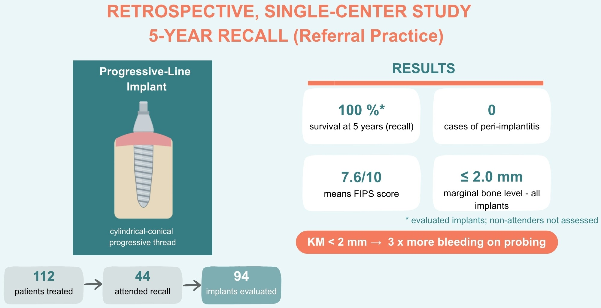
Peri-Implant Tissue Health and Prosthodontic Outcomes of a Cylindrical-Conical Implant with a Progressive Thread Design at Five-Year Follow-Up: A Single-Center Cross-Sectional Retrospective Study


Abstract
1. Introduction
2. Materials and Methods
2.1. Study Design and Setting
2.2. Patient and Implant Selection
2.3. Implant System
2.4. Surgical and Prosthetic Protocol
2.5. Clinical and Radiographic Examination
- -
- Implant status (in situ/failed/not evaluable).
- -
- Percussion sensitivity (negative/positive).
- -
- Functional Implant Prosthodontic Score (FIPS): five subscales (interproximal, occlusion, design, mucosa, bone), each scored 0–2 (maximum total 10) [22]. In this cohort the FIPS was applied at the implant level, with each implant treated as an individual restorative unit; for splinted restorations and 3-unit fixed dental prostheses (FDPs), every supporting implant was scored separately, so that a finding such as chipping was attributed only to the affected abutment. As FDPs were included, the interproximal score was limited to 0 or 2 for terminal single-tooth implants and mesial bridge abutments and was not assessable at terminal bridge abutments. The total FIPS was derived by summing the subscale means. As the cohort also included splinted restorations and 3-unit FDPs, this implant-level application represents a pragmatic, not independently validated modification and is treated as exploratory.
- -
- Bleeding on probing (BOP): recorded as the number of bleeding sites per implant (0–6) on gentle probing at a standardized force of 0.25 N, and categorized, for pragmatic reporting, as 0/6 (healthy), 1–2/6 (possible mild mucositis), or ≥3/6 (clear inflammatory sign).
- -
- Suppuration (Pus): binary (present/absent).
- -
- Mucosal dehiscence: binary (present/absent).
- -
- Width of keratinized mucosa (KM): measured at the mid-buccal aspect and categorized as <2 mm or ≥2 mm.
- -
- Additional signs of inflammation: binary (present/absent).
- -
- Plaque Score (PS): at each of six peri-implant sites (three buccal, three oral), visible plaque or deposits were scored as present or absent based on visual assessment (without disclosing agents), giving a count of 0–6 per implant. Plaque load was categorized per implant as 0/6 (plaque-free), 1–2/6 (low), or ≥3/6 (moderate-to-high), mirroring the bleeding-on-probing categories.
- -
- Periapical radiograph: marginal bone level measured mesially and distally on right-angle periapical radiographs as the distance from the implant shoulder to the most coronal bone-to-implant contact, recorded to the nearest 0.1 mm (a value of 0 mm denotes bone at the implant shoulder). A 0.5-mm threshold was used for categorization and emergence-profile comparison. Radiographs unsuitable for exact measurement were recorded as not evaluable.
- -
- Emergence profile: classified retrospectively on periapical radiographs as concave or convex, immediately coronal to the implant, separately at the mesial and distal aspects; radiographs not suitable for an exact measurement were recorded as not evaluable.
2.6. Statistical Analysis
3. Results
3.1. Patient Flow and Implant Attrition
3.2. Baseline Implant Characteristics
3.3. Prosthodontic Outcomes: FIPS
3.4. Peri-Implant Tissue Health
3.5. Marginal Bone Level
3.6. Prosthodontic Emergence Profile
4. Discussion
5. Conclusions
Author Contributions
Funding
Institutional Review Board Statement
Informed Consent Statement
Data Availability Statement
Acknowledgments
Conflicts of Interest
References
- Elani, H.W.; Starr, J.R.; Da Silva, J.D.; Gallucci, G.O. Trends in Dental Implant Use in the U.S., 1999–2016, and Projections to 2026. J. Dent. Res. 2018, 97, 1424–1430. [Google Scholar] [CrossRef] [Scilit] [PubMed]
- Peres, M.A.; Lalloo, R. Tooth loss, denture wearing and implants: Findings from the National Study of Adult Oral Health 2017–18. Aust. Dent. J. 2020, 65, S23–S31. [Google Scholar] [CrossRef] [Scilit] [PubMed]
- Moraschini, V.; Poubel, L.A.d.C.; Ferreira, V.F.; Barboza, E.d.S.P. Evaluation of survival and success rates of dental implants reported in longitudinal studies with a follow-up period of at least 10 years: A systematic review. Int. J. Oral Maxillofac. Surg. 2015, 44, 377–388. [Google Scholar] [CrossRef] [Scilit] [PubMed]
- Howe, M.-S.; Keys, W.; Richards, D. Long-term (10-year) dental implant survival: A systematic review and sensitivity meta-analysis. J. Dent. 2019, 84, 9–21. [Google Scholar] [CrossRef] [Scilit] [PubMed]
- French, D.; Ofec, R.; Levin, L. Long term clinical performance of 10,871 dental implants with up to 22 years of follow-up: A cohort study in 4247 patients. Clin. Implant Dent. Relat. Res. 2021, 23, 289–297. [Google Scholar] [CrossRef] [Scilit] [PubMed]
- Kupka, J.R.; König, J.; Al-Nawas, B.; Sagheb, K.; Schiegnitz, E. How far can we go? A 20-year meta-analysis of dental implant survival rates. Clin. Oral Investig. 2024, 28, 541. [Google Scholar] [CrossRef] [Scilit] [PubMed]
- von Stein-Lausnitz, M.; Nickenig, H.-J.; Wolfart, S.; Neumann, K.; Von Stein-Lausnitz, A.; Spies, B.C.; Beuer, F. Survival rates and complication behaviour of tooth implant-supported, fixed dental prostheses: A systematic review and meta-analysis. J. Dent. 2019, 88, 103167. [Google Scholar] [CrossRef] [Scilit] [PubMed]
- Papaspyridakos, P.; Chen, C.-J.; Singh, M.; Weber, H.-P.; Gallucci, G.O. Success criteria in implant dentistry: A systematic review. J. Dent. Res. 2012, 91, 242–248. [Google Scholar] [CrossRef] [Scilit] [PubMed]
- Schwarz, F.; Derks, J.; Monje, A.; Wang, H.-L. Peri-implantitis. J. Clin. Periodontol. 2018, 45, S246–S266. [Google Scholar] [CrossRef] [Scilit] [PubMed]
- Scarano, A.; Khater, A.G.A.; Gehrke, S.A.; Serra, P.; Francesco, I.; Di Carmine, M.; Tari, S.R.; Leo, L.; Lorusso, F. Current Status of Peri-Implant Diseases: A Clinical Review for Evidence-Based Decision Making. J. Funct. Biomater. 2023, 14, 210. [Google Scholar] [CrossRef] [Scilit] [PubMed]
- Berglundh, T.; Armitage, G.; Araujo, M.G.; Avila-Ortiz, G.; Blanco, J.; Camargo, P.M.; Chen, S.; Cochran, D.; Derks, J.; Figuero, E.; et al. Peri-implant diseases and conditions: Consensus report of workgroup 4 of the 2017 World Workshop on the Classification of Periodontal and Peri-Implant Diseases and Conditions. J. Clin. Periodontol. 2018, 45, S286–S291. [Google Scholar] [CrossRef] [Scilit] [PubMed]
- Schwarz, F.; Herten, M.; Bieling, K.; Becker, J. Crestal bone changes at nonsubmerged implants (Camlog) with different machined collar lengths: A histomorphometric pilot study in dogs. Int. J. Oral Maxillofac. Implant. 2008, 23, 335–342. [Google Scholar]
- Derks, J.; Tomasi, C. Peri-implant health and disease. A systematic review of current epidemiology. J. Clin. Periodontol. 2015, 42, S158–S171. [Google Scholar] [CrossRef] [Scilit] [PubMed]
- Schwarz, F.; Becker, K.; Sahm, N.; Horstkemper, T.; Rousi, K.; Becker, J. The prevalence of peri-implant diseases for two-piece implants with an internal tube-in-tube connection: A cross-sectional analysis of 512 implants. Clin. Oral Implant. Res. 2017, 28, 24–28. [Google Scholar] [CrossRef] [Scilit] [PubMed]
- Katafuchi, M.; Weinstein, B.F.; Leroux, B.G.; Chen, Y.; Daubert, D.M. Restoration contour is a risk indicator for peri-implantitis: A cross-sectional radiographic analysis. J. Clin. Periodontol. 2018, 45, 225–232. [Google Scholar] [CrossRef] [Scilit] [PubMed]
- Strauss, F.J.; Park, J.-Y.; Lee, J.-S.; Schiavon, L.; Smirani, R.; Hitz, S.; Chantler, J.G.; Mattheos, N.; Jung, R.; Bosshardt, D.; et al. Wide Restorative Emergence Angle Increases Marginal Bone Loss and Impairs Integrity of the Junctional Epithelium of the Implant Supracrestal Complex: A Preclinical Study. J. Clin. Periodontol. 2024, 51, 1677–1687. [Google Scholar] [CrossRef] [Scilit] [PubMed]
- Strauss, F.J.; Monje, A.; Park, J.-Y.; Lee, J.S.; Hitz, S.; Jung, R.E.; Cha, J.K.; Thoma, D.S. Peri-Implant Inflammation Varies With Different Restorative Emergence Angles: An Histological Evaluation. J. Esthet. Restor. Dent. 2026, 38, 1009–1016. [Google Scholar] [CrossRef] [Scilit] [PubMed]
- Prpic, V.; Gojsovic, S.; Kosec, P.; Skec, S.; Catic, A. Emergence Angle and Emergence Profile in Implant-Supported Restorations: A Scoping Review. Dent. J. 2026, 14, 236. [Google Scholar] [CrossRef] [Scilit] [PubMed]
- Wennström, J.L.; Derks, J. Is there a need for keratinized mucosa around implants to maintain health and tissue stability? Clin. Oral Implant. Res. 2012, 23, 136–146. [Google Scholar] [CrossRef] [Scilit] [PubMed]
- Ramanauskaite, A.; Schwarz, F.; Sader, R. Influence of width of keratinized tissue on the prevalence of peri-implant diseases: A systematic review and meta-analysis. Clin. Oral Implant. Res. 2022, 33, 8–31. [Google Scholar] [CrossRef] [Scilit] [PubMed]
- Mahardawi, B.; Jiaranuchart, S.; Damrongsirirat, N.; Arunjaroensuk, S.; Mattheos, N.; Somboonsavatdee, A.; Pimkhaokham, A. The lack of keratinized mucosa as a risk factor for peri-implantitis: A systematic review and meta-analysis. Sci. Rep. 2023, 13, 3778. [Google Scholar] [CrossRef] [Scilit] [PubMed]
- Joda, T.; Ferrari, M.; Brägger, U. A prospective clinical cohort study analyzing single-unit implant crowns after three years of loading: Introduction of a novel Functional Implant Prosthodontic Score (FIPS). Clin. Oral Implant. Res. 2017, 28, 1291–1295. [Google Scholar] [CrossRef] [Scilit] [PubMed]
- Joda, T.; Zarone, F.; Zitzmann, N.U.; Ferrari, M. The Functional Implant Prosthodontic Score (FIPS): Assessment of reproducibility and observer variability. Clin. Oral Investig. 2018, 22, 2319–2324. [Google Scholar] [CrossRef] [Scilit] [PubMed]
- Ruppin, J.-M. One-year clinical experience with Progressive-Line implants. EDI 2020, 16, 54–63. [Google Scholar]
- Ruhstorfer, M.; Güth, J.-F.; Stimmelmayr, M.; Waltenberger, L.; Schubert, O.; Graf, T. Systematic review of peri-implant conditions and aesthetic outcomes of customized versus conventional healing abutments. Int. J. Implant Dent. 2024, 10, 61. [Google Scholar] [CrossRef] [Scilit] [PubMed]
- Laurell, L.; Lundgren, D. Marginal bone level changes at dental implants after 5 years in function: A meta-analysis. Clin. Implant Dent. Relat. Res. 2011, 13, 19–28. [Google Scholar] [CrossRef] [Scilit] [PubMed]
- Memenga-Nicksch, S.; Marschner, F.; Thomas, N.H.; Holzwart, D.; Staufenbiel, I. Systematic review and meta-analysis on marginal bone loss of dental implants placed in augmented or pristine bone sites: Findings from clinical long-term studies. J. Dent. 2025, 158, 105808. [Google Scholar] [CrossRef] [Scilit] [PubMed]
- Schubert, O.; Goob, J.; Schweiger, J.; Güth, J.-F.; Edelhoff, D.; Graf, T. Clinical performance of monolithic lithium disilicate hybrid abutment crowns over at least 3.5 years. J. Prosthodont. 2024, 33, 34–40. [Google Scholar] [CrossRef] [Scilit] [PubMed]
- Naumann, M.; Scholz, P.; Krois, J.; Schwendicke, F.; Sterzenbach, G.; Happe, A. Monolithic hybrid abutment crowns (screw-retained) versus monolithic hybrid abutments with adhesively cemented monolithic crowns. Clin. Oral Implant. Res. 2023, 34, 209–220. [Google Scholar] [CrossRef] [Scilit] [PubMed]
- Zhang, Y.; Wei, D.; Tian, J.; Zhao, Y.; Lin, Y.; Di, P. Clinical evaluation and quantitative occlusal change analysis of posterior implant-supported all-ceramic crowns: A 3-year randomized controlled clinical trial. Clin. Oral Implant. Res. 2023, 34, 1188–1197. [Google Scholar] [CrossRef] [Scilit] [PubMed]


| Parameter | Value |
|---|---|
| Patient-level (n = 44) | |
| Women/men | 30 (68.2%)/14 (31.8%) |
| Age at placement, years (mean ± SD) | 58.3 ± 13.1 (range 19–84) |
| Implant-level (n = 94) | |
| Insertion torque, Ncm (mean ± SD) | 27.2 ± 8.0 (range 15–45; n = 76) |
| Bone quality, D1/D2/D3/D4 | 2 (2.1%)/16 (17.0%)/41 (43.6%)/35 (37.2%) |
| Augmentation performed | 82 (87.2%) |
| Internal sinus floor elevation | 49 (52.1%) |
| Lateral bone augmentation (bone chips) | 16 (17.0%) |
| External sinus floor elevation | 13 (13.8%) |
| Block augmentation | 3 (3.2%) |
| Combined techniques | 9 (9.6%) |
| Bone spreading | 1 (1.1%) |
| Transmucosal healing | 56 (59.6%) |
| Submerged healing | 38 (40.4%) |
| FIPS Subscale | Maximum Score | Mean Score (5-Year) | n |
|---|---|---|---|
| Interproximal | 2 | 1.29 | 68 |
| Occlusion | 2 | 1.77 | 90 |
| Design | 2 | 1.60 | 94 |
| Mucosa | 2 | 1.43 | 94 |
| Bone | 2 | 1.48 | 87 |
| Total FIPS | 10 | 7.57 | — |
| Restoration Type | Interproximal | Occlusion | Design | Mucosa | Bone | Total FIPS |
|---|---|---|---|---|---|---|
| Single crown (48 implants) | 1.30 (43) | 1.81 (47) | 1.68 (47) | 1.70 (47) | 1.54 (41) | 8.03 |
| Fixed dental prosthesis/bridge (46 implants) | 1.28 (25) | 1.72 (43) | 1.52 (47) | 1.15 (47) | 1.43 (46) | 7.11 |
| All fixed restorations (94 implants) | 1.29 (68) | 1.77 (90) | 1.60 (94) | 1.43 (94) | 1.48 (87) | 7.58 |
| Parameter | n | % |
|---|---|---|
| Bleeding on probing (BOP) (n = 94) | ||
| BOP 0/6 (healthy) | 35 | 37.2% |
| BOP 1–2/6 (possible mild mucositis) | 30 | 31.9% |
| BOP ≥ 3/6 (clear inflammatory sign) | 29 | 30.9% |
| Soft-tissue findings | ||
| Suppuration (Pus) | 0 | 0% |
| Mucosal dehiscence | 7 | 7.5% |
| Keratinized mucosa < 2 mm | 13 | 13.8% |
| Additional signs of inflammation | 4 | 4.3% |
| All parameters negative | 47 | 50.0% |
| Plaque (n = 94) | ||
| Plaque 0/6 (plaque-free) | 25 | 26.6% |
| Plaque 1–2/6 (low) | 33 | 35.1% |
| Plaque ≥3/6 (moderate–high) | 36 | 38.3% |
| Marginal Bone Level Category | Mesial n (%) | Distal n (%) |
|---|---|---|
| 0–0.5 mm | 56 (67.5%) | 46 (55.4%) |
| 0.5–1.5 mm | 25 (30.1%) | 32 (38.6%) |
| ≥1.5 mm | 2 (2.4%) | 5 (6.0%) |
| Not radiographically evaluable | 11 | 11 |
| Total implants | 94 | 94 |
| Aspect | Profile | n | Bone Level > 0.5 mm | BOP ≥ 3/6 |
|---|---|---|---|---|
| Mesial | Concave | 74 | 25% (17/67) | 30% (22/74) |
| Mesial | Convex | 18 | 50% (8/16) | 28% (5/18) |
| Distal | Concave | 76 | 37% (25/68) | 30% (23/76) |
| Distal | Convex | 16 | 67% (10/15) | 25% (4/16) |
Disclaimer/Publisher’s Note: The statements, opinions and data contained in all publications are solely those of the individual author(s) and contributor(s) and not of MDPI and/or the editor(s). MDPI and/or the editor(s) disclaim responsibility for any injury to people or property resulting from any ideas, methods, instructions or products referred to in the content. |
© 2026 by the authors. Licensee MDPI, Basel, Switzerland. This article is an open access article distributed under the terms and conditions of the Creative Commons Attribution (CC BY) license.
Share and Cite
Ruppin, J.-M.; Graf, T. Peri-Implant Tissue Health and Prosthodontic Outcomes of a Cylindrical-Conical Implant with a Progressive Thread Design at Five-Year Follow-Up: A Single-Center Cross-Sectional Retrospective Study. Dent. J. 2026, 14, 594. https://doi.org/10.3390/dj14090594
Ruppin J-M, Graf T. Peri-Implant Tissue Health and Prosthodontic Outcomes of a Cylindrical-Conical Implant with a Progressive Thread Design at Five-Year Follow-Up: A Single-Center Cross-Sectional Retrospective Study. Dentistry Journal. 2026; 14(9):594. https://doi.org/10.3390/dj14090594
Chicago/Turabian StyleRuppin, Jörg-Martin, and Tobias Graf. 2026. "Peri-Implant Tissue Health and Prosthodontic Outcomes of a Cylindrical-Conical Implant with a Progressive Thread Design at Five-Year Follow-Up: A Single-Center Cross-Sectional Retrospective Study" Dentistry Journal 14, no. 9: 594. https://doi.org/10.3390/dj14090594
APA StyleRuppin, J.-M., & Graf, T. (2026). Peri-Implant Tissue Health and Prosthodontic Outcomes of a Cylindrical-Conical Implant with a Progressive Thread Design at Five-Year Follow-Up: A Single-Center Cross-Sectional Retrospective Study. Dentistry Journal, 14(9), 594. https://doi.org/10.3390/dj14090594