Article

In Vitro Evaluation of the Antibacterial Properties and Cellular Response of Liquid-Leukocyte Platelet-Rich Fibrin Products on Barrier Membranes: A Pilot Study

by
Nichol Chun Wai Tsang
1,
Aneesha Acharya
1,2 and
Georgios Pelekos
1,*
1
Division of Periodontology and Implant Dentistry, Faculty of Dentistry, The University of Hong Kong, Hong Kong SAR, China
2
Dr. D. Y. Patil Dental College & Hospital, Dr. D. Y. Patil Vidyapeeth, Pimpri, Pune 411018, India
*
Author to whom correspondence should be addressed.
Dent. J. 2025, 13(6), 228; https://doi.org/10.3390/dj13060228
Submission received: 25 February 2025 / Revised: 16 April 2025 / Accepted: 27 April 2025 / Published: 22 May 2025
(This article belongs to the Special Issue New Perspectives in Periodontology and Implant Dentistry)

Abstract

:
Background: Barrier membranes (BMs) have been used in dental surgical procedures for decades, but their exposure can increase the risk of infections and compromise healing from regenerative procedures. Liquid-leukocyte platelet-rich fibrin (LPRF) products have shown antimicrobial effects and enhance wound healing. This in vitro study aimed to evaluate the antimicrobial effects and cellular responses of LPRF products as adjunctive treatments for barrier membranes, hypothesizing that the two liquid LPRF products could improve antibacterial activity against selected oral pathogen species and augment human gingival fibroblast cellular proliferation on BM. Methods: LPRF exudate (LPRF-EX) and liquid fibrinogen (PLyf), human LPRF products, were prepared with recommended centrifugation protocols and used to treat resorbable (Bio-gide®) and non-resorbable (Cyto-plast™) BMs. Human gingival fibroblasts (HGFs) were cultured on the treated and untreated BMs. Scanning electron microscopy (SEM) was applied to observe cell adhesion, and CCK-8 assays were used to study cell proliferation. Oral P. gingivalis and A. naeslundii were incubated with the BMs. Bacterial adhesion was visualized using SEM, and colony-forming unit (CFU) counts were obtained. Results: SEM images showed markedly greater fibrin network formation after 7 days on resorbable BM (Bio-gide®) treated with PLyF, but with no notable differences in other resorbable BM or non-resorbable BM groups with both treatments. CCK-8 assays showed non-significant effects on HGF proliferation at 3 and 5 days. SEM showed A. naeslundii growth inhibition in the LPRF-EX- and PLyf-treated BMs, and the greatest reduction in CFU counts of both P. gingivalis and A. naeslundii was noted with treated Cytoplast™. Conclusions: Within the limitations of this preliminary study, it can be concluded that the LPRF-EX and PLyf treatment of BM induced an antimicrobial effect. Their effects on cellular response were unclear due to the lack of significant findings on SEM analysis.

1. Introduction

Several biomaterials, scaffolds, biomodulators, and other approaches have been developed and introduced for use in regenerative dentistry, including the use of genes, stem cells, bone substitutes, and growth factors [1]. Regenerative dentistry has stimulated the development of several biomaterials for use in various techniques such as sinus augmentation, ridge preservation, guided bone regeneration (GBR) and periodontally regenerative procedures [2]. Amongst these, platelet-rich plasma (PRP) was first introduced in the mid-1990s and is commonly regarded as a first-generation platelet concentrate. PRP, derived from whole blood, promotes healing in the maxillofacial area owing to its presence of growth factors [3,4]. However, the preparation of PRP is a costly and relatively complicated procedure. Leukocyte- and platelet-rich fibrin (LPRF), also known as a second-generation platelet concentrate, was developed in 2001, and is in gel form, with a much more straightforward and cost-effective preparation procedure [5]. It involves a single centrifugation step [6], and this simplified procedure also minimizes the risk of trans-contamination. Blood is collected in vacuum tubes and subsequently centrifuged at a standard rate, producing three layers, including a fibrin-rich LPRF clot [5]. Further evolution of PRF products has included advanced PRF (A-PRF) and advanced PRF plus (A-PRF+), which involve longer centrifugation times [7].
The LPRF clot is formed after centrifugation, and three layers are separated: the platelet-poor plasma as the first layer (PPP); a fibrin clot (i.e., LPRF) forming the second layer, which is used in surgical procedures; and red blood cells forming the third layer [5]. The fibrin clot is a three-dimensional fibrin network with a thin whitish layer in between termed the “buffy coat”, which corresponds to the accumulated platelets trapped in the PRF matrix [8,9]. Following the centrifugation, different types of cells are trapped within this three-dimensional matrix, including platelets, leukocytes, macrophages, granulocytes, and neutrophils. The incorporation of white blood cells, especially leukocytes, also adds antibacterial and immune regulatory properties to LPRF [10]. The LPRF clot contains several growth factors, such as platelet-derived growth factor (PDGF), vascular endothelial growth factor (VEGF), and transforming growth factor (TGF), which contribute to accelerated soft and hard tissue healing [11] by facilitating rapid proliferation, regulating cell migration, and promoting angiogenesis. In addition, osteogenic cells are also found in the LPRF, which can differentiate to osteocytes and osteoblasts and facilitate hard tissue healing [12]. Antimicrobial effects of LPRF preparations have been attributed to the release of antimicrobial peptides, peroxide, and biofilm inhibition [13], and liquid LPRF has shown improved antimicrobial effects.
A lower centrifugation force can cause more leukocytes to be trapped in the PRF matrix [14]. Thus, modifications of centrifugation speed and time have a significant impact on the type and number of cells and growth factors, as well as the fibrin architecture of the LPRF clot [15]. Several LPRF products have been reported using such modifications. During the transformation of an LPRF clot to membrane by compression, LPRF exudate (LPRF-EX) is produced as a by-product. The exudate derived is rich in various plasma proteins, such as fibronectin [16], and angiogenic growth factors and proteins [16]. By shortening the centrifugation time, another platelet concentrate can be produced, namely liquid fibrinogen (PLyF) [15]. This liquid form of concentrate contains platelets, leukocytes, plasma proteins, and fibrinogen, with an almost 1.5-fold mean accumulation of platelets along with slow release of various growth factors such as TGF-β1, PDGF-AB, FGF-2, and VEGF [15,17]. Other LPRF products include injectable platelet-rich fibrin [18,19] and advanced platelet-rich fibrin (A-PRF), produced by adjusting the centrifugation time and rate.
Guided bone regeneration (GBR) involves the creation of a mechanical barrier that prevents the soft tissue from growing into an osseous defect, thus allowing osteoprogenitor cells to proliferate and differentiate such that osteogenic cells can repopulate the wound [20]. A number of barrier membranes (BMs) have been developed, serve to “guide” the type of tissue in the healing site by functioning as a barrier against epithelial tissue, and can include resorbable and non-resorbable BMs. Resorbable BMs usually comprise collagen, which may be of xenogeneic or human origin [21].
The most frequently encountered clinical complication in the use of BMs remains their exposure, which may lead to infection and compromised wound healing. While the rate of non-resorbable BM exposure varies across clinical studies, it is well documented, and complication rates of up to 45.5% are reported in cases of their application in vertical bone augmentation [22,23]. When BMs are exposed, oral bacteria rapidly colonize the membrane, which can subsequently cause infection and necessitate early membrane removal [24]. The colonizing species and biofilm formation vary depending on the type and composition of the BM [25]. BM exposure raises the risk of infection and compromised healing, potentially leading to a negative effect on the outcome of GBR (guided bone regeneration) and GTR (guided tissue regeneration) [25,26,27]. BM exposure is associated with substantially lower bone gain, with 74% lower gain reported in edentulous ridge augmentation [28]. A meta-analysis reported a 22.7% complication with resorbable BMs in vertical ridge augmentations [29]. Commonly used BMs do not possess antibacterial properties [26]. BMs can act as carriers of bioactive molecules [30,31], and as the antibacterial property of BMs can be very advantageous, attempts to induce antibacterial effects in BMs have included incorporation of antibiotics, antimicrobial peptides, and metallic nanoparticles, but antibiotics in particular raise concerns of antimicrobial resistance and potential cytotoxicity [32]. Clinical evidence supporting the effectiveness of antimicrobial BM treatments is very scarce [33]. Taken together, these findings point to a need for bifunctional BM treatments that confer both antimicrobial and regenerative properties. Considering these properties of LPRF products, they could be attractive adjuncts to BMs.
The specific aims of this study were to characterize the antimicrobial effects and cellular responses induced by two LPRF products: LPRF-EX and PLyF treatment of resorbable and non-resorbable barrier membranes. We hypothesized that the two liquid LPRF products could improve antibacterial activity and enhance human gingival fibroblast cellular proliferation on barrier membranes. To the best of our understanding, the effects of different LPRF products as an additive treatment for BMs are yet to be investigated. Therefore, we hypothesized that liquid LPRF products can be used as a treatment of resorbable and non-resorbable barrier membranes, Bio-gide® (BG) and Cytoplast™ (CP), to provide a simple means of inducing an antimicrobial effect while improving wound healing-related cellular events. The results could clinically translate to the reduced incidence of bacterial infection and compromised healing and improved wound healing properties in cases of BM exposure.

2. Results

2.1. HGF Cell-Culture Experiments

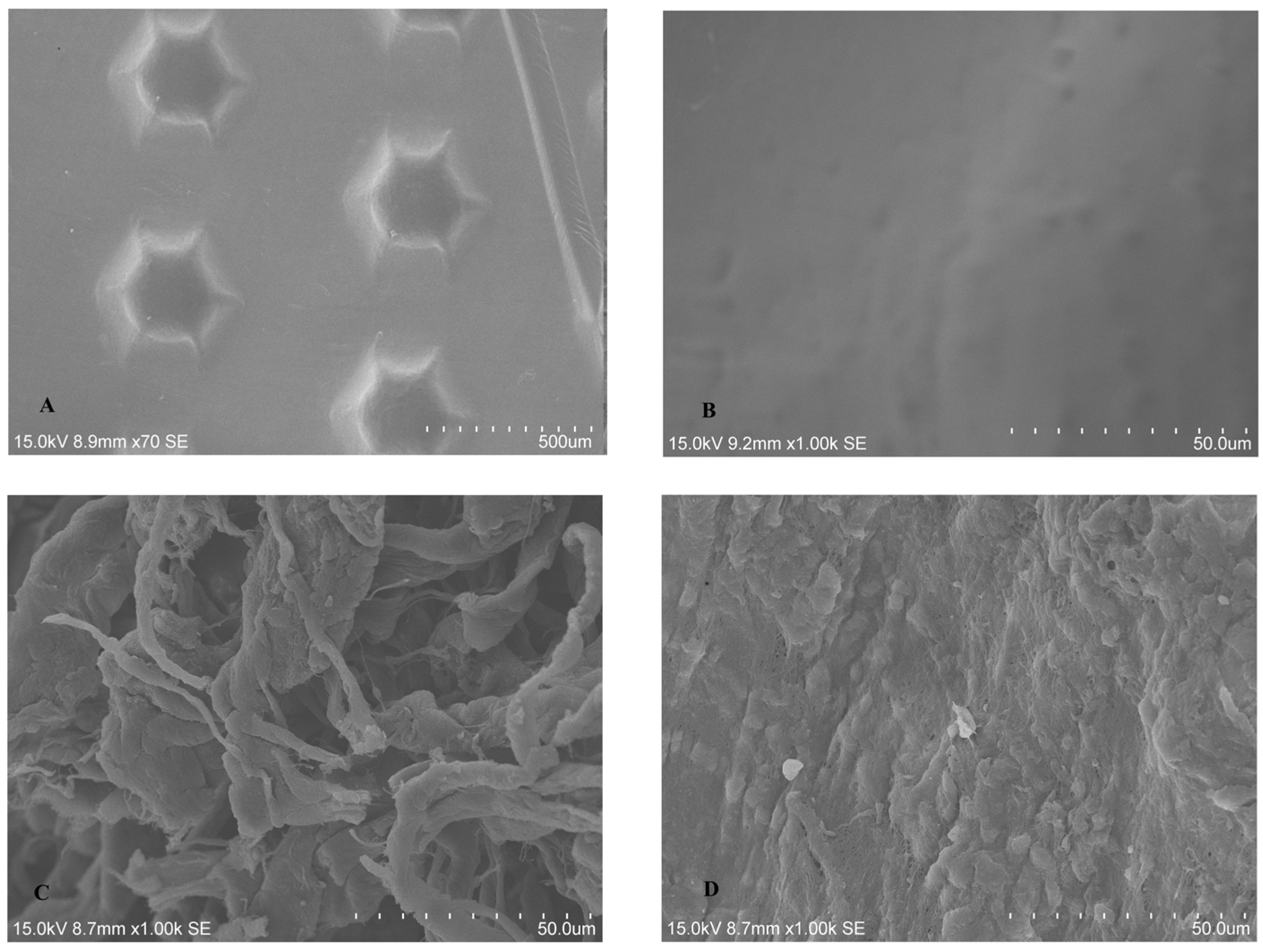
2.1.1. SEM Analysis of Untreated BM

The two BMs displayed distinct appearances under SEM. The CP showed hexagonal surface, which purportedly increases the area available for cellular attachment without increasing porosity, whereas coarse fibers were evident on the rough surface of BG, which has high similarities to human collagen (Figure 1A–D).

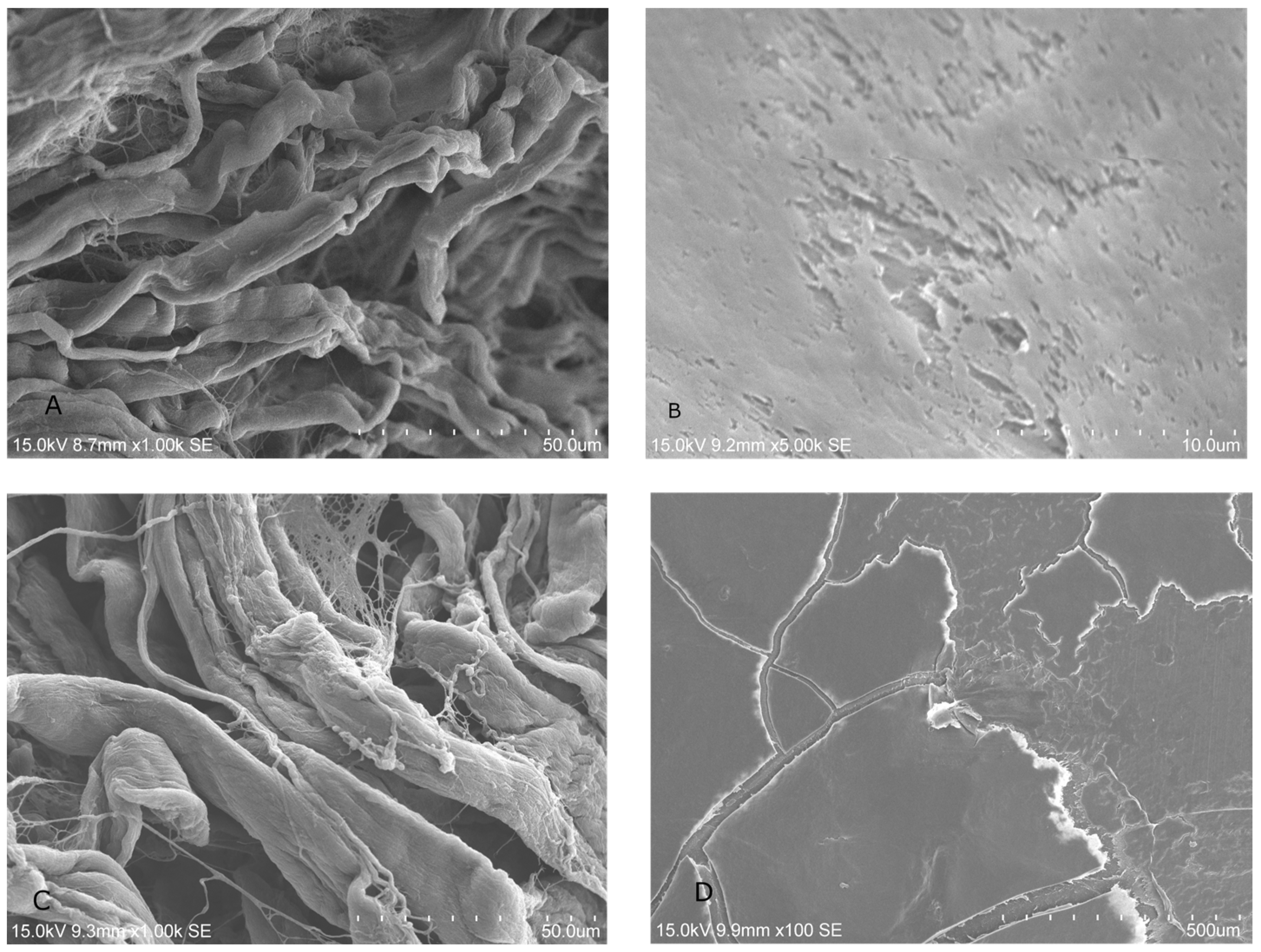
2.1.2. SEM Analysis of Treated Membrane with HGF Cell Culture

The treatment with LPRF-EX (Figure 2A,B) showed no obvious changes in cell growth on either BM after treatment. SEM showed robust fibrin network on BG treated with PLyF on day 7 (Figure 3).

2.1.3. HGF Cell Proliferation Assays

Results of HGF cell proliferation investigated with CCK-8 (Cell Counting Kit-8) assays are depicted in Figure 4. No significant intergroup differences were found. Trend analysis showed that in the absence of membrane and/or LPRF products, cell proliferation appeared as the highest. A trend for greater cell proliferation at CP treated with PLyF on day 1 (Figure 4) was evident, suggesting a rapid release of growth factor at the initial stage.
Conversely, a trend indicating enhanced proliferation of HGF cells under LPRF-EX was noted at day 3 and day 5 (Figure 4), suggesting a potential slow release of growth factor. However, upon statistical analysis, no significant difference was noted in cell proliferation between different LPRF treatments (LPRF-EX or PLyF) (Table 1). Further studies extending the duration of investigation may yield more conclusive results.

2.2. Oral Bacterial Culture Experiments

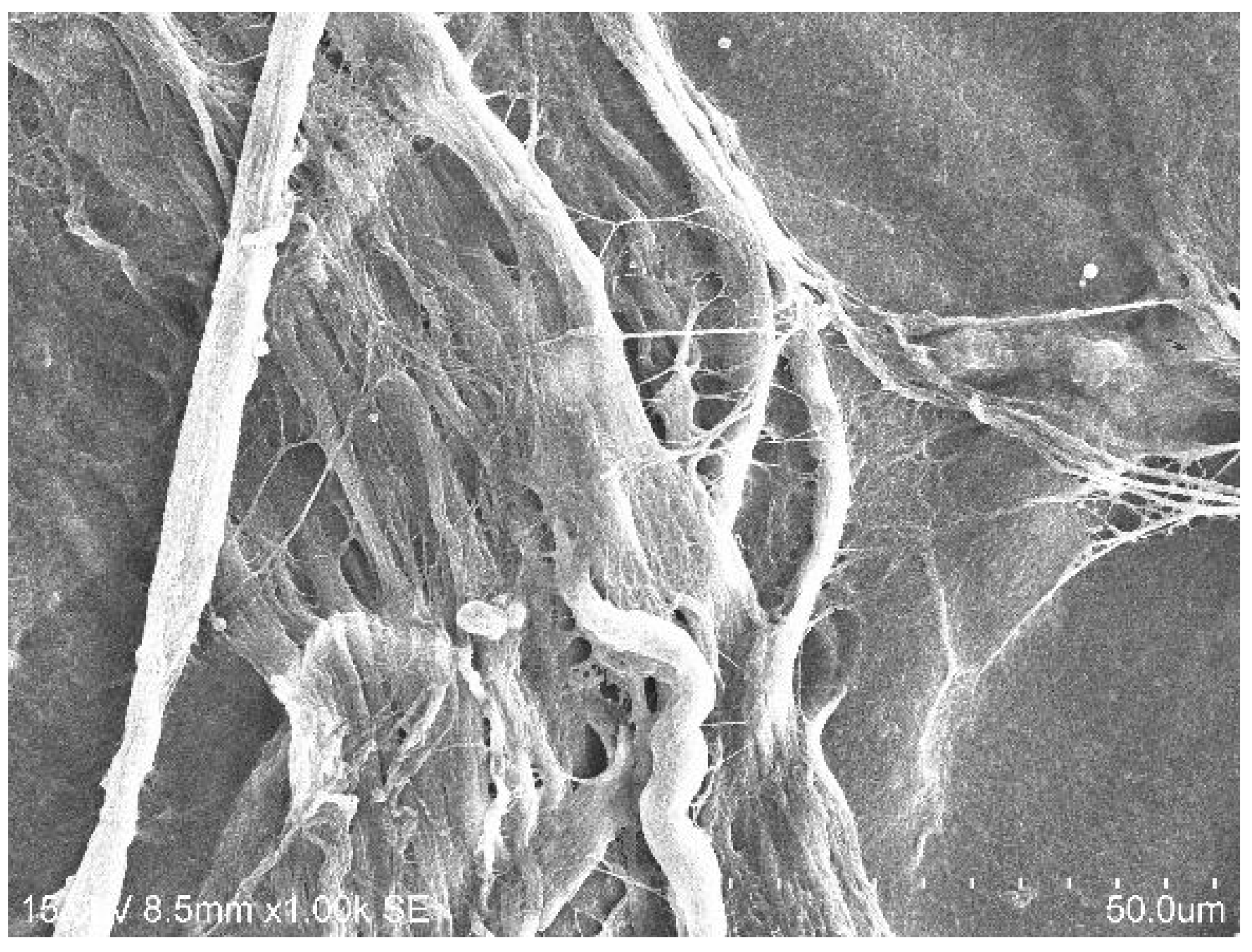
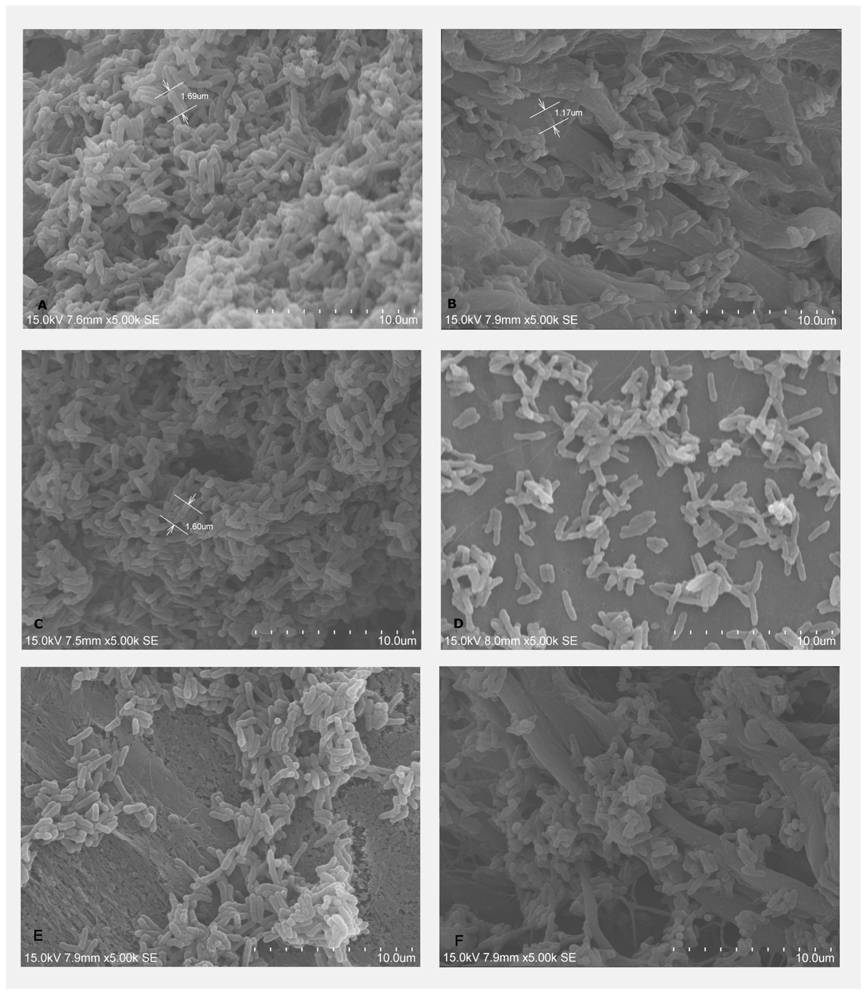
2.2.1. Bacterial Adhesion on BMs Examined by SEM

Bacterial adhesion of A. naeslundii on the treated and untreated BMs was visualized under SEM, showing that A. naeslundii could adhere to both BG and CP. BM treated with LPRF-EX demonstrated fewer adherent cells. The subjective assessment of quantity of adherent bacterial cells appeared lower on the BG treated with PLyF (Figure 5E) than that of BG treated with LPRF-EX (Figure 5C). Overall, it was apparent that the adherent bacteria decreased, demonstrating the possible antimicrobial effects of PLyF and LPRF-EX on A. naeslundii growth on BMs.

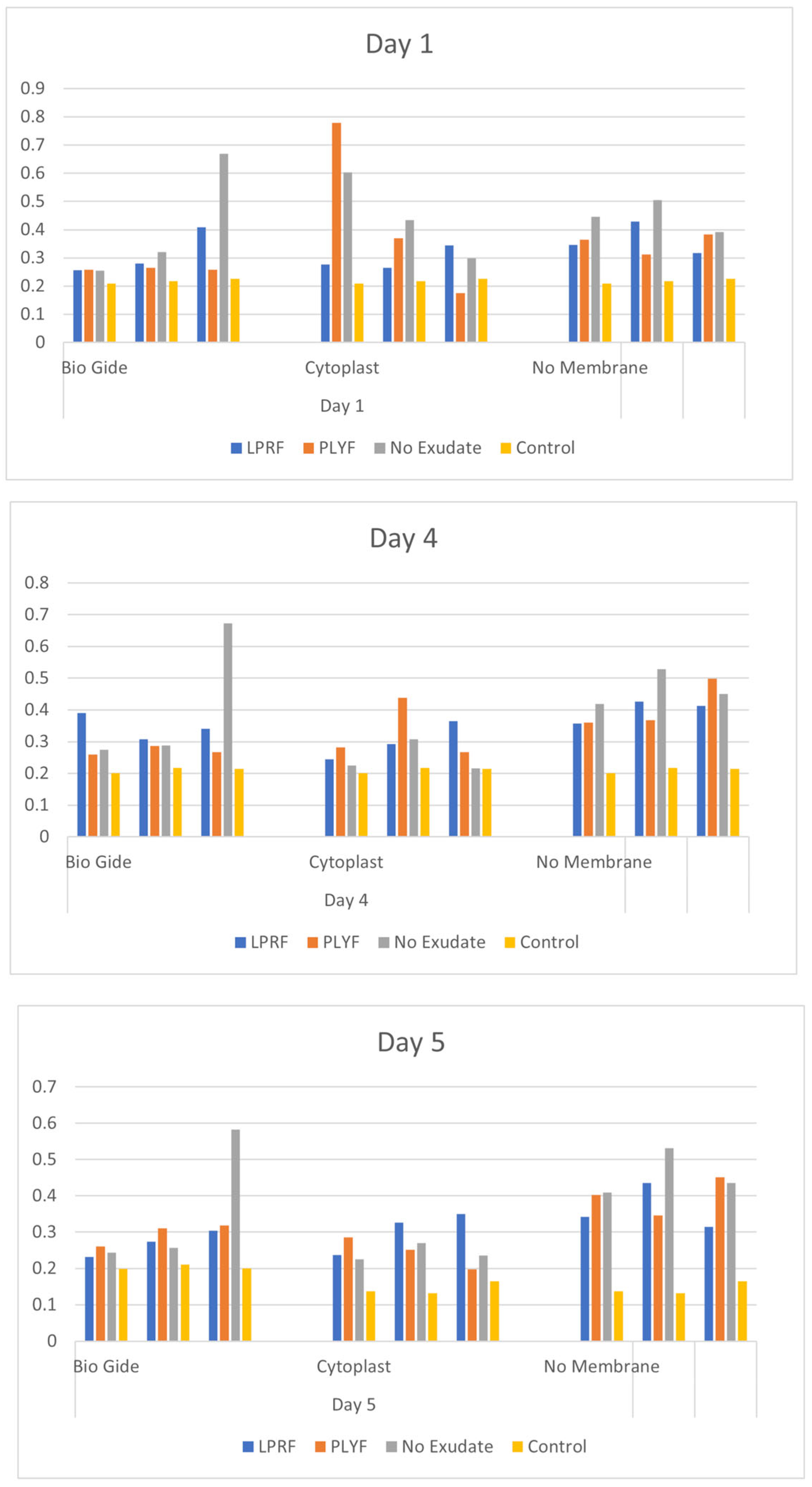
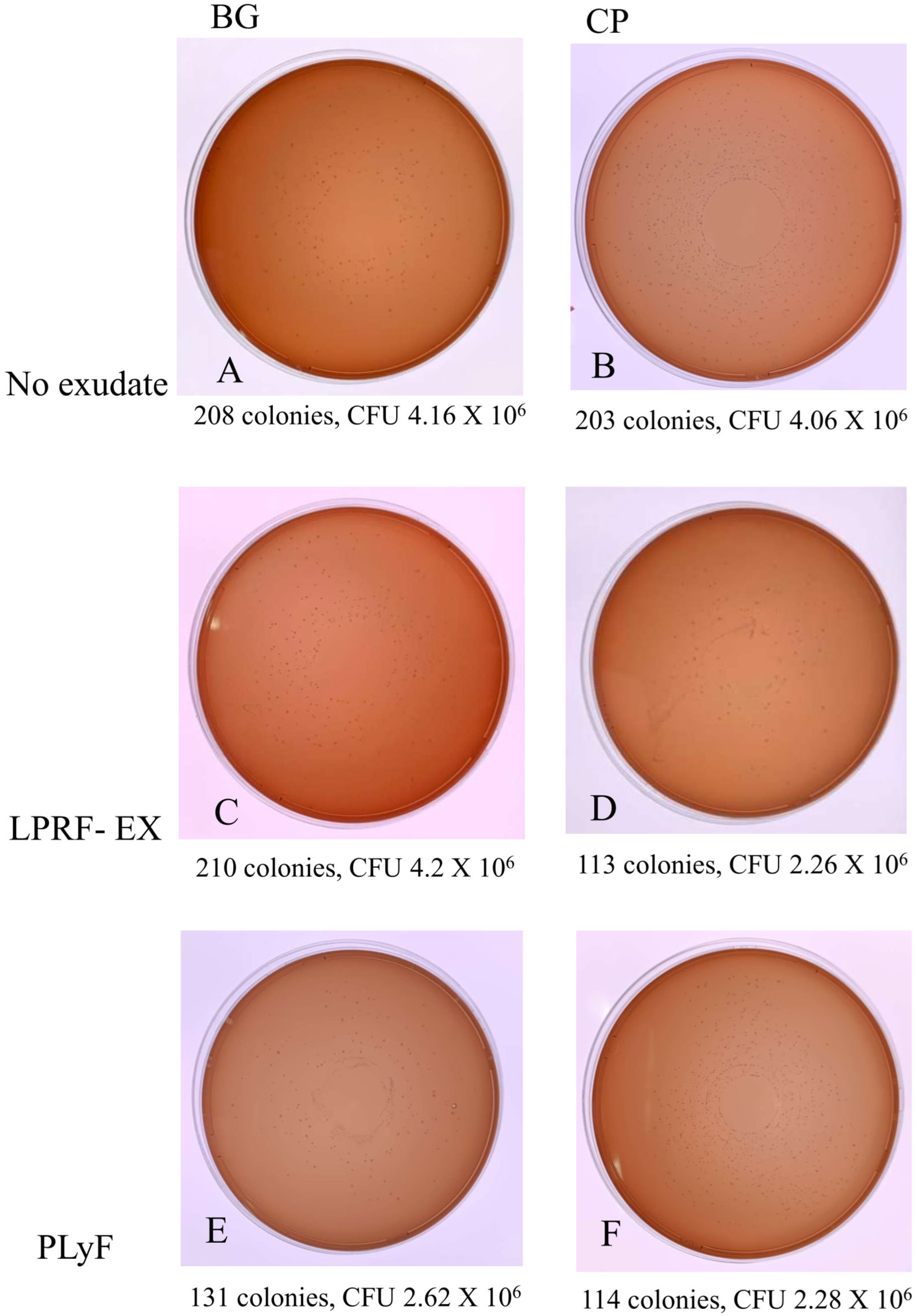
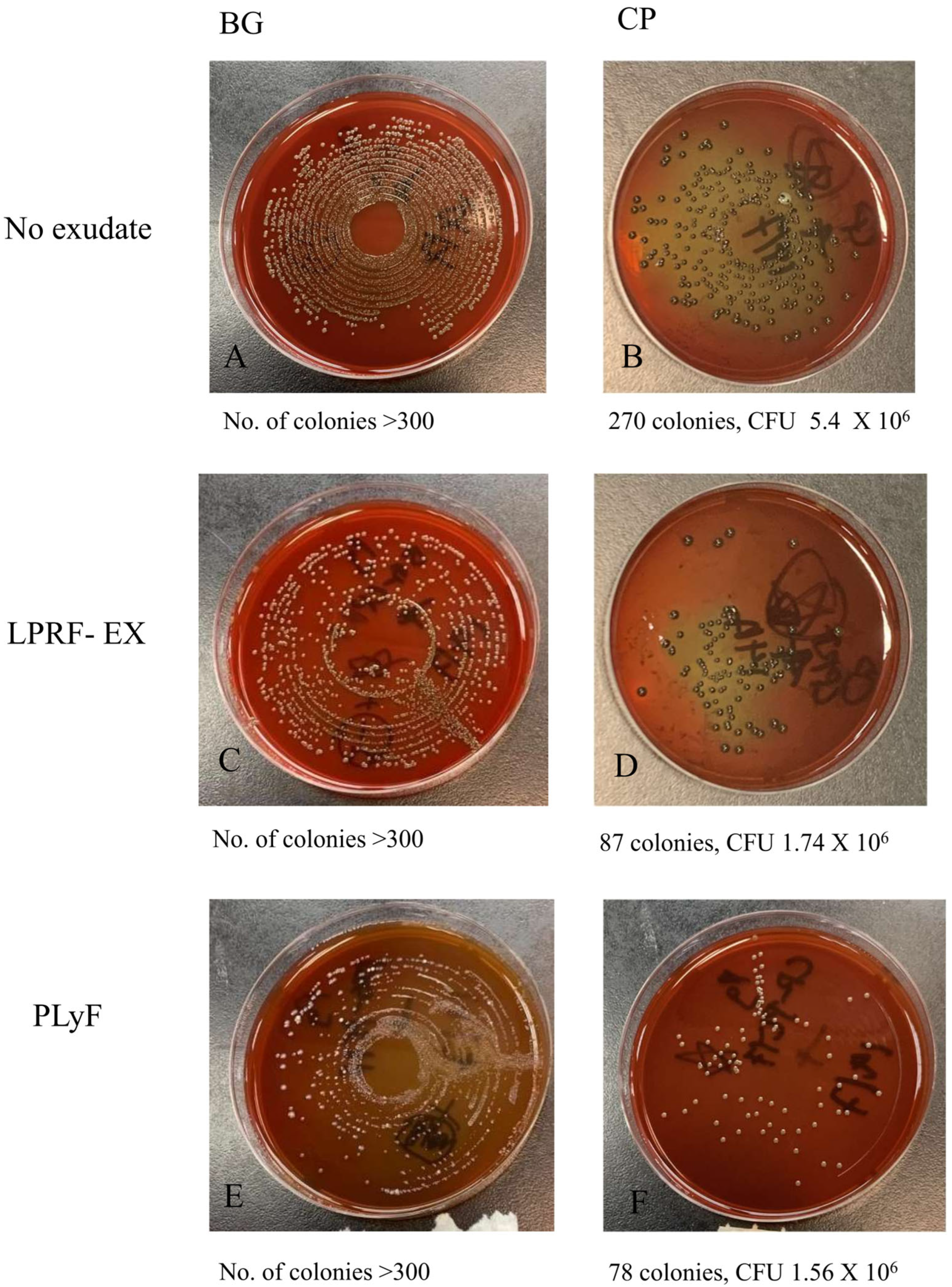
2.2.2. Bacterial CFU Counts

From the results of CFU counts (Figure 6 and Figure 7), it was apparent that the treatment with LPRF-EX or PLyF on both BMs produced antibacterial properties, in which the inhibitory effect of LPRF-EX on A. naeslundii growth on CP was the most marked.

3. Discussion

Within its limitations, the present study provides preliminary in vitro evidence suggestive of antimicrobial effects of the two different LPRF products when used in combination with resorbable and non-resorbable barrier membranes, while biomodulator effects were unclear. A dose-dependent effect of LPRF on HGF proliferation has been shown [34,35]. The comparison of two different LPRF products and two different barrier membranes has not been reported until now. LPRF enhances bone augmentation outcomes [36]. Clinically, liquid LPRF applied in combination with bovine-derived xenograft improved GBR outcomes in peri-implant bone augmentation [37]. Preclinical animal histological data have shown that the coating of Biogide® with LPRF improved new bone formation [38]. The consistency of the findings across the treatment groups verified the primary research assumptions while raising the need for studies to optimize specific LPRF treatment protocols for improved outcomes. Others have shown differences in penetration depth of different collagen membranes biofunctionalized by liquid LPRF products [39] and improved angiogenesis [40], further highlighting the need for investigations focused on specific BM and LPRF preparation combinations.
Antimicrobial activity was shown in this study: the growth and adhesion of A. naeslundii was inhibited by the LPRF treatments. A more marked inhibitory effect upon P. gingivalis and A. naeslundii growth was seen on CP, suggesting a potentially greater benefit. The two oral species chosen, A naeslundi and P. gingivalis, are implicated in periodontitis as an early colonizer [41] and keystone species [42], respectively. The antimicrobial effect of LPRF products has been attributed to the increased release of oxygen metabolites and leucocytes in previous work that showed iPRF (injectable platelet-rich fibrin, other forms of LPRF products) had greater antimicrobial activity against P. gingivalis [43,44].
In addition to the antimicrobial properties, we assessed the early-phase cell response during the first week and quantified the HGF proliferation on days 1, 3, and 5, representing initial seeding and proliferation [45,46]. SEM analysis showed an increased presence of fibrin networks on BG treated with PLyF at 3 and 7 days. The resorbable BM treated with PLyF showed denser fibrin network formation by cultured HGFs compared to the control, suggestive of a potential increase in cellular activity.
However, the differences in HGF proliferation were insignificant, pointing to the need for longer-term and larger-sample studies. A trend for greater cell proliferation for CP treated with PLyF on day 1 was evident and may suggest a rapid release of growth factors in initial stages that may have led to greater early HGF proliferation, whereas the trend for increased proliferation of HGF cells under LPRF-EX on days 3 and 5 may indicate a slower growth factor release. The characterization of growth factor release from PLyF showed increased levels of TGF-β1, PDGF-AB, FGF-2, and VEGF levels over 7 days, with sustained high release of PDGF-AB till 14 days, which are key growth factors in wound healing [15]. LPRF-EX can stimulate platelet activation and promote wound healing and immune modulation via an interleukin 8-dominated cytokine response [47]. Liqiud LPRF treatment shows different chemokine gene expression patterns for HGF compared to PRF clots [48]. Comparative molecular investigations are warranted to understand the biological mechanisms underlying host cell responses to BMs treated with various LPRF preparations.
Taken together, these preliminary findings support the positive biofunctional and antimicrobial effects of LPRF preparations as treatments for both resorbable and non-resorbable barrier membranes (BMs), broadly aligning with previous findings. Liquid LPRF treatment of implant surfaces has been shown to promote fibrin network formation [49]. Blatt et al. [50] showed increased pro-angiogenesis and vessel growth on CP treated with liquid PRF. Specifically, we showed that the application of LPRF-EX and PLyF can confer antimicrobial effects and enhance cellular wound healing processes, which may clinically translate to a reduced incidence of bacterial infections and compromised healing in cases of barrier membrane exposure. The antibacterial potential of liquid LPRF products may have important clinical relevance in regenerative dentistry. The improved wound healing properties could be particularly valuable for patients with systemic conditions, such as diabetes, or in areas with a high risk of infection. Clinical research that investigates the benefits of the putative antimicrobial property of these treatments in specific subpopulations such as diabetics and immunocompromised and geriatric patients and higher-risk procedures such as vertical bone augmentation can comprise a future direction of research. Clinical implications of such findings could include LPRF treatment-based protocols of GBR and GTR that reduce the incidence of BM complications and enhance clinical outcomes.
Major limitations of the study include the small sample and duration of the study, which warrant validation in larger-sample and longitudinal studies. The lack of in vivo validation and potentially divergent clinical effects also represent key limitations. No sample size estimation was performed, and the results may be considered preliminary and warrant verification. The present study did not include any molecular analysis and SEM provided qualitative data, which can be considered preliminary. Given the nature of in vitro study, there could be variability in clinical outcomes. Further research is warranted to establish the differences in biological effects produced by various LPRF products besides LPRF-EX and PLyF and potential clinical trials to enable the selection of optimal clinical protocols for the biofunctionalization of barrier membranes and assess the short- and long-term effects clinically. Moreover, specific LPRF preparation methods should be investigated further, and the effects of centrifuge features, protocols, donor traits, and the downstream effects on wound healing warrant greater elucidation. Clinical trials that assess the long-term effects of these LPRF-based treatments on wound healing and treatment outcomes are essential.

4. Materials and Methods

4.1. Subject Recruitment, Inclusion and Exclusion Criteria

Blood was collected from the antecubital vein of 4 healthy volunteers recruited as a convenience sample, with standard aseptic precautions after obtaining informed consent. The inclusion criterion was healthy males or females aged 20–45 years. The exclusion criteria were (i) pregnancy or lactation; (ii) diagnosed medical conditions or diseases (e.g., diabetes); (iii) hematological diseases or bleeding disorders (e.g., platelet dysfunction or thrombocytopenia); (iv) being on anticoagulants or antiplatelet drugs; and (v) a history of smoking or alcohol consumption. These conditions were excluded, as they may affect changes in the clotting mechanisms and platelet regulatory pathways. The study protocol was reviewed and approved by the Institutional Review Board of the University of Hong Kong. Written informed consent was obtained from the included subjects undergoing blood collection procedures.

4.2. Preparation of L-PRF Products

All L-PRF products were prepared using the same centrifuge system (IntraSpin™, Intra-Lock, Boca Raton, FL, USA). For, LPRF-EX, 4 tubes of 9 mL of venous blood were collected by the same investigator (N.C.W.T) into red-capped vacuum tubes, transferred to the centrifuge machine, and centrifuged under a standardized protocol at 12-min and 2700 rpm to firstly obtain LPRF clots (Figure 2A). The LPRF clots were collected with surgical tweezers and subsequently transferred to a sterile box (Xpression™ Kit, Intra-Lock, Boca Raton, FL, USA) (Figure 2B). The clots were compressed in the kit (Figure 2C) for 5 min to form LPRF membranes, and the resulting exudate was LPRF-EX, which was collected with a sterile syringe.
Similarly to the preparation of LPRF-EX, 4 tubes of 9 mL venous blood were collected into white-capped vacuum tubes and centrifuged with a different protocol, 3 min centrifugation at 2700 rpm, as reported earlier [34]. Under this centrifugation rate, the yellow fluid (PLyF) at the top layer was carefully aspirated using a sterile syringe (5 mL) and 18-gauge needle (Figure 3).
The LPRF-EX and PLyF were brought to the laboratory and filtered with a syringe filter collected and prepared for use to treat the BMs. All procedures were performed under a tissue culture hood.
Centrifuge specific features and centrifugation protocols are known to affect the cell concentration and growth factor amounts and timing of release [35], which may have precluded the exact replication of previously reported protocols.

4.3. Preparation and Treatment of BMs

Bio-gide® (BG) (Figure 5A) (Geistlich Pharma AG, Wolhusen, Switzerland) and Cytoplast™ (CP) (Osteogenics Biomedical, Lubbock, TX, USA) (Figure 5B) BMs were sectioned into 4.7 mm circular sections using a sterile mucosa punch (Straumann Mucosa punch Ø 4.7 mm, guided—L 30 mm, Figure 5C) with finger pressure. These Bio-gide® and Cytoplast™ BM specimens were placed in 24-well plates or 96-well plates. The smooth, cell-occlusive, low-porosity compact surface was oriented facing inwards, and the rough, high-porosity, spongy surface was oriented facing outwards. Each membrane was handled aseptically upon the sealing package using sterile tweezers, prepared using a mucosal punch (Straumann™, Basel, Switzerland), and placed directly into culture plates under the laminar hood.
The sectioned BM specimens were randomized and treated by 10 min inoculation with test treatments: (i) LPRF-EX; (ii) PLyF; and control (iii) Dulbecco’s modified Eagle’s medium (DMEM) (Thermofisher Scientific®, Waltham, MA, USA). DMEM is a standard cell-culture medium and most commonly used for gingival fibroblast culture and was hence chosen as the control reagent [36]. The treatment media were added till the BM was completely submerged.

4.4. Human Gingival Fibroblast (HGF) Cell-Culture Experiments

4.4.1. Cells and Culture

A human gingival fibroblast 1 cell line (PCS-201-018, ATCC® CRL-2014™, Manassas, VA, USA) (HGF-1) was procured from ATCC® and cultures from passage 4–6 were used. Cells were seeded on treated BM specimens at a seeding density of 2 × 10³ cells/cm2 in 24-well plates for scanning electron microscopy (SEM) or 96-well plates (for CCK8-assays), and immersed in DMEM supplemented with 5% fetal bovine serum and 1 mL of antibacterial agent (Primocin®, InvivoGen, San Diego, CA, USA) (Figure 6A,B). Culture plates were placed within a humidified atmosphere (95% air, 5% carbon dioxide) and incubated at 37 °C. After 4 h, the seeded specimens were washed with phosphate-buffered saline to remove non-adherent cells and transferred to new wells. Culture media were changed at 24 h, then every 48 h. Cell seeding density was selected in accordance with ATCC recommendations.

4.4.2. Scanning Electron Microscopy for Fibroblast Adhesion

SEM was performed on day 3 and day 7. Specimens collected on day 3 included (i) BG with no treatment; (ii) CP with no treatment; (iii) BG with LPRF-EX ± HGF; (iv) BG with PLyF ± HGF; (v) CP with LPRF-EX ± HGF; and (vi) CP with PLyF ± HGF. Specimens were fixed with 2.5% glutaraldehyde overnight, followed by dehydration on the following day in graded alcohol series (70%, 85%, 95% and 100%). The specimens were stored in a desiccator at room temperature and processed to visualize under SEM. The same procedures were repeated on day 7.

4.4.3. Cell Proliferation Assays

Cell Counting Kit-8 (CCK-8, Dojindo Laboratories®, Kumamoto, Japan) colorimetric assays were used to study cell proliferation. Cell proliferation was studied on day 1, day 3 and day 5. Old culture medium was discarded, and new fresh medium (DMEM with 5% FBS and antibacterial agent) containing diluted CCK-8 reagent (10 µL CCK8 and 100 µL fresh medium) was used to incubate the specimens for 3 h. The solution was transferred to a new 96-well plate with micropipettes for measuring absorbance at 450 nm. A new culture medium was added to the original 96-well plates with BMs. All experiments were performed in triplicate: BG treated with LPRF-EX; BG treated with PLyF; BG with no treatment; CP treated with LPRF, CP treated with PLyF; CP with no treatment; and a control with no BM (Figure 6A–C). All experiments were performed in triplicate (Figure 6).

4.5. Oral Bacterial Infection Experiments

4.5.1. Bacterial Culture

Two selected oral microbial species, P. gingivalis (ATCC W83) and A. naeslundii (ATCC 12104), were procured from ATCC® (Manassas, VA, USA), incubated with the treated specimens in 24-well plates for 2 days for A. naeslundii using BHI broth and 4 days for P. gingivalis using P. gingivalis broth, and cultured in an anaerobic chamber. Before the assays, the optical density of each culture was adjusted to 0.1.

4.5.2. Scanning Electron Microscopy (SEM) (Hitachi™ VP-SEM SU1510, Hitachi, Tokyo, Japan) for Bacterial Adhesion

The treated specimens were collected at day 2 for A. naeslundii and day 3 for P. gingivalis. The specimens were rinsed with 0.9% sterile saline baths and fixed with 2.5% glutaraldehyde overnight (12 h), followed by dehydration the next day in increasing grades of alcohol (70%, 85%, 95%, and 100%) for 15 min each. Specimens were then stored in a desiccator at room temperature for 1 h and SEM was used to visualize adherent bacteria. Four representative areas were selected for assessment randomly and images were photographed at magnifications from 70 to 5000×.

4.5.3. Colony-Forming Unit (CFU) Counts

The treated specimens collected were washed and sonicated for 5 min to detach the adherent microbes into a recovery medium. Serial dilutions were plated onto tryptic soy agar and incubated for another 2 to 4 days, and the numbers of colonies were counted.

4.6. Data Analysis

Qualitative analysis of the SEM images was performed by the same examiner. CFU counts were compared. The CCK-8 assay absorbance values were used to calculate the mean of triplicate experimental samples and two-way ANOVA with Bonferroni’s multiple comparison test was applied using Prism 9.4.0 software (GraphPad Software, San Diego, CA, USA). A p-value < 0.05 was considered statistically significant. Normality distribution was verified with the Shapiro–Wilk test.

Author Contributions

Conceptualization, A.A. and G.P.; methodology, A.A.; formal analysis, N.C.W.T.; investigation, N.C.W.T.; resources, G.P.; data curation, N.C.W.T.; writing—original draft preparation, N.C.W.T.; writing—review and editing, G.P. and A.A.; supervision, G.P.; project administration, G.P.; funding acquisition, G.P. All authors have read and agreed to the published version of the manuscript.

Funding

This research was funded by the Division of Periodontology and Implant Dentistry Faculty of Dentistry, University of Hong Kong. The APC was funded by the Division of Periodontology and Implant Dentistry, Faculty of Dentistry, University of Hong Kong.

Institutional Review Board Statement

The study was conducted in accordance with the Declaration of Helsinki, and approved by Institutional Review Board of the University of Hong Kong/Hospital Authority Hong Kong West Cluster (HKU/HA HKW IRB) (protocol code: UW 19-137; date of approval: 7 September 2020).

Informed Consent Statement

Informed consent was obtained from all subjects involved in the study.

Data Availability Statement

The raw data generated in this study can be provided by the corresponding author upon reasonable request.

Acknowledgments

The authors acknowledge the administrative and technical support provided by the Central Research Laboratories, Faculty of Dentistry, University of Hong Kong.

Conflicts of Interest

The authors declare no conflicts of interest. The funders had no role in the design of the study; in the collection, analyses, or interpretation of data; in the writing of the manuscript; or in the decision to publish the results.

Abbreviations

Bio-gide® (BG); Cytoplast™ (CP); barrier membrane (BM); human gingival fibroblast (HGF); LPRF exudate (LPRF-EX); liquid fibrinogen (PLyf); platelet-rich fibrin (PRF); platelet-rich plasma (PRP); scanning electron microscopy (SEM).

References

  1. Amrollahi, P.; Shah, B.; Seifi, A.; Tayebi, L. Recent advancements in regenerative dentistry: A review. Mater. Sci. Eng. C Mater. Biol. Appl. 2016, 69, 1383–1390. [Google Scholar] [CrossRef] [PubMed]
  2. Mehta, D.B.; Deshpande, N.C.; Dandekar, S.A. Comparative Evaluation of Platelet-Rich Fibrin Membrane and Collagen Membrane along with Demineralized Freeze-Dried Bone Allograft in Grade II Furcation Defects: A Randomized Controlled Study. J. Indian Soc. Periodontol. 2018, 22, 322–327. [Google Scholar] [PubMed]
  3. Marx, R.E.; Carlson, E.R.; Eichstaedt, R.M.; Schimmele, S.R.; Strauss, J.E.; Georgeff, K.R. Platelet-rich plasma: Growth factor enhancement for bone grafts. Oral Surg. Oral Med. Oral Pathol. Oral Radiol. Endod. 1998, 85, 638–646. [Google Scholar] [CrossRef]
  4. Whitman, D.H.; Berry, R.L.; Green, D.M. Platelet gel: An autologous alternative to fibrin glue with applications in oral and maxillofacial surgery. J. Oral Maxillofac. Surg. 1997, 55, 1294–1299. [Google Scholar] [CrossRef]
  5. Dohan Ehrenfest, D.M.; Del Corso, M.; Diss, A.; Mouhyi, J.; Charrier, J.B. Three-dimensional architecture and cell composition of a Choukroun’s platelet-rich fibrin clot and membrane. J. Periodontol. 2010, 81, 546–555. [Google Scholar] [CrossRef]
  6. Dohan Ehrenfest, D.M. How to optimize the preparation of leukocyte- and platelet-rich fibrin (L-PRF, Choukroun’s technique) clots and membranes: Introducing the PRF Box. Oral Surg. Oral Med. Oral Pathol. Oral Radiol. Endod. 2010, 110, 275–280. [Google Scholar] [CrossRef]
  7. Ghanaati, S.; Booms, P.; Orlowska, A.; Kubesch, A.; Lorenz, J.; Rutkowski, J.; Landes, C.; Sader, R.; Kirkpatrick, C.; Choukroun, J. Advanced Platelet-Rich Fibrin: A New Concept for Cell-Based Tissue Engineering by Means of Inflammatory Cells. J. Oral Implantol. 2014, 40, 679–689. [Google Scholar] [CrossRef]
  8. Dohan, D.M.; Choukroun, J.; Diss, A.; Dohan, S.L.; Dohan, A.J.; Mouhyi, J.; Gogly, B. Platelet-rich fibrin (PRF): A second-generation platelet concentrate. Part II: Platelet-related biologic features. Oral Surg. Oral Med. Oral Pathol. Oral Radiol. Endod. 2006, 101, e45–e50. [Google Scholar] [CrossRef]
  9. Bielecki, T.; Dohan Ehrenfest, D.M.; Everts, P.A.; Wiczkowski, A. The role of leukocytes from L-PRP/L-PRF in wound healing and immune defense: New perspectives. Curr. Pharm. Biotechnol. 2012, 13, 1153–1162. [Google Scholar] [CrossRef]
  10. Crisci, A. The L-PRF membrane (fibrin rich in platelets and leukocytes) and its derivatives (A-PRF, I-PRF) are useful as a source of stem cells in regenerative wound therapy: Experimental work on the horse. Regen. Med. Ther. 2019, 3, 37–45. [Google Scholar] [CrossRef]
  11. He, L.; Lin, Y.; Hu, X.; Zhang, Y.; Wu, H. A comparative study of platelet-rich fibrin (PRF) and platelet-rich plasma (LPRF) on the effect of proliferation and differentiation of rat osteoblasts in vitro. Oral Surg. Oral Med. Oral Pathol. Oral Radiol. Endod. 2009, 108, 707–713. [Google Scholar] [CrossRef] [PubMed]
  12. Fujioka-Kobayashi, M.; Miron, R.J.; Hernandez, M.; Kandalam, U.; Zhang, Y.; Choukroun, J. Optimized platelet-rich fibrin with the low-speed concept: Growth factor release, biocompatibility, and cellular response. J. Periodontol. 2017, 88, 112–121. [Google Scholar] [CrossRef]
  13. Rodríguez Sánchez, F.; Verspecht, T.; Castro, A.B.; Pauwels, M.; Andrés, C.R.; Quirynen, M.; Teughels, W. Antimicrobial Mechanisms of Leucocyte- and Platelet Rich Fibrin Exudate Against Planktonic Porphyromonas gingivalis and Within Multi-Species Biofilm: A Pilot Study. Front. Cell. Infect. Microbiol. 2021, 11, 722499. [Google Scholar] [CrossRef]
  14. Dohan Ehrenfest, D.M.; Pinto, N.R.; Pereda, A.; Jiménez, P.; Corso, M.D.; Kang, B.S.; Nally, M.; Lanata, N.; Wang, H.L.; Quirynen, M. The impact of the centrifuge characteristics and centrifugation protocols on the cells, growth factors, and fibrin architecture of a leukocyte-and platelet-rich fibrin (L-PRF) clot and membrane. Platelets 2018, 29, 171–184. [Google Scholar] [CrossRef]
  15. Serafini, G.; Lopreiato, M.; Lollobrigida, M.; Lamazza, L.; Mazzucchi, G.; Fortunato, L.; Mariano, A.; Scotto d’Abusco, A.; Fontana, M.; De Biase, A. Platelet Rich Fibrin (PRF) and Its Related Products: Biomolecular Characterization of the Liquid Fibrinogen. J. Clin. Med. 2020, 9, 1099. [Google Scholar] [CrossRef]
  16. Ratajczak, J.; Vangansewinkel, T.; Gervois, P.; Merckx, G.; Hilkens, P.; Quirynen, M.; Lambrichts, I.; Bronckaers, A. Angiogenic Properties of ‘Leukocyte- and Platelet-Rich Fibrin’. Sci. Rep. 2018, 8, 14632. [Google Scholar] [CrossRef]
  17. Blanco, J.; García Alonso, A.; Hermida-Nogueira, L.; Castro, A.B. How to explain the beneficial effects of leukocyte-and platelet-rich fibrin. Periodontol. 2000 2025, 97, 74–94. [Google Scholar] [CrossRef]
  18. Miron, R.J.; Fujioka-Kobayashi, M.; Hernandez, M.; Kandalam, U.; Zhang, Y.; Ghanaati, S.; Choukroun, J. Injectable platelet rich fibrin (i-PRF): Opportunities in regenerative dentistry? Clin. Oral Investig. 2017, 21, 2619–2627. [Google Scholar] [CrossRef]
  19. Wang, X.; Zhang, Y.; Choukroun, J.; Ghanaati, S.; Miron, R.J. Effects of an injectable platelet-rich fibrin on osteoblast behavior and bone tissue formation in comparison to platelet-rich plasma. Platelets 2018, 29, 48–55. [Google Scholar] [CrossRef]
  20. Dahlin, C.; Linde, A.; Gottlow, J.; Nyman, S. Healing of bone defects by guided tissue regeneration. Plastic and reconstructive surgery. Plast. Reconstr. Surg. 1988, 81, 672–676. [Google Scholar] [CrossRef]
  21. Bianchi, S.; Bernardi, S.; Simeone, D.; Torge, D.; Macchiarelli, G.; Marchetti, E. Proliferation and morphological assessment of human periodontal ligament fibroblast towards bovine pericardium membranes: An in vitro study. Materials 2022, 15, 8284. [Google Scholar] [CrossRef]
  22. Pistilli, R.; Simion, M.; Barausse, C.; Gasparro, R.; Pistilli, V.; Bellini, P.; Felice, P. Guided Bone Regeneration with Nonresorbable Membranes in the Rehabilitation of Partially Edentulous Atrophic Arches: A Retrospective Study on 122 Implants with a 3- to 7-Year Follow-up. Int. J. Periodontics Restor. Dent. 2020, 40, 685–692. [Google Scholar] [CrossRef] [PubMed]
  23. Rocchietta, I.; Fontana, F.; Simion, M. Clinical outcomes of vertical bone augmentation to enable dental implant placement: A systematic review. J. Clin. Periodontol. 2008, 35 (Suppl. S8), 203–215. [Google Scholar] [CrossRef] [PubMed]
  24. Benic, G.I.; Hämmerle, C.H. Horizontal bone augmentation by means of guided bone regeneration. Periodontol. 2000 2014, 66, 13–40. [Google Scholar] [CrossRef] [PubMed]
  25. Abdo, V.L.; Suarez, L.J.; de Paula, L.G.; Costa, R.C.; Shibli, J.; Feres, M.; Barão, V.-R.; Bertolini, M.; Souza, J.-S. Underestimated microbial infection of resorbable membranes on guided regeneration. Colloids Surf. B Biointerfaces 2023, 226, 113318. [Google Scholar] [CrossRef]
  26. Slutzkey, S.; Kozlovsky, A.; Artzi, Z.; Matalon, S. Collagen barrier membranes may accelerate bacterial growth in vitro: A potential clinical risk to regenerative procedures. Quintessence Int. 2015, 46, 43–50. [Google Scholar] [CrossRef]
  27. Machtei, E.E. The effect of membrane exposure on the outcome of regenerative procedures in humans: A meta-analysis. J. Periodontol. 2001, 72, 512–516. [Google Scholar] [CrossRef]
  28. Garcia, J.; Dodge, A.; Luepke, P.; Wang, H.L.; Kapila, Y.; Lin, G.H. Effect of membrane exposure on guided bone regeneration: A systematic review and meta-analysis. Clin. Oral Implant. Res. 2018, 29, 328–338. [Google Scholar] [CrossRef]
  29. Urban, I.A.; Montero, E.; Monje, A.; Sanz-Sánchez, I. Effectiveness of vertical ridge augmentation interventions: A systematic review and meta-analysis. J. Clin. Periodontol. 2019, 46 (Suppl. S21), 319–339. [Google Scholar] [CrossRef]
  30. Omar, O.; Elgali, I.; Dahlin, C.; Thomsen, P. Barrier membranes: More than the barrier effect? J. Clin. Periodontol. 2019, 46, 103–123. [Google Scholar] [CrossRef]
  31. Mozgan, E.M.; Edelmayer, M.; Janjić, K.; Pensch, M.; Fischer, M.B.; Moritz, A.; Agis, H. Release kinetics and mitogenic capacity of collagen barrier membranes supplemented with secretome of activated platelets—The in vitro response of fibroblasts of the periodontal ligament and the gingiva. BMC Oral Health 2017, 17, 66. [Google Scholar] [CrossRef] [PubMed]
  32. Huang, Q.; Huang, X.; Gu, L. Periodontal Bifunctional Biomaterials: Progress and Perspectives. Materials 2021, 14, 7588. [Google Scholar] [CrossRef]
  33. Toledano-Osorio, M.; Vallecillo, C.; Vallecillo-Rivas, M.; Manzano-Moreno, F.J.; Osorio, R. Antibiotic-Loaded Polymeric Barrier Membranes for Guided Bone/Tissue Regeneration: A Mini-Review. Polymers 2022, 14, 840. [Google Scholar] [CrossRef]
  34. Mudalal, M.; Wang, Z.; Mustafa, S.; Liu, Y.; Wang, Y.; Yu, J.; Wang, S.; Sun, X.; Zhou, Y. Effect of Leukocyte-Platelet Rich Fibrin (L-PRF) on tissue regeneration and proliferation of human gingival fibroblast cells cultured using a modified method. Tissue Eng. Regen. Med. 2021, 18, 895–904. [Google Scholar] [CrossRef]
  35. Goel, A.; Windsor, L.J.; Gregory, R.L.; Blanchard, S.B.; Hamada, Y. Effects of platelet-rich fibrin on human gingival and periodontal ligament fibroblast proliferation from chronic periodontitis versus periodontally healthy subjects. Clin. Exp. Dent. Res. 2021, 7, 436–442. [Google Scholar] [CrossRef]
  36. Quirynen, M.; Siawasch, S.; Temmerman, A.; Cortellini, S.; Dhondt, R.; Teughels, W.; Castro, A.B. Do autologous platelet concentrates (APCs) have a role in intra-oral bone regeneration? A critical review of clinical guidelines on decision-making process. Periodontol. 2000 2023, 93, 254–269. [Google Scholar] [CrossRef]
  37. Işık, G.; Özden Yüce, M.; Koçak-Topbaş, N.; Günbay, T. Guided bone regeneration simultaneous with implant placement using bovine-derived xenograft with and without liquid platelet-rich fibrin: A randomized controlled clinical trial. Clin. Oral Investig. 2021, 25, 5563–5575. [Google Scholar] [CrossRef]
  38. Apaza Alccayhuaman, K.A.; Heimel, P.; Tangl, S.; Lettner, S.; Kampleitner, C.; Panahipour, L.; Kuchler, U.; Gruber, R. Human versus Rat PRF on Collagen Membranes: A Pilot Study of Mineralization in Rat Calvaria Defect Model. Bioengineering 2024, 11, 414. [Google Scholar] [CrossRef]
  39. Al-Maawi, S.; Herrera-Vizcaíno, C.; Orlowska, A.; Willershausen, I.; Sader, R.; Miron, R.J.; Choukroun, J.; Ghanaati, S. Biologization of Collagen-Based Biomaterials Using Liquid-Platelet-Rich Fibrin: New Insights into Clinically Applicable Tissue Engineering. Materials 2019, 12, 3993. [Google Scholar] [CrossRef]
  40. Blatt, S.; Schröger, S.-V.; Pabst, A.; Kämmerer, P.W.; Sagheb, K.; Al-Nawas, B. Biofunctionalization of Xenogeneic Collagen Membranes with Autologous Platelet Concentrate—Influence on Rehydration Protocol and Angiogenesis. Biomedicines 2022, 10, 706. [Google Scholar] [CrossRef]
  41. Sato, T.; Watanabe, K.; Kumada, H.; Toyama, T.; Tani-Ishii, N.; Hamada, N. Peptidoglycan of Actinomyces naeslundii induces inflammatory cytokine production and stimulates osteoclastogenesis in alveolar bone resorption. Arch. Oral Biol. 2012, 57, 1522–1528. [Google Scholar] [CrossRef] [PubMed]
  42. Lunar Silva, I.; Cascales, E. Molecular Strategies Underlying Porphyromonas gingivalis Virulence. J. Mol. Biol. 2021, 433, 166836. [Google Scholar] [CrossRef]
  43. Karde, P.A.; Sethi, K.S.; Mahale, S.A.; Khedkar, S.U.; Patil, A.G.; Joshi, C.P. Comparative evaluation of platelet count and antimicrobial efficacy of injectable platelet-rich fibrin with other platelet concentrates: An in vitro study. J. Indian Soc. Periodontol. 2017, 21, 97–101. [Google Scholar] [CrossRef]
  44. Sindhusha, V.B.; Ramamurthy, J. Comparison of Antimicrobial Activity of Injectable Platelet-Rich Fibrin (i-PRF) and Leukocyte and Platelet-Rich Fibrin (l-PRF) Against Oral Microbes: An In Vitro Study. Cureus 2023, 15, e46196. [Google Scholar] [CrossRef]
  45. Wu, J.; Yu, P.; Lv, H.; Yang, S.; Wu, Z. Nanostructured Zirconia Surfaces Regulate Human Gingival Fibroblasts Behavior Through Differential Modulation of Macrophage Polarization. Front. Bioeng. Biotechnol. 2021, 8, 611684. [Google Scholar] [CrossRef]
  46. Osman, M.A.; Kushnerev, E.; Alamoush, R.A.; Seymour, K.G.; Yates, J.M. Two Gingival Cell Lines Response to Different Dental Implant Abutment Materials: An In Vitro Study. Dent. J. 2022, 10, 192. [Google Scholar] [CrossRef]
  47. Melo-Ferraz, A.; Coelho, C.; Miller, P.; Criado, M.B.; Monteiro, M.C. Comprehensive analysis of L-PRF exudate components and their impact on whole blood platelets. Clin. Oral Investig. 2024, 28, 470. [Google Scholar] [CrossRef]
  48. Imani, A.; Panahipour, L.; Kühtreiber, H.; Mildner, M.; Gruber, R. RNAseq of Gingival Fibroblasts Exposed to PRF Membrane Lysates and PRF Serum. Cells 2024, 13, 1308. [Google Scholar] [CrossRef]
  49. Lollobrigida, M.; Maritato, M.; Bozzuto, G.; Formisano, G.; Molinari, A.; De Biase, A. Biomimetic implant surface functionalization with liquid L-PRF products: In vitro study. BioMed Res. Int. 2018, 2018, 9031435. [Google Scholar] [CrossRef]
  50. Blatt, S.; Krüger, M.; Kämmerer, P.W.; Thiem, D.G.; Matheis, P.; Eisenbeiß, A.K.; Wiltfang, J.; Al-Nawas, B.; Naujokat, H. Non-Interventional Prospective Observational Study of Platelet Rich Fibrin as a Therapy Adjunctive in Patients with Medication-Related Osteonecrosis of the Jaw. J. Clin. Med. 2022, 11, 682. [Google Scholar] [CrossRef]
Figure 1. SEM images showing (A) hexagonal surface of CP; (B) smooth surface side of CP; (C) the rough, high-porosity surface of BG; and (D) the smooth cell occlusive surface of BG.
Dentistry 13 00228 g001
Figure 2. SEM images of HGF cultures on day 3: (A) treatment of LPRF-EX on BG; (B) treatment of LPRF-EX on CP; (C) treatment of PLyF EX on BG; (D) treatment of PLyF on CP.
Dentistry 13 00228 g002
Figure 3. SEM image showing HGF proliferation on BG treated with PLyF on day 7.
Dentistry 13 00228 g003
Figure 4. Bar plots depicting HGF cell proliferation represented by CCK-8 assay absorbance values on days 1, 3, and 5.
Dentistry 13 00228 g004
Figure 5. SEM images of A. naeslundi culture on: (A) untreated BG; (B) untreated CP; (C) LPRF-EX-treated BG; (D) LPRF-EX-treated CP; (E) PLyF-treated BG; (F) PLyF-treated CP.
Dentistry 13 00228 g005
Figure 6. CFU counts of A. naeslundi culture on: (A) untreated BG; (B) untreated CP; (C) LPRF-EX-treated BG; (D) LPRF-EX-treated CP; (E) PLyF-treated BG; (F) PLyF-treated CP.
Dentistry 13 00228 g006
Figure 7. CFU counts of P. gingivalis culture on: (A) untreated BG; (B) untreated CP; (C) LPRF-EX-treated BG; (D) LPRF-EX-treated CP; (E) PLyF-treated BG; (F) PLyF-treated CP.
Dentistry 13 00228 g007
Table 1. Comparison of CCK-8 absorbance values for HGF proliferation on differently treated BMs. ns means no significant findings.
Day 1Day 3Day 5
Bonferroni’s Multiple Comparison TestMean Diff. Adjusted p ValueMean Diff. Adjusted p ValueMean Diff. Adjusted p Value
BG
LPRF vs. PLYF0.0549ns0.88590.07477ns>0.9999−0.02683ns>0.9999
LPRF vs. No Exudate−0.09857ns0.6808−0.0658ns>0.9999−0.09123ns0.5255
PLYF vs. No Exudate−0.1535ns0.4051−0.1406ns0.2464−0.0644ns0.9969
CP
LPRF vs. PLYF−0.1459ns0.4402−0.0286ns>0.99990.05997ns>0.9999
LPRF vs. No Exudate−0.15ns0.42110.05177ns>0.99990.06123ns>0.9999
PLYF vs. No Exudate−0.0041ns0.99930.08033ns0.92020.001267ns>0.9999
No Membrane
LPRF vs. PLYF0.009933ns0.996−0.0098ns>0.9999−0.03547ns>0.9999
LPRF vs. No Exudate−0.0844ns0.753−0.0674ns>0.9999−0.09417ns0.4871
PLYF vs. No Exudate−0.09433ns0.7026−0.0575ns>0.9999−0.0587ns>0.9999
Disclaimer/Publisher’s Note: The statements, opinions and data contained in all publications are solely those of the individual author(s) and contributor(s) and not of MDPI and/or the editor(s). MDPI and/or the editor(s) disclaim responsibility for any injury to people or property resulting from any ideas, methods, instructions or products referred to in the content.

