Article

Biogenic ZnO Nanoparticles Derived from Eichhornia crassipes: Synthesis and Application in the Degradation of Multiple Organic Dyes and Chlorpyrifos Ethyl

by
Nelson Nagles-Vergara
1,2,
Jose Alejandro Villegas-Fuentes
3,
Alfredo Rafael Vilchis-Nestor
4,
Yuber Palacios-Torres
2,
Efraím A. Serna-Galvis
5,6,
Jorge L. Gallego
1,* and
Priscy Alfredo Luque-Morales
3,*
1
Biodiversity, Biotechnology, and Bioengineering Research Group—GRINBIO, Department of Engineering, University of Medelín, Medellín 050026, Antioquia, Colombia
2
Grupo de Investigación en Recursos Naturales y Toxicología Ambiental, Facultad de Ciencias Naturales, Universidad Tecnológica del Chocó “Diego Luis Córdoba”, Ciudadela Universitaria, Quibdó 270001, Chocó, Colombia
3
Facultad de Ingeniería, Arquitectura y Diseño, Universidad Autónoma de Baja California, Ensenada C.P. 22860, Baja California, Mexico
4
Centro Conjunto de Investigación de Química Sustentable, Universidad Autónoma del Estado de Mexico—Universidad Nacional Autónoma de Mexico, Toluca C.P. 50200, Estado de Mexico, Mexico
5
Departamento de Química, Facultad de Ciencias Naturales y Exactas, Universidad de Valle, Calle 12 No. 100-00, Santiago de Cali 760032, Valle del Cauca, Colombia
6
Grupo de Investigación en Remediación Ambiental y Biocatálisis (GIRAB), Instituto de Química, Facultad de Ciencias Exactas y Naturales, Universidad de Antioquia, UdeA, Calle 70. 52-21, Medellín 050010, Antioquia, Colombia
*
Authors to whom correspondence should be addressed.
Inorganics 2026, 14(9), 243; https://doi.org/10.3390/inorganics14090243 (registering DOI)
Submission received: 25 August 2026 / Revised: 12 September 2026 / Accepted: 14 September 2026 / Published: 18 September 2026
(This article belongs to the Special Issue Inorganic Photocatalysts for Environmental Applications, 2nd Edition)

Abstract

The green synthesis of zinc oxide (ZnO) nanoparticles using plant biomass offers a sustainable approach for developing photocatalytic materials for water treatment. In this study, ZnO nanoparticles were biosynthesized using aqueous extracts of Eichhornia crassipes from the Medio Atrato region, Chocó, Colombia, at 1%, 2%, and 4% (w/v). The materials were characterized by FTIR, UV-Vis, XRD, and SEM–EDX. All samples exhibited the hexagonal wurtzite structure of ZnO without detectable secondary crystalline phases. Average crystallite sizes were 38.66, 38.16, and 31.52 nm for EC-1%, EC-2%, and EC-4%, respectively, decreasing with increasing extract concentration. Photocatalytic activity was evaluated under UV irradiation using six organic dyes: amido black 10B, eosin yellow, methylene blue, methyl orange, methyl red, and rhodamine B. Performance depended on both pollutant type and extract concentration. EC-2% showed the most consistent overall performance, achieving 87% degradation of amido black 10B, 92% of methyl orange, 81% of methyl red, and 93% of rhodamine B. Eosin yellow reached approximately 97% removal at 90 min, while EC-4% achieved 95% methylene blue degradation after 180 min. EC-2% also removed approximately 56.78% of chlorpyrifos ethyl after 180 min. Overall, E. crassipes-mediated ZnO nanoparticles demonstrate promising photocatalytic activity toward diverse organic pollutants and provide a potential route for valorizing invasive aquatic biomass.

1. Introduction

Water pollution by synthetic organic dyes and pesticides remains a major environmental challenge due to their high chemical stability, toxicity, and persistence in aquatic ecosystems [1,2,3]. Colored effluents from textile, paper, pharmaceutical, and related industries often contain complex aromatic dyes (azo and xanthene structures that frequently resist conventional biological and physicochemical treatments and may generate toxic by-products [4]. In parallel, organophosphorus pesticides like chlorpyrifos ethyl contribute to the continuous input of hazardous compounds into surface waters and sediments, where they can persist and form transformation products of ecotoxicological concern, such as 3,5,6-trichloro-2-pyridinol [5]. These contaminants compromise water quality, affect aquatic biota, and pose health risks to exposed populations, particularly in regions with limited wastewater treatment infrastructure [6,7].
Advanced oxidation processes based on semiconductor photocatalysts have emerged as promising alternatives for the removal of recalcitrant organic pollutants, since they enable the in situ generation of reactive oxygen species capable of degrading dyes and pesticides under irradiation [8]. Among available semiconductors, zinc oxide (ZnO) stands out due to its suitable band gap, high photochemical stability, abundance, and relatively low cost, as well as its versatility for structural and surface modification [9]. However, conventional routes for ZnO synthesis often involve harsh conditions, toxic reagents, and high energy inputs, limiting their sustainability [10]. Green synthesis strategies using plant extracts as reducing, capping, and stabilizing agents have therefore gained increasing attention as a means to tailor ZnO nanoparticles while reducing the environmental footprint of the material [11,12,13,14].
Aquatic macrophytes such as Eichhornia crassipes (water hyacinth) are particularly attractive for this purpose because they combine invasive behavior and high biomass productivity with a rich phytochemical composition [15,16,17,18,19]. The phytochemical profile of E. crassipes includes diverse bioactive compounds, such as phenolic compounds, flavonoids, tannins, terpenoids, alkaloids, and other reducing and antioxidant constituents, which can participate in the reduction, complexation, nucleation, and stabilization processes involved in the formation of metal and metal oxide nanoparticles, as well as molecules such as catechol, resorcinol, kaempferol, quercetin, among other phytochemical constituents [20,21,22]. This chemical richness makes E. crassipes not only a readily available biomass resource but also a promising biological precursor for the sustainable synthesis of functional nanomaterials.
E. crassipes is recognized as one of the world’s worst invasive aquatic plants, but its abundant biomass has been successfully used for water remediation and for the biogenic synthesis of ZnO-based photocatalysts, achieving efficient removal of model dyes under UV or visible irradiation [23,24]. In tropical river–floodplain systems such as the Atrato basin in Colombia, the proliferation of E. crassipes aggravates local management problems yet offers an opportunity to generate value-added materials for water treatment. Despite numerous reports on plant-mediated ZnO synthesis, the influence of E. crassipes extract concentration on the structural, optical, and photocatalytic properties of ZnO nanomaterials, particularly for the simultaneous degradation of multiple dyes and a representative organophosphorus pesticide, remains insufficiently explored [19,25]. In this work, we address this gap by synthesizing ZnO nanocomposites using E. crassipes extracts at different concentrations, characterizing their structural and optical features, and evaluating their photocatalytic performance in the degradation of several dyes and chlorpyrifos ethyl.

2. Results and Discussion

2.1. FTIR

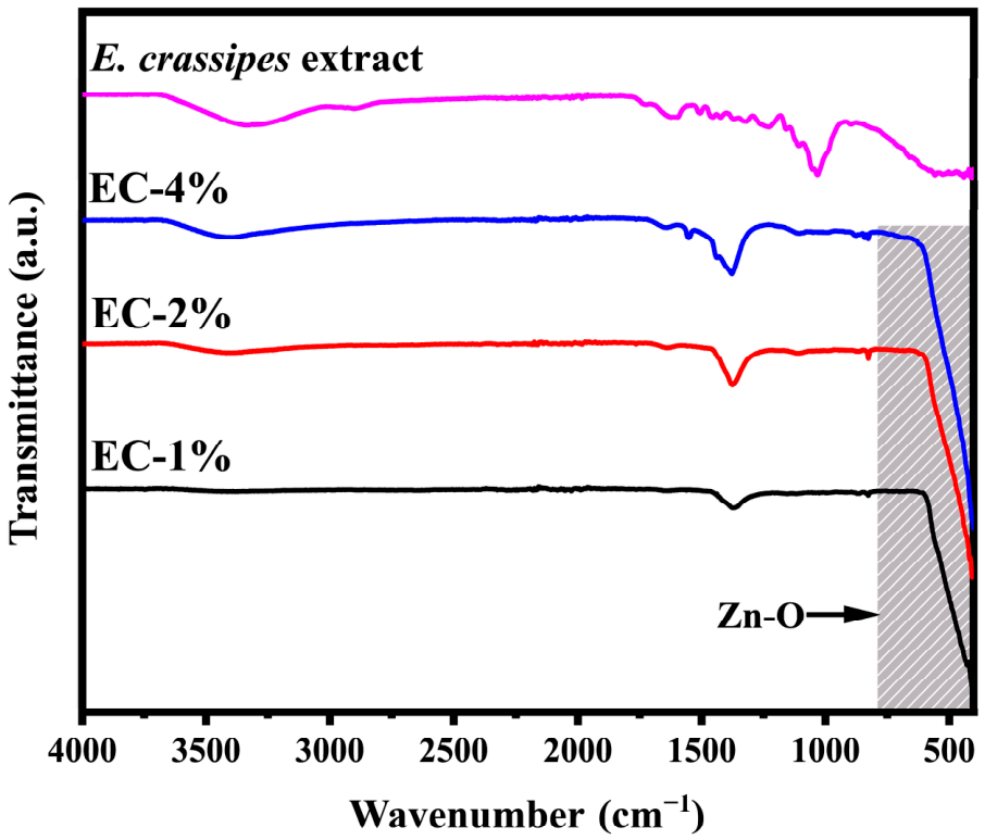
Figure 1 shows the FTIR spectra of the E. crassipes extract and the ZnO nanoparticles synthesized with 1%, 2%, and 4%. In the extract, a broad band at approximately 3410 cm−1 is observed, attributed to O–H stretching vibrations of hydroxyl groups present in phenols and alcohols [26,27]. The spectra of the ZnO nanoparticles display a characteristic band near 435 cm−1, corresponding to Zn–O stretching, confirming the formation of zinc oxide. These results are consistent with previously reported values [28]. The band at 1632 cm−1 is associated with C=O and C=C stretching vibrations, typical of carbonyl and aromatic compounds such as flavonoids and other phenolic metabolites in the plant extract [29]. Additionally, the band near 1045 cm−1 is related to C–O stretching in alcohols, ethers, or phenolic groups, as reported in earlier studies [30,31].

2.2. UV-Vis Spectroscopy

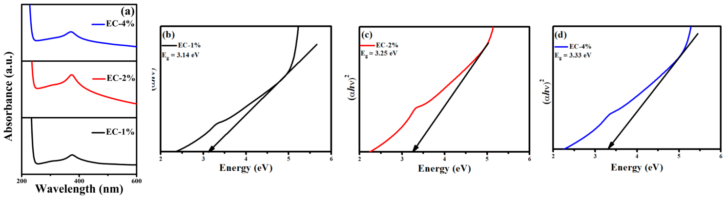
The UV-Vis absorption spectra of ZnO nanoparticles synthesized using aqueous extracts of Eichhornia crassipes at different concentrations are presented in Figure 2a. A characteristic absorption band is observed in the ultraviolet region, with maxima located at 375 nm, 373 nm, and 370 nm for EC-1%, EC-2%, and EC-4%, respectively. These absorption features are consistent with the characteristic optical response of ZnO and are associated primarily with electronic transitions involving the valence and conduction bands of the semiconductor [32,33,34]. The slight shift in the absorption maximum toward shorter wavelengths with increasing extract concentration indicates changes in the optical response of the synthesized materials. Such variations may be associated with differences in crystallinity, particle or crystallite size, defect states, and surface interactions resulting from the phytochemical composition of the extracts [35,36]. The band-gap energy (Eg) was estimated using the Tauc method, considering a direct allowed electronic transition (n = 1/2), which is characteristic of ZnO as a direct band-gap semiconductor, as presented in Equation (1) [37].
( α h ν ) n = A ( h ν E g )
where α represents the absorption coefficient, hν is the energy of the incident photon, and A is a constant.
The Tauc plots illustrated in Figure 2b–d yielded band-gap values of 3.14 eV, 3.25 eV, and 3.33 eV for EC-1%, EC-2%, and EC-4%, respectively. These values are consistent with those reported for ZnO nanoparticles and show a slight reduction compared to bulk ZnO (~3.37 eV), suggesting the presence of structural defects, surface states, or residual organic species introduced during the green synthesis process [38]. The differences among the samples indicate that the concentration of E. crassipes extract used during synthesis influences the optical properties of the resulting ZnO. The lower band-gap value observed for EC-1% may be associated with structural defects, surface states, lattice imperfections, or differences in the concentration of residual organic species introduced during the green synthesis process.

2.3. XRD

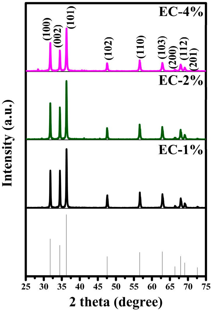
The XRD diffractogram presented in Figure 3 shows diffraction peaks at 2θ values which correspond to different crystallographic peaks: 31.83 (100), 34.34 (002), 36.40 (101), 47.58 (102), 56.64 (110), 62.86 (103), 66.33 (200), 68.06 (112), and 69.32 (201) [39]. The obtained reflections are characteristic of ZnO nanoparticles with a hexagonal wurtzite phase (JCPDS No. 96-900-4179) [40]. No additional diffraction reflections attributable to crystalline impurity phases or secondary crystalline compounds were observed in the analyzed samples. This result indicates that the synthesized materials predominantly consisted of crystalline ZnO [41].
The crystallite size (D) of the obtained ZnO nanoparticles was calculated employing the Scherrer equation, where K is the Scherrer constant, λ is the wavelength of the incident X-rays, β is the full width at half maximum, and θ corresponds to the Bragg angle [42].
D = K λ β C o s ( θ )
The calculated average crystallite sizes were 38.66, 38.16, and 31.52 nm for EC-1%, EC-2%, and EC-4%, respectively. These results reveal a decrease in the average crystallite size with increasing E. crassipes extract concentration, particularly for the EC-4% sample. The reduction in crystallite size is consistent with the greater broadening of the diffraction reflections observed for EC-4%, since increased peak broadening is generally associated with smaller coherent crystallite domains. The observed reduction in crystallite size with increasing extract concentration may be related to the greater availability of phytochemical constituents during nanoparticle formation. These biomolecules can interact with zinc-containing species and the developing ZnO nuclei, potentially increasing the number of nucleation sites while limiting subsequent crystal growth through surface adsorption or stabilization effects [43].

2.4. SEM/EDX

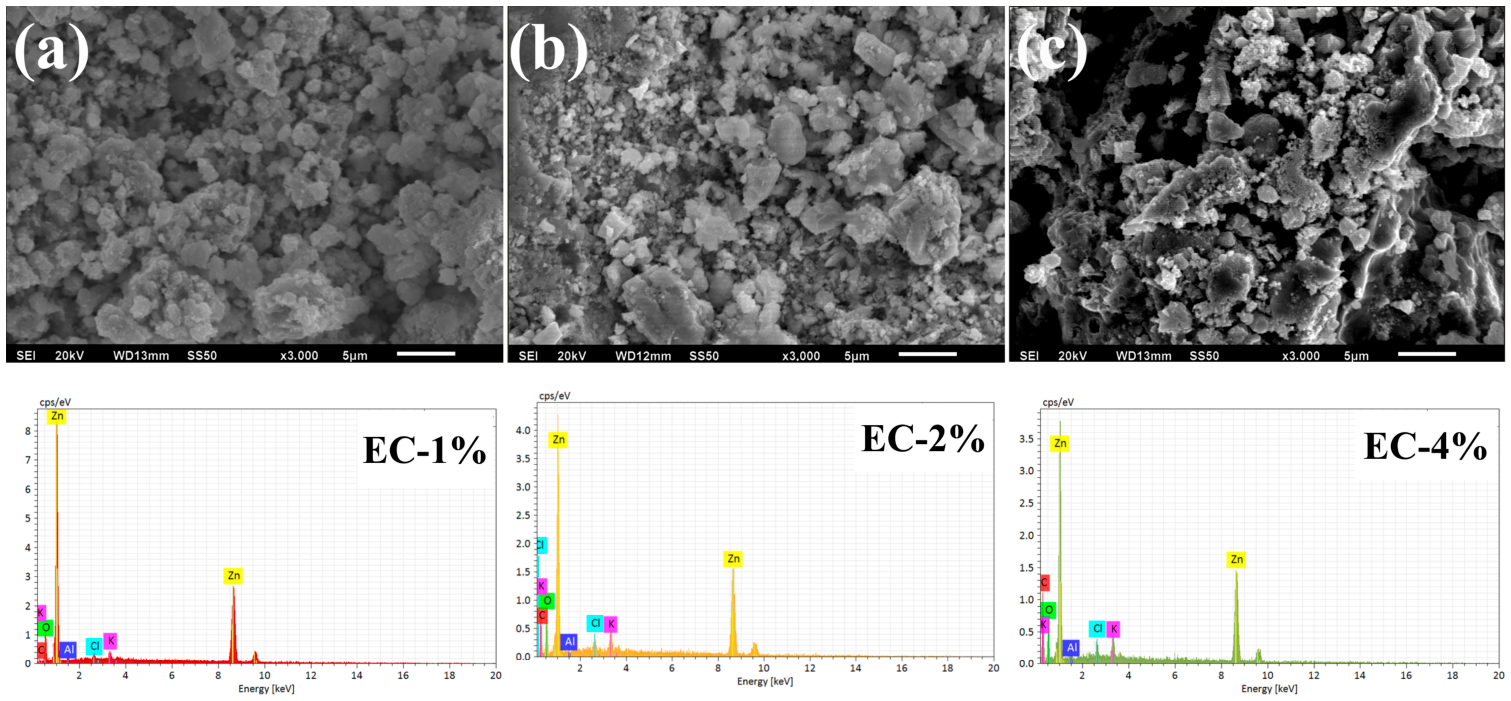
SEM was employed to investigate the surface morphology of the green-synthesized ZnO nanoparticles, as shown in Figure 4. The micrographs revealed predominantly quasi-spherical to irregularly shaped particles, with noticeable differences in the degree of aggregation among the synthesized samples. The EC-1% sample exhibited a relatively high degree of particle agglomeration, whereas a progressive reduction in aggregation was observed with increasing E. crassipes extract concentration. The EC-4% sample exhibited a comparatively more dispersed morphology, with more clearly distinguishable particle domains. The observed differences in aggregation may be associated with the increasing concentration of phytochemical constituents present in the E. crassipes extracts. During the biosynthesis process, these compounds can interact with zinc-containing species and the surface of the developing ZnO nuclei, potentially acting as capping and stabilizing agents. Such interactions may limit the excessive aggregation during nucleation and crystal growth [44,45]. Energy-dispersive X-ray spectroscopy (EDX) was used to determine the elemental composition of the synthesized materials. The EDX spectra showed the characteristic signals corresponding primarily to zinc (Zn) and oxygen (O), confirming the elemental composition expected for ZnO [46,47]. A carbon (C) signal was also detected in the analyzed samples. The presence of carbon may be associated with residual organic compounds derived from the E. crassipes extract. Therefore, the EDX results primarily confirm the presence of Zn and O in the synthesized materials, while the detected carbon signal may indicate the presence of residual plant-derived species on the nanoparticle surface.

2.5. Evaluation of the Photocatalytic Activity

2.5.1. Amido Black 10-B

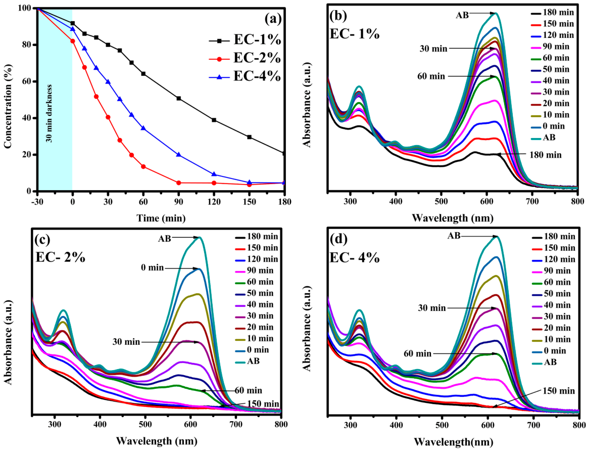
Amido Black 10B (AB 10-B) is an azo dye widely used in the textile industry and in biochemical applications. Its complex aromatic structure and high chemical stability make it resistant to conventional degradation processes, resulting in its persistence as a pollutant in wastewater [48]. The photocatalytic degradation of AB under UV irradiation was evaluated using ZnO nanoparticles synthesized via a green method using E. crassipes extract at concentrations of 1%, 2%, and 4%. The degradation profiles presented in Figure 5 show an initial decrease in AB concentration, which can be attributed to adsorption onto the ZnO surface. This effect is likely enhanced by the presence of organic functional groups from the plant extract, which promote interactions between the dye molecules and the catalyst surface. The relatively low initial degradation observed for AB may also be related to its large aromatic structure and multiple functional groups, which can influence its orientation and accessibility to the photocatalyst surface. In addition, as an anionic azo dye, its interaction with the ZnO surface may differ from that of cationic dyes, affecting both adsorption and subsequent access to photogenerated reactive species [49]. After 60 min of irradiation, degradation efficiencies of approximately 35%, 87%, and 65% were achieved for EC-1%, EC-2%, and EC-4%, respectively, indicating a clear improvement in photocatalytic activity for EC-2%. The higher efficiency observed for the 2% sample suggests the presence of an optimal extract concentration that enhances surface reactivity and the availability of active sites [50,51]. In contrast, the lower degradation observed for the EC-1% sample may be due to insufficient surface functionalization, while the reduced performance at 4% may be due to excessive organic content that partially blocks active sites or hinders effective interaction with the dye [52].

2.5.2. Eosin Yellow

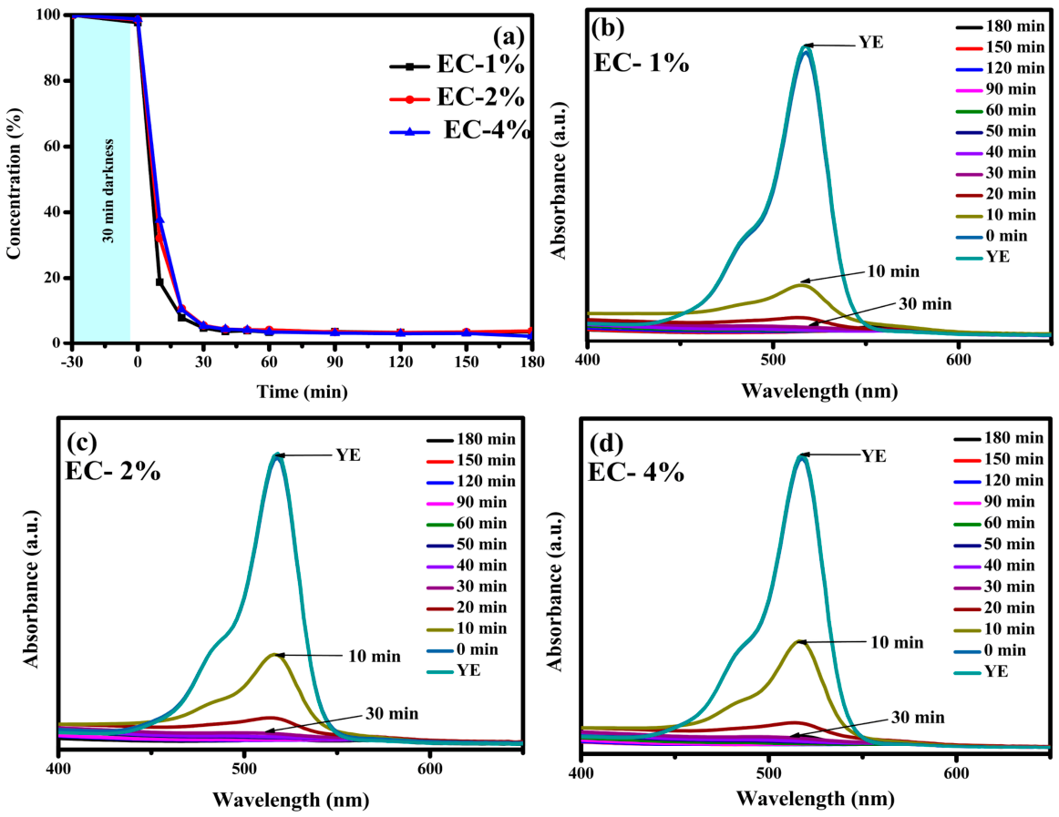
Eosin Yellow (EY) is an anionic xanthene dye widely used in textile, pharmaceutical, and biological staining applications [53]. Its high stability and resistance to biodegradation have led to growing concern about its persistence in aquatic environments [54]. The photocatalytic degradation of EY under UV irradiation was investigated using ZnO nanoparticles synthesized with E. crassipes extract at 1%, 2%, and 4%, as presented in Figure 6. During the initial dark period (30 min), only a minor decrease in EY concentration (below 10%) was observed, indicating limited adsorption onto the ZnO surface. However, upon UV irradiation, a rapid degradation process occurred, as evidenced by a sharp decline in the characteristic absorption band around 515 nm, associated with π-π* transitions in the xanthene structure [55,56,57]. The rapid degradation of EY compared with AB and some of the other dyes may be associated with differences in molecular structure and the susceptibility of its xanthene chromophore to attack by photogenerated reactive species. Its anionic character and limited dark adsorption also suggest that the high removal observed under irradiation was not primarily controlled by adsorption. In contrast to the behavior observed for other dyes, EY exhibited a very fast initial degradation rate. Within the first 10 min, removal efficiencies of approximately 82%, 67%, and 64% were achieved for EC-1%, EC-2%, and EC-4%, respectively, highlighting the superior early-stage performance of the 1% material. As irradiation time increased, all samples converged to a similar degradation level of ~97% at 90 min, after which no further significant changes were detected up to 180 min, suggesting that the system reached a steady state. This behavior indicates that EY degradation is primarily governed by rapid photoinduced processes rather than adsorption. The faster initial response of EC-1% suggests that lower extract content may favor more accessible, reactive surface sites, enabling faster interaction with photogenerated species. In contrast, higher extract concentrations (2% and 4%) appear to slightly delay the initial degradation, possibly due to increased surface coverage by organic residues [58,59]. Despite these initial differences, the similar final efficiencies suggest that all materials possess sufficient photocatalytic capability to effectively degrade EY once the reaction progresses.

2.5.3. Methylene Blue

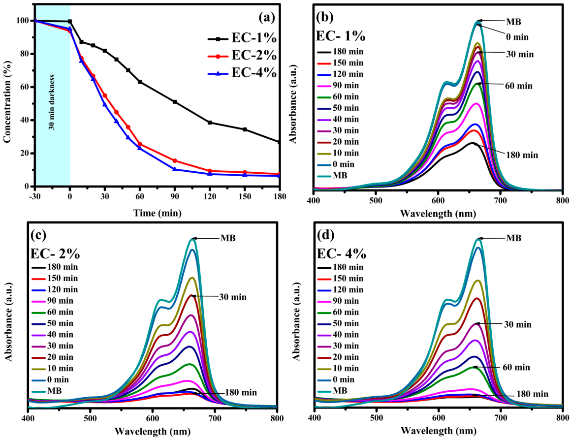
Methylene blue (MB) is a cationic thiazine dye widely used in the textile, paper, and pharmaceutical industries. Its presence in wastewater is of environmental concern due to its high chemical stability and potential toxicity [60,61]. The degradation process is presented in Figure 7. An initial decrease in MB concentration is observed, primarily attributed to adsorption onto the photocatalyst’s active sites. This process is driven by electrostatic interactions between the cationic dye molecules and negatively charged surface functional groups (–OH, –COOH) present on ZnO–EC [62,63]. The cationic nature of MB therefore favors its interaction with negatively charged surface sites, which may facilitate its proximity to the photocatalyst and subsequently increase its exposure to photogenerated reactive species. Its relatively smaller molecular structure compared with dyes such as AB may also favor access to surface active sites [64]. After 30 min of irradiation, degradation efficiencies of approximately 18%, 44%, and 54% were obtained for EC-1%, EC-2%, and EC-4%, respectively, indicating a clear dependence on extract concentration at early stages. Upon continued UV exposure, the UV-Vis spectra show a gradual decrease in the main absorption band centered at approximately 664 nm, which is associated with the π–π* transitions of MB [65,66]. This progressive reduction confirms the disruption of the chromophoric structure and the effective degradation of the dye. At 90 min, removal efficiencies reached 49%, 85%, and 90% for the 1%, 2%, and 4% samples, respectively. After 180 min, the degradation further increased to 74%, 92%, and 95%, demonstrating the superior performance of the 4% material, followed by EC-2% and EC-1%. The observed trend indicates that increasing extract concentration enhances photocatalytic performance for MB, likely due to improved surface functionalization and greater availability of active sites, which facilitate dye adsorption and subsequent degradation.

2.5.4. Methyl Orange

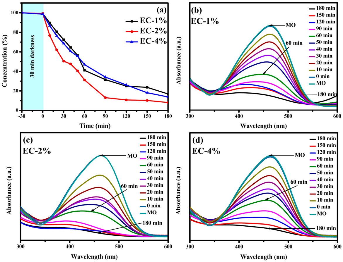
Methyl orange (MO) is an anionic azo dye commonly used in textile processing and as a pH indicator, and its persistence in wastewater makes it a relevant environmental pollutant. The photocatalytic degradation of MO under UV irradiation was evaluated using ZnO nanoparticles synthesized with E. crassipes extract at different concentrations. The degradation profiles presented in Figure 8 show a progressive decrease in dye concentration over time, indicating effective photocatalytic activity for all materials. The gradual degradation observed for MO may be related to its anionic character and azo structure, which can influence its interaction with the ZnO surface and its susceptibility to oxidation. Compared with the rapid degradation observed for EY, the azo bond and molecular structure of MO may require more sustained interaction with the photogenerated reactive species. After 60 min of irradiation, removal efficiencies of approximately 59%, 69%, and 54% were achieved for EC-1%, EC-2%, and EC-4%, respectively, highlighting the improved performance of the EC-2% sample at intermediate reaction times. As the reaction proceeds, the degradation continues steadily. By 120 min, removal efficiencies increased to about 76% for EC-1%, 91% for EC-2%, and 74% for EC-4%. The system approaches its maximum efficiency around 180 min, with final degradation values of approximately 87% for EC-1%, 92% for EC-2%, and 87% for EC-4%, indicating that all materials achieve high removal, albeit at different rates. The results suggest that MO degradation follows a gradual pathway, where the EC-2% sample consistently exhibits the best performance. This behavior indicates that an intermediate extract concentration provides favorable conditions for photocatalysis, likely due to a balance between surface reactivity and accessibility of active sites. In contrast, lower or higher extract contents may limit efficiency either by insufficient surface modification or by partial blockage of active sites.

2.5.5. Methyl Red

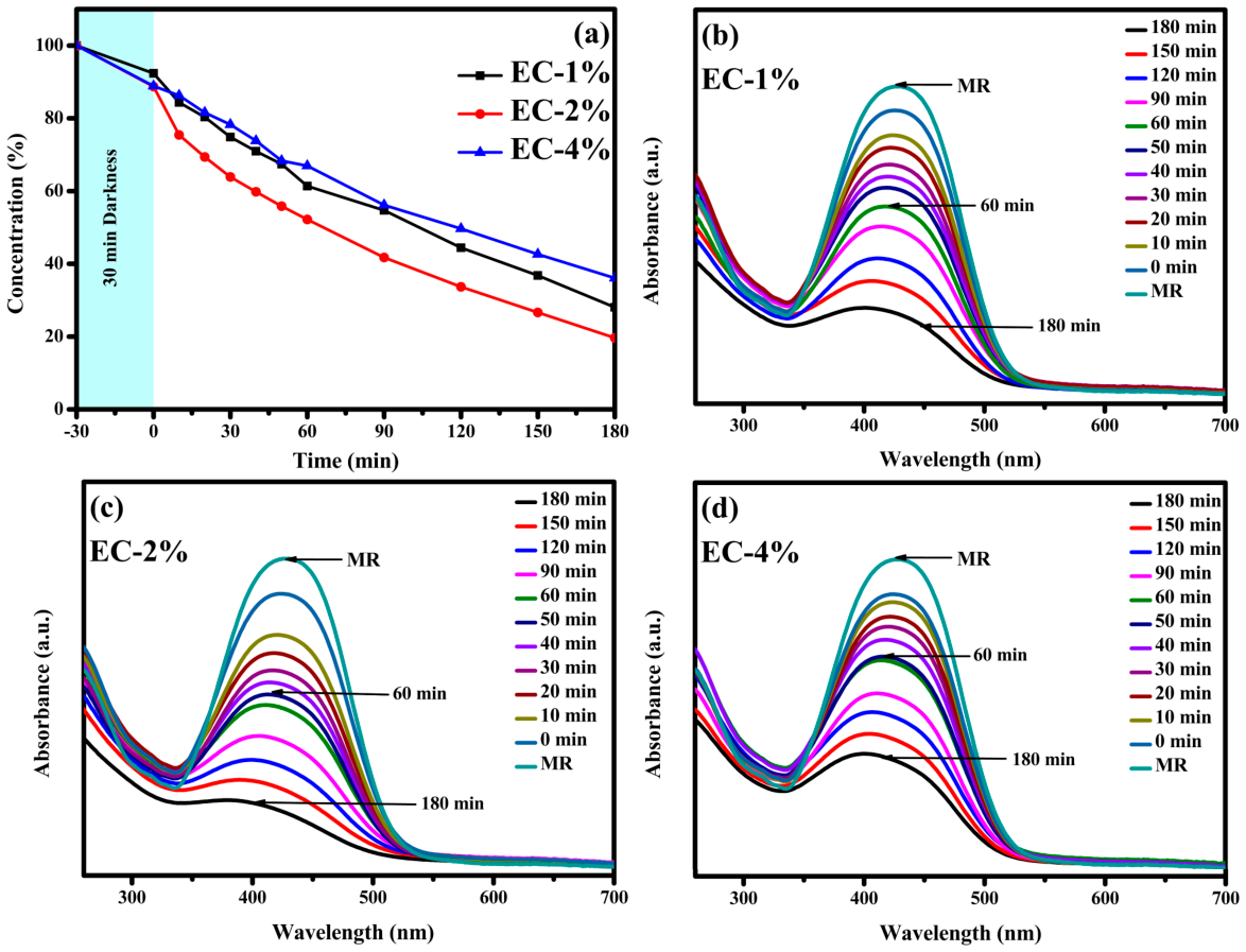
Methyl red (MR) is an azo dye widely used in textile and dyeing processes, and its presence in wastewater is of concern due to its persistence and potential toxicity [67,68]. The photocatalytic degradation of MR under UV irradiation was evaluated using ZnO nanoparticles synthesized with E. crassipes extract at different concentrations as presented in Figure 9. The evolution of MR concentration during irradiation exhibits a gradual, sustained degradation pattern compared to other dyes. This slower behavior may be associated with the molecular structure of MR, particularly its azo group and aromatic rings, which can provide greater chemical stability and require prolonged exposure to reactive species for effective degradation. Its molecular structure and ionization state may also influence its interaction with the ZnO surface and the accessibility of reactive sites [69]. After 60 min, removal efficiencies of approximately 40%, 48%, and 33% were achieved for EC-1%, EC-2%, and EC-4%, respectively, indicating moderate photocatalytic activity during the early stages. As the reaction progressed, the degradation continued steadily, reaching final removal values of about 79% for EC-1%, 81% for EC-2%, and 65% for EC-4% after 180 min.
These results suggest that MR degradation occurs more slowly and progressively, without the rapid initial removal observed for other systems. The EC-2% sample showed the best overall performance, although the difference with the 1% material is relatively small. In contrast, the lower efficiency observed for the 4% sample may be attributed to an excess of organic species in the extract, which could reduce the availability of active sites or limit interactions between the catalyst surface and the dye [70]. Overall, the degradation of MR appears to be less sensitive to rapid surface processes and more dependent on sustained photocatalytic activity over time. The relatively similar performance of EC-1% and EC-2% further suggests that, for MR, dye structure and reactivity may play a more important role than differences in initial surface interaction alone. This behavior highlights the importance of maintaining an appropriate balance in synthesis conditions to ensure effective long-term performance rather than only fast initial degradation.

2.5.6. Rhodamine B

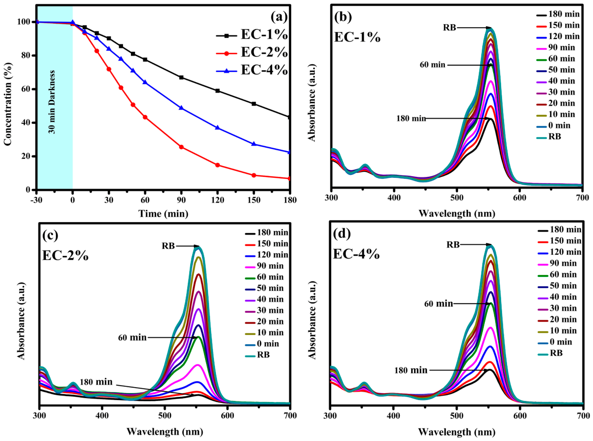
Rhodamine B (RB) is a cationic dye widely used in the textile and food industries. Despite its practical applications, its high stability and toxicity make it a concerning pollutant in aquatic environments [71,72]. Figure 10 presents the degradation of RB through time under UV radiation. During the stirring in darkness, only a slight decrease in RB concentration (below 10%) was observed, indicating limited adsorption onto the catalyst surface. Upon UV irradiation, a gradual degradation occurs. The behavior of RB can be associated with its cationic character and xanthene structure. Although its positive charge can favor electrostatic interactions with negatively charged surface sites, its relatively large and structurally complex molecule may influence its accessibility to reactive sites and contribute to the gradual degradation observed [73]. After 90 min, removal efficiencies of approximately 33%, 76%, and 51% were achieved for EC-1%, EC-2%, and EC-4%, respectively, showing a clear improvement in performance for the EC-2% sample. As the reaction progressed, the degradation continued steadily, reaching final removal values of about 57% for EC-1%, 93% for EC-2%, and 78% for EC-4% after 180 min. These results indicate that EC-2% exhibits the highest overall photocatalytic efficiency, while EC-4% shows intermediate behavior and EC-1% the lowest activity. The observed trend suggests that an intermediate extract concentration provides a more favorable balance for photocatalytic performance. While the presence of organic compounds in the extract can enhance surface reactivity, excessive amounts may partially hinder the process by limiting access to active sites. In contrast, lower extract content may not provide sufficient surface modification to maximize efficiency.
The photocatalytic performance of the ZnO-EC materials varies depending on both the dye and the extract concentration. Among the pollutants, EY showed the fastest degradation, achieving near-complete removal in a short time, whereas AB-10B and MR exhibited more gradual degradation, indicating a greater dependence on sustained photocatalytic activity. MB and RB displayed a clearer dependence on extract concentration, with improved performance at intermediate or higher extract contents, likely due to enhanced surface interactions. MO showed intermediate behavior, with steady degradation and smaller differences among samples at longer times. Overall, the EC-2% material consistently demonstrated the most balanced performance across all dyes, suggesting that an intermediate extract concentration optimally balances surface reactivity and active site availability.
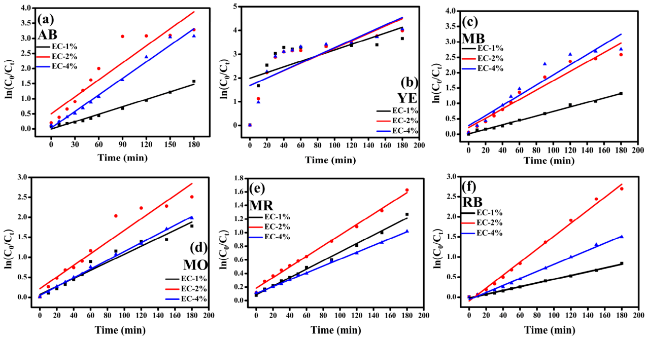
To further evaluate the photocatalytic degradation rate, the experimental data were analyzed using the pseudo-first-order kinetic model, expressed as ln (A0/At) = kt, where A0 and At are the absorbance values at the initial and irradiation times, respectively, and k is the apparent pseudo-first-order rate constant. The corresponding linear plots are presented in Figure 11, while the calculated k values are summarized in Table 1.

2.5.7. Chlorpyrifos Ethyl

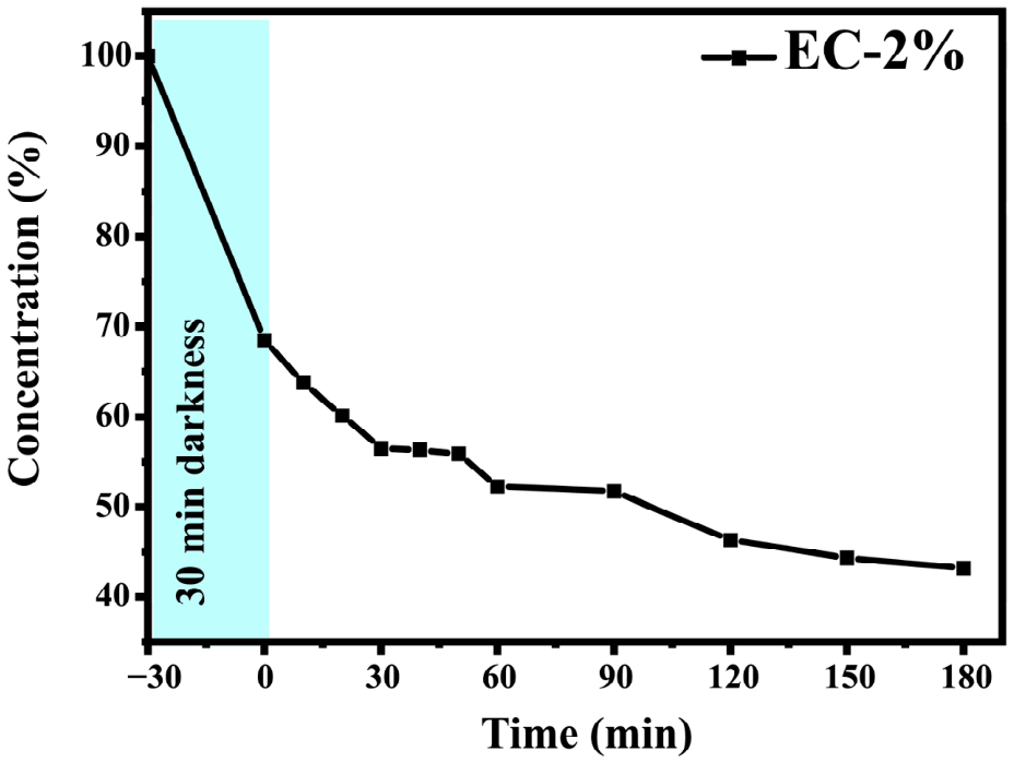
EC-2% was selected for the degradation of chlorpyrifos-ethyl due to its consistently superior and balanced photocatalytic performance across all tested dyes. It showed high efficiency without the limitations observed for EC-1% and EC-4%. Therefore, EC-2% provides the most reliable and representative conditions for evaluating pollutant degradation, as presented in Figure 12. The degradation of chlorpyrifos ethyl using EC-2% ZnO nanoparticles follows a two-stage behavior consisting of an initial adsorption step followed by a slow photocatalytic process. During the first 30 min in the dark, the concentration decreases to 68.33%, indicating significant adsorption of the pesticide onto the catalyst surface [74]. Upon UV irradiation, a gradual decrease in concentration is observed (55.87% at 30 min, 52.43% at 60 min, 46.67% at 120 min, and 43.22% at 180 min), reflecting a relatively slow degradation compared to organic dyes. Once the nanoparticles and the pollutant are irradiated with UV light, the ZnO surface becomes active, attacking the pesticide molecules already clinging to it, including bonds such as phosphorothioate (P=S) or ester bonds, breaking the molecule and transforming it into intermediates such as 3,5,5-trichloro-2-pyridinol [75]. The degradation of the pesticide is slower than that of dyes due to the high stability of its trichloropyridinyl ring, which resists chemical breakdown. Furthermore, although the pesticide strongly adheres to the surface of ZnO nanoparticles, excessive adsorption can hinder the process. By crowding the catalyst surface, pesticide molecules may block the active sites required to generate reactive oxygen species (ROS), thereby slowing the overall degradation rate.

3. Materials and Methods

3.1. Materials

Fresh Eichhornia crassipes (water hyacinth) plants were collected from wetlands in the Medio Atrato region (Chocó, Colombia). Zinc nitrate hexahydrate (Zn(NO3)2·6H2O, 99% purity) was used as a zinc precursor. Analytical-grade dyes used for photocatalytic evaluation included methylene blue (MB, 98% purity), methyl orange (MO, 97% purity), rhodamine B (RhB, 95% purity), eosin yellow (EY, 90% purity), methyl red (MR, 95% purity), and amido black (AB, 97% purity), all purchased from Sigma-Aldrich (St. Louis, MO, USA). Chlorpyrifos ethyl (CE, 77% purity) was purchased from Innovación Agrícola, Álvaro Obregón, Mexico. Deionized water was used in all experiments.

3.2. Eichhornia Crassipes Extract Preparation

Freshly collected E. crassipes biomass was thoroughly washed with deionized water to remove adhering soil, debris, and other surface contaminants. The plant material was air-dried for 48 h and subsequently dried in an oven at 60 °C until a dry mass was obtained. The dried biomass was then ground into a fine powder.
Aqueous E. crassipes extracts were prepared at concentrations of 1%, 2%, and 4% (w/v). Briefly, 0.5, 1.0, and 2.0 g of dried E. crassipes powder were separately dispersed in 50 mL of distilled water to obtain the 1%, 2%, and 4% (w/v) preparations, respectively. Each suspension was stirred at 400 rpm for 2 h at room temperature and subsequently heated at 60 °C for an additional 2 h. After extraction, the mixtures were filtered through Whatman No. 4 filter paper to remove residual plant material. The resulting filtrates were collected and used immediately for the biosynthesis of ZnO nanoparticles.

3.3. Biosynthesis of ZnO Nanoparticles

ZnO nanoparticles were biosynthesized by dissolving 2 g of zinc nitrate hexahydrate in 50 mL of each E. crassipes extract (1%, 2%, and 4% w/v). The mixtures were stirred at 400 rpm for 1 h, then heated in a water bath at 60 °C for 12 h until a paste-like consistency was achieved. The resulting material was then calcined at 400 °C for 1 h, ground into a fine powder, and stored for further use. The synthesized nanoparticle samples were labeled as EC-1%, EC-2%, and EC-4%.

3.4. ZnO Nanoparticle Characterization

The biosynthesized ZnO nanoparticles were characterized using multiple analytical techniques. Fourier transform infrared spectroscopy (FTIR) was employed to identify surface functional groups, utilizing a PerkinElmer spectrophotometer (4000–400 cm−1). Ultraviolet–visible (UV-Vis) spectroscopy was conducted to evaluate the optical properties of the nanomaterial using a PerkinElmer Lambda 365 spectrophotometer (200–800 nm) (PerkinElmer, Inc., Waltham, MA, USA). X-ray diffraction (XRD) analysis was performed with a Bruker D2-Phaser diffractometer (Bruker Daltonics, Billerica, MA, USA)to confirm the crystalline structure (Cu Kα= 1.5406 Å, over a 2θ range of 25–75). Scanning electron microscopy (SEM) coupled with energy-dispersive X-ray spectroscopy (EDX) was used to examine surface morphology and elemental composition, respectively, using a JEOL JSM-6310LV microscope (JEOL USA, Inc., Peabody, MA, USA).

3.5. Photocatalytic Activity

Photodegradation experiments of organic dyes were conducted at an initial concentration of 15 ppm. For photocatalytic evaluation, 50 mg of ZnO nanoparticles from each synthesis condition (EC-1%, EC-2%, and EC-4%) was dispersed in 50 mL of dye solution and stirred in the dark at 400 rpm for 30 min to establish adsorption–desorption equilibrium. Subsequently, the suspensions were transferred to a photoreactor and irradiated using a 10 W UV light source (18 mJ/cm2). During irradiation, 1 mL aliquots were withdrawn at 10 min intervals during the first 60 min, and thereafter every 30 min until a total reaction time of 180 min was reached. Photodegradation of chlorpyrifos ethyl was performed at an initial concentration of 150 ppm using only the EC-2% sample. The same experimental procedure used for dye degradation was applied to evaluate chlorpyrifos ethyl. The collected aliquots were analyzed by UV-Vis spectroscopy to monitor the temporal evolution of dye and chlorpyrifos ethyl degradation. The degradation of the organic pollutants was calculated employing Equation (3), where C0 is the initial dye concentration, and Ct is the dye concentration at time t.
R e m o v a l   % = C 0 C t C 0     100

4. Conclusions

ZnO nanoparticles were successfully biosynthesized using aqueous Eichhornia crassipes extracts from the Medio Atrato region, Chocó, Colombia, demonstrating the potential of this invasive aquatic biomass as a sustainable resource for photocatalyst production. FTIR, XRD, and SEM analyses confirmed the formation of crystalline ZnO with a hexagonal wurtzite structure, while increasing extract concentration from 1% to 4% reduced the average crystallite size from 38.66 to 31.52 nm and decreased particle agglomeration.
The synthesized materials exhibited photocatalytic activity toward structurally diverse organic dyes under UV irradiation, with performance dependent on both pollutant type and extract concentration. EC-2% showed the most consistent overall performance, while EC-4% achieved the highest methylene blue degradation (95%). Eosin yellow showed approximately 97% removal with all photocatalysts after 90 min. EC-2% also promoted chlorpyrifos ethyl removal, with approximately 56.78% degradation after 180 min.
Overall, E. crassipes-mediated ZnO nanoparticles show promise for the photocatalytic treatment of diverse organic pollutants while providing a potential strategy for valorizing invasive aquatic biomass. Further studies should address mineralization, transformation products, catalyst reusability, and performance in real water matrices.

Author Contributions

N.N.-V.: Writing—review & editing, Writing—original draft, Methodology, Investigation, Conceptualization. J.A.V.-F.: Methodology, Writing—original draft, Writing—review & editing, Formal analysis. J.L.G.: Supervision, Methodology, and Writing—review. E.A.S.-G.: Formal analysis, Investigation, Methodology. A.R.V.-N.: Methodology, Writing—review & editing, Formal analysis. Y.P.-T.: Formal analysis, Investigation, Methodology. P.A.L.-M.: Formal analysis, Methodology, Writing—original draft, Writing—review & editing. All authors have read and agreed to the published version of the manuscript.

Funding

The authors thank the projects of the Autonomous University of Baja California (UABC) 402/3391 and 402/3751, as well as 402/1/C/58/24. This research was funded by the Frontera Science Project with number CF-2023-I-1805 of SECIHTI.

Data Availability Statement

The original contributions presented in this study are included in the article. Further inquiries can be directed to the corresponding authors.

Acknowledgments

The authors acknowledge Uvaldo Hernández and María Citlalit Martínez Soto for technical assistance.

Conflicts of Interest

The authors declare no conflicts of interest.

References

  1. Islam, M.M.; Aidid, A.R.; Mohshin, J.N.; Mondal, H.; Ganguli, S.; Chakraborty, A.K. A Critical Review on Textile Dye-Containing Wastewater: Ecotoxicity, Health Risks, and Remediation Strategies for Environmental Safety. Clean. Chem. Eng. 2025, 11, 100165. [Google Scholar] [CrossRef] [Scilit]
  2. Baig, A.; Siddique, M.; Panchal, S. A Review of Visible-Light-Active Zinc Oxide Photocatalysts for Environmental Application. Catalysts 2025, 15, 100. [Google Scholar] [CrossRef] [Scilit]
  3. Ramesh, N.; Lai, C.W.; Bin Johan, M.R.; Mousavi, S.M.; Badruddin, I.A.; Kumar, A.; Sharma, G.; Gapsari, F. Progress in Photocatalytic Degradation of Industrial Organic Dye by Utilising the Silver Doped Titanium Dioxide Nanocomposite. Heliyon 2024, 10, e40998. [Google Scholar] [CrossRef] [Scilit] [PubMed]
  4. Trinidad-Urbina, R.E.; Hernández-Orozco, M.M.; Ramírez-Bon, R. Photodegradation of Methylene Blue, Rhodamine 6G and Methyl Orange under Sunlight/H2O2 Irradiation. Int. J. Environ. Sci. Technol. 2026, 23, 474. [Google Scholar] [CrossRef] [Scilit]
  5. Sami, S.A.; Akter, B.; Nirob, M.D.N.H.; Jahan, I.; Lokman, M.D.; Khatun, M.H.; Hossain, M.K.; Barmon, J. A Comprehensive Review on Environmental Dynamics of Chlorpyrifos: Fate, Toxicity, Remediation, and Regulatory Aspects. Ecotoxicol. Environ. Saf. 2025, 308, 119484. [Google Scholar] [CrossRef] [Scilit] [PubMed]
  6. Gavrilaș, S.; Burescu, F.L.; Chereji, B.D.; Munteanu, F.D. The Impact of Anthropogenic Activities on the Catchment’s Water Quality Parameters. Water 2025, 17, 1791. [Google Scholar] [CrossRef] [Scilit]
  7. Sefali, S.; Ruby, R.; Dimple, D.; Giri, A. Toxicological Implications of Emerging Pollutants on Aquatic Organisms. Discov. Environ. 2026, 4, 43. [Google Scholar] [CrossRef] [Scilit]
  8. Sumantrao, S.K.; Kariyajjanavar, P.; Shridhar, A.H.; Ghagane, S.C.; Chigari, S.S.; Bonageri, G.; Ansari, M.Z.; Alsubaie, A.S. Sunlight-Driven GO/ZnO Nanocomposite for Photocatalytic Degradation of Chlorpyrifos Insecticide and Its Biological Activities. J. Environ. Chem. Eng. 2025, 13, 115437. [Google Scholar] [CrossRef] [Scilit]
  9. Mia, M.S.; Farook, O.; Hossain, M.T.; Azad, M.A.S.; Bhowmik, S.; Alam, M.A. Green Synthesis of ZnO and Ag- ZnO Nanocrystalline Materials: Optical Band Gap Engineering and Multi-Model XRD Size–Strain Investigation. Results Chem. 2026, 20, 103022. [Google Scholar] [CrossRef] [Scilit]
  10. Zeghoud, S.; Hemmami, H.; Ben Seghir, B.; Ben Amor, I.; Kouadri, I.; Rebiai, A.; Messaoudi, M.; Ahmed, S.; Pohl, P.; Simal-Gandara, J. A Review on Biogenic Green Synthesis of ZnO Nanoparticles by Plant Biomass and Their Applications. Mater. Today Commun. 2022, 33, 104747. [Google Scholar] [CrossRef] [Scilit]
  11. Mutukwa, D.; Taziwa, R.T.; Khotseng, L. A Review of Plant-Mediated ZnO Nanoparticles for Photodegradation and Antibacterial Applications. Nanomaterials 2024, 14, 1182. [Google Scholar] [CrossRef] [Scilit] [PubMed]
  12. Kolotygina, V.Y.; Zhilyakov, A.Y.; Bukharinova, M.A.; Khamzina, E.I.; Stozhko, N.Y. Green Synthesis of ZnO Nanoparticles: Effect of Synthesis Conditions on Their Size and Photocatalytic Activity. ChemEngineering 2026, 10, 15. [Google Scholar] [CrossRef] [Scilit]
  13. Rahman, F.; Majed Patwary, M.A.; Bakar Siddique, M.A.; Bashar, M.S.; Haque, M.A.; Akter, B.; Rashid, R.; Haque, M.A.; Royhan Uddin, A.K.M. Green Synthesis of Zinc Oxide Nanoparticles Using Cocos Nucifera Leaf Extract: Characterization, Antimicrobial, Antioxidant and Photocatalytic Activity. R. Soc. Open Sci. 2022, 9, 220858. [Google Scholar] [CrossRef] [Scilit] [PubMed]
  14. Mousa, S.A.; Wissa, D.A.; Hassan, H.H.; Ebnalwaled, A.A.; Khairy, S.A. Enhanced Photocatalytic Activity of Green Synthesized Zinc Oxide Nanoparticles Using Low-Cost Plant Extracts. Sci. Rep. 2024, 14, 16713. [Google Scholar] [CrossRef] [Scilit] [PubMed]
  15. Villamagna, A.M.; Murphy, B.R. Ecological and Socio-Economic Impacts of Invasive Water Hyacinth (Eichhornia crassipes): A Review. Freshw. Biol. 2010, 55, 282–298. [Google Scholar] [CrossRef] [Scilit]
  16. Harun, I.; Pushiri, H.; Amirul-Aiman, A.J.; Zulkeflee, Z. Invasive Water Hyacinth: Ecology, Impacts and Prospects for the Rural Economy. Plants 2021, 10, 1613. [Google Scholar] [CrossRef] [Scilit] [PubMed]
  17. Amalina, F.; Razak, A.S.A.; Krishnan, S.; Zularisam, A.W.; Nasrullah, M. Water Hyacinth (Eichhornia crassipes) for Organic Contaminants Removal in Water—A Review. J. Hazard. Mater. Adv. 2022, 7, 100092. [Google Scholar] [CrossRef] [Scilit]
  18. Lemecho, B.A.; Sabir, F.K.; Andoshe, D.M.; Gultom, N.S.; Kuo, D.-H.; Chen, X.; Mulugeta, E.; Desissa, T.D.; Zelekew, O.A. Biogenic Synthesis of Cu-Doped ZnO Photocatalyst for the Removal of Organic Dye. Bioinorg. Chem. Appl. 2022, 2022, 8081494. [Google Scholar] [CrossRef] [Scilit] [PubMed]
  19. Zelekew, O.A.; Fufa, P.A.; Sabir, F.K.; Duma, A.D. Water Hyacinth Plant Extract Mediated Green Synthesis of Cr2O3/ZnO Composite Photocatalyst for the Degradation of Organic Dye. Heliyon 2021, 7, e07652. [Google Scholar] [CrossRef] [Scilit] [PubMed]
  20. Hasnat, H.; Riti, S.J.; Shompa, S.A.; Alam, S.; Islam, M.M.; Kabir, F.; Khan, M.S.; Shao, C.; Zeng, C.; Wang, S.; et al. Unveiling the Therapeutic Potentials of Water Hyacinth (Eichhornia crassipes (Mart.) Solms) Flower against Oxidative Stress, Inflammation and Depressive Disorders: GC-MS/MS, In Vitro, In Vivo and In Silico Approaches. Chem. Biodivers. 2024, 21, e202401268. [Google Scholar] [CrossRef] [Scilit] [PubMed]
  21. Ahmed, F.E.; Wodag, A.F.; Gelebo, G.G.; Gebre, B.M.; Ayana, M.T. Investigating the Tannin Content and Phytochemical Composition of Water Hyacinth (Eichhornia crassipes) in Lake Tana, Ethiopia. J. Eng. 2025, 2025, 3967820. [Google Scholar] [CrossRef] [Scilit]
  22. Ben Bakrim, W.; Ezzariai, A.; Karouach, F.; Sobeh, M.; Kibret, M.; Hafidi, M.; Kouisni, L.; Yasri, A. Eichhornia crassipes (Mart.) Solms: A Comprehensive Review of Its Chemical Composition, Traditional Use, and Value-Added Products. Front. Pharmacol. 2022, 13, 842511. [Google Scholar] [CrossRef] [Scilit] [PubMed]
  23. Sayeem, N.R.; Podder, U.; Alam, M.A.; Shishir, M.K.H.; Miah, M.A.R.; Anik, F.K.; Ghosh, P.; Ahmed, S.; Islam, M.; Jahan, S.A. Critical Perspective Review on Green Synthesis of Zinc Oxide Nanoparticles: Explored Wurtzite Phase, Characterization and Real World Applications. Next Nanotechnol. 2026, 9, 100412. [Google Scholar] [CrossRef] [Scilit]
  24. Santhosh Nair, A.; Syama, A.; Ramachandran Prabhu, R. A Comprehensive Review on Green Synthesis of Nanoparticles from Eichhornia Crassipes as a Potential Management Strategy to Negate the Adverse Effects of the Noxious Aquatic Weed. Lett. Appl. NanoBioSci. 2025, 14, 141. [Google Scholar] [CrossRef]
  25. Sonthongphithak, P.; Muangnapoh, C.; Ratanatawanate, C.; E-Kobon, T.; Piyasaengthong, A.; Hongsachart, P.; Sriuttha, M.; Sengkhamparn, N.; Dhanapal, A.C.T.A.; Wittayanarakul, K. Biosynthesis of Zinc Oxide Nanoparticles Using Water Hyacinth Extracts: Characterization, Evaluation of Antimicrobial and Dye Removal. J. Met. Mater. Miner. 2024, 34, 1979. [Google Scholar] [CrossRef] [Scilit]
  26. Pasieczna-Patkowska, S.; Cichy, M.; Flieger, J. Application of Fourier Transform Infrared (FTIR) Spectroscopy in Characterization of Green Synthesized Nanoparticles. Molecules 2025, 30, 684. [Google Scholar] [CrossRef] [Scilit] [PubMed]
  27. Camez-Cota, J.A.; Gómez-López, P.; Gaxiola, A.; Várgas-Ortiz, R.A.; Orozco-Carmona, V.M.; Ramírez-Bon, R.; Castro-Beltrán, A.; Alvarado-Beltrán, C.G. A Green Approach Modifications of TiO2 Thin Films for UV Photophysical Applications. Opt. Mater. 2025, 165, 117153. [Google Scholar] [CrossRef] [Scilit]
  28. Mohammadzadeh Kakhki, R.; YereyehZadeh, F.; Mohammadpoor, M. Machine Learning Optimized Photocatalytic Degradation of Tetracycline Using Sulfur-Doped Zinc Oxide/Chitosan Nanocomposites. J. Polym. Environ. 2025, 33, 5042–5065. [Google Scholar] [CrossRef] [Scilit]
  29. Halevas, E.; Mavroidi, B.; Varna, D.; Zahariou, G.; Litsardakis, G.; Pelecanou, M.; Hatzidimitriou, A.G. Structurally Characterized Cobalt and Nickel Complexes of Flavonoid Chrysin as Potential Radical Scavenging Compounds. Inorganics 2025, 13, 230. [Google Scholar] [CrossRef] [Scilit]
  30. Mounica, P.; Ramya, K.; Sangeetha, T.; Arivazhagan, G. The OH and C = O Stretching Bands of Ethanol, Benzaldehyde, and Their Binary Solutions: FTIR Spectral Studies. Spectrosc. Lett. 2025, 58, 975–991. [Google Scholar] [CrossRef] [Scilit]
  31. Hamaamin, O.K.; Ghareeb, H.O.; Mohammed, S.J. Eco-Friendly Enhancement of Optical and Structural Properties in Polyvinyl Alcohol Films via Eggplant Peel Dye Doping. Sci. Rep. 2025, 15, 28891. [Google Scholar] [CrossRef] [Scilit] [PubMed]
  32. Wijesinghe, U.; Thiripuranathar, G.; Iqbal, H.; Menaa, F. Biomimetic Synthesis, Characterization, and Evaluation of Fluorescence Resonance Energy Transfer, Photoluminescence, and Photocatalytic Activity of Zinc Oxide Nanoparticles. Sustainability 2021, 13, 2004. [Google Scholar] [CrossRef] [Scilit]
  33. Chatterjee, K.; Dutta, A.; Mishra, S.; Bairy, B.; Sen, M.B.; Gorai, A.; Saha, S.K.; Akhtar, A.J. Impact of Tailoring of the Defect States and the Band Gap towards Extreme Photocatalytic Performance and Photo-Induced Conductivity in Cobalt Doped ZnO QD. Ceram. Int. 2023, 49, 32768–32778. [Google Scholar] [CrossRef] [Scilit]
  34. Elabbasy, M.T.; El Bayomi, R.M.; Abdelkarim, E.A.; Hafez, A.E.-S.E.; Othman, M.S.; Ghoniem, M.E.; Samak, M.A.; Alshammari, M.H.; Almarshadi, F.A.; Elsamahy, T.; et al. Antibacterial and Antibiofilm Activity of Green-Synthesized Zinc Oxide Nanoparticles Against Multidrug-Resistant Escherichia Coli Isolated from Retail Fish. Molecules 2025, 30, 768. [Google Scholar] [CrossRef] [Scilit] [PubMed]
  35. Ruiz-Duarte, E.V.; Molina-Jiménez, J.P.; Avila, D.A.; Torres, C.O.; Horta-Piñeres, S.D. Rapid Synthesis of Highly Crystalline ZnO Nanostructures: Comparative Evaluation of Two Alternative Routes. Crystals 2025, 15, 640. [Google Scholar] [CrossRef] [Scilit]
  36. Hameed, H.; Waheed, A.; Sharif, M.S.; Saleem, M.; Afreen, A.; Tariq, M.; Kamal, A.; Al-onazi, W.A.; Al Farraj, D.A.; Ahmad, S.; et al. Green Synthesis of Zinc Oxide (ZnO) Nanoparticles from Green Algae and Their Assessment in Various Biological Applications. Micromachines 2023, 14, 928, Correction in Micromachines 2025, 16, 995. https://doi.org/10.3390/mi16090995. [Google Scholar] [PubMed]
  37. Bouderbala, I.Y.; Guessoum, A.; Rabhi, S.; Herbadji, A.; Bouras, I.-E. Layer-Dependent Photocatalytic Efficiency of ZnO Thin Films: Kinetic Analysis, Structural Insights and Optical Bandgap Characterization Using Absorption Spectrum Fitting, Tauc’s Plot, and Cody Models. J. Mater. Sci. Mater. Electron. 2025, 36, 1782. [Google Scholar] [CrossRef] [Scilit]
  38. Jadhav, S.R.; Mohite, S.V.; An, K.C.; Jang, D.H.; Erande, K.; Kim, Y.; Rajpure, K.Y. Crafting Defective ZnO Nanoparticles: A Green Synthesis for Enhanced Photocatalytic Degradation of Organic Pollutants. Ceram. Int. 2024, 50, 22783–22794. [Google Scholar] [CrossRef] [Scilit]
  39. Fouda, A.; Saied, E.; Eid, A.M.; Kouadri, F.; Alemam, A.M.; Hamza, M.F.; Alharbi, M.; Elkelish, A.; Hassan, S.E.-D. Green Synthesis of Zinc Oxide Nanoparticles Using an Aqueous Extract of Punica Granatum for Antimicrobial and Catalytic Activity. J. Funct. Biomater. 2023, 14, 205. [Google Scholar] [CrossRef] [Scilit] [PubMed]
  40. Hasan, D.M.; Kassim Al-Hussainawy, M.; Mohamed, A.; Alqaraghuli, H.G.J.; Saud, H.R.; Kyhoiesh, H.A.K. Silica-Coated Zinc Oxide Nanocomposite: A Novel Green Synthesis Using Rice Husk as a Source of Silica Nanoparticles. Phosphorus Sulfur Silicon Relat. Elem. 2026, 201, 263–271. [Google Scholar] [CrossRef] [Scilit]
  41. Sedefoglu, N. Characterization and Photocatalytic Activity of ZnO Nanoparticles by Green Synthesis Method. Optik 2023, 288, 171217. [Google Scholar] [CrossRef] [Scilit]
  42. Kem, A.; Ansari, M.R.; Prathap, P.; Jayasimhadri, M.; Peta, K.R. Eco-Friendly Green Synthesis of Stable ZnO Nanoparticles Using Citrus Limon: X-Ray Diffraction Analysis and Optical Properties. Phys. Scr. 2022, 97, 085814. [Google Scholar] [CrossRef] [Scilit]
  43. Nandhini, A.; Anilkumar, P.; Jasmin, J.; Balamurali, S. Green Synthesis, Characterization, Structural, Morphological, Antibacterial, and Cytotoxicity Evaluation of Zinc Oxide Nanoparticles Using Fioria Vitifolia Extract. Biophys. Chem. 2025, 323, 107440. [Google Scholar] [CrossRef] [Scilit] [PubMed]
  44. Wu, Y.S.; Sam, E.S.; Prasath, A.R.; Selvam, K.; Sangameshwaran, V.; Ragavendran, C.; El-Rayyes, A.; Tank, G. Green Synthesis of Zinc Oxide Nanoparticles Using Catharanthus Pusillus: Characterization, Antibacterial, Cytotoxicity of A549 Cells, and Photocatalytic Degradation of Eosin (E) Dye. J. Water Process Eng. 2025, 77, 108606. [Google Scholar] [CrossRef] [Scilit]
  45. Abdulqodus, A.N.; Abdulrahman, A.F.; Mostafa, S.H.; Kareem, A.A.; Hamad, S.M.; Ahmed, S.M.; Almessiere, M.A.; Shaikhah, D. Green Synthesis of ZnO Nanoparticles: Effect of PH on Morphology and Photocatalytic Degradation Efficiency. Appl. Phys. A 2025, 131, 720. [Google Scholar] [CrossRef] [Scilit]
  46. Hayat, K.; Din, I.U.; Alam, K.; Khan, F.U.; Khan, M.; Mohamed, H.I. Green Synthesis of Zinc Oxide Nanoparticles Using Plant Extracts of Fumaria Officinalis and Peganum Harmala and Their Antioxidant and Antibacterial Activities. Biomass Convers. Biorefin. 2025, 15, 9565–9579. [Google Scholar] [CrossRef] [Scilit]
  47. Gaikwad, D.N.; Gaikwad, S.T.; Manjul, R.K.; Rajbhoj, A.S.; Varade, G.A.; Patil, A.S.; Patane, M.S.; Chandanshive, A.A.; Kamble, S.B. Green Synthesis of Zinc Oxide Nanoparticles Using Mirabilis Jalapa Flower Extract as a Recyclable and Heterogeneous Nanocatalyst for the Microwave Assisted Synthesis of 1, 8-Dioxooctahydroxanthenes. Res. Chem. Intermed. 2026, 52, 241–261. [Google Scholar] [CrossRef] [Scilit]
  48. Ramamurthy, K.; Madesh, S.; Priya, P.S.; Ayub, R.; Aljawdah, H.M.; Arokiyaraj, S.; Guru, A.; Arockiaraj, J. Textile Azo Dye, Sudan Black B, Inducing Hepatotoxicity Demonstrated in in Vivo Zebrafish Larval Model. Fish Physiol. Biochem. 2024, 50, 1811–1829. [Google Scholar] [CrossRef] [Scilit] [PubMed]
  49. Ben Ameur, W.; Chouchene, B.; Schneider, R.; Hajjaji, A.; Zouaoui, M. Biosynthesis of MgAl2O4 Nanoparticles and Their Use as Photocatalyst for Sunlight-Driven Degradation of Amido-Black. Solid State Commun. 2025, 404, 116044. [Google Scholar] [CrossRef] [Scilit]
  50. Sun, Y.; Zhang, W.; Li, Q.; Liu, H.; Wang, X. Preparations and Applications of Zinc Oxide Based Photocatalytic Materials. Adv. Sens. Energy Mater. 2023, 2, 100069. [Google Scholar] [CrossRef] [Scilit]
  51. Nagaraja, K.; Mallika, B.; Arunpandian, M.; Oh, T.H. Enhanced Photocatalytic Degradation of Norfloxacin and Amido Black 10B Using Bael Gum-Infused ZnO/Ag2CO3 Nanocomposites for Sustainable Approach to Environmental Remediation and Antimicrobial Activity. Int. J. Biol. Macromol. 2025, 305, 140962. [Google Scholar] [CrossRef] [Scilit] [PubMed]
  52. Mutukwa, D.; Taziwa, R.T.; Tichapondwa, S.M.; Khotseng, L. Optimisation, Synthesis, and Characterisation of ZnO Nanoparticles Using Leonotis Ocymifolia (L. ocymifolia) Leaf Extracts for Antibacterial and Photodegradation Applications. Int. J. Mol. Sci. 2024, 25, 11621. [Google Scholar] [CrossRef] [Scilit] [PubMed]
  53. Umukoro, E.H.; Akintunde, T.A.; Jeje, S.O.; Shongwe, M.B.; Afolabi, O.R.; Adekunle, A.S.; Oluwafemi, O.S. Photocatalytic Removal of Eosin Yellow Dye in Wastewater Using Silver-Nickel Oxide Modified Zinc Oxide (Ag-NiO/ZnO) Nanocomposite. Next Mater. 2025, 8, 100896. [Google Scholar] [CrossRef] [Scilit]
  54. Abdulhameed, A.S.; Al Omari, R.H.; Younes, M.K.; Abualhaija, M.; Algburi, S. Highly Efficient Adsorption of Eosin Yellow Dye from Aqueous Solutions Using Polymer-Based Nanocomposite Developed from Cross-Linked Chitosan-Citrate and Co2O3 Nanoparticles. Surf. Interfaces 2024, 54, 105146. [Google Scholar] [CrossRef] [Scilit]
  55. Villagrán, Z.; Anaya-Esparza, L.M.; Velázquez-Carriles, C.A.; Silva-Jara, J.M.; Ruvalcaba-Gómez, J.M.; Aurora-Vigo, E.F.; Rodríguez-Lafitte, E.; Rodríguez-Barajas, N.; Balderas-León, I.; Martínez-Esquivias, F. Plant-Based Extracts as Reducing, Capping, and Stabilizing Agents for the Green Synthesis of Inorganic Nanoparticles. Resources 2024, 13, 70. [Google Scholar] [CrossRef] [Scilit]
  56. Haleem, A.; Ullah, M.; ur Rehman, S.; Shah, A.; Farooq, M.; Saeed, T.; Ullah, I.; Li, H. In-Depth Photocatalytic Degradation Mechanism of the Extensively Used Dyes Malachite Green, Methylene Blue, Congo Red, and Rhodamine B via Covalent Organic Framework-Based Photocatalysts. Water 2024, 16, 1588. [Google Scholar] [CrossRef] [Scilit]
  57. Huang, X.; Ye, W.; Zhuang, J.; Hu, C.; Dong, H.; Lei, B.; Liu, Y. π-Conjugated Structure Enhances the UV Absorption Performance of Carbon Dots and Application in the Design of Light-Colored Sunglasses. ACS Sustain. Chem. Eng. 2024, 12, 10399–10410. [Google Scholar] [CrossRef] [Scilit]
  58. Ashiegbu, D.C.; Pilane, P.; Moma, J.; Potgieter, H. Enhanced Photocatalytic Degradation of Cyanide in Mining Wastewater Using a ZnO-BiOI Heterojunction Catalyst. Surfaces 2025, 8, 3. [Google Scholar] [CrossRef] [Scilit]
  59. De Lima Rezende, T.K.; Carneiro, J.A.; Barbosa, H.P.; Sardela Junior, M.R.; Ferrari, J.L. Recent Advances in Rare Earth Oxides Applied to Energy Conversion: A Short Review. Ceram. Int. 2025, 51, 5583–5596. [Google Scholar] [CrossRef] [Scilit]
  60. Hong, J.; Bao, J.; Liu, Y. Removal of Methylene Blue from Simulated Wastewater Based upon Hydrothermal Carbon Activated by Phosphoric Acid. Water 2025, 17, 733. [Google Scholar] [CrossRef] [Scilit]
  61. Muteeb, G.; Ansari, K.; Eyvaz, M.; Farhan, M.; Aatif, M.; Agrawal, D.; El Oirdi, M.; Dehghani, M.H. Removal of Methylene Blue (MB) Dye from Water and Wastewater Using Acid-Activated Chicken Bone in a Batch Adsorption Process. Sci. Rep. 2025, 15, 23098. [Google Scholar] [CrossRef] [Scilit] [PubMed]
  62. Pandian, A.; Rameesha, L.; Boobalan, C.; Sanjnaa, S. An Expanded Review of Green Synthesis Nanoparticles for the Removal of Industrial Effluents, Specifically Methylene Blue. Environ. Technol. Rev. 2025, 14, 885–909. [Google Scholar] [CrossRef] [Scilit]
  63. Greco, E.; De Spirt, A.; Miani, A.; Piscitelli, P.; Trombin, R.; Barbieri, P.; Marin, E. Nanomaterials in Photocatalysis: An In-Depth Analysis of Their Role in Enhancing Indoor Air Quality. Appl. Sci. 2025, 15, 1629. [Google Scholar] [CrossRef] [Scilit]
  64. El-Sayed, F.; Hussien, M.S.A.; Alabdulaal, T.H.; Abdel-Aty, A.H.; Zahran, H.Y.; Yahia, I.S.; Abdel-Wahab, M.S.; Ibrahim, E.H.; Ibrahim, M.A.; Elhaes, H. Study of Catalytic Activity of G-SrO Nanoparticles for Degradation of Cationic and Anionic Dye and Comparative Study Photocatalytic and Electro & Photo-Electrocatalytic of Anionic Dye Degradation. J. Mater. Res. Technol. 2022, 20, 959–975. [Google Scholar] [CrossRef] [Scilit]
  65. Ali, M.A.; Maafa, I.M.; Qudsieh, I.Y. Photodegradation of Methylene Blue Using a UV/H2O2 Irradiation System. Water 2024, 16, 453. [Google Scholar] [CrossRef] [Scilit]
  66. Desch, N.; Rheindorf, A.; Fassbender, C.; Sloot, M.; Lake, M. Photocatalytic Degradation of Methylene Blue by Anatase TiO2 Coating. Appl. Res. 2024, 3, e202300081. [Google Scholar] [CrossRef] [Scilit]
  67. Liu, Z.; Qian, M.; Cheng, X.; Liu, Z. Recent Advances in the Photocatalytic Degradation of Phenol over Bi-Based Oxide Catalysts. Processes 2024, 12, 1799. [Google Scholar] [CrossRef] [Scilit]
  68. Ikram, M.; Naeem, M.; Zahoor, M.; Rahim, A.; Hanafiah, M.M.; Oyekanmi, A.A.; Shah, A.B.; Mahnashi, M.H.; Al Ali, A.; Jalal, N.A.; et al. Biodegradation of Azo Dye Methyl Red by Pseudomonas Aeruginosa: Optimization of Process Conditions. Int. J. Environ. Res. Public Health 2022, 19, 9962. [Google Scholar] [CrossRef] [Scilit] [PubMed]
  69. Arif, M. Catalytic and Photocatalytic Reduction/Degradation of Methyl Red Dye by Metal Nanoparticles, Metal Oxides, and Their Composites: A Critical Review. RSC Adv. 2026, 16, 43161–43186. [Google Scholar] [CrossRef] [Scilit] [PubMed]
  70. Sasikruba, S.; Siranjeevi, R.; Muthuvel, I.; Thirunarayanan, G.; Chinnasamy, S.; Rajachandrasekar, T. Evaluation of LaVO4/ZnO Nanohetero-Junction for the Photocatalytic Degradation of Carcinogenic Acid Dye. Inorg. Chem. Commun. 2025, 174, 113907. [Google Scholar] [CrossRef] [Scilit]
  71. Ashfaq, M.H.; Imran, M.; Haider, A.; Shahzadi, A.; Mustajab, M.; Ul-Hamid, A.; Nabgan, W.; Medina, F.; Ikram, M. Antimicrobial Potential and Rhodamine B Dye Degradation Using Graphitic Carbon Nitride and Polyvinylpyrrolidone Doped Bismuth Tungstate Supported with in Silico Molecular Docking Studies. Sci. Rep. 2023, 13, 17847. [Google Scholar] [CrossRef] [Scilit] [PubMed]
  72. Priya, P.S.; Pratiksha Nandhini, P.; Vaishnavi, S.; Pavithra, V.; Almutairi, M.H.; Almutairi, B.O.; Arokiyaraj, S.; Pachaiappan, R.; Arockiaraj, J. Rhodamine B, an Organic Environmental Pollutant Induces Reproductive Toxicity in Parental and Teratogenicity in F1 Generation in Vivo. Comp. Biochem. Physiol. Part C Toxicol. Pharmacol. 2024, 280, 109898. [Google Scholar] [CrossRef] [Scilit] [PubMed]
  73. Nguyen, L.T.T.; Nguyen, H.T.T.; Nguyen, L.T.H.; Vu, N.V.; Vu, H.T.; Tran, H.T.; Nguyen, H.Q.; Dinh, H.T.T.; Tran, T.V. Facile Synthesis of ZnFe2O4/Bentonite Composites and Their Utilization for Photocatalytic Degradation of Rhodamine B Dye. Int. J. Environ. Sci. Technol. 2025, 22, 10689–10698. [Google Scholar] [CrossRef] [Scilit]
  74. Samy, M.; Gar Alalm, M.; Ezeldean, E.; El-Dissouky, A.; Badr, N.B.E.; Al-Muhtaseb, A.; Alhajeri, N.S.; Osman, A.I.; Tawfik, A. Solar-light-driven ZnO/Biochar Treatment of Pesticides Contaminated Wastewater: A Practical and Computational Study. Energy Sci. Eng. 2022, 10, 4708–4725. [Google Scholar] [CrossRef] [Scilit]
  75. Figueroa-Careaga, A.; Rangel-Mendez, R.; Cervantes, F.J.; Razo-Flores, E.; Ontiveros-Valencia, A. Microbial Communities Produced Palladium Nanoparticles for the Degradation of 3,5,6-Trichloro-2-Pyridinol, Metabolite of the Insecticide Chlorpyrifos. J. Nanopart. Res. 2026, 28, 34. [Google Scholar] [CrossRef] [Scilit]
Figure 1. FTIR spectra of E. crassipes extract and ZnO nanoparticles (EC-1%, EC-2%, and EC-4%), showing the characteristic functional groups and their changes after biosynthesis, indicating the involvement of phytochemicals in nanoparticle formation.
Inorganics 14 00243 g001
Figure 2. (a) UV-Vis absorption spectra of ZnO nanoparticles synthesized at different extract concentrations (EC-1%, EC-2%, and EC-4%), and (bd) corresponding Tauc plots used to estimate the optical band-gap energy (Eg). The arrows indicate the extrapolation of the linear region toward the energy axis for the determination of Eg.
Inorganics 14 00243 g002
Figure 3. XRD spectra of the synthesized ZnO nanoparticles employing different concentrations of E. crassipens.
Inorganics 14 00243 g003
Figure 4. SEM micrographs of the synthesized ZnO nanoparticles employing E. crassipens: (a) EC-1%, (b) EC-2%, and (c) EC-4%.
Inorganics 14 00243 g004
Figure 5. (a) Photocatalytic degradation of AB through time, and (bd) UV-Vis spectra of AB degradation employing EC-1%, EC-2%, and EC-4%, respectively.
Inorganics 14 00243 g005
Figure 6. (a) Photocatalytic degradation of EY through time, and (bd) UV-Vis spectra of EY degradation employing EC-1%, EC-2%, and EC-4%, respectively.
Inorganics 14 00243 g006
Figure 7. (a) Photocatalytic degradation of MB through time, and (bd) UV-Vis spectra of MB degradation employing EC-1%, EC-2%, and EC-4%, respectively.
Inorganics 14 00243 g007
Figure 8. (a) Photocatalytic degradation of MO through time, and (bd) UV-Vis spectra of MO degradation employing EC-1%, EC-2%, and EC-4%, respectively.
Inorganics 14 00243 g008
Figure 9. (a) Photocatalytic degradation of MR through time, and (bd) UV-Vis spectra of MR degradation employing EC-1%, EC-2%, and EC-4%, respectively.
Inorganics 14 00243 g009
Figure 10. (a) Photocatalytic degradation of RB through time, and (bd) UV-Vis spectra of RB degradation employing EC-1%, EC-2%, and EC-4%, respectively.
Inorganics 14 00243 g010
Figure 11. Pseudo-first-order kinetic plots for the photocatalytic degradation of organic dyes: (a) AB, (b) YE, (c) MB, (d) MO, (e) MR, and (f) RB.
Inorganics 14 00243 g011
Figure 12. Photocatalytic degradation of chlorpyrifos ethyl through time employing nanoparticles synthesized from E. crassipes.
Inorganics 14 00243 g012
Table 1. Pseudo-first-order rate constants (k) for the photocatalytic degradation of organic dyes.
Dyek (min−1)
EC-1%EC-2%EC-4%
AB0.008220.018770.018270
EY0.011870.015650.01594
MB0.007290.015260.01653
MO0.010020.014560.01092
MR0.006260.007800.00504
RB0.004650.016120.00874
Disclaimer/Publisher’s Note: The statements, opinions and data contained in all publications are solely those of the individual author(s) and contributor(s) and not of MDPI and/or the editor(s). MDPI and/or the editor(s) disclaim responsibility for any injury to people or property resulting from any ideas, methods, instructions or products referred to in the content.