Article

Mixed-Valence Pentadecavanadate with Ca2+-ATPase Inhibition Potential and Anti-Breast Cancer Activity

by
Bianca R. Brito
1,†,
Heloísa de S. Camilo
1,†,
Anderson F. da Cruz
2,
Ronny R. Ribeiro
1,
Eduardo L. de Sá
1,
Carolina Camargo de Oliveira
2,
Gil Fraqueza
3,4,
Giseli Klassen
5,
Manuel Aureliano
3,6,* and
Giovana G. Nunes
1,*
1
Departamento de Química, Universidade Federal do Paraná, Curitiba 81531-980, Brazil
2
Departamento de Biologia Celular, Universidade Federal do Paraná, Curitiba 81531-980, Brazil
3
Centro de Ciências do Mar, Universidade do Algarve, 8000-139 Faro, Portugal
4
Instituto Superior de Engenharia, Universidade do Algarve, 8000-139 Faro, Portugal
5
Departamento de Patologia Básica, Universidade Federal do Paraná, Curitiba 82590-300, Brazil
6
Faculdade de Ciências e Tecnologia, Universidade do Algarve, 8005-139 Faro, Portugal
*
Authors to whom correspondence should be addressed.
These authors contributed equally to this work.
Inorganics 2025, 13(9), 306; https://doi.org/10.3390/inorganics13090306
Submission received: 14 August 2025 / Revised: 6 September 2025 / Accepted: 9 September 2025 / Published: 12 September 2025

Abstract

Polyoxovanadates are a subclass of polyoxometalates (POMs) known to interact with proteins and to present anticancer, antimicrobial, and antiviral activities. Herein, we aimed to pursue the study of the breast anticancer activity of a mixed-valence polyoxovanadate, [Cl@VV7VIV8O36]6− (V15) against MCF-7 and MDA-MB-231 cancer cell lines and to analyze its Ca2+-ATPase inhibition potential. 51V NMR and UV-Vis/NIR studies of V15 indicated its stability in HEPES and RPMI media. For the Ca2+-ATPase activity, V15 showed an IC50 value of 14.2 μM and a mixed type of inhibition. The electrostatic potential map of V15 and other POMs were correlated with the enzyme activity inhibition. V15 also exhibited cytotoxicity against MDA-MB-231 (IC50 = 17.2 μM) and MCF-7 (IC50 = 15.1 μM) breast cancer cell lines. Using V15 concentrations equivalent to half and 1/4 of the IC50, it was observed that MDA-MB-231 cell migration was reduced by 90 and 70%, after 24 h, respectively. Moreover, V15 caused morphological changes from fusiform to an epithelial-like (amoeboid) shape. Finally, V15 induced the increase in RIPK1, MLKL, and RIPK3 gene expression, up to 3, 10, and 15-fold, respectively, pointing out that the mechanisms of cell death in the triple-negative breast cancer cell line may occur by necroptosis.

Graphical Abstract

1. Introduction

Polyoxovanadates (POVs), an important subclass of polyoxometalates (POMs), are polyatomic oxide anions that contain vanadium in its highest oxidation states. Decavanadate, [HnV10O18](6–n)– (V10), is the most extensively studied POV regarding its biological activity, such as antitumor and antibacterial activity, and is used in protein crystallography, serving as a model compound for understanding the interactions between POVs and proteins [1]. The potential of V10 as an antitumor agent has been explored with promising results, for example, for the hybrid compound Metformin-V10 that showed stronger antiproliferative effects on melanoma cells [2]. Many of the in vivo and in vitro biological effects of V10 have been linked to inhibition of P-type ATPases [1].
Calcium ions are universal secondary messengers that play a vital role in the functioning of mitochondrial processes such as adenosine triphosphate (ATP) production, reactive oxygen species (ROS) generation, and apoptosis [3]. An imbalance in Ca2+ levels is associated with several diseases, including cancer, diabetes, Parkinson’s disease, and Alzheimer’s disease [4,5,6]. The Ca2+-ATPase of the sarcoplasmic reticulum, found in muscle, is a transmembrane ion transporter. It is one of the most representative enzymes belonging to the family of P-type ATPases involved in cytosolic Ca2+ homeostasis. This enzyme is vital for the proper functioning of cells, and in the specific case of Ca2+-ATPase, it regulates muscle contraction and relaxation [7]. Ca2+-ATPase inhibitors have been used as drugs for cardioprotection, immunosuppression, and antitumor agents. The latter effect occurs due to the increase in cytoplasmic Ca2+ levels, which consequently activates apoptotic factors, leading to cell death [8,9]. As previously described, these P-type ATPases were considered one of the potential targets for POMs, which are expected to develop into the next generation of anticancer drugs that selectively target cancer cells [10]. Moreover, the calcium homeostasis dysfunction, a well-known hallmark of cancer, has recently been reviewed for its role in chemoresistance [11].
Recently, a POV with all vanadium centers in +V oxidation state, PV14 ox, was tested regarding its effect on U87 glioblastoma cells, and it was also reported to inhibit Ca2+-ATPase and Na+/K+-ATPase in vitro and ex vivo, respectively [12]. Besides targeting these enzymes, also classified as P-type ATPases, POMs were also described to act as agonists of P2X receptors in hippocampal neuronal HT-22 cells, with the activation of metabotropic purinergic P2 receptors responsible for about 80% contribution to the cytosolic Ca2+ increase [13]. Therefore, by affecting cellular Ca2+ homeostasis implicitly, POMs might interfere with several physiological and pathological processes, among others [13,14].
Mixed-valence polyoxovanadates (MV-POVs) can adopt various structural patterns, including wheel and bowl-type structures, Lindqvist-type [15], and a range of pseudo-spherical, host–guest vanadium-oxido {V14}, {V15}, {V16}, {V17}, and {V18} clusters, with different combinations of +III, +IV, and +V oxidation states. Studies using Density Functional Theory, DFT, have demonstrated that in such compounds, the electrons in the aggregate tend to be uniformly distributed over all the vanadium centres [16]. The pentadecavanadates, [X@V15O36]q–, where X = Cl, Br, and CO32−, and net charge q = 4, 6, or 8, consist of fifteen edge-shared {O=VO4} square pyramids arranged in a pseudo-spherical structure with approximate C2v symmetry, encapsulating a single anion. These cage-like MV-POVs have been studied for their magnetic and electrochemical properties and applied to build a wood-based solar evaporation generator that achieves 90% efficiency for water purification [17].
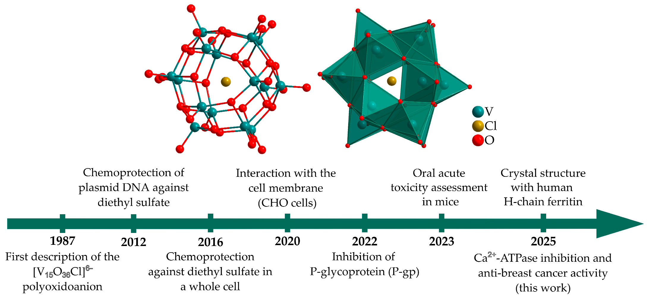
Although MV-POVs encompass the oxidation states most frequently observed under physiological conditions, their therapeutic potential remains comparatively underexplored in relation to other polyoxometalates [18]. Specifically, the [(CH3)4N]6[Cl@VIV8VV7O36] (V15) [19] has been evaluated in its interaction with different biological targets in the last 15 years (Figure 1). In studies developed by our research group, V15 showed a chemoprotective effect of pUC19 plasmid DNA of up to 70%, minimizing the action of potentially carcinogenic agents such as the alkylating agent diethyl sulfate. Moreover, Escherichia coli cultures have been used as a more complex cell model system, resulting in approximately 40% chemoprotection for V15 and [I@VIV12VV6O42]7− (V18I) [20]. Correlation with spectroscopic studies has suggested a relationship between stability in solution and chemoprotective performance, where MV-POVs containing encapsulated halides V15 and V18I have shown more promising results than V10 and those containing phosphate encapsulated [20]. The studies developed in vitro have enabled us to evaluate the reactivity patterns of MV-POVs in a controlled biological environment, thereby establishing structure-activity-reactivity relationships.
Studies involving ABC transporter P-glycoprotein showed that V15 can inhibit the multidrug resistance associated with this membrane transporter. Currently, the resistance to multiple drugs with different chemical structures and mechanisms of action is a major challenge in conventional cancer therapies [21]. Recently, it was demonstrated that V15 interacts with Chinese Hamster Ovary cells, decreasing the packing of membrane lipids and increasing signal transduction pathways mediated by the aggregation of the luteinizing hormone receptor (LHR) [22]. The LHR is a G-protein-coupled receptor that plays a key role in reproductive physiology in both males and females [22]. In addition, it was recently reported that a crystallographic structure of a model lysozyme, in which different MV-POV, including [(H2O)@V15O36]5–, non-covalently interact with enzyme surface residues [23]. Recently, the crystalline structure of a Cl@V15-ferritin adduct was reported as obtained from a suspension containing [VIVO(acac)2], where acac = acetylacetonato, and H-chain ferritin in basic medium, under mild conditions [24]. This physiologically relevant protein was involved in the spontaneous formation and stabilization of the mixed-valence [Cl@V15O36]6−, containing 8V(IV): 7V(V), which corresponds to the classical polyoxidoanion described by Muller and used in this work, differing only in the cations. These achievements should encourage further research into mixed-valence cage-like chemistry and delve into their biological properties.
Among the few in vivo studies with POVs accessing anticancer activity, V10 prevented and suppressed tumor growth (B16-F10 melanoma allografted mice) in 70% [25]. A heterometallic Mo/V-POV reduced breast tumor growth in BALB/c female mice [26]. The V10 was evaluated for its toxicity to Sparus aurata fish [27], while V15 was assessed in mice through an acute 28-day repeated toxicity study. To our knowledge, no other POV has been studied so far, finding a moderated toxicity following a single oral dose of V15 from 25 to 2000 mg kg−1, even after 14 days of exposure. However, repeated daily exposure over 28 days caused high toxicity at doses above 25 mg/kg, and V15 was classified as level 5 in the oral assay [28]. Taking all together, not only V15, but also other compounds with physiologically accessible oxidation states of vanadium must be considered to better understand MV-POV chemistry, with a view to the safe future use of drugs based on vanadium.
Figure 1. Timeline of the cage-like MV-POV [(CH3)4N]6[Cl@V15O36] (V15), emphasizing its role in biological applications, with ball-and-stick and polyhedral representations (the cations were omitted for clarity) [19,20,21,22,24,28,29].
Inorganics 13 00306 g001
The previous studies motivated us to further explore the biological activities of V15 to analyze its anticancer potential. Breast cancer is the most common cancer and causes the highest mortality among women worldwide, and is subdivided according to molecular mechanisms and aggressiveness [30]. The metastatic disease, responsible for more than 90% of cancer-related deaths, is defined by the ability of cancer cells to detach from the primary tumor and, through the bloodstream, invade nearby tissues, forming secondary tumors in different organs. The molecular classification of breast cancer subtypes mainly involves the estrogen and progesterone receptor genes (RE and RP, respectively) and the HER2 protein [31]. There are 4 molecular subtypes, with 70% of tumors classified as luminal A and B, characterized by the expression of hormone receptors RE and RP and negative for HER2 (Luminal A) or weakly positive for HER2 (Luminal B). Luminal subtypes are the least aggressive, with luminal B being more aggressive than luminal A. In order of aggressiveness, 15–20% are HER2 positive (overexpressing the HER2 protein) and 15% are triple negative (TNG), i.e., they do not have hormone receptors or overexpress HER2 [31]. Despite optimal systemic chemotherapy, fewer than 30% of women with metastatic breast cancer survive 5 years after diagnosis, and virtually all women with metastatic triple-negative breast cancer will ultimately die of their disease [32].
For luminal tumors, therapy uses tamoxifen (hormone therapy) or aromatase inhibitors, and for HER2 tumors, the monoclonal antibody trastuzumab (anti-HER2 antibody) is used. Advanced breast cancer, especially TNG, presents a highly metastatic profile and drug resistance. The lack of specific therapeutic targets means that TNG tumors are treated only with chemotherapy agents (e.g., doxorubicin and/or paclitaxel). However, chemotherapy agents present severe acute side effects ranging from vomiting and intense malaise to adverse effects such as cardiotoxicity, neurotoxicity, and neuropathy [33,34]. In this sense, vanadium compounds, including POVs, are promising chemotherapy agents, besides the well-known inorganic-based drugs containing cisplatin [35,36], as several studies have shown that they are active at different stages of carcinogenesis, such as prevention, therapy, and early diagnosis [37].
Nowadays, in breast cancer, we have found many research fields and/or perspectives such as cancer genomics, cancer therapy resistance, artificial intelligence tools, DNA repair and immunosurveillance, tumor microenvironment, clinical cancer biomarkers, non-invasive diagnostics, signaling in triple-negative breast cancer, chemotherapeutic agents for breast cancer, association with other cancer types, among many others [38]. Additionally, cancer research is conducted to discover new inorganic compounds, such as POMs, that combine inhibitory effects and/or reversal of drug resistance, as well as for breast cancer photothermal therapy using POMs-based nanoparticles [26,35,36,39].
The aim of this work is to pursue the studies of the putative biological activities of the mixed-valence pentadecavanadate, [(CH3)4N]6[Cl@V15O36] (V15), analyzing the Ca2+-ATPase inhibition potential and its anti-breast cancer activity in the luminal breast cancer cell line (MCF-7) and the TNG breast cancer cell line (MDA-MB-231). Specifically, in this work, we undertook the following tasks: (i) the evaluation of the V15 stability and speciation under the experimental conditions employed using 51V NMR and UV/Vis spectroscopy; (ii) the determination of the IC50 value for inhibition of Ca2+-ATPase activity, using a enzymatic couple assay; (iii) the determination of Vmax and Km kinetic parameters, regarding the native ligand ATP, using the couple enzymatic assay and the Lineweaver-Burk plot of Ca2+-ATPase in the absence or presence of inhibitor for determination of the type of enzyme inhibition; (iv) comparison of the electrostatic potential map of [V10O28]6− (V10), [MnV13O38]7−(MnV13), [VV14O38(PO4)]9− (PV14ox) and also [Nb10O28]6− (Nb10) with [Cl@VV7VIV8O36]6− (V15); (v) determination of the V15 IC50 values against MDA-MB-231 and MCF-7 cell lines, at 24 h; (vi) analysis of the effect of V15 in MDA-MB-231 cell migration; (vii) to verify V15 modulation of epithelial–mesenchymal transition (EMT) in MDA-MB-231 by cell morphology alteration; (viii) the putative mechanism of cell death induced by V15 searching for necroptosis and apoptosis.

2. Results and Discussion

2.1. Stability Studies

Although the characterization of POV structures in the solid state is important, understanding their behavior in solution is essential to identify the vanadium species present in equilibrium, which may include bioactive forms that contribute to the overall biological activity. It is known that the stability of POVs depends on the counter cation [40], the encapsulated anion [16], and the buffer or culture medium compositions used in the biological assay [41]. In the physiological medium, interaction of POVs with peptides and proteins may also change the nuclearity and oxidation state of the vanadium compound [42]. The stability study of V15 was already performed in solution containing DMEM medium (pH 7.4) [22], water (pH 6.3) [29], PIPES (pH 7.5) [29], and LB (Luria–Bertani) medium [20]. These correlations are essential for the rational selection of new biological applications.
Herein, the speciation of 1.0 mM V15 in enzymatic medium at pH 7.0 (25 mM HEPES, 100 mM KCl, 5.0 mM MgCl2, 50 µM CaCl2), and in RPMI medium (pH 7.4) after incubation at room temperature, was monitored by 51V NMR (Figure 2). Spectra were also recorded in water for comparison. In water/D2O and RPMI, NMR spectra were recorded at 0, 3, 10, 24, 48, and 72 h, reflecting the conditions used in the cellular viability assay. In the enzymatic medium, spectra were acquired at 0 and 1 h, aligning with the time course of the Ca2+-ATPase inhibition assay.
After dissolving V15 in H2O, only a signal at δ –560 ppm was detected, attributed to the mononuclear [H2VO4] complex (V1), indicating that V15 hydrolyses partially [43]. Since V15 contains seven V(V) and eight V(IV) centers, the intact mixed-valence cluster was not detected by 51V NMR. Over time, the concentration of vanadium(V) species increases, and after approximately 10 h, the signals characteristic of the three vanadium(V) environments of V10 appeared (Figure 2c) [43]. The intensity of these signals increased over time, with isomeric shifts at—423 (VA), –504 (VB), and –522 (VC) ppm, consistent with the protonated [HV10O28]5− species [43].
In enzymatic medium, the 51V NMR of V15 reveals signals for V1, [H2V2O7]2− (V2) and [V4O12]4− (V4) at –560, –574 and –578 ppm, respectively (Figure 2a). The speciation of POVs in solution is strongly pH-dependent, with V10 forming predominantly under acidic environments. This explains why V10 forms upon V15 hydrolysis in water but does not form in neutral buffered solutions. Given that RPMI medium contains nutrients such as amino acids, phosphate, and glucose, which are capable of binding metal ions, a more complex speciation profile was expected. Indeed, NMR signals corresponding to oligovanadates V2, V4, and [V5O15]5- (V5) at δ –585 ppm were observed (Figure 2d) during the time of the experiment. Additionally, the signal at δ –558 ppm of V1 represents a combination of V1 and a phosphate-vanadate (PV) complex (V1 + PV), as these species rapidly exchange on the NMR timescale [22].
The electronic paramagnetic resonance (EPR) spectroscopy at X-band complements the 51V NMR studies, as EPR detects the paramagnetic V(IV), while 51V NMR detects V(V) species. The 1.00 mM freshly prepared solutions of V15 in water, enzymatic, and RPMI media at 77 K (Figure S1) gave weak broad lines compatible with those previously described for the mixed-valence polyoxidoanions [20,22,29]. Although some variations in the spectra can be observed over 72 h, the poor signal-to-noise ratio prevented detailed analysis. However, no resolvable hyperfine structure, characteristic of vanadium(IV) mononuclear species, was detected. These results reinforce V15 stability in different conditions and a possible oxidation of vanadium(IV) to vanadium(V) upon hydrolysis.
Additionally, the stability of V15 in enzymatic and RPMI medium was monitored using UV/Vis/NIR spectroscopy over a period of 168 h, and the results were compared with those registered for the aqueous solution (Figure 3). In the initial time, the spectra displayed a broad band centered at 950 nm assigned to a partially delocalized intervalence charge-transfer transition (IVCT, type II, V(IV)→V(V)) [44] characteristic of MV-POVs [29]. A second, higher-intensity band, starting at approximately 500 nm and extending to the ultraviolet region, was also observed, and attributed to ligand-metal charge transfers (LMCT, pπ (O, oxo) → d(V)). In the aqueous solution, some oxidation of V(IV) to V(V) occurred, causing a gradual reduction in IVCT absorbance. After 168 h, the V15 concentration had dropped to 46% of its initial value, as found by the calibration curve (Figure S2). The aggregate also undergoes partial hydrolysis, resulting in a mixture of V15, V1, and V10, as suggested by NMR data along with the evolution of UV/Vis/NIR spectrum profiles and the change in the color of the solution from intense green to yellowish green. It is well known that the octahedral V(V) in V10 causes a bathochromic shift in the LMCT bands compared to V(V) in other geometries, and that this transition is responsible for the yellow color of the anion [45].
Despite the speciation observed in NMR spectra in the initial time, V15 showed greater stability in the enzymatic medium containing HEPES pH 7.0, a buffer with a weak affinity for binding metal ions, in RPMI medium pH 7.4 (Figure 2b,d), reducing the MLCT band intensity by ca. 10% after 168 h. RPMI, on the other hand, has a composition rich in amino acids, including aspartate, that might interact with the POV and stabilize it or lead to redox reactions, depending on the conditions (temperature, pH, concentration, and ionic force of the medium). In the face of the above, V15 is more kinetically stable to hydrolysis for days under physiological conditions and is not completely oxidised or broken down into oligovanadates before exerting the biological effect reported here.

2.2. Inhibition of Ca2+-ATPase

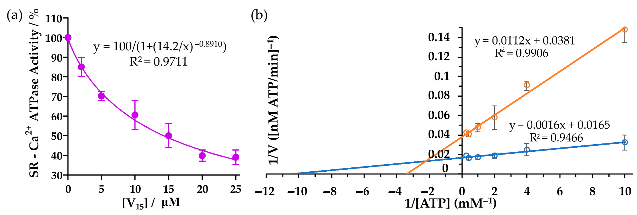
The effect of V15 on the activity of sarcoplasmic reticulum Ca2+-ATPase from skeletal muscle was evaluated in vitro. V15 inhibited enzyme activity in a concentration-dependent manner, with a half-maximal inhibitory concentration (IC50) value of 14.2 μM (Figure 4a).
Among other POVs, decavanadate (V10) exhibits an IC50 of 15 μM [46]. In previously reported studies, the bicapped Keggin-type [VV14O38(PO4)]9− (PV14 ox) showed an IC50 of 5 μM [12], while K7MnV13O38 (MnV13) and K5MnV11O33 (MnV11) presented IC50 values of 31 μM and 58 μM, respectively [47]. Notably, both V15 and V10 carry the same net charge of −6 at physiological pH. Although their sizes and structures differ, the similarity in IC50 values may be attributed to their charge and the predominance of electrostatic interactions and hydrogen bonds with the target protein, as previously described for other POMs [48]. V15 solution at the IC50 concentration (15 μM) was incubated in the enzymatic medium for 60 min prior to measuring its inhibitory effect on Sarcoplasmic/Endoplasmic Reticulum Calcium ATPase (SERCA) hydrolysis activity. After incubation, V15 ATPase inhibitory activity was reduced to 42.4%, instead of 50% without incubation, indicating that the POV remains relatively stable in solution, even after 60 min. We emphasize that the IC50 values were determined during the first two minutes after the POV addition. In fact, no incubation was made with the POMs in the medium to prevent any decomposition. Therefore, it is suggested that V15 is the most probable species responsible for the observed inhibitory effects on the Ca2+-ATPase, as described elsewhere for similar experimental conditions with other POMs [12,46,47,48,49,50]. On the other hand, if putative V15 decomposition into V1 species occurs, it would lead to a lower inhibition potential activity for this ATPase, considering that the IC50 values for V1 species were reported to be lower (IC50 = 80 μM) [49].
It was determined that V15 exerts a mixed-type inhibition on the ATPase activity, as evidenced by a decrease in Vmax and an increase in Km relative to the control measured by the slope and intercept of the double-reciprocal plot in Figure 4b. This suggests that V15 interacts with at least two distinct binding sites on the enzyme. In addition to the ATP-binding site, V15 may also bind to other domains of the Ca2+-ATPase, regardless of whether or not the enzyme is bound to its substrate. Mixed-type inhibition has also been reported for other POVs, such as PV14ox and MnV13. In contrast, V10 exhibits non-competitive inhibition with respect to ATP, suggesting mechanistic differences despite similar IC50 values between V10 and V15. It is known that V10 binds to all conformational states of the enzyme—E1, E2, phosphorylated or not—whereas monomeric vanadate binds exclusively to the E2 conformation [49].
Furthermore, V10 can induce cysteine oxidation accompanied by the reduction of V(V) to V(IV), implying a potential role for V(IV) species in biological activity [51]. MV-POVs have already been observed, for instance, in lysozyme crystals [42]. X-ray structure studies have shown that [(OH2)@V15O36]5− and [(OH2)@V15O33]+, both described as mixed-valence (VV7/VIV8), crystallize with lysozyme in a solution treated with monomeric vanadyl complex, [VIVO(acac)2] (acac = acetylacetonato). The interaction with the protein occurs through hydrogen bonds with lysine, tyrosine, and arginine residues [42]. Moreover, [V15O33(OH2)]+ species was observed to form covalent bonds with the protein, where two POV oxygen atoms are replaced by those of aspartyl carboxylates, and a third is replaced by a glycine carbonyl oxygen. These interactions suggest that lysozyme structure provides a stabilizing environment for the V15-like-POVs [42,52].
These observations indicate that not only the surface charge, but also the POV specific structure and the presence of vanadium in the +IV oxidation state contribute to its interaction with Ca2+-ATPase and the resulting inhibition of its enzymatic activity.

Electrostatic Potential (ESP) Map

The inhibition of Ca2+-ATPase by POMs is frequently associated with their high negative surface charge and has been correlated with their charge density and size [48,53]. In fact, it was described that Preyssler-type anion [NaP5W30O110]14− (P5W30) [50] and [H10Se2W29O103]14− (Se2W2914−) with a similar charge density (q/m = 0.47 and 0.48, respectively) shows the highest POMs Ca2+-ATPase inhibitory potential, respectively, IC50 = 0.37 and 0.3 μM whereas [H2P2W12O48]12− (P2W12) with a charge density of 1.00 presents a higher IC50 value (11 μM) [48]. Besides the charge of the POM, charge density, and size, other features may contribute to POMs’ interactions with proteins and the inhibition of enzymatic activities. For example, the isostructural decavanadate (V10) and decaniobate (Nb10) with analogous structure, net charge, and approximately the same size but with different inhibition potential for the Ca2+-ATPase activity (See Table 1 presented below), have been shown to present different binding sites for G-actin [51]. Thus, it was shown that V10 binds preferentially to the alpha binding site surrounded by Arg and Lys residues, being this the binding site of the natural substrate of the protein (ATP), whereas Nb10 binds to a binding site beta [54]. Besides this specific POV-actin interaction, several distinct modes of POVs' interactions with proteins have been recently described [54]. Regarding the Ca2+-ATPase, V10 was found to crystallize in a highly positively charged groove formed by three protein domains [52], highlighting its ability to bind electrostatically to this region.
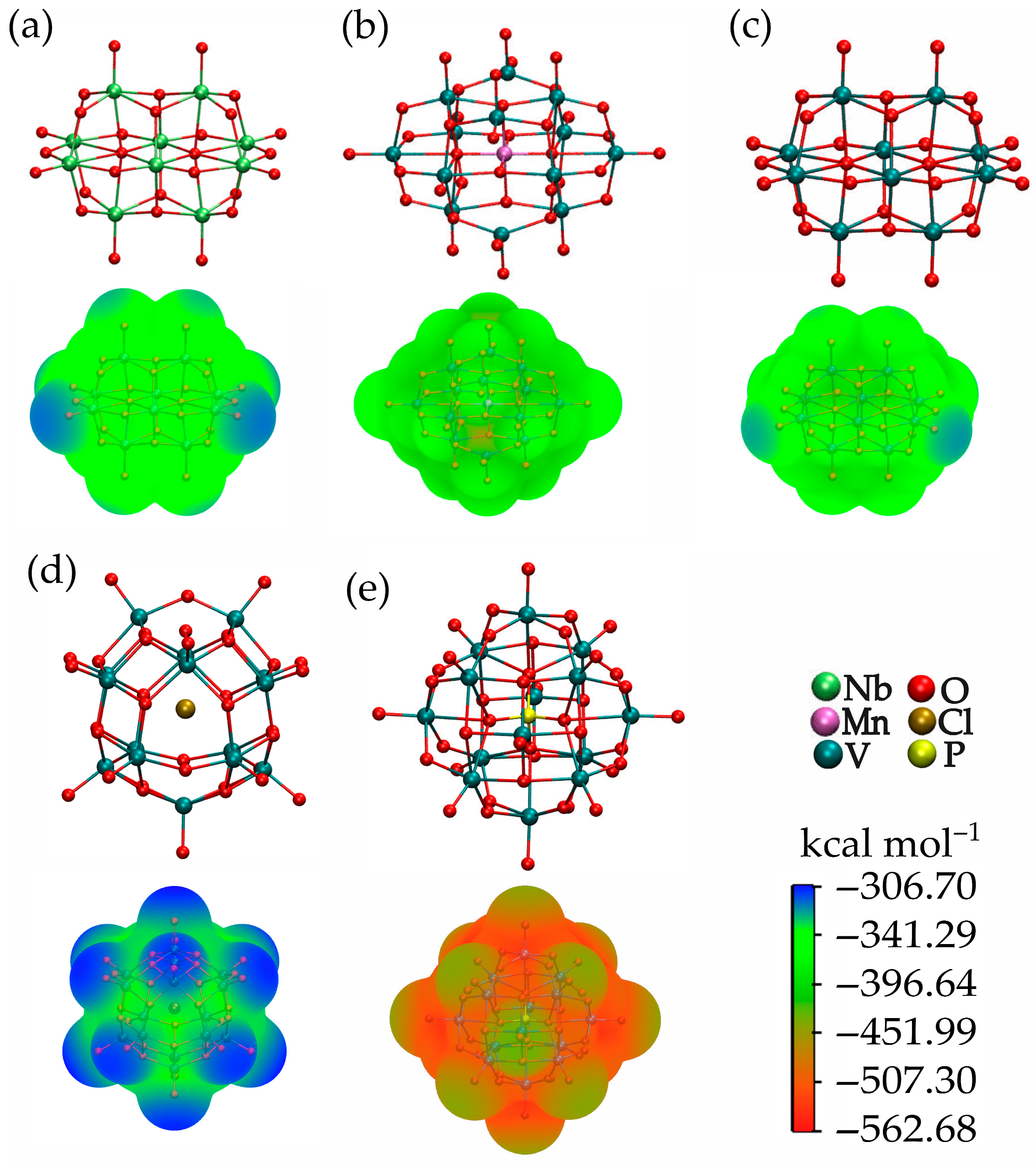
The Electrostatic Potential (ESP) map is a useful tool for examining the most probable sites of non-covalent interactions, such as electrostatic attractions and hydrogen bonds. The ESP is broadly used to visualize how the electrostatic potential varies across the surface of a molecule, and it also aids in analyzing intermolecular interactions based on electrostatic complementarity. ESP calculations were performed for V15 and for other selected discrete POVs that have been previously evaluated for their Ca2+-ATPase inhibitory activity [V10O28]6− (V10), [Nb10O28]6− (Nb10), [MnV13O38]7− (MnV13), which are expected to exist as deprotonated species under the enzymatic assay conditions [12]. The fully oxidized bicapped Keggin anion [VV14O38(PO4)]9− (PV14 ox) was also assessed despite its low kinetic stability in aqueous solution at pH 7, because it coexists in equilibrium with oligovanadates in freshly prepared solutions [55].
For better comparison, all volume electron densities were represented as isosurfaces (Visosurface, Figure 5), and are drawn using a continuous color gradient at the same electrostatic potential scale. All values on the isosurfaces are negative, with the most electron-rich areas in red, indicating regions of highest attraction for a positively charged species or ions. The blue areas represent the less negative potential values, while the green regions indicate intermediate electron-rich areas. Except for the heterometallic MnV13, the most negative electrostatic surface potential values of the POMs are localized in the vicinity of the doubly and triply bridging oxygen atoms, while the terminal oxygen atoms exhibited the least negative electrostatic potential. For MnV13, the most negative potentials are positioned in the centre of the aggregate.
To shed light on the factors involved in the Ca2+-ATPase inhibitory activity promoted by POVs, some molecular and electrostatic features were attempted to correlate with IC50 values depicted in Table 1. The relative order for both Emax and Emin is V15 < Nb10 < V10 < MnV13 < PV14 ox, while the IC50 order is Nb10 < MnV13 < V10~V15 < PV14 ox. The analysis of the results reveals two important parameters: the surface maxima and minima values in ESP (Emax and Emin), and the volume enclosed by the isosurface (Visosurface).
Regarding the border inhibitors, PV14 ox shows the most negative Emin, the highest ΔE value (123 kcal mol−1), and the biggest Visosurface of 835.19 Å3, being the most nucleophilic POV and exhibiting the strongest inhibition of Ca2+-ATPase (Table 1). On the other hand, the poorest inhibitor, Nb10, presents Emin of—382.80 kcal mol−1 and a ΔE value of only 59 kcal mol−1; moreover, its Visosurface is 25% smaller than the PV14 ox one. The Emax and Emin for V10 are slightly more negative than those for Nb10, but its Visosurface being 18% larger might be associated with its inhibitory activity, which is 1.75 times more potent. The MnV13, in turn, exhibits the second-lowest Emin value, but its IC50 is comparable to that of Nb10. Here, the impact of the volume is significant because it showed the lowest Visosurface, i.e., 29% and 33% smaller than V15 and PV14 ox.
The V15 is a key aggregate to demonstrate the correlation between the Ca2+-ATPase activity with the ESP and the Visosurface, because even showing the lowest Emax, Emin, and a ΔE of only 48 kcal mol−1, its IC50 value is close to the V10 (15 and 14.2 μM). The Visosurface calculated for V15 is only 5.9% bigger than V10 and 5.4% smaller than PV14 ox, but considerably higher than Nb10, achieving 25%. These results showed that, despite their structural differences and variations in vanadium oxidation states, V10 and V15 display similar availability of negative charge to participate in electrostatic interactions with the expected binding site of this enzyme [48]. However, the relationship between delocalization and electron density distribution in ESP is not straightforward, and there are still few systematic structural studies with mixed valence POVs to trace a linear correlation. As can be observed in Table 1, V15 presents a charge density (q/m) of 0.40. It was reported that POMs with moderate charge density (q/m = 0.33) interact strongly with protein surfaces due to a process named chaotropic effect [56]. It was suggested that this chaotropic effect contributes to a structure–activity relationship for the affinity of POMs towards proteins [56]. Still, further studies are needed to unravel at the molecular level the POMs features that correlate with specific biological activities.

2.3. Inhibition of Cell Viability in Breast Cancer Cells

The MTT assay was performed to evaluate cell viability using V15 in the non-tumorigenic, human mammary epithelial cell line (HB4a), luminal breast cancer cell line (MCF-7), and the triple-negative breast cancer cell line (MDA-MB-231). The cells were initially introduced into the wells to allow cell adhesion for 2 h, followed by the addition of increasing amounts of V15 (Figure S3). The results of cell viability inhibition of the cell lines treated with V15 showed a dose-dependent effect for the three cell lines, with an IC50 value of 1.02 μM for HB4a, 15.1 μM for MCF-7, and 17.2 μM for MDA-MB-231 (Figure 6 and Table 2). The IC50 value for luminal breast cancer cells was 16 times more potent than the commercial drug 5-fluorouracil under similar conditions (24 h, RPMI) [57].
The non-tumorigenic cell line showed higher sensitivity to V15 compared to the two cancer cell lines, indicating that the POV does not exhibit selectivity regarding this experimental model. Despite the lack of selectivity, in our previous studies with V15 using peripheral blood mononuclear cells (PBMC), an IC50 value of 12.9 μM was determined in RPMI after 48 h [28]. These data, along with early toxicity studies in mice, may guide future in vivo research when focusing on the urgent need to develop new therapies for the triple-negative breast cancer subtype. In such studies, a V15 concentration in the order of 1.0 μM (see Figure S2) would be reasonable, because it produced a cytotoxic effect of approximately 30–40% on MDA-MB-231 cells.
The IC50 value of V15 in MCF-7 and MDA-MB-231 cell lines was selected to monitor cell viability over 24, 48, and 72 h. The graphs presented in Figure S4 demonstrate that the POV is a time-dependent potent inhibitor of cell growth in both cell lines, corroborating the value determined for the IC50 after 24 h of incubation. The time curve showed a significant decrease in the viability of MDA-MB-231 cells after incubation, with only 45.4% (24 h), 15.3% (48 h), and 8.3% (72 h) of cells remaining alive. Similar results were observed with MCF-7, showing 64.3% (24 h), 12.1% (48 h), and 3.03% (72 h). Therefore, V15 showed a cumulative effect in the interference of cell viability over time.
The IC50 values obtained for V15 in the MCF-7 and MDA-MB-231 cell lines are promising, as they are lower than those found in the literature for other polyoxovanadates. For MCF-7 cells, a literature POV hybrid covalently bonded to amino acid esters, [V6O13{(OCH2)3C10H14O5}2]2−, exhibited a cytotoxic effect (IC50 = 53.01 μM) twice as strong as that of the traditional commercial drug 5-fluorouracil, in DMEM at 48 h [58]. Two decavanadates with betaine showed anticarcinogenic activity against MCF-7 cells at 24 h, with IC50 values close to 200 μM [59] while the super-Keggin MV-POV [(H2O)@V18O42]12− (H2O@V18) presented IC50 values of 45.95 µM and >500 µM for 24 h, respectively [60]. According to spectroscopic studies, V15 is stable in RPMI medium, suggesting that the activity could be attributed to this MV-POV. To the best of our knowledge, no speciation studies are available for H2O@V18. The H2O@V18 presents the lowest cytotoxic effect, being approximately 3 and 30 times less potent than V15 for MCF-7 and MDA-MB-231 cells, respectively.

2.4. Inhibition of Cell Migration Using the Wound Healing Assay in MDA-MB-231 Cell Line
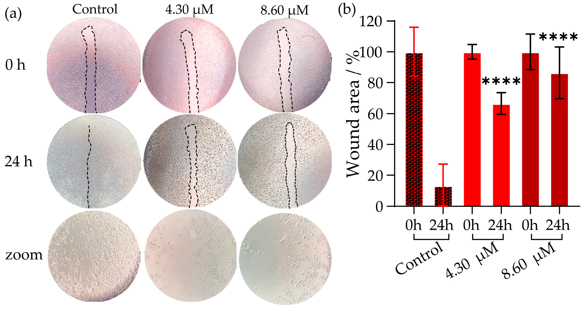
The Wound Healing assay is a simple method used to study cell migration. Reducing migration is desirable for a therapy that aims not only to diminish a tumor but also to delay or even prevent metastasis. The MDA-MB-231 cell line exhibits mesenchymal characteristics (fusiform shape), which is indicative of its migratory and invasive potential [61,62]. The cancer cells were treated with V15 at concentrations of 8.60 and 4.30 μM, equivalent to half and a quarter of the IC50 value, and the wound area was photographed after removing non-viable cells. Figure 7a displays selected images where, in the control, the typical migration of the cell line over the scratch is observed. In both samples treated with V15, the wound remained open, indicating that cell migration was impaired.
The wounds in the wells containing cells treated with both concentrations of V15 for 24 h were compared with the wounds of the control (non-treated cells) incubated for 24 h (Figure 7b). In the control samples, the wound area remained open by only 13%. For V15, a dose-dependent cell migration was observed after 24 h, and consequently, the gap area remained 66% and 86% open for the lower and higher concentrations, respectively. Similar results were reported for decavanadate with tetra-(benzylammonium), which also inhibited cell migration of MDA-MB-231 at concentrations of 0.70 and 2.8 μM [63].
Figure 7. Inhibition of cell migration as analyzed by the Wound Healing Assay in the MDA-MB-231 cell line without treatment (negative control) and treated with V15 for 0 and 24 h. (a) Selected photos of cell migration over the wound, from a total of 6 repetitions. The zoom is related to the wound area. (b) The graph was generated by taking six images in each condition, with the aid of ImageJ.JS 1.53m software [64]. The asterisks indicate the statistical treatment for six repetitions compared to the control at 24 h. **** means p < 0.001. The bars represent the standard deviation.
Inorganics 13 00306 g007

2.5. Scanning Electron Microscopy (SEM Images) in MDA-MB-231 Cell Line

SEM analysis was performed on the MDA-MB-231 cell line at the same concentrations as those used in the Wound Healing assay (4.30 and 8.60 µM). The SEM images showed cells with a fusiform shape, characteristic of this mesenchymal subtype of triple-negative cell line (Figure 8a), which changed to a more epithelial-like (amoeboid) morphology (Figure 8b,c). This morphology is generally related to a lower migratory capacity, which corroborates the cell migration assays. Finally, the microvesicles that appeared after treatment with the highest dose of V15 might be associated with the beginning of the cell death process. These morphological changes in the cells may be related to interactions with the cytoskeleton and possibly with the actin [65].
A process that is related to the progression of the disease to the metastatic form is called epithelial–mesenchymal transition (EMT). EMT is a reversible process, transitioning from epithelial to mesenchymal cells through a change in morphology. EMT occurs naturally in several processes, such as wound healing and embryonic development. However, when associated with the formation of fibroblasts (tissue regeneration, trauma, or inflammation), it can cause fibrosis and organ damage. When in neoplastic cells, there is an increase in cell proliferation and invasive and metastatic phenotypes, increasing the malignancy and aggressiveness of the disease [65]. The EMT phenotypical alteration involves genetic and epigenetic modifications in cancer cells when they initiate the metastasis process. Here, we observed a potential reverse process (mesenchymal–epithelial transition), indicating that V15 can switch off one of the most important hallmarks of cancer, thereby reducing the mechanism of progression to metastatic moving cells [65]. However, assays carried out at concentrations up to 1.0 μM of V15 caused only minor changes in cell morphology (Figure S5).

2.6. Gene Expression in MDA-MB-231 Cell Line

There are three classic mechanisms of cell death: apoptosis, necrosis, and necroptosis. Apoptosis is a programmed and “organized” cell death, essential for the development and homeostasis of the organism, and it does not trigger an inflammatory response. It is characterized by cell contraction and formation of apoptotic bodies, and since the cell contents are not released into the bloodstream, there is no inflammatory response. This mechanism can be triggered by internal signals, such as DNA damage or cell death signalling [66]. Necrosis is an unprogrammed cell death, usually triggered by injury, anoxia, or extreme tissue damage. Unlike apoptosis, it is an inflammatory process that results in the release of cellular contents into the bloodstream. It is characterized by cell swelling and rupture of the plasma membrane, leading to the release of cellular contents and the consequent inflammatory response. This mechanism can be caused mainly by a lack of blood supply [66].
Necroptosis is a cell death that resembles classical necrosis in morphology; however, it is a programmed inflammatory cell death. Unlike necrosis, it depends on a signaling pathway mediated by kinases called RIPK1 and RIPK3, with the pseudokinase MLKL acting as its effector molecule [66]. It is characterized by the controlled rupture of the plasma membrane and the release of cellular contents, but with some regulation compared to necrosis [66].
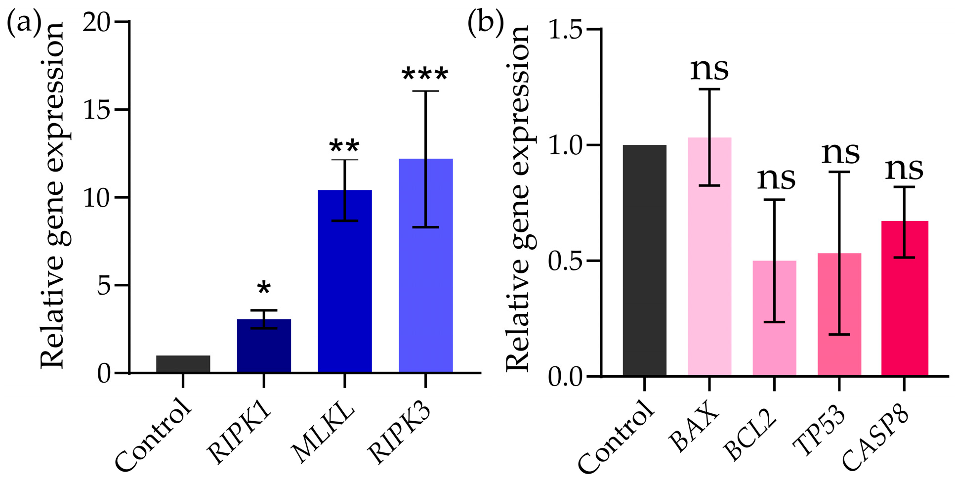
Based on the results presented above for the POVs, it was decided to investigate the cell death mechanism only for V15 in the MDA-MB-231 cell line, since it alters cell morphology, which may indicate interaction with the membrane and the triggering of processes inside the cell. The selected genes were those related to the mechanisms of apoptosis (BCL2, antiapoptosis; TP53, apoptosis mediated by DNA damage; CASP8, apoptosis via extrinsic pathway; and BAX, pro-apoptosis), and necroptosis (RIPK1, MLKL, and RIPK3). The graphs of relative gene expression compared with control without treatment and normalized by GAPDH constitutive gene (Figure 9) showed that those referred to necroptosis mechanism, RIPK1, MLKL, and RIPK3, increased 3, 10, and 15-fold, respectively (Figure 9a). The expression of these genes indicates that one of the mechanisms of cell death caused by V15 in the TNG breast cancer cell line may occur by necroptosis. On the other hand, no statistically significant effect on the general apoptosis mechanism could be related to V15 in this model (Figure 9b).
Interaction with V15 can trigger the necroptosis pathway, signaled by the activation of the RIPK3, RIPK1, and MLKL genes, which are factors involved in the formation of the necrosome, activated when caspase inactivation or inhibition occurs. The necrosome, along with other factors, migrates to the cell membrane, causing rupture and leakage, leading to cell death [67]. Some studies with POMs confirm that this class of compounds can cross the cell membrane of cancer cells, localizing itself within the cytoplasm; however, the mechanism of action of POMs has not yet been thoroughly explored [68,69,70,71,72,73]. About forty years ago, Yamase performed the first study on the mechanism of POMs in cancer cells [74]. The proposed mechanism involves the reduction and reoxidation of POM, as well as of the cellular components of the electron transport chain, which can inhibit the formation of ATP, leading to cell death by apoptosis [74]. This proposed mechanism is based on a series of studies that suggested that the cytotoxicity of bioactive POMs directly relates to their redox potential; however, other factors, such as size, structure, and composition, also significantly influence the activity of POMs against cancer cells [10].
POMs might affect cancer cells through several mechanisms, including the ones referred above, such as inducing apoptosis and affecting bioenergetics processes, but other mechanisms were described, such as interfering with DNA replication and gene expression, cell cycle arrest, generating reactive oxygen species (ROS), disrupting membrane transports, and/or inactivating essential enzymes within cancer cells [10,75]. Of course, those specific mechanisms for anticancer activity might depend on the POM’s structure and composition in order to selectively target cancer cells and reduce toxicity to healthy cells. When comparing and sorting the cell viability IC50 values in ascending order for 20 human cancer cell lines tested for different types of POMs, the first polyoxovanadates (POVs), polyoxomolybdates (POMos), polyoxopaladates (POPds), and polyoxotungstates (POTs) [75]. When comparing clinically approved drugs and POMs, in many cases, better results were observed for POMs in relation to the drugs [75]. Therefore, POVs such as V15 could become in the future an alternative to existing drugs in cancer therapy [75].
Alterations in calcium levels related to cell death by necroptosis have recently been revised, and it has been proposed that classical necroptosis is mediated by death receptors that work in synergy with caspase inhibitory signals [76]. However, a non-classical model of necroptosis has emerged. Based on the argument that cytosolic calcium signaling can drive the necroptotic mode of cell death in both dependent/independent receptor mechanisms when the cytoplasmic calcium levels are strictly related. Here we have found an inhibition of Ca2+-ATPase activity and a putative correlation with a decrease in cell proliferation, a phenotypic switch from mesenchymal to epithelial, and cell death by necroptosis. More detailed studies are in progress to clarify this probably new mechanism of drug interaction with promising therapeutic future intervention [76].

3. Materials and Methods

3.1. Generalities

The synthesis reactions were carried out in air, in deionized water (ultrapure water, resistivity less than 3.5 μS·cm−1 at 25 °C). All the chemicals purchased were of reagent grade and were used without further purification. Roswell Park Memorial Institute (RPMI) medium and fetal bovine serum (FBS), both from Thermo Fisher Scientific (Waltham, MA, USA); 4-(2-hydroxyethyl)1-piperazine ethane sulfonic acid (HEPES) from Sigma-Aldrich (St. Louis, MO, USA); 3-(4,5-dimethyltiazol-2-yl)-2,5-diphenyl-tetrazolium bromide (MTT), sodium cacodylate, glutaraldehyde, and osmium tetroxide were provided by Merck (Darmstadt, Germany). The AllPrep DNA/RNA Mini Kit (Qiagen, Hilden, Germany), SuperScript IV from Invitrogen (Walthan, MA, USA), and SYBR Green Power Master Mix from Applied Biosystems (Foster City, CA, USA) were used as received.
Powder X-ray diffractogram (PXRD) was registered on a Shimadzu XRD-6000 equipment (Kyoto, Japan), with Cu-Kα radiation (λ = 1.5418 Å) using silicon powder as an internal standard, using a voltage of 40 kV, current of 30 mA, and scan speeds of 0.02°s−1 (at 2ϴ). The 51V NMR spectra were recorded using a Bruker Avance 400 MHz (9.4 T) equipment (Ettlingen, Germany) , equipped with a direct detection multinuclear probe (5 mm). The spectra were acquired at 303 K with 500 μL of each sample solution, using 90° pulses. The spectra were registered with 2048 scans, with a recycle delay of 0.100 s and acquisition times of 0.218 s, along a spectral window of 714 ppm (+ 50 ppm to −650 ppm) with a 51V core acquisition frequency of 105.25 MHz. Electron Paramagnetic Resonance spectra were recorded in an X-band Bruker ELEXSYS E-500 spectrometer (Ettlingen, Germany) from solution frozen at 77 K. The spectra were referenced using pure VOCl3 in a capillary (external reference), fixing the signal at 0.00 ppm. UV-vis/NIR spectra were recorded on a PerkinElmer LAMBDA 1050 UV-Vis/NIR spectrophotometer (Waltham, MA, USA) equipped with three PMT/InGaAs/PbS detectors at room temperature in a wavelength range of 265–1300 nm. Scanning Electron Microscopy (SEM) analyses were performed using a TESCAN VEGA3 LMU equipment (Brno, Czech Republic) , with a resolution of 3.0 nm, at magnifications ranging from 500× to 10,000×.

Synthesis and Stock Solution of V15

The (Me4N)6[Cl@V15O36], V15, was synthesized following the procedure described in the literature [29] and in the Supplementary Material. A detailed scaled-up synthesis was described for some of us in reference [28]. IR spectra were registered for a freshly synthesized sample, after two months and two years of aging. Main bands in IR spectrum (Figure S6) of dark green crystals: νas and νs (CH3) weak bands at 3024 and 2952 cm−1 respectively, strong δas and δs(CH3) absorptions at 1485 cm−1 and 1446 cm−1 respectively, and ν(V=O) at 983 cm−1; ν(V−O−V) at 580, 660 and 725 cm−1. The diffraction peaks at angles 2θ of 7.4, 8.6, 15.4, 17.6, 19.9, 22.3, 23.1, 25.7, 26.7, 27.2, 30.7, 32.8, 33.2, 41.2 and 41.7° (Figure S7) are in good agreement with those previously reported, confirming that the solid was isolated free of the fresnoite-type oxide (NH4)2V3O8, a common insoluble byproduct in the synthesis of mixed-valence POVs [28].
New data on V15 stability in solid state over a period of 2 years were obtained by the infrared spectroscopic analysis, indicating that this MV-POV remained stable for a long time of storage (Figure S6). Stock solutions of V15 were prepared at a concentration of 1.0 mM (pH 4.67) in heated water, considering a molecular weight of 1820.42 g mol−1 to yield a green solution. Freshly prepared solutions were used in all experiments to prevent oxidation of the vanadium. The aqueous solution was diluted in RPMI culture medium or enzymatic medium to the desired concentration specified in each biological assay. The enzymatic medium contained: 25 mM HEPES (pH 7.0), 100 mM KCl, 5 mM MgCl2, and 50 μM CaCl2.

3.2. Spectroscopic Studies in Solution

The vanadium species formed in a 1.0 mM V15 solution were determined using vanadium nuclear magnetic resonance (51V NMR) spectroscopy. For sample preparation, 450 μL of 1.11 mM stock solution of V15 in water, RPMI pH 7.4 (9:1), without the addition of fetal bovine serum or enzymatic medium, received 50 μL of D2O, to render a final concentration of 1.0 mM. The spectra in aqueous solution and in RPMI medium were recorded at several points (0, 3, 10, 24, 48, and 72 h) to determine whether there were changes in the speciation during the time of the cellular cytotoxicity experiment. The spectra in the enzymatic medium were recorded at 0 h and 1 h.
Ultraviolet, visible, and near-infrared (UV/Vis/NIR) spectroscopy was used to follow the consumption of V15 at times of 0, 3, 10, 24, 48, 72 and 168 h, measuring the absorption band from 265 to 1300 nm in a PerkinElmer LAMBDA 1050 UV/Vis/NIR spectrophotometer (Waltham, MA, USA) equipped with three PMT/InGaAs/PbS. All spectra were recorded at ambient temperature in water, RPMI, and enzymatic media. A 1.0 mM solution of V15 in each condition was incubated for up to 168 h at 25 °C, and the spectrum was recorded at 24 h intervals. Due to the high molar absorptivity of V15, all samples had to be diluted to 0.020 mM prior to analysis to guarantee a detection within the linear range.

3.3. Computational Methods

The crystallographic data for the selected polyoxovanadates were available in the Cambridge Crystallographic Data Centre with CCDC numbers 722216 [Nb10O28]6− [77], 1610770 [MnV13O38]7− [78], 1839623 [V10O28]6− [79], 794586 [Cl@VV7VIV8O36]6− [29], 1886317 [VV14O38(PO4)]9− [12]. The structure of V10 was subject to geometric optimisation after removal of hydrogens, using Density Functional Theory (DFT) with the WB97X-D3 functional [80,81] and DEF2-TZVP basis set [82], utilising ORCA 5.0.4 software [83]. Electrostatic potential (ESP) map calculations were conducted using Multiwfn 3.8 software with an isosurface defined as 0.001 e/bohr3 [84,85,86]. The isosurfaces were rendered using the VMD—Visual Molecular Dynamics 1.9.3 software [87]. For [Nb10O28]6−, the wave function was calculated using the relativistic Hamiltonian Douglas–Kroll–Hess (DKH), with the basis SARC-DKH-TZVP for niobium and DKH-def2-TZVP with the auxiliary set SARC/J for the oxygens [88].

3.4. Inhibition of Ca2+-ATPase Activity by V15

Isolated sarcoplasmic reticulum (SR) vesicles prepared from rabbit skeletal muscles as described elsewhere [46] were suspended in 0.1 M KCl, 10 mM HEPES (pH 7.0), diluted 1:1 with 2.0 M sucrose, and frozen in liquid nitrogen prior to storage at −80 °C.
The activity of Ca2+-ATPase and its inhibition of the POV solution were measured spectrophotometrically by monitoring the change in absorbance at 340 nm at room temperature, both in the absence (100% activity) and in the presence of several concentrations of V15 (2.0, 5.0, 10, 15, 20, and 25 μM) for determination of IC50 (Figure 2a), for determination of the type of inhibition (Figure 2b) the activity was measured without inhibitor and with inhibitor (V15 at 15 μM) using increasing ATP concentrations (0.10 mM, 0.25 mM, 0.50 mM, 1.0 mM, 2.5 mM, and 3.75 mM).
The measurements were performed using the coupled enzyme pyruvate kinase/lactate dehydrogenase assay, as described elsewhere [46]. Aqueous stock solutions of V15 were prepared prior to use at 1.0 mM in water. The reaction medium contained the following: 25 mM HEPES (pH 7.0), 100 mM KCl, 5 mM MgCl2, and 50 μM CaCl2. For the coupled enzyme assay, the following components were added: 0.42 mM phosphoenolpyruvate, 18 IU lactate dehydrogenase, 7.5 IU pyruvate kinase, 0.25 mM NADH, and 2.5 mM ATP. The POV solution was added to the assay medium, and the experiment was initiated immediately after the addition of SR vesicles containing the Ca2+-ATPase (10 μg mL−1), and the absorbance was followed for about 1 min to measure the basal activity. Subsequently, 4% (w/w) calcium ionophore A23187 was added to the cuvette, and the decrease in absorbance was recorded for 3 min (uncoupled ATPase activity). ATPase activity and inhibition were calculated based on the rate of absorbance decrease per minute in the absence and presence of V15. Data are presented as means standard deviation (± SD). The results represent the average of triplicate experiments. The inhibitory effect of V15 was expressed as the IC50 value, defined as the concentration of V15 that induces 50% inhibition of Ca2+-ATPase activity.

3.5. Cell Culture

The human breast cancer cell lines, MDA-MB-231 and MCF-7, were provided by the ATCC (American Type Culture Collection) from Manassas, VA, USA). The cells were cultivated in RPMI 1640 medium supplemented with 10% FBS at 5% CO2 and 37 °C in a humidified atmosphere.

3.6. MTT Assay

The MDA-MB-231 and MCF-7 cells were cultured in RPMI medium containing approximately 5 × 10³ cells/well in a 96-well plate. The V15 was solubilized in water and included after 2 h of cellular adhesion. After incubating for 24 h, the in vitro cytotoxicity was measured by the MTT assay [89], and the results were analyzed in a MULTISKAN SkyHigh multiplate spectrophotometer (Vantaa, Finland) at a wavelength of 540 nm. IC50 values were calculated using GraphPad Prism 7 Software for Windows. Cell viability values were expressed as arithmetic means, with standard deviation calculated considering three independent experiments. Differences between multiple groups and controls were detected by one-way ANOVA followed by Tukey’s multiple comparisons test. A value of p < 0.05 was considered statistically significant [90].

3.7. Cell Migration by Wound Healing Assay

The MDA-MB-231 cell line was cultured in 6-well plates with the addition of 6 × 10⁵ cells per well. After cells confluence, FBS was removed for 4 h, and using a tip, a straight opening through the cell monolayer was made [91]. V15 (4.30 and 8.60 μM) was added, and the cultures were incubated with RPMI medium containing 1% FBS. Each well was washed 3 times with 1.0 mL of PBS 1x to remove the dead cells. The area of the opening in the cultures was photographed using an inverted microscope at 0, 24 h, and the wound area was measured using ImageJ® 1.53m software to analyze cell migration.

3.8. Scanning Electron Microscopy (SEM) Images

The preparation of cell line samples for SEM was carried out following Gonçalves’ protocol [92]. Briefly, circular coverslips measuring 0.130 mm in diameter were placed in a 24-well plate, and 30 × 103 cells per well were cultivated. After 2 h of cell adhesion, the control (same water volume) and V15 (4.30 and 8.60 μM) were added. After 24 h, the cells were washed with 0.100 M sodium cacodylate and fixed with 2.5% glutaraldehyde in 0.100 M sodium cacodylate buffer at pH 7.20 and kept for 1 h at room temperature. Then, the coverslips were washed twice with 0.100 M sodium cacodylate with a 10 min interval between each wash and fixed with 1% osmium tetroxide in 0.100 M sodium cacodylate buffer at pH 7.2 for 30 min in the absence of light. They were washed again twice with 0.100 M sodium cacodylate with a 10 min interval between washes. In the dehydration step, the cells were washed with ethanol at different concentrations (from 50% up to 100%) with a 10-min interval between washes. After the critical point was performed, the samples were metallized with gold, and SEM images were obtained with the Electronic Scanning Microscope TESCAN ORSAY HOLDING MIRA3 (Brno, Czech Republic).

3.9. Gene Expression Quantification

The MDA-MB-231 cells (8 × 10⁵) were plated in P100 culture plates in quintuplicate and treated for 48 h with V15 at the obtained IC50 concentration. The total RNA was extracted using the AllPrep DNA/RNA Mini Kit (Qiagen, Hilden, Germany), and Reverse transcription was performed with SuperScript IV Reverse Transcription Kits (Invitrogen, Waltham, MA, USA) according to the manufacturer’s instructions. Quantitative PCR experiments were performed using SYBR Green Power Master Mix, following the manufacturer’s protocols in the Step One Plus equipment (Applied Biosystems, Foster City, CA, USA). The reference transcript used for gene normalization was GAPDH. In terms of efficiency, we used the pattern curve for each primer, and the acceptable value was more than 98% efficiency. The obtained results were analyzed using the_Ct method and further processed using the 2−ΔΔCt [93]. The primer sequences are listed in Table S1.

4. Conclusions

In the present study, a mixed-valence polyoxovanadate, [ClVV7VIV8O36] (V15), was studied against the MDA-MB-231 cancer cell line and analyzed its Ca2+-ATPase inhibition potential. Moreover, the electrostatic potential map of V15, V10, PV14 ox, MnV13, and Nb10 was correlated with the Ca2+-ATPase activity inhibition. We believed that this kind of interdisciplinary collaborations, although not always easy to be achieved, are needed and represents a key cog in the “clock of the knowledge”, dynamically joining several scientific wheels such as chemistry, biology, and medicine, among others, for achieving perfect shared innovation and development [94], particularly for the understanding of the effects of POMs in biological systems and their putative applications in medicine.
Thus, we first analyzed the V15 inhibitory potential for Ca2+-ATPase activity using a coupled enzymatic method. It was determined that V15 presented an IC50 value of 14.2 μM and a mixed type of inhibition (Figure 10a). By affecting a key protein involved in cellular Ca2+ homeostasis, V15 might interfere with several physiological and pathological processes. 51V-NMR and UV/Vis studies of V15 indicated its stability in the enzymatic and RPMI media used. In fact, V15 also exhibited cytotoxicity against MDA-MB-231 (IC50 = 17.2 μM) and MCF-7 (IC50 = 15.1 μM) breast cancer cells, determined after 24 h (Figure 10b). Furthermore, using V15 concentrations equivalent to half and 1/4 of the IC50, MDA-MB321 cell migration was prevented by 70% and 90%, after 24 h, respectively (Figure 10c). Also, the fusiform morphology characteristic of the MDA-MB-231 cell line was lost to an epithelial-like (amoeboid) morphology upon exposure to V15, which is a potential advantage to control the tumor growth and progression (Figure 10d). Finally, it was observed that V15 induces an increase in the expression of the genes RIPK1, MLKL, and RIPK3, up to 3, 10, and 15-fold, respectively, pointing out that the mechanisms of cell death caused by V15 in the triple-negative breast cancer cell line may occur by necroptosis (Figure 10e). Putting it all together, V15, which targets Ca2+-ATPase with high affinity, has shown promising anticancer activities against breast cancer cells, and is worthy of being explored for therapeutic applications as well as for other areas of research.

Supplementary Materials

The following supporting information can be downloaded at: https://www.mdpi.com/article/10.3390/inorganics13090306/s1, Table S1: Primers sequence used for gene expression quantification in Homo sapiens; Figure S1: EPR spectra recorded at 77 K for V15 at 1.00 mM. (a) in aqueous solution, (b) in an enzymatic medium HEPES, and (c) in RPMI cell medium. Figure S2: Calibration curve constructed by plotting absorbance measured at λ = 950 nm vs. concentration of V15 in aqueous solution; Figure S3: Effects of V15 on cell viability using the MTT assay. In (a), the concentration curve and (b) IC50 curve fit for MCF-7 cells. In (c) concentration curve and (d) IC50 curve fit for MDA-MB-231 cells; Figure S4: Time curve of V15 in incubation times of 24, 48, and 72 h. In (a) to MCF-7 cells and in (b) to MDA-MB-231 cells. OD (Optic Density) was measured at 540 nm. **** means p < 0.001; Figure S5: Scanning Electron Microscopy (SEM) images for V15 in MDA-MB-231 cell line, at magnifications of 1000×, 2500×, 5000×, and 10,000× for (a) control; (b) concentration of 0.500 µmol L−1 and (c) 1.00 µmol L−1. Figure S6: Infrared spectra of V15 recorded in KBr pellets for: (a) freshly synthesized sample, (b) after two months of aging, and (c) after two years of aging. Figure S7: Powder X-ray diffraction patterns of V15, comparing the experimental with the simulated diffractogram generated from the single-crystal X-ray diffraction data CIF file number 794586.

Author Contributions

Conceptualization, M.A., G.G.N. and G.K.; methodology and formal analysis, M.A., G.K., G.F., E.L.d.S., R.R.R. and C.C.d.O.; investigation, G.F., B.R.B., H.d.S.C. and A.F.d.C.; writing—review and editing, G.F., M.A., G.G.N., G.K., B.R.B. and H.d.S.C.; funding acquisition, M.A., G.G.N. and G.K. All authors have read and agreed to the published version of the manuscript.

Funding

This research was funded by Portuguese national funds from FCT—Foundation for Science and Technology. This study was academically funded by government agencies with no private resources. Technology, grant number project UIDB/04326/2020, UIDP/04326/2020 and LA/P/0101/2020, and by the Conselho Nacional de Desenvolvimento Científico e Tecnológico (CNPq, Project No. 310107/2025-3 and 403533/2025-2) and pesquisa/PRPPG/UFPR (04/2023).

Data Availability Statement

The original contributions presented in the study are included in the article/Supplementary Material, further inquiries can be directed to the corresponding authors.

Acknowledgments

M.A. gives thanks to Algarve University and the Portuguese national funds from FCT—Foundation for Science and Technology through project UIDB/04326/2020 (DOI:10.54499/UIDB/04326/2020), UIDP/04326/2020 (DOI:10.54499/UIDP/04326/2020), and LA/P/0101/2020 (DOI:10.54499/LA/P/0101/2020). E.L.d.S., H.d.S.C., and G.G.N. are grateful to the Centro Nacional de Processamento de Alto Desempenho (CENAPAD-SP) for providing access to computational infrastructure. The authors thank Centro de Microscopia Eletrônica (CME-UFPR) for the SEM analysis. B. R. B. and H.d.S.C. thank Coordenação de Aperfeiçoamento de Pessoal de Nível Superior (CAPES-PROEX and CAPES-PrInt, Finance Code 001), A.F.d.C. thanks CNPq for research grants and scholarships, G.G.N. thanks to Conselho Nacional de Desenvolvimento Científico e Tecnológico (CNPq grant 310107/2025-3 and 403533/2025-2)

Conflicts of Interest

The authors declare no conflicts of interest.

Abbreviations

The following abbreviations are used in this manuscript:
MCF-7Michigan Cancer Foundantion-7
MDA-MB-231Monroe Dunaway Anderson Cancer Center-Mammary/Breast-231
FBSFetal Bovine Serum
RPMIRoswell Park Memorial Institute
DMEMDulbecco’s Modified Eagle’s Medium
HEPES4-(2-hidroxyethyl)1-piperazine ethane sulfonic acid
PBSPhosphate-Buffered Saline
PIPEPiperazine-n,n’-Bis(2-ethanesulphonicAcid)
MTT3-(4,5-dimethyltiazol-2-yl)-2,5-diphenyl-tetrazolium bromide
POMsPolyoxometalates
POVsPolyoxovanadates
MV-POVsMixed-Valence Polyoxovanadates
acacAcetylacetonate
5-Fu5-Fluorouracyl
ROSReactive Oxygen Species
51V-NMRNuclear Magnetic Resonance of Vanadium 51
UV-Vis/NIRUltraviolet-Visible/Near-Infrared spectroscopy
SEMScanning Electron Microscopy
ESPElectrostatic Surface Potential
LHRLuteinizing Hormone Receptor
ATPAdenosine Triphosphate
ABCATP-Binding Cassette
CHOChinese Hamster Ovary
P-gpP-glycoprotein
EMTEpithelial–Mesenchymal Transition
HER2Human Epidermal Growth Factor Receptor 2
RIPK1Receptor-Interacting Serine/Threonine-Protein Kinase 1
RIPK2Receptor-Interacting Serine/Threonine-Protein Kinase 2
RIPK3Receptor-Interacting Serine/Threonine-Protein Kinase 3
BAXBcl-2-Associated X Protein;
BCL2B-Cell Lymphoma 2
TP53Tumor Protein p53
CASP8Caspase-8
MLKLMixed Lineage Kinase Domain-Like Protein
GAPDHGlyceraldehyde-3-Phosphate Dehydrogenase
SRSarcoplasmic Reticulum
SERCASarcoplasmic/Endoplasmic Reticulum Calcium ATPase 
IC50Half Maximal Inhibitory Concentration
LBLuria–Bertani
TNGTriple-Negative
EREstrogen Receptor
PRProgesterone Receptor

References

  1. Aureliano, M. Decavanadate: A Journey in a Search of a Role. Dalton Trans. 2009, 42, 9093–9100. [Google Scholar] [CrossRef]
  2. De Sousa-Coelho, A.L.; Aureliano, M.; Fraqueza, G.; Serrão, G.; Gonçalves, J.; Sánchez-Lombardo, I.; Link, W.; Ferreira, B.I. Decavanadate and Metformin-Decavanadate Effects in Human Melanoma Cells. J. Inorg. Biochem. 2022, 235, 111915. [Google Scholar] [CrossRef]
  3. Soman, S.K.; Woodruff, M.R.J.; Dagda, R.K. The Mitochondrial Foundations of Parkinson’s Disease: Therapeutic Implications. Aging Dis. 2025, 16, 2695–2720. [Google Scholar] [CrossRef]
  4. Rathi, K.; Bhole, R.; Bansode, P.; Motghare, N. Navigating the Landscape of Alzheimer’s Disease: From Epidemiology to Drug Re-Purposing. Adv. Pharmacol. Pharm. 2024, 12, 135–148. [Google Scholar] [CrossRef]
  5. Hu, H.; Liang, W.; Ding, G. Ion Homeostasis in Diabetic Kidney Disease. Trends Endocrinol. Metab. 2024, 35, 142–150. [Google Scholar] [CrossRef] [PubMed]
  6. Zaichick, S.V.; McGrath, K.M.; Caraveo, G. The Role of Ca2+ Signaling in Parkinson’s Disease. Dis. Model Mech. 2017, 10, 519–535. [Google Scholar] [CrossRef]
  7. Dowling, P.; Swandulla, D.; Ohlendieck, K. Biochemical and Proteomic Insights into Sarcoplasmic Reticulum Ca2+-ATPase Complexes in Skeletal Muscles. Expert Rev. Proteom. 2023, 20, 125–142. [Google Scholar] [CrossRef] [PubMed]
  8. Aureliano, M.; Fraqueza, G.; Ohlin, C.A. Ion Pumps as Biological Targets for Decavanadate. Dalton Trans. 2013, 42, 11770–11777. [Google Scholar] [CrossRef]
  9. Sukumaran, P.; Nascimento Da Conceicao, V.; Sun, Y.; Ahamad, N.; Saraiva, L.R.; Selvaraj, S.; Singh, B.B. Calcium Signaling Regulates Autophagy and Apoptosis. Cells 2021, 10, 2125. [Google Scholar] [CrossRef] [PubMed]
  10. Bijelic, A.; Aureliano, M.; Rompel, A. Polyoxometalates as Potential Next-Generation Metallodrugs in the Combat Against Cancer. Angew. Chem. Int. Ed. 2019, 58, 2980–2999. [Google Scholar] [CrossRef]
  11. Kumari, N.; Pullaguri, N.; Rath, S.N.; Bajaj, A.; Sahu, V.; Ealla, K.K.R. Dysregulation of Calcium Homeostasis in Cancer and Its Role in Chemoresistance. Cancer Drug Resist. 2024, 7, 11. [Google Scholar] [CrossRef]
  12. Fraqueza, G.; Fuentes, J.; Krivosudský, L.; Dutta, S.; Mal, S.S.; Roller, A.; Giester, G.; Rompel, A.; Aureliano, M. Inhibition of Na+/K+-and Ca2+-ATPase Activities by Phosphotetradecavanadate. J. Inorg. Biochem. 2019, 197, 110700. [Google Scholar] [CrossRef]
  13. Poejo, J.; Gumerova, N.I.; Rompel, A.; Mata, A.M.; Aureliano, M.; Gutierrez-Merino, C. Unveiling the Agonistic Properties of Preyssler-Type Polyoxotungstates on Purinergic P2 Receptors. J. Inorg. Biochem. 2024, 259, 112640. [Google Scholar] [CrossRef]
  14. Ferraro, G.; Garribba, E.; Merlino, A. Exploring Polyoxidovanadate–Protein Interaction. Trends Chem. 2025, 7, 3–6. [Google Scholar] [CrossRef]
  15. Aronica, C.; Chastanet, G.; Zueva, E.; Borshch, S.A.; Clemente-Juan, J.M.; Luneau, D. A Mixed-Valence Polyoxovanadate(III, IV) Cluster with a Calixarene Cap Exhibiting Ferromagnetic V(III)−V(IV) Interactions. J. Am. Chem. Soc. 2008, 130, 2365–2371. [Google Scholar] [CrossRef]
  16. Solé-Daura, A.; Notario-Estévez, A.; Carbó, J.J.; Poblet, J.M.; de Graaf, C.; Monakhov, K.Y.; López, X. How Does the Redox State of Polyoxovanadates Influence the Collective Behavior in Solution? A Case Study with [I@V 18 O 42] q(q = 3, 5, 7, 11, and 13). Inorg. Chem. 2019, 58, 3881–3894. [Google Scholar] [CrossRef] [PubMed]
  17. Zhang, T.; Yan, W.; Wang, Y.; Wang, J.; Liu, C.; Ye, F.; Liu, B. An Ecofriendly and Efficient Wood-Based Polyoxovanadate Solar Evaporation Generator. Sci. China Mater. 2023, 66, 3292–3299. [Google Scholar] [CrossRef]
  18. Aureliano, M.; Gumerova, N.I.; Sciortino, G.; Garribba, E.; Rompel, A.; Crans, D.C. Polyoxovanadates with Emerging Biomedical Activities. Coord. Chem. Rev. 2021, 447, 214143. [Google Scholar] [CrossRef]
  19. Müller, A.; Krickemeyer, E.; Penk, M.; Walberg, H.; Bögge, H. Spherical Mixed-Valence [V15O36]5–, an Example from an Unusual Cluster Family. Angew. Chem. Int. Ed. 1987, 26, 1045–1046. [Google Scholar] [CrossRef]
  20. Postal, K.; Maluf, D.F.; Valdameri, G.; Rüdiger, A.L.; Hughes, D.L.; de Sá, E.L.; Ribeiro, R.R.; de Souza, E.M.; Soares, J.F.; Nunes, G.G. Chemoprotective Activity of Mixed Valence Polyoxovanadates against Diethylsulphate in E. Coli Cultures: Insights from Solution Speciation Studies. RSC Adv. 2016, 6, 114955–114968. [Google Scholar] [CrossRef]
  21. Kita, D.H.; Andrade, G.A.; Missina, J.M.; Postal, K.; Boell, V.K.; Santana, F.S.; Zattoni, I.F.; Zanzarini, I.D.S.; Moure, V.R.; Rego, F.G.D.M.; et al. Polyoxovanadates as New P-glycoprotein Inhibitors: Insights into the Mechanism of Inhibition. FEBS Lett. 2022, 596, 381–399. [Google Scholar] [CrossRef] [PubMed]
  22. Althumairy, D.; Postal, K.; Barisas, B.G.; Nunes, G.G.; Roess, D.A.; Crans, D.C. Polyoxometalates Function as Indirect Activators of a G Protein-Coupled Receptor. Metallomics 2020, 12, 1044–1061. [Google Scholar] [CrossRef]
  23. Tito, G.; Ferraro, G.; Garribba, E.; Merlino, A. Formation of Mixed-Valence Cage-Like Polyoxidovanadates at 37 °C Upon Reaction of VIVO(Acetylacetonato)2 with Lysozyme. Chem. A Eur. J. 2025, 31, e202500488. [Google Scholar] [CrossRef]
  24. Lucignano, R.; Tito, G.; Ferraro, G.; Picone, D.; Pisanu, F.; Garribba, E.; Merlino, A. Spherical Mixed-Valence Pentadecavanadate Binding to Human H-Chain Ferritin. Inorg. Chem. Front. 2025. [Google Scholar] [CrossRef]
  25. Essid, A.; Elbini, I.; Ksiksi, R.; Harrab, N.; Moslah, W.; Jendoubi, I.; Doghri, R.; Zid, M.-F.; Luis, J.; Srairi-Abid, N. Decavanadate Compound Displays In Vitro and In Vivo Antitumor Effect on Melanoma Models. Bioinorg. Chem. Appl. 2025, 2025, 6680022. [Google Scholar] [CrossRef]
  26. Yao, C.; Zhao, Z.; Tan, T.; Yan, J.; Chen, Z.; Xiong, J.; Li, H.; Wei, Y.; Hu, K. Lindqvist-Type Polyoxometalates Act as Anti-Breast Cancer Drugs via Mitophagy-Induced Apoptosis. Curr. Med. Sci. 2024, 44, 809–819. [Google Scholar] [CrossRef]
  27. Soares, S.S.; Martins, H.; Duarte, R.O.; Moura, J.J.G.; Coucelo, J.; Gutiérrez-Merino, C.; Aureliano, M. Vanadium Distribution, Lipid Peroxidation and Oxidative Stress Markers upon Decavanadate in Vivo Administration. J. Inorg. Biochem. 2007, 101, 80–88. [Google Scholar] [CrossRef]
  28. Barbosa, M.D.M.; de Lima, L.M.A.; Alves, W.A.D.S.; de Lima, E.K.B.; da Silva, L.A.; da Silva, T.D.; Postal, K.; Ramadan, M.; Kostenkova, K.; Gomes, D.A.; et al. In Vitro, Oral Acute, and Repeated 28-Day Oral Dose Toxicity of a Mixed-Valence Polyoxovanadate Cluster. Pharmaceuticals 2023, 16, 1232. [Google Scholar] [CrossRef]
  29. Nunes, G.G.; Bonatto, A.C.; de Albuquerque, C.G.; Barison, A.; Ribeiro, R.R.; Back, D.F.; Andrade, A.V.C.; de Sá, E.L.; Pedrosa, F.D.O.; Soares, J.F.; et al. Synthesis, Characterization and Chemoprotective Activity of Polyoxovanadates against DNA Alkylation. J. Inorg. Biochem. 2012, 108, 36–46. [Google Scholar] [CrossRef] [PubMed]
  30. Bray, F.; Laversanne, M.; Sung, H.; Ferlay, J.; Siegel, R.L.; Soerjomataram, I.; Jemal, A. Global Cancer Statistics 2022: GLOBOCAN Estimates of Incidence and Mortality Worldwide for 36 Cancers in 185 Countries. CA Cancer J. Clin. 2024, 74, 229–263. [Google Scholar] [CrossRef] [PubMed]
  31. Orrantia-Borunda, E.; Anchondo-Nuñez, P.; Acuña-Aguilar, L.E.; Gómez-Valles, F.O.; Ramírez-Valdespino, C.A. Subtypes of Breast Cancer. In Breast Cancer; Exon Publications: Brisbane, Australia, 2022; pp. 31–42. [Google Scholar]
  32. Bonotto, M.; Gerratana, L.; Poletto, E.; Driol, P.; Giangreco, M.; Russo, S.; Minisini, A.M.; Andreetta, C.; Mansutti, M.; Pisa, F.E.; et al. Measures of Outcome in Metastatic Breast Cancer: Insights From a Real-World Scenario. Oncologist 2014, 19, 608–615. [Google Scholar] [CrossRef]
  33. Schirrmacher, V. From Chemotherapy to Biological Therapy: A Review of Novel Concepts to Reduce the Side Effects of Systemic Cancer Treatment (Review). Int. J. Oncol. 2018, 54, 407–419. [Google Scholar] [CrossRef]
  34. Wang, J.; Wu, S.-G. Breast Cancer: An Overview of Current Therapeutic Strategies, Challenge, and Perspectives. Breast Cancer Targets Ther. 2023, 15, 721–730. [Google Scholar] [CrossRef]
  35. Lee, J.S.; Yost, S.E.; Yuan, Y. Neoadjuvant Treatment for Triple Negative Breast Cancer: Recent Progresses and Challenges. Cancers 2020, 12, 1404. [Google Scholar] [CrossRef] [PubMed]
  36. Shen, G.; Liu, Z.; Wang, M.; Zhao, Y.; Liu, X.; Hou, Y.; Ma, W.; Han, J.; Zhou, X.; Ren, D.; et al. Neoadjuvant Apatinib Addition to Sintilimab and Carboplatin-Taxane Based Chemotherapy in Patients with Early Triple-Negative Breast Cancer: The Phase 2 NeoSAC Trial. Signal Transduct. Target. Ther. 2025, 10, 41. [Google Scholar] [CrossRef]
  37. Bishayee, A.; Waghray, A.; Patel, M.A.; Chatterjee, M. Vanadium in the Detection, Prevention and Treatment of Cancer: The in Vivo Evidence. Cancer Lett. 2010, 294, 1–12. [Google Scholar] [CrossRef] [PubMed]
  38. Aureliano, M.; Camilo, H.S.; Brito, B.R.; Ribeiro, R.R.; Fraqueza, G.; Klassen, G.; Nunes, G.G. Polyoxovanadates with Anti-breastCancer Activity and Ca2+-ATPase Inhibition Potential. Eur. Assoc. Cancer Res. 2025, 19, 511–512. [Google Scholar] [CrossRef]
  39. Wang, C.; Dai, Z.; Zhang, Q.; Li, X.; Ma, M.; Shi, Z.; Zhang, J.; Liu, Q.; Chen, H. A Bifunctional Biomineralized Polyoxometalate Enabling Efficient Non-Inflammatory NIR-II Photothermal Tumor Therapy. Chem. Eng. J. 2024, 490, 151601. [Google Scholar] [CrossRef]
  40. Misra, A.; Kozma, K.; Streb, C.; Nyman, M. Beyond Charge Balance: Counter-Cations in Polyoxometalate Chemistry. Angew. Chem. Int. Ed. 2020, 59, 596–612. [Google Scholar] [CrossRef]
  41. Gumerova, N.I.; Rompel, A. Speciation Atlas of Polyoxometalates in Aqueous Solutions. Sci. Adv. 2023, 9, eadi0814. [Google Scholar] [CrossRef]
  42. Tito, G.; Ferraro, G.; Pisanu, F.; Garribba, E.; Merlino, A. Non-Covalent and Covalent Binding of New Mixed-Valence Cage-like Polyoxidovanadate Clusters to Lysozyme. Angew. Chem. Int. Ed. 2024, 63, e202406669. [Google Scholar] [CrossRef] [PubMed]
  43. Gumerova, N.I.; Rompel, A. Polyoxometalates in Solution: Speciation under Spotlight. Chem. Soc. Rev. 2020, 49, 7568–7601. [Google Scholar] [CrossRef]
  44. Day, P.; Hush, N.S.; Clark, R.J.H. Mixed Valence: Origins and Developments. Philos. Trans. R. Soc. A Math. Phys. Eng. Sci. 2008, 366, 5–14. [Google Scholar] [CrossRef]
  45. Aureliano, M. Recent Perspectives into Biochemistry of Decavanadate. World J. Biol. Chem. 2011, 2, 215–225. [Google Scholar] [CrossRef]
  46. Fraqueza, G.; Ohlin, C.A.; Casey, W.H.; Aureliano, M. Sarcoplasmic Reticulum Calcium ATPase Interactions with Decaniobate, Decavanadate, Vanadate, Tungstate and Molybdate. J. Inorg. Biochem. 2012, 107, 82–89. [Google Scholar] [CrossRef]
  47. Marques-da-Silva, D.; Fraqueza, G.; Lagoa, R.; Vannathan, A.A.; Mal, S.S.; Aureliano, M. Polyoxovanadate Inhibition of Escherichia Coli Growth Shows a Reverse Correlation with Ca2+-ATPase Inhibition. New J. Chem. 2019, 43, 17577–17587. [Google Scholar] [CrossRef]
  48. Gumerova, N.; Krivosudský, L.; Fraqueza, G.; Breibeck, J.; Al-Sayed, E.; Tanuhadi, E.; Bijelic, A.; Fuentes, J.; Aureliano, M.; Rompel, A. The P-Type ATPase Inhibiting Potential of Polyoxotungstates. Metallomics 2018, 10, 287–295. [Google Scholar] [CrossRef]
  49. Fraqueza, G.; Batista de Carvalho, L.A.E.; Marques, M.P.M.; Maia, L.; Ohlin, C.A.; Casey, W.H.; Aureliano, M. Decavanadate, Decaniobate, Tungstate and Molybdate Interactions with Sarcoplasmic Reticulum Ca2+-ATPase: Quercetin Prevents Cysteine Oxidation by Vanadate but Does Not Reverse ATPase Inhibition. Dalton Trans. 2012, 41, 12749–12758. [Google Scholar] [CrossRef]
  50. Aureliano, M.; Fraqueza, G.; Berrocal, M.; Cordoba-Granados, J.J.; Gumerova, N.I.; Rompel, A.; Gutierrez-Merino, C.; Mata, A.M. Inhibition of SERCA and PMCA Ca2+-ATPase Activities by Polyoxotungstates. J. Inorg. Biochem. 2022, 236, 111952. [Google Scholar] [CrossRef]
  51. Sciortino, G.; Aureliano, M.; Garribba, E. Rationalizing the Decavanadate(V) and Oxidovanadium(IV) Binding to G-Actin and the Competition with Decaniobate(V) and ATP. Inorg. Chem. 2021, 60, 334–344. [Google Scholar] [CrossRef] [PubMed]
  52. Toyoshima, C.; Nakasako, M.; Nomura, H.; Ogawa, H. Crystal Structure of the Calcium Pump of Sarcoplasmic Reticulum at 2.6 Å Resolution. Nature 2000, 405, 647–655. [Google Scholar] [CrossRef] [PubMed]
  53. Hua, S.; Inesi, G.; Toyoshima, C. Distinct Topologies of Mono- and Decavanadate Binding and Photo-Oxidative Cleavage in the Sarcoplasmic Reticulum ATPase. J. Biol. Chem. 2000, 275, 30546–30550. [Google Scholar] [CrossRef]
  54. Aureliano, M.; Gumerova, N.I.; Sciortino, G.; Garribba, E.; McLauchlan, C.C.; Rompel, A.; Crans, D.C. Polyoxidovanadates’ Interactions with Proteins: An Overview. Coord. Chem. Rev. 2022, 454, 214344. [Google Scholar] [CrossRef]
  55. Zarroug, R.; Artetxe, B.; Ayed, B.; López, X.; Ribeiro, N.; Correia, I.; Pessoa, J.C. New Phosphotetradecavanadate Hybrids: Crystal Structure, DFT Analysis, Stability and Binding Interactions with Bio-Macromolecules. Dalton Trans. 2022, 51, 8303–8317. [Google Scholar] [CrossRef] [PubMed]
  56. Solé-Daura, A.; Poblet, J.M.; Carbó, J.J. Structure–Activity Relationships for the Affinity of Chaotropic Polyoxometalate Anions towards Proteins. Chem. A Eur. J. 2020, 26, 5799–5809. [Google Scholar] [CrossRef] [PubMed]
  57. Aybek, H.; Temel, Y.; Ahmed, B.M.; Ağca, C.A.; Çiftci, M. Deciphering of The Effect of Chemotherapeutic Agents on Human Glutathione S-Transferase Enzyme and MCF-7 Cell Line. Protein Pept. Lett. 2020, 27, 888–894. [Google Scholar] [CrossRef]
  58. Hu, X.; Wang, H.; Huang, B.; Li, N.; Hu, K.; Wu, B.; Xiao, Z.; Wei, Y.; Wu, P. A New Scheme for Rational Design and Synthesis of Polyoxovanadate Hybrids with High Antitumor Activities. J. Inorg. Biochem. 2019, 193, 130–132. [Google Scholar] [CrossRef] [PubMed]
  59. Kioseoglou, E.; Gabriel, C.; Petanidis, S.; Psycharis, V.; Raptopoulou, C.P.; Terzis, A.; Salifoglou, A. Binary Decavanadate-Betaine Composite Materials of Potential Anticarcinogenic Activity. Z. Anorg. Allg. Chem. 2013, 639, 1407–1416. [Google Scholar] [CrossRef]
  60. Qi, W.; Zhang, B.; Qi, Y.; Guo, S.; Tian, R.; Sun, J.; Zhao, M. The Anti-Proliferation Activity and Mechanism of Action of K12[V18O42(H2O)]∙6H2O on Breast Cancer Cell Lines. Molecules 2017, 22, 1535. [Google Scholar] [CrossRef]
  61. Liu, H.; Zang, C.; Fenner, M.H.; Possinger, K.; Elstner, E. PPARγ Ligands and ATRA Inhibit the Invasion of Human Breast Cancer Cells in Vitro. Breast Cancer Res. Treat. 2003, 79, 63–74. [Google Scholar] [CrossRef]
  62. Chavez, K.J.; Garimella, S.V.; Lipkowitz, S. Triple Negative Breast Cancer Cell Lines: One Tool in the Search for Better Treatment of Triple Negative Breast Cancer. Breast Dis. 2011, 32, 35–48. [Google Scholar] [CrossRef]
  63. Ksiksi, R.; Essid, A.; Kouka, S.; Boujelbane, F.; Daoudi, M.; Srairi-Abid, N.; Zid, M.F. Synthesis and Characterization of a Tetra-(Benzylammonium) Dihydrogen Decavanadate Dihydrate Compound Inhibiting MDA-MB-231 Human Breast Cancer Cells Proliferation and Migration. J. Mol. Struct. 2022, 1250, 131929. [Google Scholar] [CrossRef]
  64. Image, J. JS Software. Available online: https://Ij.Imjoy.Io/ (accessed on 16 June 2025).
  65. Ghafoor, S.; Garcia, E.; Jay, D.J.; Persad, S. Molecular Mechanisms Regulating Epithelial Mesenchymal Transition (EMT) to Promote Cancer Progression. Int. J. Mol. Sci. 2025, 26, 4364. [Google Scholar] [CrossRef] [PubMed]
  66. Kumar, V.; Abbas, A.K.; Aster, J.C. Robbins & Cotran Patologia: Bases Patológicas Das Doenças, 9th ed.; Elsevier: Rio de Janeiro, Brazil, 2016. [Google Scholar]
  67. Liu, X.; Zhou, M.; Mei, L.; Ruan, J.; Hu, Q.; Peng, J.; Su, H.; Liao, H.; Liu, S.; Liu, W.; et al. Key Roles of Necroptotic Factors in Promoting Tumor Growth. Oncotarget 2016, 7, 22219–22233. [Google Scholar] [CrossRef]
  68. Zheng, W.; Yang, L.; Liu, Y.; Qin, X.; Zhou, Y.; Zhou, Y.; Liu, J. Mo Polyoxometalate Nanoparticles Inhibit Tumor Growth and Vascular Endothelial Growth Factor Induced Angiogenesis. Sci. Technol. Adv. Mater. 2014, 15, 035010. [Google Scholar] [CrossRef] [PubMed][Green Version]
  69. Dianat, S.; Bordbar, A.K.; Tangestaninejad, S.; Zarkesh-Esfahani, S.H.; Habibi, P.; Abbasi Kajani, A. CtDNA Interaction of Co-Containing Keggin Polyoxomolybdate and in Vitro Antitumor Activity of Free and Its Nano-Encapsulated Derivatives. J. Iran. Chem. Soc. 2016, 13, 1895–1904. [Google Scholar] [CrossRef]
  70. Dianat, S.; Bordbar, A.-K.; Tangestaninejad, S.; Yadollahi, B.; Amiri, R.; Zarkesh-Esfahani, S.-H.; Habibi, P. In Vitro Antitumor Activity of Free and Nano-Encapsulated Na5[PMo10V2O40]·nH2O and Its Binding Properties with CtDNA by Using Combined Spectroscopic Methods. J. Inorg. Biochem. 2015, 152, 74–81. [Google Scholar] [CrossRef]
  71. Dianat, S.; Bordbar, A.K.; Tangestaninejad, S.; Yadollahi, B.; Zarkesh-Esfahani, S.H.; Habibi, P. In Vitro Antitumor Activity of Parent and Nano-Encapsulated Mono Cobalt-Substituted Keggin Polyoxotungstate and Its CtDNA Binding Properties. Chem. Biol. Interact. 2014, 215, 25–32. [Google Scholar] [CrossRef] [PubMed]
  72. Geisberger, G.; Gyenge, E.B.; Maake, C.; Patzke, G.R. Trimethyl and Carboxymethyl Chitosan Carriers for Bio-Active Polymer–Inorganic Nanocomposites. Carbohydr. Polym. 2013, 91, 58–67. [Google Scholar] [CrossRef]
  73. Dianat, S.; Bordbar, A.K.; Tangestaninejad, S.; Yadollahi, B.; Zarkesh-Esfahani, S.H.; Habibi, P. CtDNA Binding Affinity and in Vitro Antitumor Activity of Three Keggin Type Polyoxotungestates. J. Photochem. Photobiol. B Biol. 2013, 124, 27–33. [Google Scholar] [CrossRef]
  74. Yamase, T. Polyoxometalates for Molecular Devices: Antitumor Activity and Luminescence. Mol. Eng. 1994, 3, 241–262. [Google Scholar] [CrossRef]
  75. Carvalho, F.; Aureliano, M. Polyoxometalates Impact as Anticancer Agents. Int. J. Mol. Sci. 2023, 24, 5043. [Google Scholar] [CrossRef] [PubMed]
  76. Faizan, M.I.; Ahmad, T. Altered Mitochondrial Calcium Handling and Cell Death by Necroptosis: An Emerging Paradigm. Mitochondrion 2021, 57, 47–62. [Google Scholar] [CrossRef]
  77. Ohlin, C.A.; Villa, E.M.; Fettinger, J.C.; Casey, W.H. A New Titanoniobate Ion—Completing the Series [Nb10O28]6−, [TiNb9O28]7− and [Ti2Nb8O28]8−. Dalton Trans. 2009, 15, 2677–2678. [Google Scholar] [CrossRef]
  78. Li, D.; Liu, S.; Sun, C.; Xie, L.; Wang, E.; Hu, N.; Jia, H. A Novel 2D Layered Network Based on 13-Vanadomanganate(IV): K3(HABOB)4[MnV13O38]·9H2O (ABOB=N-Amidino-4-Morpholincarboxamidine). Inorg. Chem. Commun. 2005, 8, 433–436. [Google Scholar] [CrossRef]
  79. Missina, J.M.; Gavinho, B.; Postal, K.; Santana, F.S.; Valdameri, G.; de Souza, E.M.; Hughes, D.L.; Ramirez, M.I.; Soares, J.F.; Nunes, G.G. Effects of Decavanadate Salts with Organic and Inorganic Cations on Escherichia coli, Giardia intestinalis, and Vero Cells. Inorg. Chem. 2018, 57, 11930–11941. [Google Scholar] [CrossRef]
  80. Grimme, S.; Ehrlich, S.; Goerigk, L. Effect of the Damping Function in Dispersion Corrected Density Functional Theory. J Comput. Chem. 2011, 32, 1456–1465. [Google Scholar] [CrossRef]
  81. Chai, J.-D.; Head-Gordon, M. Long-Range Corrected Hybrid Density Functionals with Damped Atom–Atom Dispersion Corrections. Phys. Chem. Chem. Phys. 2008, 10, 6615–6620. [Google Scholar] [CrossRef]
  82. Weigend, F.; Ahlrichs, R. Balanced Basis Sets of Split Valence, Triple Zeta Valence and Quadruple Zeta Valence Quality for H to Rn: Design and Assessment of Accuracy. Phys. Chem. Chem. Phys. 2005, 7, 3297–3305. [Google Scholar] [CrossRef]
  83. Neese, F. Software Update: The ORCA Program System—Version 5.0. WIREs Comput. Mol. Sci. 2022, 12, e1606. [Google Scholar] [CrossRef]
  84. Lu, T.; Chen, F. Multiwfn: A Multifunctional Wavefunction Analyzer. J. Comput. Chem. 2012, 33, 580–592. [Google Scholar] [CrossRef]
  85. Lu, T.; Chen, F. Quantitative Analysis of Molecular Surface Based on Improved Marching Tetrahedra Algorithm. J. Mol. Graph. Model. 2012, 38, 314–323. [Google Scholar] [CrossRef] [PubMed]
  86. Lu, T. A Comprehensive Electron Wavefunction Analysis Toolbox for Chemists, Multiwfn. J. Chem. Phys. 2024, 161, 082503. [Google Scholar] [CrossRef]
  87. Humphrey, W.; Dalke, A.; Schulten, K. VMD: Visual Molecular Dynamics. J. Mol. Graph. 1996, 14, 33–38. [Google Scholar] [CrossRef]
  88. Pantazis, D.A.; Chen, X.-Y.; Landis, C.R.; Neese, F. All-Electron Scalar Relativistic Basis Sets for Third-Row Transition Metal Atoms. J. Chem. Theory Comput. 2008, 4, 908–919. [Google Scholar] [CrossRef]
  89. Van de Loosdrecht, A.A.; Beelen, R.H.J.; Ossenkoppele, G.J.; Broekhoven, M.G.; Langenhuijsen, M.M.A.C. A Tetrazolium-Based Colorimetric MTT Assay to Quantitate Human Monocyte Mediated Cytotoxicity against Leukemic Cells from Cell Lines and Patients with Acute Myeloid Leukemia. J. Immunol. Methods 1994, 174, 311–320. [Google Scholar] [CrossRef]
  90. GraphPad Software, Inc. GraphPad Prism—Version 7.0 for Windows; GraphPad Prism: Boston, MA, USA, 2018. [Google Scholar]
  91. Serio, F.; da Cruz, A.F.; Chandra, A.; Nobile, C.; Rossi, G.R.; D’Amone, E.; Gigli, G.; del Mercato, L.L.; de Oliveira, C.C. Electrospun Polyvinyl-Alcohol/Gum Arabic Nanofibers: Biomimetic Platform for in Vitro Cell Growth and Cancer Nanomedicine Delivery. Int. J. Biol. Macromol. 2021, 188, 764–773. [Google Scholar] [CrossRef]
  92. Gonçalves, J.P.; de Oliveira, C.C.; da Silva Trindade, E.; Riegel-Vidotti, I.C.; Vidotti, M.; Simas, F.F. In Vitro Biocompatibility Screening of a Colloidal Gum Arabic-Polyaniline Conducting Nanocomposite. Int. J. Biol. Macromol. 2021, 173, 109–117. [Google Scholar] [CrossRef] [PubMed]
  93. Livak, K.J.; Schmittgen, T.D. Analysis of Relative Gene Expression Data Using Real-Time Quantitative PCR and the 2−ΔΔCT Method. Methods 2001, 25, 402–408. [Google Scholar] [CrossRef]
  94. Aureliano, M.; Ma, B. Feature Papers in BioChem. BioChem 2025, 5, 17. [Google Scholar] [CrossRef]
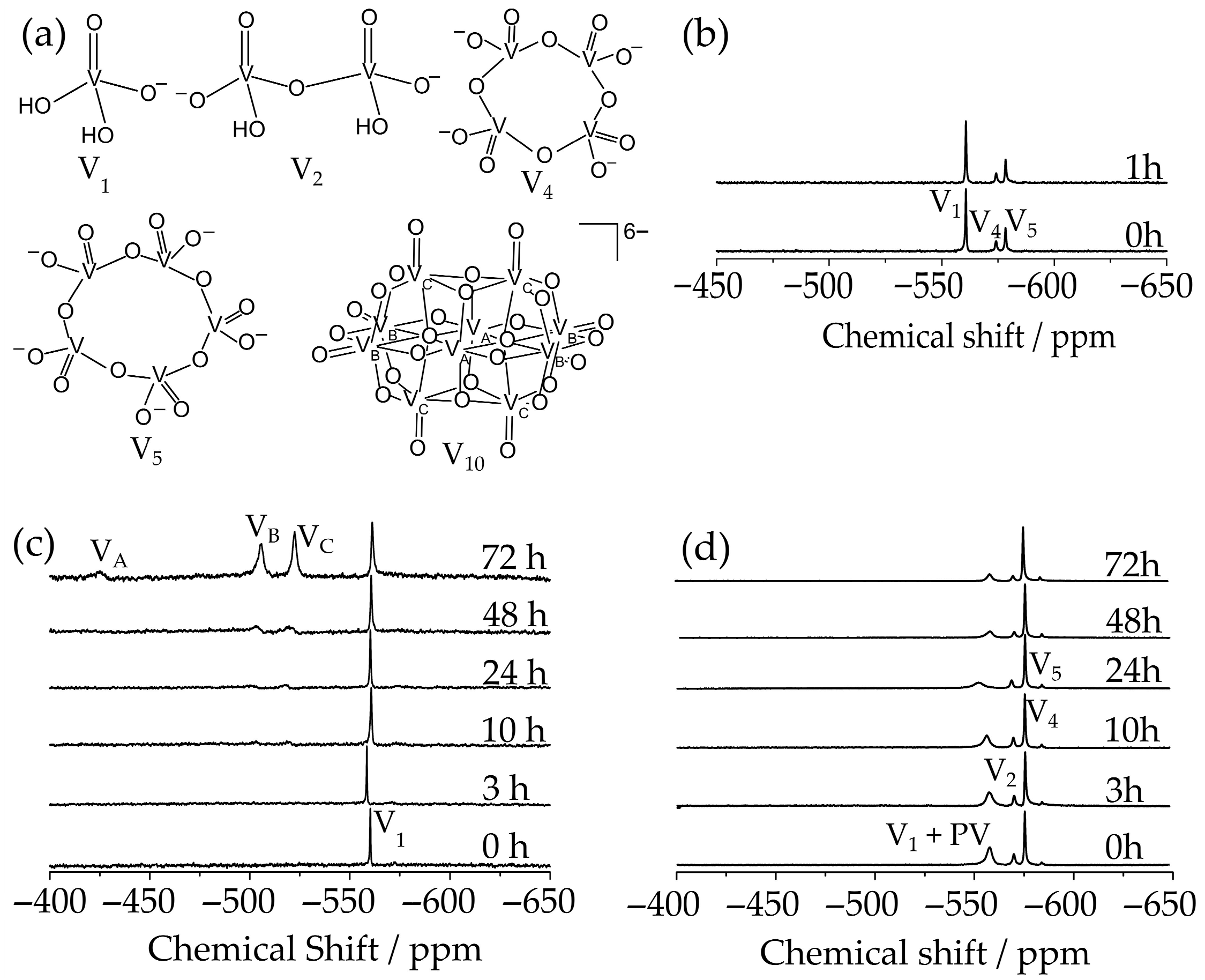
Figure 2. 51V NMR spectra recorded at room temperature of 1.0 mM V15 solutions at different times. In (a) structure of the vanadium species. In (b), a freshly prepared solution in enzymatic medium and after incubation for 1 h; (c) water/D2O (9:1), pH 6.3, and (d) in RPMI (pH 7.4). In (c,d), the spectra were registered from freshly prepared solutions and after incubation for 3, 10, 24, 48, and 72 h.
Inorganics 13 00306 g002
Figure 3. UV/Vis/NIR absorption spectra of V15 solutions at room temperature. The 1.0 mM solutions were incubated for 24, 48, 72, and 168 h: (a) in water; (b) in enzymatic medium; (c) in RPMI medium (pH 7.4).
Inorganics 13 00306 g003
Figure 4. (a) Inhibition of Ca2+-ATPase activity. (b) Lineweaver-Burk plot of Ca2+-ATPase activity in the absence (blue) and in the presence (orange) of 15 µM de V15.
Inorganics 13 00306 g004
Figure 5. Ball and stick structures and ESP map of the anions (a) [Nb10O28]6− (Nb10) (b) [MnV13O38]7− (MnV13) (c) [V10O28]6− (V10) (d) [Cl@VV7VIV8O36]6− (V15) (e) [VV14O38(PO4)]9− (PV14 ox).
Inorganics 13 00306 g005
Figure 6. Evaluation of the cytotoxic effect on (a) HB4a, (b) MCF-7, and (c) MDA-MB-231 cell lines at different concentrations of V15 at 24 h. The graphs show the dose–response inhibition (IC50 determination) for all the cell lines, with the R-squared and linear curve fitting.
Inorganics 13 00306 g006
Figure 8. SEM images for V15 in MDA-MB-231 cell line, at magnifications of 500×, 2500×, 5000×, and 10,000× for (a) control; (b) concentration of 4.30 µM, and (c) 8.60 µM.
Inorganics 13 00306 g008
Figure 9. Evaluation of relative gene expression in the MDA-MB-231 line treated with 17.2 µM of V15 for 48 h. (a) Relative necroptosis gene expression and (b) relative apoptosis gene expression, compared to the constitutive gene GAPDH. The statistical treatment is compared to the control, ns (non-significant), * p < 0.01; ** p < 0.001; *** p < 0.0001.
Inorganics 13 00306 g009
Figure 10. Compilation of the results obtained for V15 in this work: (a) Inhibition of Ca2+-ATPase activity; (b) Cytotoxicity to breast cancer cell lines MCF-7 and MDA-MB-231; (c) Inhibition of MDA-MB-231 cell migration; (d) Potentially reverse epithelial–mesenchymal transition, and (e) Showed necroptosis mechanism of cell death.
Inorganics 13 00306 g010
Table 1. Surface maxima and minima of ESP (Emax and Emin, respectively), ΔE, and the volume enclosed by the calculated isosurface (Visosurface 0.001 e/bohr3) and charge density (q/m).
ParameterNb10MnV13V10V15PV14 ox
IC50 μM35311514.25
Emax/kcal mol−1−323.31−350.29−329.76−306.01−439.53
Emin/kcal mol−1−382.80−468.82−412.55−354.20−562.68
ΔE/kcal mol−159.5118.582.7948.19123.1
Visosurface3629.94558.32744.23790.49835.19
q/m0.60.540.60.40.64
(q/m)/Visosurface/10–49.5249.6448.0625.0607.697
ΔE = Emax − Emin; q/m = is expressed as charge (q) of the POM divided by its number of metal atoms (m); Visosurface = volume of electron density in the POM.
Table 2. IC50 values determined for V15 regarding Ca2+-ATPase inhibition, breast cancer cell lines (MCF-7 and MDA-MB-231), and normal breast cell line (HB4a) viabilities, after 24 h of incubation.
AssayIC50 (µM)
Ca2+-ATPase14.2
HB4a1.02
MCF-7 15.1
MDA-MB-231 17.2
Disclaimer/Publisher’s Note: The statements, opinions and data contained in all publications are solely those of the individual author(s) and contributor(s) and not of MDPI and/or the editor(s). MDPI and/or the editor(s) disclaim responsibility for any injury to people or property resulting from any ideas, methods, instructions or products referred to in the content.