Abstract
Lanthanide rare earth elements possess significant promise for material applications owing to their distinctive optical and magnetic characteristics, as well as their versatile coordination capabilities. This study introduced a lanthanide-functionalized magnetic nanocellulose composite (NNC@Fe3O4@La(OH)3) for effective phosphorus/nitrogen (P/N) ligand separation. The hybrid material employs the adaptable coordination geometry and strong affinity for oxygen of La3+ ions to show enhanced DNA-binding capacity via multi-site coordination with phosphate backbones and bases. This study utilized cellulose as a carrier, which was modified through carboxylation and amination processes employing deep eutectic solvents (DES) and polyethyleneimine. Magnetic nanoparticles and La(OH)3 were subsequently incorporated into the cellulose via in situ growth. NNC@Fe3O4@La(OH)3 showed a specific surface area of 36.2 m2·g−1 and a magnetic saturation intensity of 37 emu/g, facilitating the formation of ligands with accessible La3+ active sites, hence creating mesoporous interfaces that allow for fast separation. NNC@Fe3O4@La(OH)3 showed a significant affinity for DNA, with adsorption capacities reaching 243 mg/g, mostly due to the multistage coordination binding of La3+ to the phosphate groups and bases of DNA. Simultaneously, kinetic experiments indicated that the binding process adhered to a pseudo-secondary kinetic model, predominantly dependent on chemisorption. This study developed a unique rare-earth coordination-driven functional hybrid material, which is highly significant for constructing selective separation platforms for P/N-containing ligands.
1. Introduction
Lanthanide elements (Ln) consist of 15 metals ranging from lanthanum (La) to lutetium (Lu), all of which have highly similar chemical properties [1,2]. Because of their unusual internal 4f electron structures, which give them unique optical, catalytic, and magnetic capabilities, lanthanide elements hold a prominent place in inorganic chemistry [2]. Furthermore, because of their distinct electron localization, varied oxidation states, and adaptable coordination skills, lanthanide ions can combine with a variety of ligands to generate complexes that have potential uses in chemical catalysis, biosensing, energy development, and high-resolution imaging.
Lanthanide ions (Ln3+) exhibit an excellent coordination capacity with oxygen and nitrogen atoms; hence, molecules with O and N donor atoms are frequently selected as ligands for the synthesis of lanthanide complexes [3,4]. For instance, ligands with imine, amide, carboxylate, or phosphate groups can readily bind with Ln3+. Lanthanide elements have a higher affinity for polydentate ligands because of their high coordination number (8–12 in solution) [5]. Recent studies have demonstrated exceptional binding affinities between Ln and biomolecules [6], such as peptides, proteins, and nucleic acids. Ln3+ can form stable complexes with peptides and stabilize proteins, potentially substituting for Ca2+ in biological systems [7]. Furthermore, Ln may both sense or identify nucleic acid molecules and attach to the phosphate ester bonds of nucleic acids, demonstrating considerable hydrolytic activity. Therefore, the integration of lanthanide metals with biomolecules to create innovative functional materials utilizing rare earth elements has emerged as a focal point in inorganic and materials chemistry research.
With the rapid development of nanotechnology, inorganic nanoparticles and nanocomposites have found widespread application in various fields of modern industry due to their regular shapes, hard textures, biodegradability, and surface functionalization. The design and synthesis of lanthanide metal coordination nanoparticles are essential for the utilization of lanthanide elements. Lanthanide metal nanoparticles generally manifest as oxides or hydroxides [8]. Nonetheless, their elevated surface energy renders them susceptible to agglomeration, leading to hindering stability and controllability, which considerably obstructs their practical applicability [8,9]. It is simple to produce a stable structure by fixing metal nanoparticles onto a substrate through in situ growth. A great carrier platform for creating organic-inorganic hybrid materials is cellulose, a naturally occurring, renewable, hydroxyl-rich biopolymer. Nano-cellulose, in particular, has a high specific surface area, multi-level pore structure, and is simple to functionalize [10]. Cellulose has been shown to improve the stability and effectiveness of metal nanoparticles [11]. Nowadays, a lot of research has been performed on hybrid cellulose composites such as TiO2, CuO, ZnO, and Fe3O4 [12]. Furthermore, the integration of inorganic or organic materials with magnetic nanoparticles can enhance the recyclability and manipulability of compliant materials, offering novel insights for the advancement of sustainable functional materials.
Deoxyribonucleic acid (DNA) is not only a crucial hereditary material but also functions as a notable ligand for various metal ions [13,14]. Its interaction with Ln3+ transpires via both the phosphate backbone and the bases. The phosphate groups in DNA can interact with Ln3+ by electrostatic forces, whereas the nitrogenous ligands from the bases can create complexes with Ln3+ nucleotides, especially with adenosine or guanosine phosphates [15,16,17]. Considering the robust affinity of Ln3+ for DNA, exploiting the coordination benefits of rare earth elements to construct inorganic–organic hybrid nanomaterials can enhance comprehension of the coordination dynamics between rare earth metals and biomolecules.
This research employed a sustainable and efficient approach to create a controlled inorganic–organic hybrid nanocomposite material consisting of lanthanum-doped magnetic nano-cellulose. The production, structure, and specific interaction with DNA were thoroughly examined. The binding mechanism between lanthanum-doped magnetic nano-cellulose and biomolecules was clarified through adsorption analysis and structural confirmation, providing mechanistic insights for the development of innovative inorganic–organic composite materials.
2. Results and Discussion
2.1. Structure and Characteristics of Lanthanum Hybrid Magnetic Nanocellulose
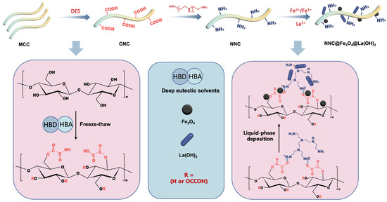
A schematic diagram of the rare earth metal-hybrid magnetic nano-cellulose composite material (NNC@Fe3O4@La(OH)3) preparation procedure is presented in Figure 1. To create carboxylated cellulose, microcrystalline cellulose (MCC) is first treated with a deep eutectic solvent to shrink it and change some of its hydroxyl groups into carboxyl groups (CNC). After that, polyethyleneimine is used to create amino-modified cellulose (NNC). Lastly, rare earth metal hybrid magnetic nano-cellulose composite materials are created by using metal salts to create a metal-hybrid structure on the cellulose surface in situ in an alkaline environment.
Figure 1.
Schematic illustration of the NNC@Fe3O4@La(OH)3 preparation.
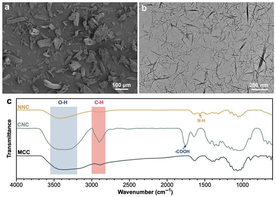
Figure 2a,b display the micrographs of microcrystalline cellulose and nano-cellulose. Figure 2a illustrates the original microcrystalline cellulose, characterized by greater dimensions and an irregular shape, with a diameter of roughly 50 μm. Figure 2b illustrates nanocellulose derived from treatment with deep eutectic solvent (DES). The image illustrates a notable decrease in cellulose size, suggesting that the cellulose may have undergone partial dissolution and subsequent reprecipitation, resulting in the formation of finer fibers. Figure 2c presents the infrared spectrum, revealing alterations in the cellulose structure. MCC displays the conventional cellulose structure, whereas CNC reveals a distinctive peak of carboxyl groups at 1730 cm−1. In NNC, the distinctive peak of carboxyl groups vanishes, but a characteristic peak for -NH2 at 1600 cm−1 emerges, indicating additional chemical alteration and the successful incorporation of amino groups [18].
Figure 2.
(a) SEM of microcrystalline cellulose; (b) TEM of carboxylated cellulose; (c) FTIR of MCC, CNC, and NNC.
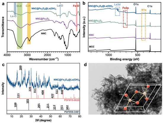
Figure 3a displays the infrared spectrum of metal-hybridized NNC, highlighting alterations in chemical structure throughout the preparation process. NNC@Fe3O4 displays a pronounced Fe-O vibration at 580 cm−1 in contrast to NNC. The introduction of La(OH)3 results in a peak at 1383 cm−1 (La-O) and broadens the O-H stretching band at 3609 cm−1, indicating the presence of hydroxyl groups in La(OH)3. Infrared data further validate the association of La with NNC@Fe3O4. Figure 3b illustrates the chemical composition and characteristic peaks of binding energy corresponding to the electronic energy levels of MCC, NNC, NNC@Fe3O4, and NNC@Fe3O4@La(OH)3. All samples display distinct C1s signals from C-C, C-O, and C=O bonds in cellulose, along with notable O1s signals from hydroxyl or carboxyl groups in cellulose molecules [18]. The N1s peak (400 eV) is attributed to nitrogen derived from amino functional groups in NNC. The Fe 2p peak at 710 eV is indicative of iron derived from the magnetic nanoparticles Fe3O4. The detection of Fe 2p signals in NNC@Fe3O4 and NNC@Fe3O4@La(OH)3 signifies the effective integration of Fe3O4; La 3d (834.7 eV) pertains to lanthanum, derived from La(OH)3 nanoparticles, confirming the successful inclusion of La [19]. Figure 3c illustrates the X-ray diffraction pattern of NNC@Fe3O4@La(OH)3. Cellulose displays a specific diffraction peak at 2θ = 22.6°, indicative of its crystalline structure [20]. NNC@Fe3O4@La(OH)3 exhibits distinct peaks at 30.1° (220), 35.5° (311), 43.1° (400), 53.7° (422), 57.0° (511), and 62.6° (440), predominantly ascribed to Fe3O4 (PDF#19-0629), thereby validating the spinel crystal structure. NNC@Fe3O4@La(OH)3 displays specific crystalline peaks at 15.3° (100), 27.8° (101), 39° (201), 47.9° (211), and 54.5° (112), aligning with the crystalline peaks of La(OH)3 (PDF#36-1481). The crystal structure, characteristic of rare earth hydroxides, is part of the hexagonal crystal system and exhibits a rod-like configuration (Figure 3d), signifying that lanthanum has effectively integrated with cellulose as La(OH)3 [21].
Figure 3.
(a) FTIR and (b) XPS of modified cellulose and NNC@Fe3O4@La(OH)3; (c) XRD and (d) TEM of NNC@Fe3O4@La(OH)3.
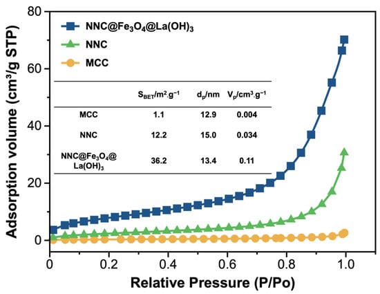
The specific surface area of nanoparticles plays a crucial role in determining the adsorption performance of materials. Figure 4 illustrates the BET isotherm spectra for MCC, NNC, and NNC@Fe3O4@La(OH)3. The specific surface area of MCC measures 1.1 m2·g−1. In contrast, NNC has a specific surface area of 12.2 m2·g−1, largely due to the smaller size of NNC particles, which are usually stored in liquid form. Upon drying, these particles tend to agglomerate, resulting in an increase in size without a corresponding rise in specific surface area. When compared to NNC, the specific surface area of NNC@Fe3O4@La(OH)3 rises to 36.2 m2·g−1, primarily attributed to the deposition of Fe3O4 and La(OH)3, which enhances the roughness of the cellulose structure. In addition, the pore diameter (dp) of MCC, NNC, and NNC@Fe3O4@La(OH)3 are mesoporous structures, and the pore volume (Vp) of NNC@Fe3O4@La(OH)3 is significantly increased compared to cellulose without rare earth metals. Therefore, NNC@Fe3O4@La(OH)3 has a high specific surface area and high pore volume, which is favorable for DNA adsorption.
Figure 4.
BET of cellulose and NNC@Fe3O4@La(OH)3.
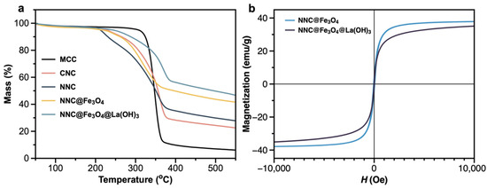
The thermal stability of materials is intricately linked to their structural composition. Figure 5a illustrates the variation in mass with temperature during the heating process for MCC, CNC, NNC, NNC@Fe3O4, and NNC@Fe3O4@La(OH)3. MCC exhibits excellent thermal stability up to 320 °C; however, it undergoes rapid decomposition at approximately 350 °C, resulting in a char yield of less than 10%. The thermal stability of CNC and NNC is lower than that of MCC, mainly because of the reduced cellulose particle size and the presence of carboxyl and amino side chains, which increase their vulnerability to thermal decomposition. Nonetheless, NNC shows an increased char residue rate (30%), mainly due to the incorporation of amino groups, which could modify the thermal decomposition pathway of cellulose [18]. Furthermore, amino groups produce non-reactive flame-retardant gases when subjected to thermal decomposition, thereby improving the char formation potential of NNC. The composite NNC@Fe3O4@La(OH)3 demonstrates enhanced thermal stability and an increased char yield (47%) in comparison to NNC, mainly attributed to the physical barrier created by the metal oxide coating on the cellulose surface. NNC@Fe3O4@La(OH)3 exhibits superior thermal stability compared to NNC@Fe3O4, suggesting that the incorporation of La(OH)3 results in a denser and more stable structure.
Figure 5.
(a) TG and (b) VSM of cellulose and NNC@Fe3O4@La(OH)3.
Magnetic nanoparticles provide swift recovery, the ability to be reused, and adaptable handling capabilities. This investigation incorporates Fe3O4 into the composite material to improve its manipulability. Figure 5b illustrates the hysteresis loops of NNC@Fe3O4 and NNC@Fe3O4@La(OH)3, with both demonstrating characteristic S-shaped hysteresis loops. While the magnetic induction of NNC@Fe3O4@La(OH)3 is marginally lower than that of NNC@Fe3O4, both demonstrate zero coercivity and remanence, signifying that they both exhibit superparamagnetic behavior.
2.2. Adsorption Behavior and Kinetics
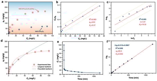
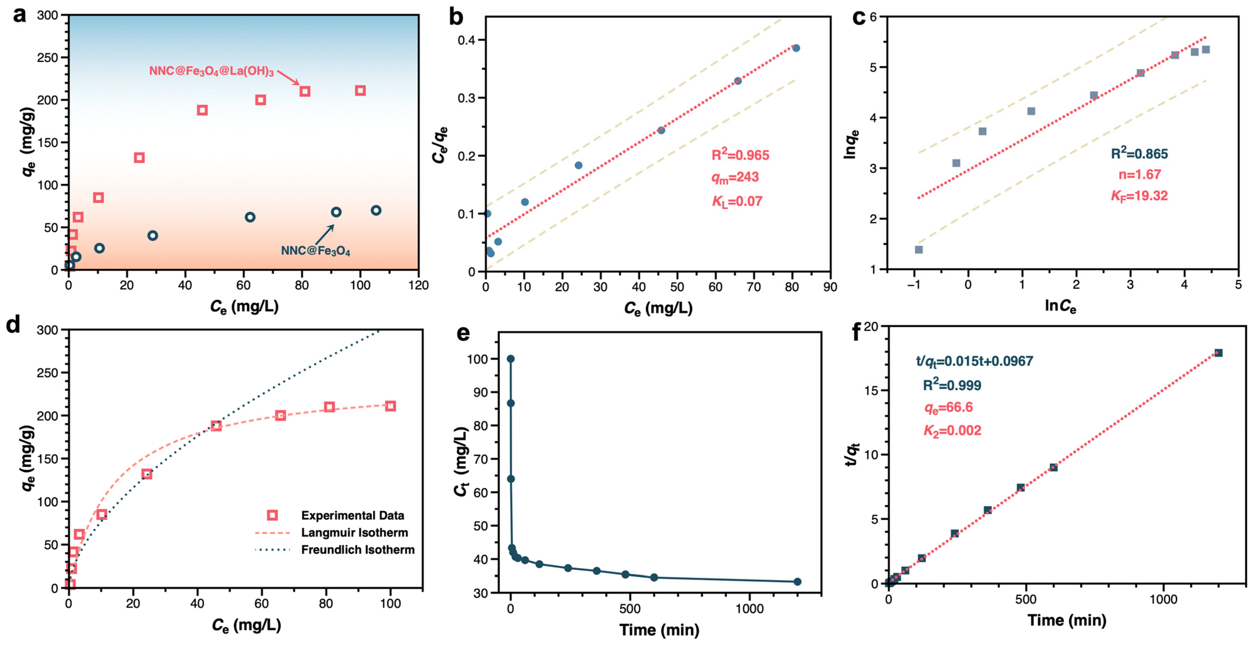
To explore the interaction behavior between lanthanum hybrid magnetic nano-cellulose nanocomposites and DNA, adsorption isotherm tests for DNA adsorption were performed on NNC@Fe3O4 and NNC@Fe3O4@La(OH)3 at 25 °C (Figure 6a). The DNA adsorption capacity of NNC@Fe3O4 was found to be 67 mg/g, primarily due to the electrostatic attraction between the positively charged amino groups of NNC and the negatively charged DNA. In contrast, for NNC@Fe3O4@La(OH)3, the maximum adsorption capacity increased to 211 mg/g, suggesting that the incorporation of rare earth elements significantly improves DNA adsorption performance. The enhancement in adsorption capacity of the NNC@Fe3O4@La(OH)3 composite is ascribed to the active La(OH)3, which improves the specific surface area and offers more active adsorption sites [22]. In addition, compared with DNA adsorption materials reported in the literature (Table S1), NNC@Fe3O4@La(OH)3 has a higher DNA adsorption capacity.
Figure 6.
(a) Adsorption isotherms; (b) Langmuir model: Ce/qe vs. Ce plot; (c) Freundlich model: lnqe vs. lnCe plot; (d) curve fitting of Langmuir model and Freundlich model; (e) kinetics of the adsorption process; (f) pseudo-second-order kinetics of NNC@Fe3O4@La(OH)3.
To enhance the understanding of the adsorption mechanism of NNC@Fe3O4@La(OH)3, two established isotherm models—Langmuir (Equation (S3)) and Freundlich (Equation (S4))—were employed to analyze the adsorption isotherm data [23]. The parameters qm and kL were calculated from the intercept and slope of the linear plot of Ce/qe against Ce (Figure 6b). The Langmuir isotherm indicates that the maximum adsorption capacity of NNC@Fe3O4@La(OH)3 for DNA is 243 mg/g. Furthermore, the n value derived from the Freundlich model was 1.67 (Figure 6c), which lies within the favorable range of 1 to 10, suggesting that the adsorption process is indeed beneficial [22,24]. The results and analysis suggest that both the Langmuir and the Freundlich isotherm models demonstrate the NNC@Fe3O4@La(OH)3 composite’s strong adsorption affinity for DNA (Figure 6d). The data presented in Figure 6b,c reveal that the correlation coefficient for the Langmuir model (0.965) surpasses that of the Freundlich model. This result indicates a dominance of surface adsorption sites, implying that chemical adsorption plays a significant role. Lanthanum engages in multi-coordinate interactions with the phosphate backbone or bases of DNA, leading to adsorption behavior that aligns with single-layer adsorption. The saturation capacity of monolayer adsorption is influenced by the availability of surface sites for coverage [24]. NNC@Fe3O4@La(OH)3 provides an increased number of active sites and a greater specific surface area.
In order to clarify the adsorption mechanism of DNA on the NNC@Fe3O4@La(OH)3 composite material, both pseudo-first-order (Equation (S5)) and pseudo-second-order kinetic (Equation (S6)) models were utilized to analyze the experimental data [22,23]. Figure 6e,f illustrate the kinetics of the adsorption process and fitting plots for the pseudo-second-order model, respectively. The pertinent parameters identified from the previously discussed kinetic models are illustrated in Figure 6f and Figure S1. The R2 values of the two fitting equations indicate that the correlation coefficient for the first-order kinetic fitting is relatively low, and there is a significant difference between the equilibrium adsorption capacity obtained from the fitting and the experimental equilibrium adsorption capacity. The second-order kinetic fitting values align well with the experimental data, showing a high correlation coefficient for DNA adsorption on NNC@Fe3O4@La(OH)3 (R2 > 0.999). Consequently, the second-order model most accurately characterizes the adsorption behavior of NNC@Fe3O4@La(OH)3. Furthermore, the theoretical values of qe derived from the second-order model show greater alignment with the experimental values obtained. The findings suggest that the adsorption behavior of NNC@Fe3O4@La(OH)3 adheres to the second-order kinetic model. The results and analysis indicate that the adsorption of DNA by NNC@Fe3O4@La(OH)3 is mainly influenced by chemisorption via coordination.
2.3. Mechanism Analysis
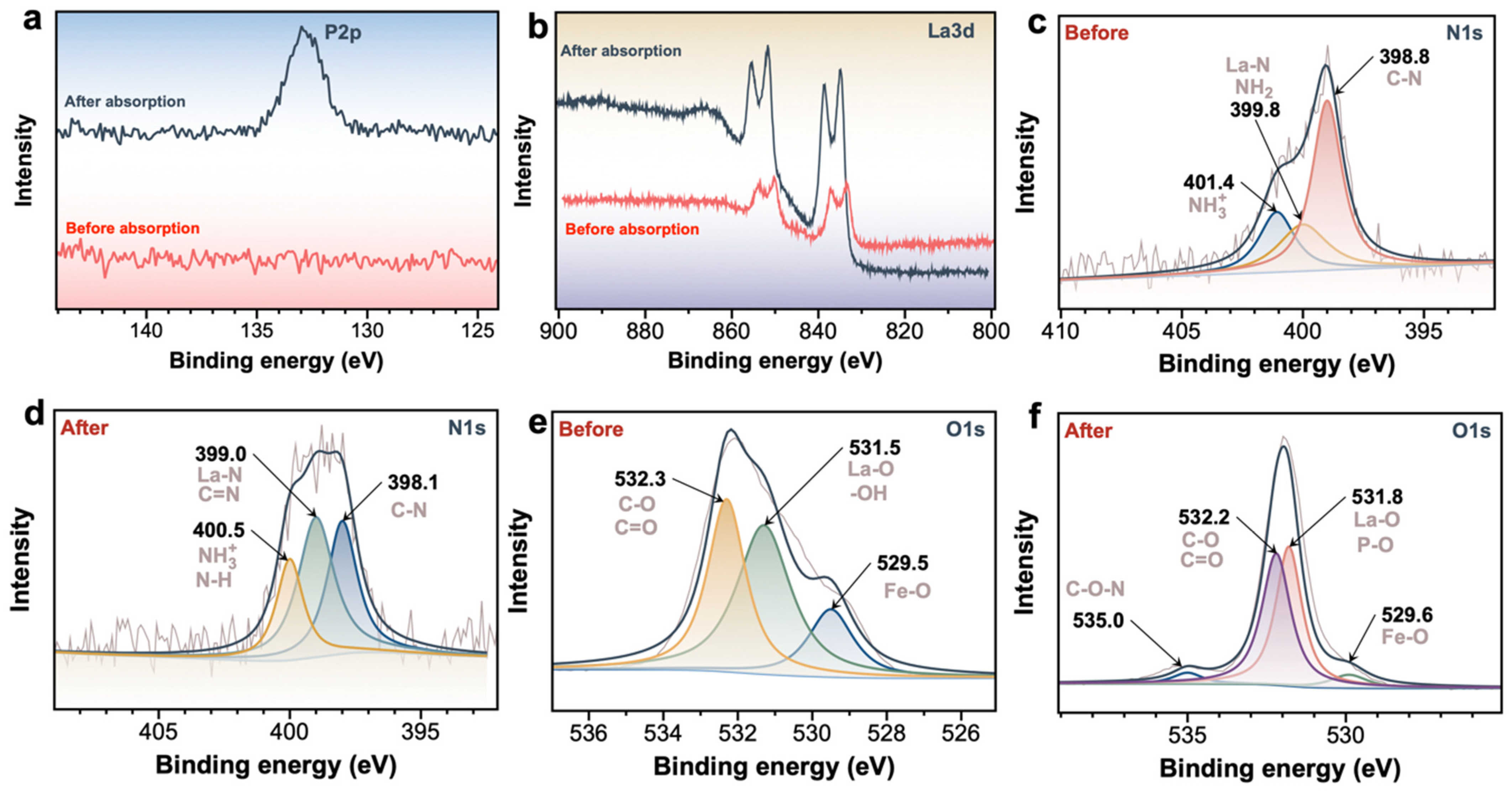
To elucidate the coordination behavior of NNC@Fe3O4@La(OH)3 with respect to DNA, XPS was utilized to examine the interaction between NNC@Fe3O4@La(OH)3 and DNA. DNA primarily consists of a phosphate backbone, sugar molecules, and nitrogenous bases. Figure 7a presents the XPS P2p curves of NNC@Fe3O4@La(OH)3 prior to and following DNA adsorption. Figure 7a illustrates that following DNA adsorption, a new element P emerged at a binding energy of 133.2 eV, suggesting a strong attachment of DNA to NNC@Fe3O4@La(OH)3. Furthermore, as illustrated in Figure 7b, the binding energy of La3d displays a red shift following adsorption, suggesting that NNC@Fe3O4@La(OH)3 has experienced significant coordination interactions with the phosphate backbone nucleobases. This interaction modifies the chemical environment of La3d, resulting in alterations to its binding energy [25]. Figure 7c,d illustrate the XPS N1s spectra prior to and following adsorption. Figure 7c clearly indicates that prior to adsorption, the N1s binding energy is predominantly associated with the amino groups on NNC. Following adsorption, the chemical environment of N1s experiences notable alterations, encompassing not just the nitrogen from the bases in DNA but also the emergence of protonated amino groups and La-N bonds (Figure 7d) [25]. This suggests that NNC@Fe3O4@La(OH)3 could demonstrate electrostatic attraction and metal coordination interactions with DNA [4,5]. Furthermore, Figure 7e,f illustrate the XPS O1s spectra prior to and following adsorption. In the pre-adsorption phase, the O1s binding energy predominantly relates to the C-O bonds in cellulose, the Fe-O bonds in Fe3O4, and the La-O bonds in La(OH)3. Following DNA adsorption, the binding energy at 535.5 eV reveals the C-O-N structure of DNA, highlighting the interaction between NNC@Fe3O4@La(OH)3 and DNA.
Figure 7.
(a) XPS P2p spectral; (b) XPS La3d spectral, (c,d) XPS N1s spectral; (e,f) XPS O1s spectral of NNC@Fe3O4@La(OH)3.
3. Materials and Methods
3.1. Chemicals and Reagents
Microcrystalline cellulose of premium purity (diameter 20–50 μm) was purchased from Sinopharm Chemical Reagents Co., Ltd. (Shanghai, China) Sodium chloride, polyethyleneimine (Mw 10,000), ammonium iron (II) sulfate hexahydrate, ammonium iron (III) sulfate dodecahydrate, aqueous ammonia solution (25−28 wt%), sodium hydroxide, lanthanum nitrate hexahydrate, sodium chloride, polyethylene glycol (PEG 6000), anhydrous ethanol, choline chloride, oxalic acid dihydrate, nickel oxide, hydrochloric acid, and dimethyl sulfoxide are all analytical grade, purchased from Shanghai Macklin Biochemical Technology Co., Ltd. (Shanghai, China) Deoxyribonucleic acid (salmon extract, purity > 90%) was purchased from Shanghai Yuanye Biotechnology Co., Ltd. (Shanghai, China).
3.2. Preparation of Carboxylated Nano-Cellulose (CNC)
First, 3 g of microcrystalline cellulose was incorporated with 30 g of a deep eutectic solvent (DES, with a molar ratio of choline chloride to oxalic acid dihydrate of 1:2) and mixed at 80 °C until a clear and transparent solution was achieved. Subsequently, the mixture was agitated at 400 rpm in a water bath maintained at 95−100 °C for a duration of 2 h. The reaction mixture was ultimately frozen at −20 °C for 24 h and subsequently thawed at ambient temperature. The material was subjected to two washes with anhydrous ethanol and three washes with ultra-pure water until colorless, resulting in carboxylated nano-cellulose (CNC).
3.3. Preparation of Amino-Modified Nanocellulose (NNC)
The 2 g of CNC were dispersed in 100 mL of water. Then, 4 g of polyethyleneimine was added, and the mixture was mixed completely. The pH was adjusted to 1.5, and the mixture was stirred at ambient temperature for 1.5 h. The material was washed three times, freeze-dried for 48 h, and amino-modified cellulose (NNC) was yielded.
3.4. Synthesis of NNC@Fe3O4
First, 6.8 g of ammonium iron (II) sulfate hexahydrate and 10.4 g of ammonium iron (III) sulfate dodecahydrate were dissolved in 500 mL of deionized water. The 4.0 g of NNC were added, and the mixture was exposed to ultrasonic stirring for 30 min. Subsequently, the pH was modified to 10 utilizing a 6 mol/L ammonia solution. After stabilization of the pH, the mixture was mechanically stirred at 50 °C in a water bath for 1 h. Following the reaction, the mixture was allowed to cool to ambient temperature. Following segregation of the solution layers, an external magnetic field was applied to facilitate their separation. The material was rinsed several times with water until neutral, then freeze-dried under vacuum until constant weight was achieved to obtain magnetic nano-cellulose (NNC@Fe3O4).
3.5. Synthesis of NNC@Fe3O4@La(OH)3
First, 1.5 g of magnetic nano-cellulose (NNC@Fe3O4) was dispersed in 100 mL of 10% ethanol, and 5 mmol of lanthanum nitrate hexahydrate was incorporated. The mixture was sonicated for 15 min, stirred at ambient temperature for 2 h, heated to 60 °C, and then 10 mL of a 2 M sodium hydroxide solution was gradually introduced. The reaction was continued for 2 h after the addition. Heating was subsequently discontinued, and the mixture was stirred at room temperature for 24 h. Magnetic separation, washing, and vacuum drying were then performed, resulting in La(OH)3-hybridized magnetic nanofibers (NNC@Fe3O4@La(OH)3).
3.6. Characterization
Scanning electron microscopy (SEM, SU1510 model, Hitachi, Tokyo, Japan) and transmission electron microscopy (TEM, JEM-2100 model, JEOL, Tokyo, Japan) were used to examine sample structures. A Fourier transform infrared spectrometer (FTIR, Vertex 70 model, Molecular Devices, San Jose, CA, USA) was used for infrared examination, having a scanning range of 4000–400 cm−1. The material surface’s elemental composition and chemical state were examined using X-ray photoelectron spectroscopy (XPS, Thermo Fisher Scientific, Waltham, MA, USA). X-ray diffraction (XRD) analysis was performed by XRD-7000 (Suzhou Sailiwei, Suzhou, China). For testing, samples were placed in XRD plates with a 10–80° scanning range and 5°/min scanning speed. A thermal gravimetric analyzer (TG209F3 model, Netzsch, Selb, Germany) was used to evaluate the material’s thermal stability. The sample mass was 10 mg, and the testing temperature was 50–800 K in nitrogen. We used a specialized surface area tester (ASAP2460 model, Micromeritics, Norcross, GA, USA) to analyze surface area. The saturation magnetization of samples was measured at 300 K using a vibrating sample magnetometer (VSM, 4HF model, ADE, Chicago, IL, USA) with a magnetic field strength of ±10,000 Oe. The resulting magnetization curves were plotted to evaluate the material’s characteristics.
3.7. Preparation of DNA Solution
Salmon sperm DNA was used as the model compound to assess the separation effectiveness of NNC@Fe3O4@La(OH)3 toward DNA in order to examine the DNA capture capability of metal-hybrid magnetic chitosan. To create DNA standard solutions with varying quantities, salmon sperm DNA was first dissolved in sterile deionized water.
3.8. Adsorption Behavior of NNC@Fe3O4@La(OH)3
In a 1.5 mL EP tube, 900 μL of binding buffer (10 wt% PEG, 2 M NaCl, pH = 4.0), 100 μL of an aliquot of the pre-determined concentration DNA solution, and 1 mg NNC@Fe3O4@La(OH)3 were combined. After that, the mixture was shaken sporadically for 10 min at room temperature. An external magnet is then used to accomplish magnetic separation. A Nanodrop 2000 UV detector (Thermo Fisher Scientific, Waltham, MA, USA) is used to determine the DNA concentration at 260 nm after the supernatant has been thoroughly removed. The difference between the concentration of DNA before and after separation is used to compute the yield of DNA extraction. The adsorption capacity of DNA was determined by the following Equation (S1) (Refer to Supplementary Materials).
3.9. Kinetics of Adsorption
The 5 mg of NNC@Fe3O4@La(OH)3, 500 μL of a 30 mg/mL DNA standard solution, and 4.5 mL of binding buffer (10 wt% PEG, 2 M NaCl, pH = 4.0) were put into a 5 mL centrifuge tube. And the mixture was shaken. Samples were collected at the specified adsorption periods. The volume of each sample was roughly 10 μL. Equation (S2) was used to determine the DNA adsorption capacity qt at various adsorption times.
4. Conclusions
This study achieved the successful synthesis of a novel lanthanum (III)-functionalized magnetic nano-cellulose hybrid material (NNC@Fe3O4@La(OH)3). The systematic characterization demonstrated that La3+ effectively hybridized with magnetic nano-cellulose, showing a high specific surface area and superparamagnetism. The findings from the adsorption studies indicate that the material demonstrates a remarkable affinity for DNA, achieving a maximum adsorption capacity of 243 mg/g. The fundamental mechanism is related to the chemical adsorption arising from the strong P/N coordination of La3+. This study highlights the considerable promise of rare earth ions (La3+) in the creation and advancement of innovative inorganic hybrid materials with targeted element recognition and capture capabilities. It offers new materials and theoretical insights that could broaden the use of rare earth-based materials in fields such as separation science and the synthesis of inorganic–organic functional molecular materials.
Supplementary Materials
The following supporting information can be downloaded at: https://www.mdpi.com/article/10.3390/inorganics13080257/s1, Figure S1. The pseudo-first-order kinetic of NNC@Fe3O4@La(OH)3; Table S1. Adsorption effects of different materials on DNA. References [26,27,28,29,30] are cited in Supplementary Materials.
Author Contributions
J.Y.: Writing—Original draft preparation, Data curation, Methodology, Formal analysis. J.F.: Writing—review & Formal analysis. H.W.: Project administration, Funding acquisition. Y.L.: Conceptualization, Writing—review & editing. All authors have read and agreed to the published version of the manuscript.
Funding
This research was supported by the National Key Research and Development Program of China (2021YFF0602801). “Pioneer” and “Leading Goose” R&D Program of Zhejiang (2022C02023).
Institutional Review Board Statement
Not applicable.
Informed Consent Statement
Not applicable.
Data Availability Statement
Data is contained within the article and Supplementary Materials.
Conflicts of Interest
The authors declare that the research was conducted in the absence of any commercial or financial relationships that could be construed as a potential conflict of interest.
References
- Youssef, H.; Sedykh, A.E.; Becker, J.; Taydakov, I.V.; Mueller-Buschbaum, K. 3-(2-Pyridyl)pyrazole Based Luminescent 1D-Coordination Polymers and Polymorphic Complexes of Various Lanthanide Chlorides Including Orange-Emitting Cerium(III). Inorganics 2022, 10, 10120254. [Google Scholar] [CrossRef]
- Zhao, C.; Sun, Y.; Ren, J.; Qu, X. Recent progress in lanthanide complexes for DNA sensing and targeting specific DNA structures. Inorganica Chim. Acta 2016, 452, 50–61. [Google Scholar] [CrossRef]
- Chan, M.; Doan, H.; Dang-Vu, T. An Investigation of Lanthanum Recovery from an Aqueous Solution by Adsorption (Ion Exchange). Inorganics 2024, 12, 12090255. [Google Scholar] [CrossRef]
- Lin, G.; Xiong, Y.; Wang, G.; Li, S.; Jiang, R.; Lu, B.; Chen, Y.; Huang, B. Selective and efficient removal of lanthanum ion in aqueous solution by P-doped porous carbon. J. Water Process Eng. 2023, 55, 104116. [Google Scholar] [CrossRef]
- Kaczmarek, M.T.; Zabiszak, M.; Nowak, M.; Jastrzab, R. Lanthanides: Schiff base complexes, applications in cancer diagnosis, therapy, and antibacterial activity. Coord. Chem. Rev. 2018, 370, 42–54. [Google Scholar] [CrossRef]
- Alexander, C.; Guo, Z.; Glover, P.B.; Faulkner, S.; Pikramenou, Z. Luminescent Lanthanides in Biorelated Applications: From Molecules to Nanoparticles and Diagnostic Probes to Therapeutics. Chem. Rev. 2025, 125, 2269–2370. [Google Scholar] [CrossRef] [PubMed]
- Cho, U.; Chen, J.K. Lanthanide-Based Optical Probes of Biological Systems. Cell Chem. Biol. 2020, 27, 921–936. [Google Scholar] [CrossRef]
- Feng, J.; Zhang, H.J. Hybrid materials based on lanthanide organic complexes: A review. Chem. Soc. Rev. 2013, 42, 387–410. [Google Scholar] [CrossRef]
- Fremy, G.; Raibaut, L.; Cepeda, C.; Sanson, M.; Boujut, M.; Seneque, O. A novel DOTA-like building block with a picolinate arm for the synthesis of lanthanide complex-peptide conjugates with improved luminescence properties. J. Inorg. Biochem. 2020, 213. [Google Scholar] [CrossRef]
- Abolore, R.S.; Jaiswal, S.; Jaiswal, A.K. Green and sustainable pretreatment methods for cellulose extraction from lignocellulosic biomass and its applications: A review. Carbohydr. Polym. Technol. Appl. 2024, 7, 100396. [Google Scholar] [CrossRef]
- Li, Y.; Jiao, H.; Zhang, H.; Wang, X.; Fu, Y.; Wang, Q.; Liu, H.; Yong, Y.-c.; Guo, J.; Liu, J. Biosafety consideration of nanocellulose in biomedical applications: A review. Int. J. Biol. Macromol. 2024, 265, 130900. [Google Scholar] [CrossRef]
- Deng, Y.; Zhu, T.; Cheng, Y.; Zhao, K.; Meng, Z.; Huang, J.; Cai, W.; Lai, Y. Recent advances in functional cellulose-based materials: Classification, properties, and applications. Adv. Fiber Mater. 2024, 6, 1343–1368. [Google Scholar] [CrossRef]
- Pages, B.J.; Ang, D.L.; Wright, E.P.; Aldrich-Wright, J.R. Metal complex interactions with DNA. Dalton Trans. 2015, 44, 3505–3526. [Google Scholar] [CrossRef]
- Qin, Z.x.; Wang, C.y.; Zhang, J.s.; Wang, Z.y.; Wei, Y.; Li, Y.t.; Dai, S.q.; Tay, F.R.; Niu, L.n. DNA-Based Materials Inspired by Natural Extracellular DNA. Adv. Funct. Mater. 2023, 33, 2211669. [Google Scholar] [CrossRef]
- Lee, J.; Lee, Y.; Choi, M.-H.; Ok, K.M.; You, T.-S. Complex Structure, Chemical Bonding, and Electrical Transport Properties of a La-Doped Zintl Phase. Inorganics 2024, 12, 12120333. [Google Scholar] [CrossRef]
- Li, H.; Li, W.; Nie, Z.; Yao, S.Z. A label-free and time-resolved luminescence strategy for the detection of proteins based on DNA-Tb3+ luminescence quenched by graphene oxide. Analyst 2015, 140, 6386–6391. [Google Scholar] [CrossRef]
- Ru, X.-M.; Yang, Z.-Y.; Ran, S.-Y. Lanthanide ions induce DNA compaction with ionic specificity. Int. J. Biol. Macromol. 2022, 210, 292–299. [Google Scholar] [CrossRef] [PubMed]
- Antunes, B.D.; Santana, L.R.; Oliveira, R.M.; Valério, A.; Carreno, N.L.V.; Wolke, S.I.; da Silva, R.; Fajardo, A.R.; Dias, A.R.G.; Zavareze, E.D. Cellulose, cellulose nanofibers, and cellulose acetate from Butia fruits (Butia odorata): Chemical, morphological, structural, and thermal properties. Int. J. Biol. Macromol. 2024, 281, 136151. [Google Scholar] [CrossRef]
- Velinova, R.; Kaneva, N.; Ivanov, G.; Kovacheva, D.; Spassova, I.; Todorova, S.; Atanasova, G.; Naydenov, A. Synthesis and Characterization of Pd/La2O3/ZnO Catalyst for Complete Oxidation of Methane, Propane and Butane. Inorganics 2025, 13, 13010017. [Google Scholar] [CrossRef]
- Salem, K.S.; Kasera, N.K.; Rahman, M.A.; Jameel, H.; Habibi, Y.; Eichhorn, S.J.; French, A.D.; Pal, L.; Lucia, L.A. Comparison and assessment of methods for cellulose crystallinity determination. Chem. Soc. Rev. 2023, 52, 6417–6446. [Google Scholar] [CrossRef]
- Ozturkkan, F.E.; Teymouri, E.; Yuksek, M.; Yildiz, E.A.; Sertcelik, M.; Alemi, A.; Necefoglu, H.; Hokelek, T. Synthesis, structural characterization and determination of the nitro-containing explosives detection potential of supramolecular lanthanum complex and Lanthana nanoparticles. Mater. Sci. Eng. B-Adv. Funct. Solid-State Mater. 2023, 290, 116331. [Google Scholar] [CrossRef]
- Liu, X.; Cheng, W.; Yu, Y.; Jiang, S.; Xu, Y.; Zong, E. Magnetic ZrO2/PEI/Fe3O4 functionalized MWCNTs composite with enhanced phosphate removal performance and easy separability. Compos. Part B-Eng. 2022, 237, 109861. [Google Scholar] [CrossRef]
- Zong, E.; Zhang, C.; Wu, S.; Gao, Y.; Yang, J.; Liu, X.; Song, P. Titanium dioxide nanoparticles functionalized chitosan toward bio-based antibacterial adsorbent for enhanced phosphate capture. Int. J. Biol. Macromol. 2023, 241, 124511. [Google Scholar] [CrossRef]
- Yang, R.D.; Xie, C.; Wan, X.; Li, H.R.; Ge, L.Y.; Li, X.F.; Zhao, G.L. In situ growth of ultrathin covalent triazine frameworks on unmodified cellulose II beads for enhanced dye pollutant removal. Chem. Eng. J. 2024, 498, 155017. [Google Scholar] [CrossRef]
- Zong, E.; Wang, X.; Zhang, L.; Yang, J.; Liu, X. A Recyclable Magnetic Aminated Lignin Supported Zr-La Dual-Metal Hydroxide for Rapid Separation and Highly Efficient Sequestration of Phosphate. Molecules 2023, 28, 28072923. [Google Scholar] [CrossRef] [PubMed]
- Yıldırım, E.; Arıkan, B.; Yücel, O.; Çakır, O.; Kara, N.T.; İyim, T.B.; Gürdağ, G.; Emik, S. Synthesis and characterization of amino functional poly(acrylamide) coated Fe3O4 nanoparticles and investigation of their potential usage in DNA isolation. Chem. Pap. 2022, 76, 5747–5759. [Google Scholar] [CrossRef]
- Xu, K.; Wang, Y.; Zhang, H.; Yang, Q.; Wei, X.; Xu, P.; Zhou, Y. Solid-phase extraction of DNA by using a composite prepared from multiwalled carbon nanotubes, chitosan, Fe3O4 and a poly (ethylene glycol)-based deep eutectic solvent. Microchim. Acta 2017, 184, 4133–4140. [Google Scholar] [CrossRef]
- Pan, X.; Cheng, S.; Su, T.; Zuo, G.; Zhang, C.; Wu, L.; Jiao, Y.; Dong, W. Poly (2-hydroxypropylene imines) functionalized magnetic polydopamine nanoparticles for high-efficiency DNA isolation. Appl. Surf. Sci. 2019, 498, 143888. [Google Scholar] [CrossRef]
- Niu, B.; Zhou, Y.; Wen, T.; Quan, G.; Singh, V.; Pan, X.; Wu, C. Proper functional modification and optimized adsorption conditions improved the DNA loading capacity of mesoporous silica nanoparticles. Colloid. Surface. A 2018, 548, 98–107. [Google Scholar] [CrossRef]
- Zhang, M.; Li, L.; Li, B.; Tian, N.; Yang, M.; Zhang, H.; You, C.; Zhang, J. Adsorption of DNA by using polydopamine modified magnetic nanoparticles based on solid-phase extraction. Anal. Biochem. 2019, 579, 9–17. [Google Scholar] [CrossRef]


Disclaimer/Publisher’s Note: The statements, opinions and data contained in all publications are solely those of the individual author(s) and contributor(s) and not of MDPI and/or the editor(s). MDPI and/or the editor(s) disclaim responsibility for any injury to people or property resulting from any ideas, methods, instructions or products referred to in the content. |
© 2025 by the authors. Licensee MDPI, Basel, Switzerland. This article is an open access article distributed under the terms and conditions of the Creative Commons Attribution (CC BY) license (https://creativecommons.org/licenses/by/4.0/).