Review

Metal Complexes with Hydroxyflavones: A Study of Anticancer and Antimicrobial Activities

by
Ljiljana E. Mihajlović
1,
Monica Trif
2 and
Marijana B. Živković
3,*
1
Innovation Center of the Faculty of Chemistry, Studentski trg 12–16, 11158 Belgrade, Serbia
2
Centre for Innovative Process Engineering (CENTIV) GmbH, Boschstr. 8–10, 28857 Syke, Germany
3
Department of Chemistry, Institute of Chemistry, Technology and Metallurgy, National Institute of the Republic of Serbia, University of Belgrade, Studentski trg 12–16, 11000 Belgrade, Serbia
*
Author to whom correspondence should be addressed.
Inorganics 2025, 13(8), 250; https://doi.org/10.3390/inorganics13080250
Submission received: 30 June 2025 / Revised: 13 July 2025 / Accepted: 18 July 2025 / Published: 22 July 2025

Abstract

Metal chelation to bioactive small molecules is a well-established strategy to enhance the biological activity of the resulting complexes. Among the widely explored structural motifs, the combination of prominent metal centers with naturally inspired derivatives has attracted considerable attention. One such promising platform is the flavone scaffold, derived from flavonoids and studied since ancient times. Flavones are plant-derived compounds known for their diverse biological activities and health benefits. They exhibit significant structural variability, primarily through backbone modifications such as hydroxylation. Importantly, coordination of metal ions to hydroxylated flavone cores often improves their natural bioactivities, including anticancer and antimicrobial effects. In this review, we summarize transition metal complexes incorporating hydroxyflavone (OH–F) ligands reported over the past 15 years. We provide a concise overview of synthetic approaches and structural characterization, with a particular emphasis on coordination modes (e.g., maltol-type, acetylacetonate-type, catechol-type, and others). Furthermore, we discuss biological evaluation results, especially anticancer and antimicrobial studies, to highlight the therapeutic potential of these complexes. Finally, we suggest directions for the future development of metal-based agents bearing hydroxyflavone moieties through several critical points in terms of the accuracy, reproducibility, and relevance of biological studies involving metal-based compounds.

Graphical Abstract

1. Introduction

According to the World Health Organization (WHO), cancer remains one of the leading causes of mortality, with nearly 10 million deaths reported worldwide in 2020, accounting for approximately one in six deaths [1]. The rapid mutation of cancer cells and the development of resistance to existing therapies represent major obstacles to effective treatment [2,3]. Furthermore, anticancer drugs often exhibit a limited selectivity for malignant cells over normal cells, resulting in systemic toxicity, adverse side effects, and damage to healthy tissues and organs.
Another pressing global health concern is the growing resistance to antimicrobial agents. The uncontrolled and excessive use of antibiotics may potentially return us to a time when infectious diseases were frequently fatal due to the absence of effective treatment options [4].
One promising strategy to address both anticancer and antimicrobial resistance issues involves the rational design of novel compounds through the combination of different molecular fragments. Such an approach can lead to synergistic effects, improving therapeutic selectivity while minimizing toxicity. Notably, a substantial proportion of recently approved drugs are inspired by natural products and their structural motifs [5]. Evidence from the literature indicates that researchers have been investigating the therapeutic potential of metal complexes incorporating nature-inspired ligands, such as flavones, for several decades [6,7].
Flavones are natural compounds composed of three six-membered rings: two benzene rings (A and B) and a γ-pyrone ring (C). Benzene ring A is fused to the heterocyclic oxygen-containing ring C, while benzene ring B is attached to ring C at position C-2 (Figure 1).
Hydroxyflavones (OH–F) are characterized by the presence of one or more hydroxyl groups within their molecular structure. Figure 2 illustrates representative OH–F compounds selected from the scientific literature that will be examined in the present review.
OH–F are biosynthesized through the shikimate pathway, which ultimately leads to the formation of the aromatic amino acid phenylalanine (Figure 3). Phenylalanine is subsequently deaminated by phenylalanine ammonia-lyase (PAL) to yield cinnamic acid, which is hydroxylated by cinnamate-4-hydroxylase (C4H) to form p-coumaric acid. The activation of p-coumaric acid by 4-coumarate-CoA ligase (4CL) results in the formation of p-coumaroyl-CoA. This intermediate undergoes condensation with three molecules of malonyl-CoA, catalyzed by chalcone synthase (CHS), to generate chalcones. Chalcones are then isomerized to flavanones via chalcone isomerase (CHI), which are further oxidized by flavone synthase (FNS) to produce flavones. Subsequent hydroxylation of flavones at specific positions, mediated by cytochrome P450-dependent monooxygenases, leads to the formation of hydroxyflavones. A comprehensive overview of this biosynthetic pathway is provided in the review by Lou et al. [8].
Figure 2. Characteristic UV–Vis absorption bands of hydroxyflavones [9], along with representative hydroxyflavone structures reported in the analyzed literature.
Inorganics 13 00250 g002
Figure 3. Biosynthetic pathway of hydroxyflavones derived from the shikimic acid route [8].
Inorganics 13 00250 g003
OH–F play several essential roles in the plants that synthesize them. They function as effective antioxidants, safeguarding plant tissues from oxidative stress induced by UV radiation and environmental stressors [10]. Specific OH–F, such as chrysin and 3-hydroxyflavone, contribute to UV–B absorption, thus serving as natural sunscreens [11]. Furthermore, they are involved in plant defense mechanisms by exhibiting antimicrobial activity against bacterial and fungal pathogens [12]. Some hydroxyflavones also modulate plant–microbe interactions and influence auxin transport, thereby affecting plant growth and development [13].
Over the past three decades, various biological activities of OH–F and its derivatives have been reported, including antioxidant [14,15,16,17,18], cytotoxic effects with distinct mechanisms of action [19,20,21,22,23,24], antimicrobial [18,25], antiviral [18], antidiabetic [26,27], vasorelaxant [28], antidepressant [29], antiparasitic [30], antiallergenic [31], and cholinesterase inhibitory activity [32].
This review focuses on transition metal complexes [M(OH–F)] featuring OH–F structural motifs, covering research published over the last 15 years. From a strictly chemical perspective, we outline common synthetic approaches used to obtain such compounds, highlight key techniques employed for their structural characterization, and discuss physicochemical properties relevant to biological applications, including stability and lipophilicity. The metal complexes are broadly categorized based on coordination modes, including maltol-type, acetylacetonate (acac)-type, catechol-type, mixed-type, and linear-type coordination.
Furthermore, their anticancer and antimicrobial activities are reviewed in the context of biological profiling. Ultimately, the aim of this review is to provide a comprehensive overview of recent advancements in this field and to underline how these findings may guide the development of next-generation anticancer and antimicrobial agents.

2. Metal–Hydroxyflavone Complexes and Their Biological Properties

In the coordination chemistry of OH–F, several commonly recognized bidentate (O,O) coordination modes to metal ions have been identified, including: (i) coordination via the 4-carbonyl and 3-hydroxyl groups (maltol-type, forming a five-membered chelate ring, see Section 2.1), (ii) coordination via the 4-carbonyl and 5-hydroxyl groups (acac-type, forming a six-membered chelate ring, see Section 2.2), (iii) coordination through two adjacent hydroxyl groups (catechol-type, five-membered chelate, see Section 2.3), and (iv) bridging coordination involving both acac- and catechol-type donor sites (see Section 2.4) (Figure 4). In addition to these bidentate modes, hydroxyflavones may also coordinate in a monodentate manner, as observed in some gold complexes (linear-type coordination, see Section 2.5) [33,34].
Besides mononuclear complexes, which contain only one central metal ion, bi- and polynuclear complexes with hydroxyflavones have also been reported. However, recent studies have not identified any of these multinuclear complexes exhibiting anticancer or antimicrobial activity. Only a few examples of these complexes with mixed coordination and demonstrated biological activity will be discussed [35,36].

2.1. Maltol-Type Coordination

In metal complexes based on OH–F coordinated in a maltol-type manner, the ligand typically functions as a bidentate chelator, binding the metal center through two adjacent oxygen atoms located on the C-ring, namely the hydroxyl group at position C-3 and the carbonyl group at position C-4. This (O,O)-coordination mode results in the formation of a stable five-membered, envelope-like metallocycle, which critically influences the structural and electronic characteristics of the resulting complexes.
Coordination induces pronounced changes in the spectroscopic profiles of hydroxyflavones, which can be used to confirm a complex formation. In ultraviolet–visible (UV–Vis) spectra, band I, associated with a π → π* transition involving the B-ring, typically undergoes a bathochromic shift of 10–30 nm upon metal coordination. In infrared (IR) spectra, the C=O stretching vibration (originally observed at 1650–1665 cm−1 in the free ligand) shifts to lower wavenumbers (around 1610–1630 cm−1), while the broad O–H stretching band (3200–3500 cm−1) diminishes or disappears, indicating deprotonation and coordination via the C-3 hydroxyl group. Additionally, new bands appear in the 400–600 cm−1 region, corresponding to metal–ligand vibrations. In 1H NMR spectra, the disappearance of the characteristic signal for the hydroxyl proton at ~10–12 ppm further supports coordination, while the 13C nuclear magnetic resonance (NMR) signal of the carbonyl carbon shifts downfield by a few ppm, consistent with metal binding.

2.1.1. (O,O)-Coordinated Complexes: Simple Metal–Hydroxyflavone Chelates

In 2016, Dell’Anna et al. synthesized three square-planar Pt(II) complexes bearing triphenylphosphine ligands and (O,O) bidentate donor ligands: 3-hydroxyflavone, quercetin, and ethyl gallate [37]. The complex containing ethyl gallate is not included in the present study. The complex with quercetin, coordinated in a catechol-type manner, is described in the following section, while complex 1 with 3-hydroxyflavone, exhibiting maltol-type coordination, is discussed here and shown in Figure 5. A concentrated methanolic solution of KOH was added dropwise to a dichloromethane solution containing equimolar amounts of cis-[PtCl2(PPh3)2] and 3-hydroxyflavone under continuous stirring at room temperature. The reaction mixture was stirred overnight at an ambient temperature. The formed precipitate of KCl was removed by filtration, and the resulting filtrate was concentrated. Ethanol was added to the concentrated solution, followed by the addition of three equivalents of n-pentane, which induced the precipitation of an orange solid. The solid was isolated by filtration, washed with cold n-pentane, and dried under a vacuum.
The structure of complex 1 was proposed based on data obtained from NMR and IR spectroscopy, high-resolution mass spectrometry (HRMS), and elemental analysis (EA). The coordination mode in the solution was elucidated by NMR spectroscopy, while in the solid state it was determined using single-crystal X-Ray diffraction (XRD) analysis. The HRMS data supported the proposed molecular composition and structural formulation of the complex.
The single crystal obtained by slow diffusion of n-pentane into the tetrahydrofuran (THF) reaction solution was not of sufficient quality to allow complete structural refinement, likely due to the presence of photodegradation products. Upon standing in air, the complex gradually underwent photodegradation, yielding ortho-benzoyl-salicylic acid after five days, and subsequently, benzoic and salicylic acids upon prolonged exposure.
The cytotoxicity of complex 1 was evaluated using the 3-(4,5-dimethylthiazol-2-yl)-2,5-diphenyltetrazolium bromide (MTT) assay after 72 h of incubation against two tumor cell lines, U87 (human glioblastoma astrocytoma cell line), and MCF-7 (human breast adenocarcinoma cell line), without including any non-cancerous cell line to allow for the assessment of selectivity (Table 1). Cisplatin was not tested under the same experimental conditions; instead, the literature values were used for comparison. The cytotoxicity of free 3-OH–F and its platinum (II) complex 1 was found to be similar on the U87 cell line, indicating only a minor synergistic effect upon complexation. In contrast, on the MCF-7 cell line, the platinum complex exhibited approximately twice the cytotoxic activity of the free ligand. Overall, the cytotoxicity of the complex on both cell lines was significantly lower than that of cisplatin; however, due to the reliance on data from the literature for cisplatin, the validity of this comparison remains uncertain. Theoretical calculations support the observation that the platinum complex readily releases its coordinated bioactive ligand in the solution, implying that the biological activity of the complex may largely depend on the cytotoxicity of its leaving groups. Antimicrobial activity of the synthesized complex was not evaluated.
In 2016, Raza and coworkers synthesized an octahedral complex of Fe(II) with quercetin, compound 2, as presented in Figure 6 [38]. FeSO4·7H2O was refluxed and being stirred for 6 h with a double stoichiometric amount of quercetin in methanol. The reaction mixture was cooled, washed, and vacuum dried. The blackish-brown precipitate stable at room temperature was obtained.
The authors did not succeed in obtaining a crystal suitable for XRD analysis; therefore, the complex was characterized using other experimental techniques. Based on conductometric analysis (CA), complex 2 behaves as a nonelectrolyte, indicating its neutral nature. Two bidentately coordinated quercetin molecules and two water molecules are bound to a single Fe(II) ion. Upon addition of an Fe(II) salt to a methanolic solution of quercetin, a bathochromic shift in the UV–Vis absorption bands was observed, indicating a complex formation. The main changes in the IR spectrum upon complexation include a shift in the carbonyl stretching band from 1661 to 1646 cm−1, and the appearance of a new band at 630 cm−1, corresponding to Fe–O stretching. The presence of two water molecules in the complex was confirmed by both EA and thermogravimetric analyses (TGA). The absence of the NMR signal corresponding to the hydroxyl group at the C-3 position of quercetin indicates a maltol-type coordination mode of quercetin to the Fe(II) ion. Additionally, scanning electron microscopy (SEM) of complex 2 revealed a uniform matrix morphology with ice rock-like surface structures, and the average particle size was approximately 5 μm.
The interaction of the quercetin–iron (II) complex 2 with deoxyribonucleic acid (DNA) was investigated using UV–Vis spectroscopy, fluorescence spectroscopy (FS), and agarose gel electrophoresis. The results indicated that the complex moderately intercalates into DNA, effectively quenches the fluorescence of the classical intercalator ethidium bromide (EB), and competes for intercalative binding sites. In addition, the complex was shown to cleave DNA via an oxidative mechanism. These findings suggest that the complex is capable of directly interacting with genetic material, which may underlie its cytotoxic effects and support its potential as an anticancer agent.
The antibacterial activity of the Fe(II) complex 2 was evaluated using the well diffusion method after 24 h of incubation against two bacterial strains: Staphylococcus aureus (Gram-positive, G+) and Escherichia coli (Gram-negative, G−) (Table 2). Penicillin sodium was used as the reference compound. The complex exhibited significantly stronger antibacterial activity compared to the free quercetin, as evidenced by larger inhibition zones, which may be explained by chelation theory [39]. The antibacterial effect of the complex was shown to be concentration dependent. The inhibition zones produced by complex 2 were 4–8 times wider than those observed for free quercetin, although still 2–3 times narrower than those produced by the reference compound.
Figure 6. Proposed structures of Fe(II), Rh(III), and Ru(II) complexes with quercetin and its derivatives, as reported by Raza et al., Sahyon et al., and Singh et al., respectively [38,40,41].
Inorganics 13 00250 g006
In 2022, Sahyon et al. synthesized the Rh(III) complex 3 presented in Figure 3 by reacting quercetin with RhCl3·3H2O in an ethanolic NaOH solution under stirring at room temperature [40]. After the solvent was evaporated, a dark red microcrystalline complex precipitated. The product was then filtered, washed with an ethanol/diethyl ether (1:1) mixture, and dried under anhydrous conditions.
CA revealed that the quercetin-based Rh(III) complex 3 behaves as a non-electrolyte in the solution. TGA confirmed the presence of one water molecule in the coordination sphere of the complex, in agreement with the results of EA. The coordination mode was elucidated by comparing the IR spectra of free quercetin and the synthesized Rh(III) complex 3. A shift in the carbonyl stretching frequency from 1664 cm−1 in free quercetin to 1620 cm−1 in complex 3 indicates the involvement of the carbonyl group in metal coordination. Among the hydroxyl groups, the C-3 hydroxyl was identified as the primary donor site due to its increased acidity attributed to its proximity to the carbonyl group and position within an oxygen-containing heterocyclic ring.
Rhodium(III) is a d6 transition metal with an 1A1g ground state. In the synthesized complex 3, the Rh(III) center adopts a hexa-coordinated, low-spin octahedral geometry, as evidenced by UV–Vis spectroscopy and magnetic susceptibility measurements. The diamagnetic character of the complex confirmed a t2g6 electron configuration, consistent with d2sp3 hybridization. Powder XRD (PXRD) analysis further supported this structure, showing similar bond lengths and bond angles close to 90º, indicating a quasi-ideal octahedral geometry. Good agreement was observed between the experimental PXRD data and theoretical calculations. The complex binds strongly to calf thymus DNA via a surface interaction with the phosphate backbone, inducing secondary structure changes (Kb = 2.76 × 10⁶ M−1), which may underlie its antiproliferative effect.
The cytotoxic potential of complex 3 was assessed using the MTT assay against five human cancer cell lines: cervical adenocarcinoma cell line (HeLa), hepatocellular carcinoma cell line (HepG2), colorectal adenocarcinoma cell line (Caco-2), prostate adenocarcinoma cell line (PC-3), and MCF-7 (breast), as well as one normal human lung fibroblast cell line (WI-38). Cisplatin was used as the reference drug (Table 3). In all tumor cell lines, complex 3 exhibited significantly higher cytotoxicity than free quercetin (4- to 15-fold); however, quercetin itself demonstrated markedly greater selectivity toward cancer cells. Compared to cisplatin, the complex was 2–4 times less cytotoxic but demonstrated an approximately 6-fold greater selectivity for cancer cells over normal cells. The highest selectivity indices (SI), calculated as the ratio of IC50 values for normal to tumor cells, were observed for HepG-2 (liver) and HeLa (cervix) cell lines, with values of 4.8 and 4.3, respectively.
Mechanistic studies suggest that complex 3 inhibits HeLa cell proliferation by inducing apoptosis, evidenced by cell cycle arrest in the pre-G1 phase, most likely due to inhibited DNA replication. This arrest may occur through the activation of the p53 tumor suppressor gene, as indicated by decreased levels of the anti-apoptotic Bcl-2 protein. Additionally, a reduction in MMP-9 levels was observed, supporting the complex’s antiproliferative and anti-metastatic effects. Based on these findings, a mechanistic hypothesis can be proposed: the complex 3 binds to the DNA surface, preventing replication in cancer cells and promoting p53 expression. Increased p53 levels can lead to the inhibition of both Bcl-2 and MMP-9, followed by the activation of caspase-9. Caspase-9 then activates caspase-3, initiating apoptosis. This apoptotic process results in the pre-G1 cell cycle arrest and decreased proliferation of HeLa cells, as reflected in the reduced proportion of cells in the G2/M phase.
To the best of our knowledge the antimicrobial activity of complex 3 was not investigated.
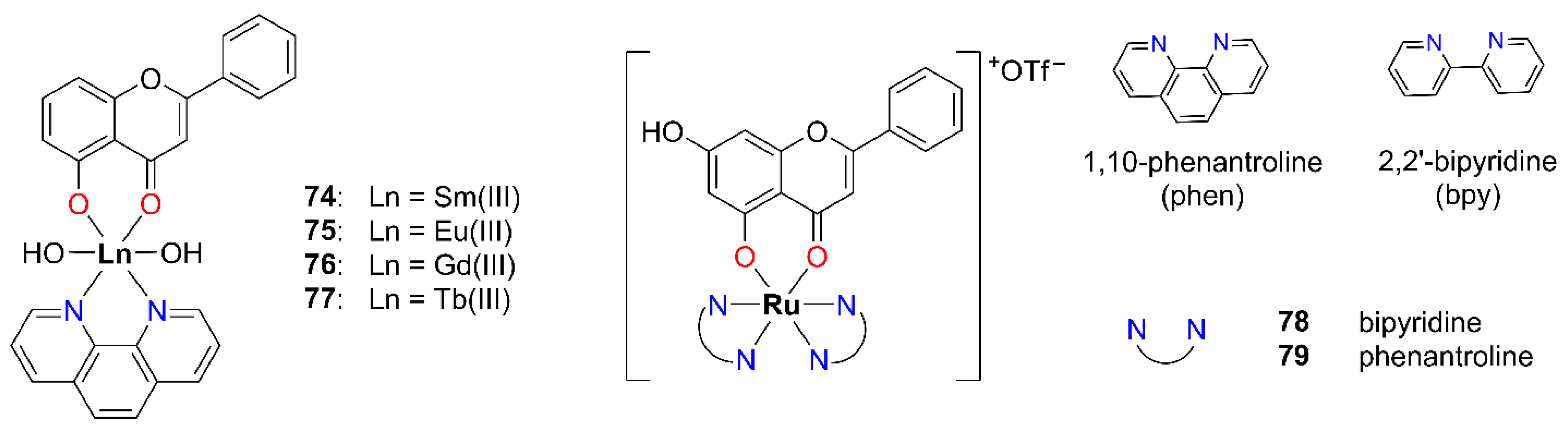
In 2017, Singh et al. synthesized a series of quercetin derivatives bearing para-substituted B-rings and subsequently prepared their [RuII(DMSO)2(querc)2] complexes (47, Figure 6) [41]. The distorted octahedral geometry structure of the complexes was proposed based on spectroscopic data (IR, NMR, and HRMS) and supported by density functional theory (DFT) calculations. In the resulting complexes 47, two dimethyl sulfoxide (DMSO) ligands were coordinated monodentately, one via the sulfur atom and the other via the oxygen atom, reflecting the ambidentate nature of DMSO.
The cytotoxicity of the synthesized complexes 47 was evaluated using the MTT assay after 24 h of exposure against MCF-7 (breast) cell line (Table 4). Among the tested compounds, complex 4, featuring a methoxy substituent on the B-ring, exhibited the highest cytotoxic activity against the MCF-7 cancer cell line (IC50 = 16 μM). In ligand–complex pairs, the IC50 values were consistently lower for the metal complexes 47 compared to their corresponding quercetin-based ligands, suggesting a potential synergistic effect between the metal center and the coordinated ligand.
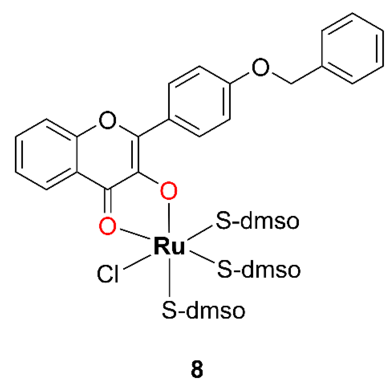
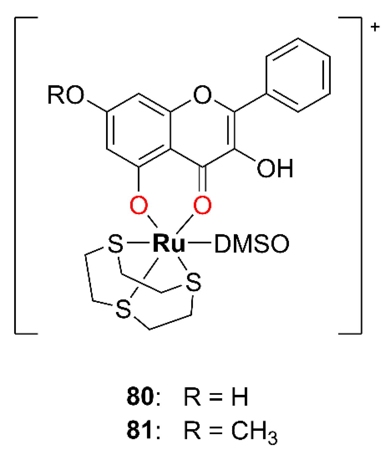
In 2010, Prajapati and coworkers synthesized two Ru(II) complexes with DMSO [42]. One of these complexes featured a chalcone ligand coordinated in a bidentate (O,O) fashion and is therefore not considered in this study. The other complex, compound 8, which incorporates a 3-OH–F derivative, is relevant to the present discussion and is shown in Figure 7.
The complex 8 was synthesized by the dropwise addition of a methanolic solution of cis-[RuIICl2(dmso)4] to a methanolic solution of the 3-OH–F derivative containing an equimolar amount of Et3N, adjusted to pH around 9.0. The resulting reaction mixture was stirred on a steam bath for 30 min. Subsequently, the solution was concentrated to one fourth of its original volume and left to stand at room temperature for 24 h. The resulting dark reddish-brown crystalline solid was collected by filtration, washed sequentially with methanol and diethyl ether, and finally dried under a vacuum. According to the authors, complex 8 is reported to be air-stable. CA indicates that it is a neutral species. The evidence for the complex 8 formation includes the appearance of a characteristic S=O stretching band around 1050 cm−1 and a Ru–S vibration band at approximately 430 cm−1. The proposed structure of complex 8 was further confirmed by XRD analysis, which revealed a monoclinic crystal system containing four discrete complex molecules arranged in a chain, with no solvent-accessible voids in the crystal packing.
Preliminary cytotoxicity screening of complex 8 was performed using the MTT assay after 48 and 72 h of incubation against Dalton’s lymphoma cell line, a murine transplantable T-cell lymphoma (DL) (Table 5). The IC50 value of complex 8 was found to be in the nanomolar range, indicating significantly higher cytotoxicity compared to the corresponding free OH–F ligand L8. This enhanced activity of complex 8 may suggest a synergistic effect between the coordinated ligand L8 and the Ru(II) metal center in mediating the cytotoxic response.
Ruthenium–DMSO complexes have attracted considerable interest due to their promising selectivity toward solid tumor metastases and relatively low toxicity to healthy tissues [43], which has led some of these compounds to enter clinical trials [44].
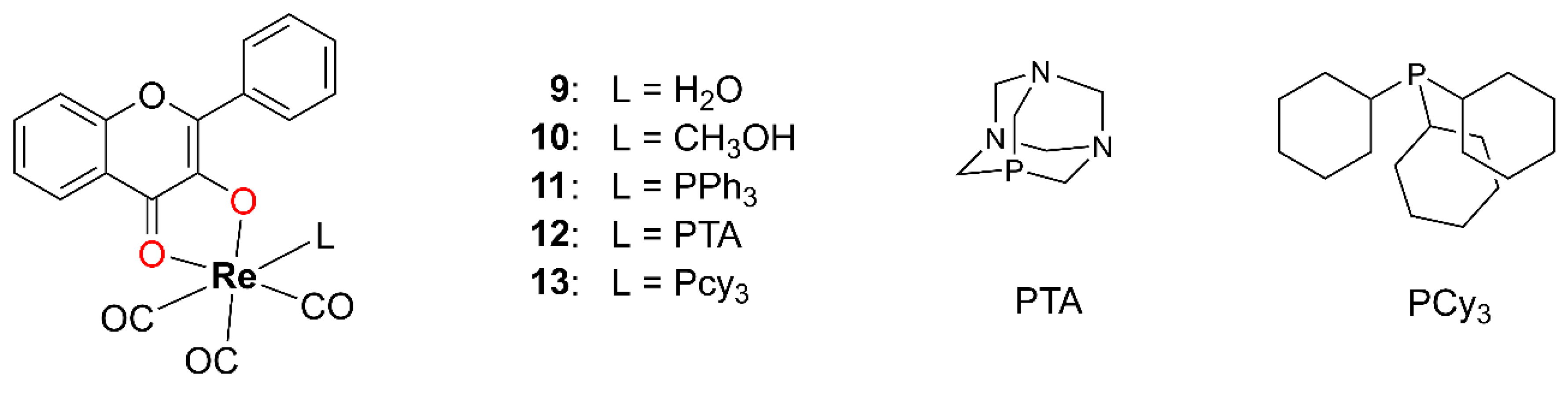
In 2020, Gantsho and coworkers synthesized 18 Re(I) carbonyl complexes incorporating tropolone, 3-OH–F, and three differently substituted quinolones as bidentate ligands [45]. Since tropolone and quinolones are not members of the OH–F family, only five Re(I) tricarbonyl complexes 913 containing 3-OH–F will be discussed here (Figure 8). Starting from [NEt4]2[Re(CO)3Br3], the addition of AgNO3 at pH 2.2 yielded the triaqua complex fac-[Re(CO)3(H2O)3]+. Subsequent substitution of two labile aqua ligands with the bidentate 3-OH–F resulted in complex 9. In methanolic solution, one of the coordinated water molecules is replaced by methanol, resulting in the complex 10. Complexes 1113 were synthesized by introducing one equivalent of a monodentate phosphine ligand. All complexes 913 were isolated as yellow precipitates or thin, needle-like crystals, which were unsuitable for XRD analysis. Structural characterization was performed using EA along with IR, UV–Vis, and NMR spectroscopy.
Complexes 9 and 10 exhibit a high tendency toward the substitution of the coordinated water or methanol ligand. In complexes 1113, elevated temperatures and an excess of the phosphine ligand promote the efficient substitution of the axially positioned carbonyl ligand trans to the phosphine. The kinetics of the substitution of complex 10 with triphenylphosphine (PPh3), 1,3,5-triaza-7-phosphaadamantane (PTA), and tricyclohexylphosphine (PCy3) were studied, and all reactions proceeded via a single-step, first-order process, with rates directly dependent on the concentration of the entering phosphine ligand.
A cytotoxicity assessment was performed exclusively for complex 11 due to the limited solubility of the other complexes in this series. The evaluation employed a fluorometric cell viability assay based on resazurin after 48 h of incubation with HeLa (cervix) as the tumor cell line, and human retinal pigment epithelial cells immortalized with hTERT (RPE-1) as the normal cell line. Cisplatin served as a positive control (Table 6). The cytotoxicity of complex 11 against HeLa cells was comparable to that of cisplatin. The SI of the complex, calculated as the ratio of IC50 values for RPE-1 and HeLa cells, was 1.5, indicating a limited selectivity toward tumor cells under the tested conditions. In contrast, cisplatin exhibited an SI of approximately five. The authors suggest that the moderate cytotoxicity observed for complex 11 may be attributed to its high kinetic stability.
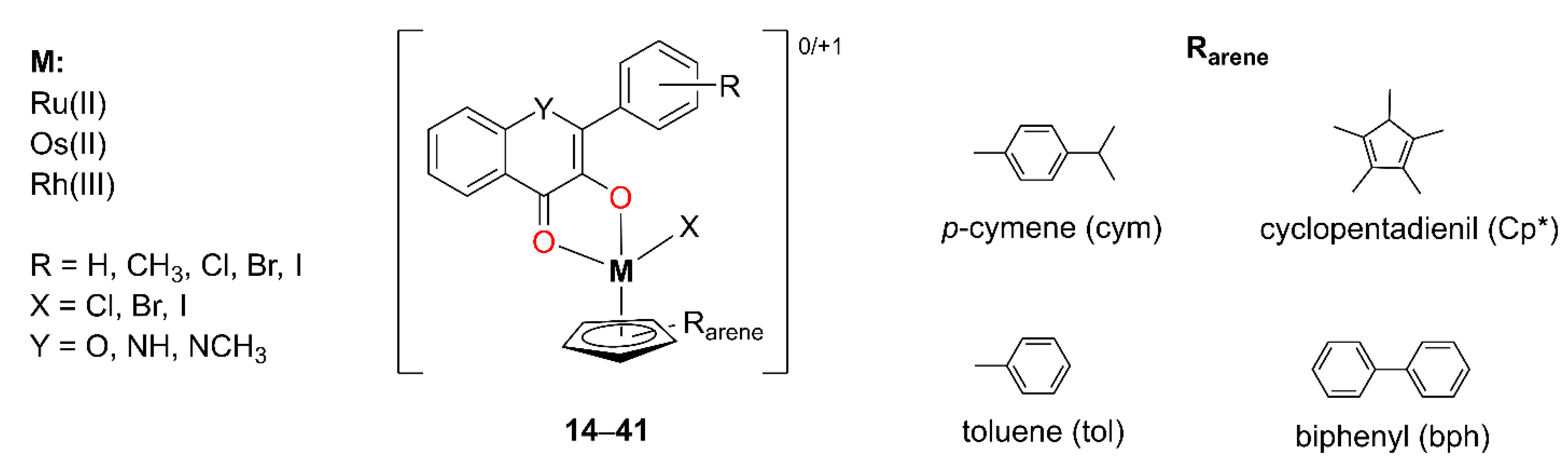
Kurzwernhart et al. synthesized 28 distinct metal complexes 1441 derived from 3-OH–F derivatives with Ru(II), Os(II), and Rh(III) as metals [46,47,48,49]. The complexes adopt a pseudo-octahedral “piano-stool” geometry, characterized by slight variations in M–O bond lengths and a moderately twisted phenyl substituent on the ligand. The general structure of the complexes is presented in Figure 9, while the individual ligand structures and corresponding cytotoxicity data are summarized in Table 7. The cytotoxicity of complexes 1441 was evaluated by an MTT assay after 96 h of exposure using three human cancer cell lines: the ovarian carcinoma cell line (CH1), the colorectal adenocarcinoma cell line (SW480), and the lung carcinoma epithelial cell line (A549). The positive control value for cisplatin was obtained from the literature.
Complexes 1423, with the general formula [RuII(Cl)(cym)(3-OH–F)], differ in both the nature and position of substituents on the B-ring of the 3-OH–F scaffold. For several of these complexes (15, 17, 18, and 21), single-crystal XRD data were obtained. Complexes bearing para-substituted B-rings exhibited the highest cytotoxic activity. In contrast, the reduced activity observed for ortho-substituted derivatives may be attributed to steric effects, as the phenyl ring in these compounds adopts a significantly more twisted conformation. This structural distortion is likely to impair molecular recognition and diminish the binding affinity to biological targets. Notably, all complexes showed a stronger inhibition of topoisomerase IIα compared to the corresponding free OH–F derivatives, which was attributed to their multitargeted mechanism of action. Furthermore, the extent of topoisomerase IIα inhibition correlated well with the observed cytotoxicity.
In a subsequent set of compounds (2433, [RuII(X)(Rarene)(3-OH–F)]), the aromatic ligand coordinated to the metal center (Rarene) was varied. The results showed that changes in lipophilicity were more strongly influenced by the 3-OH–F ligand than by the Rarene moiety [40]. Finally, in complexes 3441([OsII/RhIII(Cl)(Rarene)(3-OH–F)]0/+1), ruthenium (II) was replaced by osmium (II) or rhodium (III) as the central metal ion, but this did not significantly alter the observed cytotoxicity [41].
The complexes 1441 synthesized by Kurzwernhart et al. exhibited the highest cytotoxic activity against the CH1 (ovarian) cell line, while lower activity was observed against the SW480 (colon) and the lowest toward A549 (lung) cells, which are generally more resistant. Most IC50 values for the CH1 cell line were in the low micromolar range (≤7.9 μM), with compounds 17, 25, 29, 35, and 37 showing nanomolar potency. These five compounds share structural features including a chloride leaving group and a para-substituted B-ring of the OH–F moiety bearing either a chlorine or methyl substituent. These results suggest that both the nature and position of substituents on the B-ring, as well as the identity of the leaving group, play a critical role in enhancing cytotoxicity.

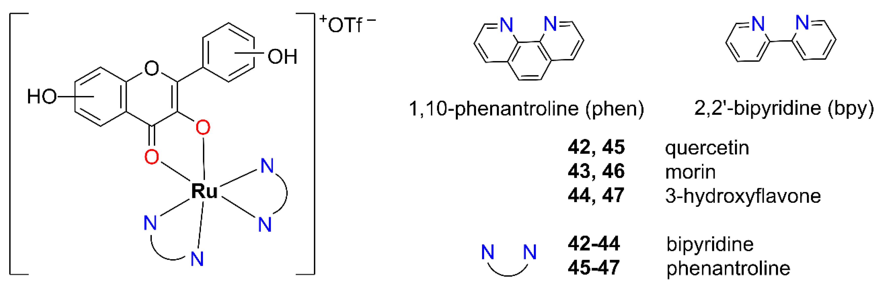
2.1.2. Mixed-Ligand Systems: Incorporating Both (O,O) and (N,N) Donor Atoms

Starting from cis-[RuII(bpy)2Cl2] or [RuII(phen)2(CO3)], Zahirović et al. synthesized a series of octahedral cationic ruthenium (II) complexes 4247 with OH–F ligands [50]. In these complexes, the diimine ligands, 2,2′-bipyridine (bpy) or 1,10-phenanthroline (phen), are coordinated in a bidentate manner through nitrogen atoms, while the OH–F ligand is coordinated via two oxygen atoms in a maltol-type binding mode (Figure 10). The resulting complexes are low-spin, diamagnetic species consistent with a t2g6 electronic configuration of the Ru(II) center.
Given that the synthesized complexes 4247 lack good leaving groups, the most probable mode of DNA interaction is intercalation. This is supported by their cationic nature and the presence of planar π-extended aromatic systems. On the other hand, the efficiency of π–π stacking interactions with nucleobases tends to decrease with the increasing number of hydroxyl groups on the OH–F ligand.
The cytotoxicity of these types of Ru(II) complexes 4247 was evaluated using the MTT assay after 72 h of incubation on four human cancer cell lines: colorectal adenocarcinoma cell line (SW620), HepG2 (liver), MCF-7 (breast), and HeLa (cervix) (Table 8). Unfortunately, the measurement errors were sufficiently high enough to raise concerns regarding the reliability and significance of the obtained results. However, two results stood out as significant: the bipyridine-based complex 44 exhibited nanomolar cytotoxicity against SW620 cells (IC50 = 0.75 μM), while the phenanthroline-based complex 47 showed micromolar activity against the MCF-7 cell line (IC50 = 8.32 μM). Both of these complexes are derivatives of 3-OH–F.
Antimicrobial activity was assessed by the disk diffusion method against four Gram-positive bacterial strains (Staphylococcus aureus ATCC 25923, Enterococcus faecalis ATCC 19433, Streptococcus β-hemolytic group A, and methicillin-resistant Staphylococcus aureus (MRSA)). Vancomycin was used as the reference drug for all Gram-positive strains, except for MRSA, where gentamicin served as the control. Four Gram-negative bacterial strains were also tested (Klebsiella pneumoniae ATCC 1705, Acinetobacter baumannii ATCC BAA-747, Pseudomonas aeruginosa, and Escherichia coli), with gentamicin as the reference drug. Additionally, the fungal strain Candida albicans was included, with nystatin used as the control. The results are presented in Table 9. Interestingly, complex 44 exhibited zones of inhibition against Gram-positive bacteria comparable in width to that of vancomycin. In the case of MRSA, the zone of inhibition was significantly wider than that of gentamicin. Against the tested fungal strain, the inhibition zone observed for 44 was identical to that measured for nystatin. Comparison of the inhibition zones of the synthesized metal complexes with those of the free hydroxyflavone ligands indicates a synergistic effect of coordination, enhancing antimicrobial activity against both Gram-positive bacteria and the fungal strain Candida albicans. Based on the obtained results, a significant improvement in the antimicrobial efficacy of complex 44 was observed against the tested Gram-positive bacteria and Candida albicans strains, relative to the uncoordinated 3-hydroxyflavone ligand.
Gençkal et al. synthesized heteroleptic complexes of Co(II), Ni(II), and Cu(II) with quercetin and diimine (phen or bpy) ligands (4851, Figure 11) [51]. A methanolic solution of metal (II) chloride x-hydrate was added dropwise to a methanolic solution of quercetin and sodium hydroxide under constant stirring at room temperature. The reaction mixture was stirred for an additional hour, after which a methanolic solution of phen or bpy was added. The mixture was then refluxed for approximately 4 h. Upon completion, it was allowed to cool to room temperature, and the resulting solid was filtered, washed with methanol, and dried under ambient conditions.
According to experimental measurements, two distinct types of complexes were obtained: an octahedral [CoII/NiII(queH-1)Cl(phen)(H2O)]∙2H2O (4849), and a square-pyramidal geometry for [CuII(queH-1)Cl(phen/bpy)]∙xH2O (5051), as illustrated in Figure 11.
EA confirms the proposed molecular formulas of the complexes 4851. CA indicates that the complexes are neutral in nature and thus behave as non-electrolytes. Their stability was verified by repeating the CA after 24 and 48 h, with consistent results. The magnetic susceptibility (χ) values for the Ni(II) and Co(II) complexes are consistent with mononuclear octahedral coordination, while those for the Cu(II) complexes support the assignment of a mononuclear square-pyramidal geometry for complexes 50 and 51. In the UV–Vis spectrum, the characteristic absorption band of free quercetin at 371 nm, assigned to the B-ring, undergoes a bathochromic shift of approximately 50 nm upon complexation, supporting a maltol-type coordination mode of the quercetin ligand. The band maximum corresponding to the carbonyl stretching vibration shifted to lower wavenumbers in all metal complexes, indicating coordination through the adjacent hydroxyl group. Since the hydroxyl group at the C3 position is significantly more acidic than the one at C5, it is expected to preferentially participate in metal coordination. Furthermore, the IR spectra reveal new bands in the 433–417 cm−1 and 652–643 cm−1 regions, corresponding to M–N and M–O bond vibrations, respectively, confirming the formation of metal–ligand bonds through nitrogen and oxygen donor atoms.
The cytotoxic activity of the compounds 4851 was initially evaluated using Sulforhodamine B (SRB) and ATP-based cell viability assays after 48 h of incubation against four different human cancer cell lines: A549 (lung), PC-3 (prostate), HeLa (cervix), and MCF-7 (breast). Improved results were obtained using the ATP assay, which is generally more sensitive than the SRB assay. Among them, the Cu(II) complexes 50 and 51 exhibited the most pronounced cytotoxicity toward the MCF-7 breast cancer cell line. Consequently, an additional breast cancer cell line, namely the human triple-negative breast cancer cell line (MDA-MB-231), was selected for further evaluation. At higher concentrations, some cytotoxic activity was also observed for the free ligands, quercetin, phen, and bpy, in breast cancer cell lines, although this activity was significantly lower compared to that of the Cu(II) complexes. Complex 50, containing phen, showed high cytotoxicity after 48 h of exposure in both MCF-7 and MDA-MB-231 cell lines, with IC50 values in the low micromolar range. One of the mechanisms underlying the cytotoxic effect of complex 50 was identified as apoptosis induction, as evidenced by an approximately 40% increase in apoptotic cells, mediated through the Caspase-3/7-dependent pathway. These findings identify complex 50 as a promising candidate for further development as an antitumor agent targeting breast cancer.
In 2014, Wang et al. synthesized four M(II) complexes 5255 (M = Cu(II) or Zn(II)) incorporating the OH–F kaempferol as an (O,O) donor ligand, along with bpy or phen as (N,N) donor ligands, as depicted in Figure 11 [52]. A methanolic solution of MCl2 was added to a methanolic solution containing equimolar amounts of kaempferol and NaOH at an ambient temperature. Subsequently, bpy or phen, dissolved in methanol, was added dropwise. The reaction mixture was filtered, and the resulting yellow precipitate was washed with ethanol and dried under a vacuum. Since single crystals suitable for XRD analysis could not be obtained, the structures of the complexes were proposed based on EA, HRMS, UV–Vis, IR, and NMR spectroscopy. A fluorometric competitive experiment using EB as a probe revealed moderate intercalation by the complexes. The increased viscosity of CT-DNA in the presence of the metal complexes 5255 further supported these findings. Notably, the complexes 5255, especially complex 52, were able to induce the cleavage of plasmid DNA (pUC19) in a dose-dependent manner.
Kaempferol and its complexes (5255) were tested for cytotoxic activity against the MDA-MB-231 (breast) cancer cell line using the MTT assay after 24 h of incubation (Table 10). All complexes showed significantly improved cytotoxicity compared to the free ligand, indicating a potential synergistic interaction between kaempferol and the coordinated metal ions.
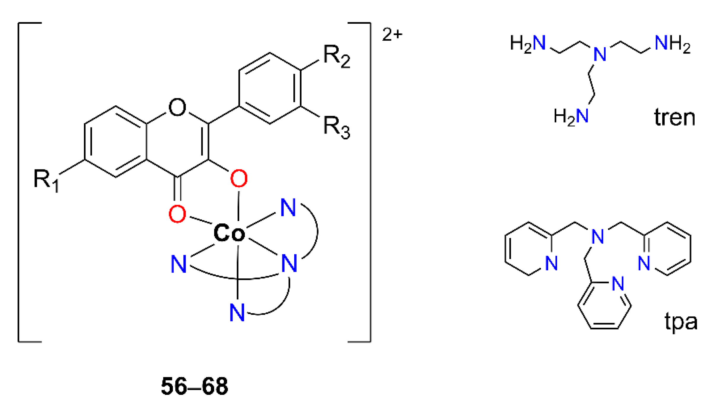
In 2021, Kozsup et al. reported the synthesis and characterization of a series of sixteen octahedral Co(III) complexes containing derivatized OH–F and tetradentate nitrogen ligands [53], namely tris(2-aminoethyl)amine (tren) or tris(2-pyridylmethyl)amine (tpa). Thirteen compounds of these (5668, [CoIII(4N)(OH–F)]2+) were obtained by maltol-type coordination (Figure 12, Table 11), using variously substituted OH–F-ligands (3-hydroxyflavone (flavH), 6-F-3′-NO2-3-hydroxyflavone (NO2FflavH), 3′-NO2-3-hydroxyflavone (NO2flavH), 4′-Me-3-hydroxyflavone (MeflavH), 6-Cl-4′-OMe-3-hydroxyflavone (ClO-MeflavH), 6-Br-3-hydroxyflavone (BrflavH), and 4′-iPr-3-hydroxyflavone (iPrflavH). Two additional complexes (69 and 70, Figure 13) were obtained by the acac method and will be discussed in the following Section 2.2. One complex, ([Co(tren)(nar)](Cl)(ClO4)), which contains naringenin (nar) from the flavanone family, will not be a subject of this review.
The [Co(4N)(flav)]2+ type of compounds (5670) was obtained in an overnight reaction by mixing the stoichiometric amount of the metal precursor ([Co(4N)Cl2]Cl (4N = tren or tpa)) and the ligand in MeOH at 60 °C with an equivalent of NaOH as a base. Most of the complexes were crystallized from water solutions after the addition of a counterion, ClO4.
The full characterization of a series of compounds 5670 included NMR, IR, and HRMS measurements, as well as crystal structure and a cyclic voltammetric (CV) analysis. Characterization data confirmed acac coordination mode (O,O) of the OH–F ligand to the metal ion and octahedral geometry in all cases. Prior to biological study, the Co(III) species were tested for their stability in a D2O:DMSO = 5:1 (v/v) mixture revealing no spectral changes in ambient conditions. Additionally, their electrochemical profile was determined using CV. The redox study indicated the influence of the substituents on the values of reduction potential as the presence of electron-donating substituents on the OH–F further shifted the cobalt complex’s reduction potential toward more negative values. The nature of the substituents demonstrated the greatest impact on the cytotoxic activity of the compounds (Table 11).
The cytotoxicity of most complexes (5665), as well as the free flavone ligands, was evaluated after 72 h of incubation against two human cancer cell lines, epithelial carcinoma cell line (A431) and A549 (lung), under both normoxic (21% O2) and hypoxic (1% O2) conditions and end-point SRB assay (Table 12). The A549 cell line was generally more sensitive to the tested compounds than A431, with the exception of compound 60, for which the EC50 values were lower in the A431 cells. Among the free ligands, MeflavH exhibited no cytotoxicity against either cell line, while flavH showed slight activity only against A549 under normoxic conditions. The ligands NO2flavH, NO2FflavH, and BrflavH were also exclusively active against A549, but their cytotoxic effects in this cell line were significant. Overall, the results indicate that the Co(III) species derived from the [Co(tpa)Cl2]Cl precursor exhibit significantly stronger cytotoxic effects compared to those obtained from [Co(tren)Cl2]Cl, highlighting the influence of substituent variation in OH–F-based ligands.

2.2. Acac-Type Coordination

The second part of our review study will focus on the acetylacetone (2,4-pentanedione, acac) type of coordination using the common acac ligand platform in coordination chemistry. Acac’s ubiquity originates from its stability, easy synthesis, and the chelate effect. Typical acac chemistry relies on two types of reactions: (a) conversions with Lewis bases when acac coordinates via both oxygens; (b) electrophilic substitution due to the nucleophilic central carbon atom forming 3-substituted acac derivatives. Besides their significant role in coordination chemistry, they are key precursors in rapidly developing fields such as nanoparticle research, polymer science, and catalysis (both homo- and heterogeneous) [54].
In hydroxyflavone-based metal complexes exhibiting a 4-carbonyl/5-hydroxyl coordination mode, the ligand acts as a bidentate chelator, coordinating the metal center through two oxygen atoms located on adjacent aromatic rings, namely the carbonyl group at position C-4 on the C-ring and the hydroxyl group at position C-5 on the A-ring. This (O,O)-coordination mode results in the formation of a six-membered chelate ring.
The spectroscopic features associated with this coordination pattern largely mirror those observed in maltol-type (3-hydroxy-4-keto) complexes. However, the key distinction lies in the 1H NMR spectrum, where the characteristic signal of the C-5 hydroxyl proton disappears, providing direct evidence of its involvement in metal coordination. In contrast to 3-OH–F complexes, where coordination primarily involves the C-ring and affects band I, the 4-carbonyl/5-hydroxyl coordination mode may also induce minor spectral shifts in band II, associated with electronic transitions in the A-ring. This subtle bathochromic or hypsochromic shift further supports the involvement of the A-ring 5-hydroxyl group in metal coordination.
The series of Co(III) complexes 5668 synthesized by Kozsup et al. (Table 11), previously reviewed in Section 2.1, has been expanded with two additional complexes, 69 and 70 (Figure 13) featuring a chrysin moiety capable of acac-type coordination (Table 13) [45]. The coordination chemistry is well established, as seen in their maltol-type analogs, and is supported by NMR, IR, and HRMS characterization data. The crystal structure analysis of complex 69 confirmed the proposed coordination mode. While chrysin, the metal precursors, and 69 exhibited no cytotoxicity against A431 and A549 cell lines, complex 70, obtained with [Co(tpa)Cl2]Cl, demonstrated improved activity in both cell lines under the tested conditions. A previously spotted trend for more active compounds with tpa moiety was confirmed for acac complexes 69 and 70 as well.
Figure 13. The chemical structures of Co(III) complexes 69 and 70, as reported by Kozsup et al. [53], and Ru(II) representatives (7173), as reported by Gasser’s group [55].
Inorganics 13 00250 g013
In the same year, the research group of Gasser published a series of monocationic Ru(II) polypyridyl complexes 7173 of the type [Ru(DIP)2(OH–F)]X (Figure 13), containing 4,7-diphenyl-1,10-phenanthroline (DIP), OH–F (5-hydroxyflavone, chrysin, or morin) and X as the counterion, PF6, and OTf [55]. These complexes were obtained by different synthetic routes which can be summarized as the following: all complexes were obtained starting from the metal precursor, Ru(DIP)2Cl2, in EtOH under N2 and protected from light. Compound 71, [Ru(DIP)2(5-OHF)](PF6), carrying 5-hydroxyflavone, involved direct coordination in the presence of NaOH. For the rest of the two complexes with chrysin (72, [Ru(DIP)2(chr)](PF6)), or morin (73, [Ru(DIP)2(mor)](PF6)), respectively, silver triflate was used prior to the addition of ligands. In the case of morin-based complex 73, the ligand was silylated with tetramethylsilane (TMS) bromide in THF before complexation, though it later appeared to be unnecessary as TMS protecting groups were hydrolyzed during the complexation. The reactions for all compounds 7173 involved refluxing (1.5–2 h), followed by cooling, solvent removal under a vacuum, and purification via DCM extraction, NH4PF6 precipitation, or Celite filtration, and preparative thin-layer chromatography (TLC) as needed. The final isolation steps included sequential washes with Et2O and heptane, sonication, centrifugation, and vacuum drying to obtain the purified Ru(II) complexes in a form of a racemic mixture of Δ and Λ enantiomers. The chemical characterization included HRMS and NMR spectroscopy while their purity was checked using EA. Prior to the cytotoxicity study, the stability of 7173 was confirmed by NMR in DMSO for 5 days.
The biological profile of Ru(II) complexes 7173 was evaluated using a fluorometric cell viability assay on four human cancer cell lines MCF-7 (breast), hypopharyngeal squamous cell carcinoma (FaDU), metastatic breast cancer cell line (MDA-MB-435S), and U87 (glioblastoma), as well as on two non-cancerous cell lines, RPE-1 (retina) and embryonic kidney (HEK293) cells. Cisplatin and doxorubicin were used as positive controls (Table 14). Among the complexes’ precursors, only chrysin was found to be active mainly against HEK293 (IC50 = 26.80 ± 2.79), while [Ru(DIP)2]Cl2 showed good activity on the RPE-1 cell line with IC50 = 3.13 ± 0.28. However, the coordination via Ru(II) significantly improved starting IC50 values for the majority of 7173 complexes. None of the complexes 7173 showed activity against the MCF-7 cell line. While they exhibited some activity against other tested cell lines, no selectivity for cancerous over normal cell lines was observed.
Munteanu et al. studied four mononuclear lanthanide-containing complexes, 7477, with 5-hydroxyflavone and phen (Figure 14) [56]. A series of complexes containing Ln(III) metal ions were synthesized by reacting the corresponding metal chlorides, LnCl3, with deprotonated 5-OH–F in the presence of triethylamine and an ethanolic solution of phen. The reactions were carried out under reflux, yielding yellow products with the general formula [Ln(OH)2(5-OH–F)(phen)]·nH2O, where n = 4, 3.5, 2, and 3 for Sm(III), Eu(III), Gd(III), and Tb(III), respectively. The physicochemical characterization of newly synthesized compounds included UV–Vis, IR, HRMS, EA, and TA. According to a DFT study, authors suggested a six-coordinated distorted octahedron geometry for all four complexes. The next step included the evaluation of the cytotoxic effects for both ligands and complexes on five human cancer cell lines (HeLa (cervix), colorectal adenocarcinoma (HT-29), colon adenocarcinoma (LoVo), MCF-7 (breast), and ovarian adenocarcinoma (SK-OV-3)) using cisplatin and doxorubicin as positive controls. Cell viability tests were performed by MTS assay within the incubation period of 48 h. Interestingly, obtained IC50 values indicated a relatively good cytotoxic activity for reported complexes, 7477, particularly against MCF-7 cancer cells (Table 15). In this case, IC50 values for 7477 were remarkably close to doxorubicin, highlighting the Eu(III)-complex 75 as the most active one. Regarding the cytotoxic profile of the precursor compounds, 5-HO–F showed an enhanced cytotoxicity in HeLa cells in comparison to Ln(III) complexes, while the Sm(III)-complex 74 demonstrated a lower IC50 in comparison to cisplatin (13.78 vs. 31.55 μM). In parallel, 5-HO–F was not active on HT-29 cells in contrast to 7477 which were more potent than cisplatin. Moreover, in the cases of LoVo and SK-OV-3, the investigated complexes 7477 displayed better activity in comparison to free ligands and cisplatin, especially against LoVo. Therefore, the authors chose HeLa and LoVo cell lines to proceed with the biological profiling that involved apoptotic effects, DNA-binding, and serum-protein-binding studies.
Moreover, all synthesized complexes demonstrated potential as DNA intercalators. This claim was supported by UV–Vis spectroscopy and various fluorescence-based assays (EB displacement assay, SYBR Gold® fluorescence quenching assay, and Alexa488 fluorescence quenching assay) and viscosimetric studies. These findings provided insights into binding dynamics, confirming effective DNA binding at micromolar concentrations highlighting the Sm(III)-complex 74 as the compound with the fastest binding kinetics and the highest activity. Molecular docking simulations further supported the intercalative binding mode. In addition to DNA interaction, the lanthanide complexes displayed effective binding to serum transport proteins, with a generally higher affinity for human serum albumin (HSA) compared to transferrin (Tf). Importantly, the authors underlined the significant role of structural flexibility in macromolecular interactions, as binding to DNA or proteins was accompanied by distinct conformational rearrangements. Overall, this study introduces four novel Ln(III) complexes capable of targeting multiple cellular components indicating how the integration of multiple (hetero)cyclic motifs and the adaptable coordination environment of trivalent lanthanide ions present a promising platform for the development of multimodal anticancer agents.
As previously discussed in the section on maltol-type coordination, Zahirović et al. synthesized mixed-ligand complexes with Ru(II) [50]. This time, complexes 78 and 79, incorporating the hydroxyflavone chrysin, which lacks a 3-hydroxyl group, formed six-membered chelate rings (Figure 14).
Cytotoxicity was evaluated using the MTT assay after 72 h of treatment, and the results are presented in Table 16. Complex 78, containing bpy, exhibited the best activity against the MCF-7 cell line, while complex 79, bearing phen, showed the highest cytotoxicity toward the HeLa cell line.
The antimicrobial activity of complex 78 is approximately two to three times lower than that of standard antibiotics, except in the case of MRSA, where its efficacy is comparable to that of gentamicin (Table 17). Compound 79 exhibits roughly half the antibacterial activity of gentamicin against Acinetobacter baumannii ATCC BAA-747, yet demonstrates slightly higher activity than the parent flavonoid, chrysin.
In 2019, Marques et al. contributed to the field of Ru(II) metal complexes with OH–F motifs coordinated in acac mode by reporting two Ru(II) trithiacyclononane ([9]aneS3) complexes, 8081, presented in Figure 15 [57]. Ru(II) complex 80 was synthesized by reacting chrysin with one equivalent of [Ru(II)([9]aneS3)Cl2(S-DMSO)] and half an equivalent of tetrabutylammonium hydroxide. The light yellow solution was refluxed in methanol for 24 h. Upon evaporation to half its volume, the mixture yielded both complex 80 and an unreacted metal precursor. The pure form of 80 was obtained by filtration of the supernatant, followed by volumization with an additional portion of diethyl ether. After one week at 4 °C, complex 80 ([Ru(II)([9]aneS3)(chrys)(S-DMSO)]Cl) was isolated as a dark orange solid, washed with cold ethanol and diethyl ether, and dried. Prior to the synthesis of the second complex (81), the ligand precursor 5-hydroxy-7-methoxyflavone (tectochrysin, tchr) was prepared by reacting chrysin with potassium carbonate (1:3 molar ratio), followed by the addition of dimethyl sulfate. The reaction mixture was refluxed in acetone for one hour, yielding a yellow precipitate that was purified via column chromatography and recrystallization from a dichloromethane/methanol mixture. In the following synthetic step, complex 81, also featuring acac-type coordination, was synthesized by reacting tchr with [Ru(II)([9]aneS3)Cl2(S-DMSO)] in a 1:1 molar ratio under a 24 h reflux in methanol, with the addition of tetrabutylammonium hydroxide. After cooling, diethyl ether and dichloromethane were added to isolate the dark orange oily product. To obtain a solid form of 81, several recrystallization steps were performed in dichloromethane, followed by washing with diethyl ether. Unlike complex 80, which was obtained in relatively low yields (23%, respectively), 2 was synthesized in a slightly improved yield of 43%. Both compounds (80 and 81) were characterized by IR, 1H, and 13C NMR spectroscopy, HRMS, and EA.
The cytotoxic effects of Ru(II) trithiacyclononane ([9]aneS3) complexes 8081 and their precursor compounds (chr and tchr) were evaluated against three cancer types using four human cancer cell lines: osteosarcoma cell line (MG-63), PC-3 (prostate), MCF-7 (breast), and MDA-MB-231 (breast) (Table 18). Cell viability was assessed using the MTT assay after a 72 h incubation period, with cisplatin as a positive control. Interestingly, the pure ligands exhibited stronger cytotoxicity, particularly against osteosarcoma cells, with the 5-methoxy derivative of chr showing the most promising effect. This finding is especially valuable given the limited availability of effective chemotherapeutic options for osteosarcoma. Moreover, OH–F compounds are generally associated with milder side effects, adding further therapeutic relevance to these results. Simultaneously, complexes 80 and 81 displayed mild cytotoxic activity, which is rather modest compared to the numerous previous studies reporting enhanced bioactivity upon metal complexation [58].

2.3. Catechol-Type Coordination

Wherever two vicinal hydroxyl groups are present within the OH–F molecule, catechol-type coordination to a metal center may occur, leading to the formation of a five-membered chelate ring. Upon coordination, the deprotonation of the hydroxyl groups is expected, which can be observed as the disappearance of the corresponding signals in the 1H NMR spectrum, along with characteristic changes in the UV–Vis spectrum.
A representative example of catechol-type coordination is described in the work of Naso et al., which reports an oxidovanadium(IV) complex with luteolin, [VO(lut)(H2O)2]Na·3H2O (compound 82; Figure 16) [59]. The background behind designing such a complex originates from both the favorable biological profile of luteolin, known for its antioxidant, anti-inflammatory, antimicrobial, and anticancer activities [60], and the well-documented therapeutic potential of oxidovanadium(IV) complexes with other flavonoids [61,62,63]. Complex 82 was synthesized by reacting VOCl2 with luteolin in a 1:2 molar ratio in ethanol, with the pH of the reaction mixture adjusted to five using 1 M NaOH. After 3 h of reflux, a dark green precipitate formed, which was isolated by filtration, washed with ethanol, and dried to yield the pure compound. The proposed structure of 82 was based on EA, UV–Vis spectroscopy, and TGA. Electron paramagnetic resonance (EPR) spectroscopy revealed the presence of two coordinated water molecules and two phenolate (ArO) groups in the metal’s coordination sphere. Moreover, EPR data indicated the formation of chains involving adjacent VO units, as supported by calculated exchange pathways. Prior to biological evaluation, the stability of 82 was confirmed by monitoring its electronic absorption spectra in DMSO under N2 atmosphere, which remained unchanged over time, indicating good solution stability.
Cell viability assays were conducted using the crystal violet assay for A549 (lung) cells and the MTT assay for MDA-MB-231 (breast) cells (Table 19). For the A549 cell line, cells were incubated at 37 °C in a growth medium for 24 h, after which the medium was replaced with fresh solutions of luteolin and complex 82. Following additional 24 h incubation, cells were stained with crystal violet. The cytotoxicity evaluation revealed comparable IC50 values for both compounds, 66.3 μM for luteolin, and 60.5 μM for complex 82. It is worth noting that significantly lower IC50 values have been reported in other studies [64].
In the next step, the MTT assay was applied to MDA-MB-231 cells treated with varying concentrations (0–100 μM) of luteolin and complex 82. After 24 h of incubation, luteolin exhibited an IC50 value of 88.3 μM, consistent with previously reported data. In contrast, its oxidovanadium(IV) complex, 82, demonstrated significantly enhanced cytotoxicity, with an IC50 of 17.0 μM. According to the authors of this study, the obtained results underscore the role of metal coordination in enhancing the anticancer activity of luteolin, as previously reported for vanadium complexes [65].
In addition to inducing cellular reactive oxygen species (ROS) generation and mitotic arrest, both luteolin and its oxidovanadium(IV) complex (VOlut) cause damage to the cytoplasmic and nuclear membranes. Their binding to bovine serum albumin (BSA) involves a combined quenching mechanism, with static quenching being the dominant process. Thermodynamic parameters, derived from the Van’t Hoff equation, revealed that the interactions are spontaneous. The luteolin–BSA complex is primarily stabilized by electrostatic forces, whereas the VOlut–BSA interaction is mainly driven by hydrogen bonding and van der Waals forces. Given the confirmed cytotoxic effects of both luteolin and 82, the complex shows potential as a promising candidate for further in vivo evaluations in cancer treatment studies.
Beside two acac-type coordinated complexes, Marques et al. (reference [57] in previous Section) prepared a catechol-type Ru(II) complex 83 (Figure 16). It was synthesized by reacting 7,3′,4′-OH–F with the metal precursor [Ru([9]aneS3)Cl2(DMSO)] in a 1:1 molar ratio, in the presence of one equivalent of tetrabutylammonium hydroxide in ethanol. The reaction mixture was refluxed for 48 h, during which the solution changed color from light yellow to orange and finally to the dark green solid product. Complex 83 was isolated by washing with ethanol and diethyl ether, then dried. According to the cytotoxicity test, complex 83 was inactive, likely due to its limited water solubility (Table 20).
As previously mentioned in Section 2.1, Dell’Anna et al. [37] synthesized Pt(II) complexes with OH–F and PPh3. One of the complexes (84), shown in Figure 16, features quercetin coordinated in a catechol-type fashion. The complex was synthesized using the same procedure as described for the maltol-type complex, with the exception that the precipitate was obtained using n-hexane instead of n-pentane. In this case, slow diffusion of n-pentane into a THF solution containing the respective complex over a period of two weeks yielded crystals suitable for XRD analysis. The complex crystallizes in the orthorhombic crystal system. Probably due to the multiple possible coordination modes of quercetin, complex 84 was obtained with approximately 5% impurities, which the authors were unable to separate. Consequently, the cytotoxicity of the complex was not evaluated.

2.4. Mixed-Type Coordination

The combination of both acac- and catechol-type coordination modes within the same complex molecule can lead to so-called mixed coordination. The observed spectral changes will reflect a combination of features characteristic of both acac- and catechol-type coordinated complexes.
One of the earliest examples of mixed-type coordination was reported by Ikeda et al., who described the synthesis and characterization of a rutin–zinc (II) flavonoid–metal complex (Figure 17) [35]. The rutin–Zn(II) complex 85 was synthesized by the slow addition of an aqueous solution of [Zn(CH3COO)2]·2H2O to a methanolic solution of dehydrated rutin. The reaction mixture was stirred at temperatures up to 40 °C for 24 h, after which the resulting complex 85 was isolated by filtration, rinsed with methanol, and dried. Structural characterization of compound 85 was carried out using IR spectroscopy, 1H NMR, UV–Vis spectroscopy, and EA. The proposed structure (Figure 17) features a mixed coordination mode, involving both catechol-type coordination via the 3′,4′-dihydroxy groups of the B-ring and acac-type coordination via the 4-keto and 5-hydroxy groups of the C-ring.
This dual chelation stabilizes the complex and is typical of flavonoid–metal interactions.
The cytotoxicity study was conducted on a panel of cancer cell lines, including murine melanoma (B16F10, derived from a C57BL/6J mouse), human skin melanoma (SK-MEL-28), acute myelogenous leukemia (KG-1), multiple myeloma (RPMI 8226), T-cell leukemia (Jurkat), and chronic myelogenous leukemia (K562), as well as on normal human cell lines, namely fibroblasts and umbilical vein endothelial cells (HUVECs). Cells were treated with compound 85 and its precursor rutin over a concentration range of 17.2–275.6 μM for 24 h. Cell viability was then assessed using the MTT assay (Table 21). While rutin alone exhibited no cytotoxic effects on either cancerous or healthy cells, and was therefore not included in the IC50 analysis, compound 85 showed significant cytotoxic activity against all tested tumor cell lines.
Importantly, 85 displayed favorable selectivity, as no cytotoxic effect was observed on normal (non-cancerous) cells. Although the compound demonstrated its strongest anticancer activity against the KG-1 leukemia cell line, it is worth noting that the study did not include a comparison with a standard positive control, which limits the interpretation of its relative potency.
In addition, Ikeda et al. reported in vivo toxicological studies in female mice. Both rutin and 85 were administered intraperitoneally (i.p.) for 7, 14 and 21 days. The toxicity was observed for 30, 60, and 90 days after the initial treatment following the parameters such as body weight measurement, hematological and biochemical analyses, and histological examination of the liver and kidneys. After 21 days of treatment, mice treated with rutin and 85 showed increased platelet, red blood cell, and white blood cell counts, which returned to normal after 90 days. A histopathological analysis revealed no abnormalities in the liver, kidneys, or lungs compared to controls. Mitochondrial membrane potential (Δψm) was evaluated by flow cytometry using Rhodamine 123. Significant loss of Δψm was observed in Ehrlich ascites carcinoma (EAC) cells treated with paclitaxel or its combinations with rutin and Rutin–zinc (II), although the single compounds had milder effects.
In vivo studies in EAC-bearing mice showed that 85 induced stronger DNA fragmentation in tumor cells than rutin alone. When combined with paclitaxel, both compounds significantly enhanced DNA fragmentation. These effects were associated with the activation of caspase-dependent apoptosis, reduced cyclin D1 and vascular endothelial growth factor (VEGF) expression, and upregulation of caspases-8 and -3. Importantly, the combination of the Rutin–zinc (II) complex, 85, with paclitaxel exhibited synergistic antitumor activity, reduced anemia and myelosuppression, common side effects of chemotherapy, and increased the survival of tumor-bearing mice.
In one of their recent studies, Wang et al. synthesized and characterized a trinuclear Zn(II) complex 86 (Figure 17) using OH–F isoorientin (iso) as a ligand [36]. This natural bioactive flavonoid C-glycoside was selected due to its broad biological activity, including anticancer and antioxidant properties [66], as well as antibacterial activity [67]. The complex was synthesized by reacting the iso ligand in ethanol with Zn(CH3COO)2·2H2O. After adjusting the pH to approximately eight, the reaction mixture was stirred and refluxed for 8 h. Upon cooling, a brown-yellow precipitate formed, which was isolated, washed with ethanol, dried, and stored at 4 °C. Comprehensive characterization of the complex was performed using 1H NMR and IR spectroscopy, EA, and TGA. To further support the proposed structure, UV–Vis spectroscopy, inductively coupled plasma atomic emission spectrometry (ICP-AES), and SEM were also employed.
The biological profiling of 86 was initiated with antibacterial activity assays, which included measurements of the bacteriostatic zone diameter, bacteriostatic rate, and bacterial growth curves. Antibacterial effects were evaluated against Escherichia coli and Staphylococcus aureus using the disk diffusion method. Initial results revealed stronger antibacterial effects of both 86 and its precursor iso against S. aureus compared to E. coli, across the same concentration range (0, 200, 400, 600, and 800 μg/mL, Table 22). Notably, 86 exhibited higher bacteriostatic rates against both bacterial strains compared to the free ligand. Additionally, a concentration-dependent increase in the diameter of the bacteriostatic zone was observed for both compounds, with 86 consistently outperforming iso (Table 23). These findings suggest that coordination with Zn(II) enhances the antibacterial efficacy of isoorientin, resulting in a stronger bacteriostatic effect. The next phase of the antibacterial study assessed the effects of iso and 86 on bacterial cell membrane integrity using SEM and transmission electron microscopy (TEM) techniques. Morphological and ultrastructural analyses revealed visible alterations in cell shape and clear signs of membrane damage, primarily induced by compound 86 and, to a lesser extent, iso. The main indication of this membrane disruption was the leakage of cytoplasmic contents, accompanied by an increase in electrical conductivity in the surrounding medium. This effect was attributed to the release of proteins and nucleic acids, as well as the continuous efflux of intracellular electrolytes such as K+ and Na+ ions. The observed damage was more pronounced in cells treated with complex 86, suggesting its superior antibacterial efficacy. This enhancement is likely due to structural and property changes upon Zn(II) coordination, which may increase the compound’s ability to interact with bacterial proteins and DNA. Finally, the authors suggested the influence of a synergistic effect, meaning that the combination of the ligand (iso) and the metal (Zn) works better than either component alone.
The cytotoxicity evaluation included both a hemolysis assay and an MTT assay for cell viability. The hemolysis assay was performed following the protocol described by Liu et al. in 2009 [68], using a microplate reader for absorbance measurements. The results provided clear evidence that both 86 and iso inhibited erythrocyte hemolysis. As lipid peroxidation of the erythrocyte membrane is typically induced by hydrogen peroxide (H2O2), the authors observed that 86 exhibited low cytotoxicity even after H2O2 exposure, indicating its favorable safety profile for potential therapeutic applications. This observation was further supported in the final stage of 86 biological profiling, which involved assessing cell viability using the MTT assay, following a slightly modified protocol based on Yuan et al. [69]. The experiments were conducted on normal human liver cells (HL-7702) and the results showed no significant difference in viability between the 86-treated group and the untreated control, indicating low cytotoxicity toward normal hepatocytes. Overall, the findings of this study classify the 86-type of compounds as primarily antibacterial agents rather than for anticancer compounds.

2.5. Linear-Type Coordination

A characteristic feature of this type of complex is coordination through a single hydroxyl group of the OH–F ligand.
In 2019, Mármol et al. described the synthesis and a full characterization of flavone-based ligands bearing propargyl ether group and their corresponding phosphane gold(I) complexes (Figure 18) as promising antiproliferative agents for colon cancer [33].
A two-step synthesis of 3-OH–F-derivatives followed a slightly changed synthetic path of Gunduz et al. [70] when 2-hydroxyacetophenone was reacted with benzaldehyde or the different para-substituted benzaldehydes in a 1:1 molar ratio under base conditions and a 3 h reflux. The obtained derivatives were further derivatized with propargyl bromide to obtain four new ligands bearing a propargyl ether group. Au(I) complexes 8794 were synthesized in MeOH after 20 h of stirring obtained ligands with the metal precursor, [AuCl(PR′3)] (PR′3 = PPh3, PTA), at room temperature. The characterization of novel compounds included NMR, IR, and XRD analysis for the OH–F derivative-bearing OMe group, while their purity was confirmed by EA. Their stability under physiological conditions (in phosphate-buffered saline (PBS) buffer at pH = 7.4 and 37 °C) was confirmed by UV–Vis absorption spectroscopy. The structural optimization of these flavonoid-inspired compounds, known for their pronounced lipophilicity, involved the introduction of a propargyl ether unit followed by coordination with a Au–phosphane moiety. This modification resulted in a well-balanced lipophilic–hydrophilic profile, with improved lipophilicity (log P) values ranging from 0.23 to 1.2.
Prior to the biological study, authors evaluated the interaction of Au(I) species with BSA adding the increasing amounts of 8794 to the BSA solution. Using fluorescence emission spectroscopy, no emission was noticed in the range from 310 to 400 nm for 8794. In addition, the binding constants and the number of binding sites for each experiment were calculated from fluorescence intensity data, identifying the hydrophobic cavity of site I in subdomain IIA of BSA as the most probable binding location.
Cell culture experiments were performed on the human colorectal adenocarcinoma subline (Caco-2/TC7), as well as on the MCF-7 (breast), HepG2 (liver), and differentiated Caco-2 cells (noncancerous colon model), in the concentration range of 0–100 μM, using auranofin and cisplatin as positive controls. As the literature reports that flavones-based compounds can influence MTT assay, Mármol et al. primarily tested the toxicity of 3-OH–F and its derivatives on undifferentiated Caco-2/TC7 cells using both MTT and SRB assays. Mármol’s study combined IC50 for four different 3-OH–F ligands obtained by MTT and SRB indicating that IC50 values from SRB were 1.2 to 5 times lower in comparison to those obtained by MTT. In addition, the SRB results were more reproducible, making this technique more reliable. In contrast, this type of behavior was not observed for eight Au(I) complexes 8794 at concentrations below 20 mM with the exception of 90 (Table 24). MTT and SRB tests resulted in similar dose–response curves and comparable IC50 values. Since all Au(I) complexes 8794 showed a reduced cell proliferation for all cell lines, the study clearly demonstrated the positive impact of metal coordination. Therefore, complexes 88 and 93, with the strongest anticancer potential and selectivity, emerged as the most promising candidates for further biological evaluations. The first step included the measurement of cyclooxygenase-1 and cyclooxygenase-2 (COX-1/2) activity, keeping in mind the flavones affinity toward cyclooxygenase [71], and the analysis of redox enzymes thioredoxin reductase (TrxR) and glutathione reductase (GR) inhibition. In both experiments, it was confirmed that Au(I) species interact with COX-1/2 and TrxR and GR redox enzymes, proceeding to ROS increase on CaCo-2 cell lines. At the stage when biological targets were identified, the type of a cell death was examined for both complexes 88 and 93. The experiments revealed that cell death occurred through the intrinsic apoptotic pathway and was accompanied by changes in cell cycle progression. Toxicity evaluation on differentiated Caco-2 cells, used as a model for noncancerous tissue, suggested that flavone-inspired gold complexes 88 and 93 may preferentially target cancer cells over normal differentiated cells, making them promising candidates for cancer treatment.
The same research group expanded the series of Au(I) complexes bearing N-heterocyclic carbene (NHC) derivatives and an OH–F moiety functionalized with a propargyl ether group (Figure 18) [34]. Mármol et al. reported eight [AuCl(NHC)]-type complexes (95102), where NHC stands for 1,3-bis(2,6-diisopropylphenyl)imidazolidin-2-ylidene (IPr; C27H38N2) or 1,3-dimethylimidazolidin-2-ylidene (IMe; C5H10N2). The compounds 95102 were synthesized by reacting four different previously obtained 3-OH–F derivatives with [AuCl(NHC)] in methanol in the presence of two equivalents of KOH as the base. The reaction mixture was stirred for 8 h at room temperature, after which the solvent was evaporated. The pure products were obtained as white, stable solids following a work-up procedure that included the addition of dichloromethane and diethyl ether. The next step involved a comprehensive characterization of complexes 95102 using NMR, IR, HRMS, and EA. Subsequently, the shake-flask method combined with UV–Vis spectroscopy was used to determine their distribution coefficients. Based on the results (log P: 0.34–1.96 for IMe; 0.42–3.4 for IPr), the complexes were classified as lipophilic species. Given that chemical stability under physiological conditions is essential for upcoming biological studies, Mármol et al. conducted stability tests in PBS at pH 7.4 and 37 °C over a 24 h period. UV–Vis monitoring of the solutions revealed no spectral changes in the main absorption bands (λ = 210–350 nm), nor any noticeable red or blue shifts. These findings confirmed that complexes 95102 remain stable under the tested experimental conditions.
The antiproliferative activity of compounds 95102 was evaluated on Caco-2 (colon) cells after a 72 h incubation period using the MTT assay in a concentration range of 0–20 µM, with auranofin as a reference drug. Despite their structural similarity to the previously reported series of Au(I) complexes 8794 with notable anticancer properties, the new series (95102) exhibited only mild to low activity in comparison to auranofin (Table 25). Nevertheless, the presence of the IMe ligand appeared to have a favorable effect, as evidenced by a significant increase in the antiproliferative activity in the corresponding complexes. Overall, the IMe-containing compounds demonstrated a higher cytotoxicity than their IPr analogs, with the exception of complexes 95 and 99. However, the antiproliferative results as a whole did not indicate sufficient potential to continue the study.
Selected compounds from the 95102 series exhibited enhanced biological activity against both Gram-positive and Gram-negative bacterial strains (Table 26). Their antimicrobial properties were evaluated using the disk diffusion assay against six strains: Escherichia coli, Pseudomonas aeruginosa, Salmonella enterica, Enterococcus faecalis, Listeria monocytogenes, Staphylococcus epidermidis, and Staphylococcus aureus. The assay was conducted following the guidelines of the Clinical and Laboratory Standards Institute (CLSI) [72]. Stock solutions of the compounds were prepared in concentrations ranging from 0 to 100 μM, and bacterial cultures were incubated for 24 h at 37 °C prior to assessing inhibition zones.
Two compounds, 96 and 100, were distinguished in the series, demonstrating greater activity in comparison to phenol. Additionally, the authors evaluated the mechanism of action for 96, relying on estimations of TrxR and dihydrofolate reductase (DHFR) activities, and further supported by SEM. In the case of Escherichia coli strain, no inhibition of the TrxR was spotted, but DHFR activity was altered in the presence of 96. This result insinuated that DHFR had a crucial role in the final antibacterial activity of 96.

3. Conclusions

3.1. Retrospective View of the Authors

In the current era of medicinal chemistry, naturally inspired compounds play a crucial role in the discovery and design of new therapeutics. Notably, nearly 50% of FDA-approved drugs are structurally related to natural products [5]. In this context, hydroxyflavone (OH–F) scaffolds combined with metal centers have gained attention as promising platforms for the development of anticancer and antimicrobial agents. Across the majority of published studies, a common feature of OH–F was recognized: its broad spectrum of biological activities, ranging from anticancer and antimicrobial to anti-inflammatory, antiviral, antimutagenic, and vasodilatory effects [73].
In our present study, we reviewed more than twenty studies published between 2010 and 2024 that investigated the biological activity of [M(OH–F)] coordination compounds, focusing on their anticancer and/or antimicrobial effects. In total, 102 complexes with various transition metals were described and classified based on their coordination mode (e.g., maltol-like, acetylacetonate, catecholate, and mixed-ligand types).
Importantly, nineteen out of twenty-two studies reported that coordination with metal centers enhanced the biological activity compared to the free ligands, justifying the incorporation of metals into OH–F-based pharmacophores. Only three studies deviated from this trend.
Despite these promising results of [M(OH–F)], their pharmaceutical application remains limited due to poor aqueous solubility. To address this challenge, we propose two key strategies. First, structural modification through the incorporation of nitrogen-containing heterocycles could enhance solubility and ligand versatility, given the prevalence of such moieties in approved drugs. Second, systematic derivatization of the OH–F platform could improve water compatibility and stability under physiological conditions, thus increasing the potential for clinical translation.
In parallel, there is a growing interest in coupling these bioactive metal complexes with biocompatible carriers and delivery matrices (e.g., chitosan, alginate, and gelatin, as well as liposomes, solid lipid nanoparticles (SLNs), and poly(lactic-co-glycolic acid) (PLGA) nanoparticles) to overcome solubility barriers and improve therapeutic outcomes. Natural polymers, lipid-based systems, and hybrid nanostructures are being actively explored for their ability to stabilize coordination compounds and modulate their release under physiological conditions. Incorporating [M(OH–F)] complexes into such delivery systems could significantly expand their biomedical utility, enabling targeted delivery, reduced toxicity, and enhanced bioavailability. These directions represent a promising frontier in medicinal chemistry, where the convergence of natural product-inspired scaffolds and metal coordination continues to unlock new therapeutic opportunities. These systems offer advantages such as controlled release, enhanced bioavailability, and targeted delivery, making them ideal candidates for the administration of coordination compounds. Combining the therapeutic potential of [M(OH–F)] scaffolds with modern drug delivery strategies has great promise for translating these complexes into clinically viable treatments.
The final part of our review addresses several common shortcomings identified in the chemical characterization and biological evaluation of the studied compounds. Some studies lacked comprehensive structural characterization of the newly synthesized complexes, with missing data such as NMR, HRMS, EA, or XRD crystallography. Others showed deviations in the biological testing, including the absence of reference compounds, lack of solubility or stability assessments, or insufficient experimental controls. These gaps can compromise the reliability of the reported biological activity and may lead to misleading conclusions at early stages of drug development and pre-clinical evaluation.
In light of the abovementioned conclusions, we would like to finalize this review by emphasizing the significant biological potential of [M(OH–F)] coordination compounds. With proper structural optimization and fine-tuning, particularly regarding solubility, stability, and target selectivity, these complexes represent promising candidates for further development into clinically relevant anticancer and antimicrobial agents.

3.2. Conclusions and Future Prospects

In this concluding section of our review, we would like to highlight several critical considerations that we believe are essential for ensuring the accuracy, reproducibility, and relevance of biological studies involving metal-based compounds.
From a strictly chemical perspective, the comprehensive structural characterization of newly synthesized compounds is strongly advised. This should definitely include techniques such as NMR spectroscopy (1H and 13C), high-resolution mass spectrometry (HRMS), infrared spectroscopy (IR), elemental analysis (EA), and X-Ray crystallography (XRD). Only the combined usage of these methods can reliably confirm the chemical structure. Despite the well-established nature of this practice, we still observe cases in the literature where characterization is incomplete, and compounds are directly subjected to biological evaluation.
Furthermore, the stability of the complexes in the culture medium and in the solvent used for cytotoxicity assays must be thoroughly assessed, ideally through 1H NMR monitoring over the incubation period. This is a crucial step, as interactions with biological media and stability under physiological conditions significantly impact the therapeutic potential and reproducibility of results.
Cytotoxicity screening should, where possible, be performed on at least five cancerous and one non-cancerous cell line. This enables the calculation of the selectivity index, which provides valuable insight into the compound’s tumor-targeting ability. The choice of a positive control must correspond to the specific tumor type under investigation, and its cytotoxicity should be evaluated under identical experimental conditions rather than cited from the literature to ensure a meaningful comparison.
Therefore, we hope this review offers constructive recommendations that will contribute to more rigorous and informative future studies on metal-based compounds with promising potential in drug development.

Funding

The authors gratefully acknowledge financial support from the European Union HORIZON-MSCA through the MET-EFFECT project under grant No. 101086373, PROMISEANG under grant agreement 101036768 funded from the Bio Based Industries Joint Undertaking (JU), and the Ministry of Science, Technological Development and Innovation of the Republic of Serbia, grant Nos. 451-03-136/2025-03/200288 and 451-03-136/2025-03/200026. The JU-CBE receives support from the European Union’s Horizon 2020 research and innovation programme and the Bio Based Industries Consortium.

Data Availability Statement

No new data were created or analyzed in this study.

Conflicts of Interest

Author Ljiljana E. Mihajlović was employed by the company Innovation Center of the Faculty of Chemistry. Author Monica Trif was employed by the company Centre for Innovative Process Engineering (CENTIV) GmbH. The remaining authors declare that the research was conducted in the absence of any commercial or financial relationships that could be construed as a potential conflict of interest.

Abbreviations

3-OH-F3-hydroxyflavone
4CL 4-coumarate-CoA ligase
5-OH-F5-hydroxyflavone
A431human epithelial carcinoma cell line
A549 human lung carcinoma epithelial cells
ATPadenosine triphosphate
apg apigenin
Bcl-2B-cell lymphoma 2
bpy2,2′-bipyridine
B16F10 murine melanoma, c57bl/6j mouse
BSA bovine serum albumin
CA conductometric analysis
Caco-2 human colorectal adenocarcinoma cell line
Caco-2/TC7 human colorectal adenocarcinoma cell subline
C4H cinnamate-4-hydroxylase
CHI chalcone isomerase
CHS chalcone synthase
CDDPcisplatin, cis-diamminedichloroplatinum(II)
chr chrysin
CH1 human ovarian carcinoma cell line
COX-1/2 cyclooxygenase-1 and cyclooxygenase-2
CVcyclic voltammetry
Compd.compound
DFTdensity functional theory
DHFR dihydrofolate reductase
DIP4,7-diphenyl-1,10-phenanthroline
DL Dalton’s lymphoma, murine transplantable t-cell lymphoma
DMSO dimethyl sulfoxide
DNA deoxyribonucleic acid
EAelemental analysis
EAC Ehrlich ascites carcinoma
EBethidium bromide
EPR electron paramagnetic resonance
FaDUhuman hypopharyngeal squamous cell carcinoma
FNS flavone synthase
FS fluorescence spectroscopy
GR glutathione reductase
HL-7702 normal human liver cells
HEK293non-cancerous human embryonic kidney cells
HeLa human cervical adenocarcinoma cell line
HepG2 human hepatocellular carcinoma cell line
HRMShigh resolution mass spectrometry
HSA human serum albumin
HT-29human colorectal adenocarcinoma
HUVECs human umbilical vein endothelial cells
ICP-AES coupled plasma atomic emission spectrometry
IMe 1,3-dimethylimidazolidin-2-ylidene
IPr 1,3-bis(2,6-diisopropylphenyl)imidazolidin-2-ylidene
i.p.intraperitoneally
IRinfrared
isoisoorientin
Jurkathuman T-lymphocyte
kaempkaempferol
K562human myelogenous leukemia
KG-1human acute myelogenous leukemia
Lligand
log Plipophilicity
LoVohuman colon adenocarcinoma
lut luteolin
NMRnuclear magnetic resonance
MCF-7 human hormone-dependant breast adenocarcinoma cell line
MDA-MB-231human triple-negative breast cancer cell line, highly metastatic
MDA-MB-435Shuman metastatic breast cancer cell line
MG63 human osteosarcoma cell line
MMP-9 matrix metalloproteinase-9
mor morin
MTT 3-(4,5-dimethylthiazol-2-yl)-2,5-diphenyltetrazolium bromide
OH-Fhydroxyflavone
p53 tumor suppressor protein involved in cell cycle regulation and apoptosis
PBS phosphate-buffered saline
phen 1,10-phenanthroline
PAL phenylalanine ammonia-lyase
PC3 human prostate adenocarcinoma cell line
PCy3 tricyclohexylphosphine
PPh3 triphenylphosphine
PTA 1,3,5-triaza-7-phosphaadamantane
PXRD powder X-Ray diffraction analyses
querc quercetin
rut rutin
ROS reactive oxygen species
RPE-1 non-cancerous human retinal pigment epithelial cells immortalized with hTERT
RPMI8226 human myeloma 
SEM scanning electron microscopy
SEM standard error of the mean
SD standard deviation
SI selectivity index
SK-MEL-28 human skin melanoma
SK-OV-3 human ovarian adenocarcinoma 
SRB sulforhodamine B
SW480 human colorectal adenocarcinoma cell line
SW620 human colorectal adenocarcinoma cell line
TA thermal analysis
tchr tectochrysin
TEM transmission electron microscopy
TGA thermogravimetric analysis
Tf transferrin
THF tetrahydrofuran
TLC thin layer chromatography
TMS tetramethylsilane
tpa tris(2-pyridylmethyl)amine
tren tris(2-aminoethyl)amine
TrxR thioredoxin reductase
WHO world health organization
U87 human glioblastoma astrocytoma cell line
UV-Visultraviolet-visible spectroscopy
VEGF vascular endothelial growth factor
WI-38 human normal lung fibroblast cell line (derived from embryonic lung tissue)
XRD X-Ray diffraction analysis
χ magnetic susceptibility

References

  1. Cancer. Available online: https://www.who.int/news-room/fact-sheets/detail/cancer (accessed on 30 June 2025).
  2. Preisler, H.D. Resistence to cytotoxic therapy: A speculative overview. Ann. Oncol. 1995, 6, 651–657. [Google Scholar] [CrossRef] [PubMed]
  3. Mansoori, B.; Mohammadi, A.; Davudian, S.; Shirjang, S.; Baradaran, B. The different mechanisms of cancer drug resistance: A brief review. Adv. Pharm. Bull. 2017, 7, 339–348. [Google Scholar] [CrossRef] [PubMed]
  4. Davies, J.; Davies, D. Origins and evolution of antibiotic resistance. Microbiol. Mol. Biol. Rev. 2010, 74, 417–433. [Google Scholar] [CrossRef] [PubMed]
  5. Newman, D.J.; Cragg, G.M. Natural products as sources of new drugs over the nearly four decades from 01/1981 to 09/2019. J. Nat. Prod. 2020, 83, 770–803. [Google Scholar] [CrossRef] [PubMed]
  6. Malešev, D.; Kuntić, V. Investigation of metal–flavonoid chelates and the determination of flavonoids via metal-flavonoid complexing reactions. J. Serb. Chem. Soc. 2007, 72, 921–939. [Google Scholar] [CrossRef]
  7. Guo, M.; Perez, C.; Wei, Y.; Rapoza, E.; Su, G.; Bou-Abdallah, F.; Chasteen, N.D. Iron-binding properties of plant polyphenols and their biological significance. Dalton Trans. 2007, 2007, 4951–4961. [Google Scholar] [CrossRef] [PubMed]
  8. Lou, H.; Hu, L.; Lu, H.; Wei, T.; Chen, Q. Metabolic Engineering of Microbial Cell Factories for Biosynthesis of Flavonoids: A Review. Molecules 2021, 26, 4522. [Google Scholar] [CrossRef] [PubMed]
  9. Kasprzak, M.M.; Erxleben, A.; Ochocki, J. Properties and applications of flavonoid metal complexes. RSC Adv. 2015, 5, 45853–45877. [Google Scholar] [CrossRef]
  10. Agati, G.; Azzarello, E.; Pollastri, S.; Tattini, M. Flavonoids as antioxidants in plants: Location and functional significance. Plant Sci. 2012, 196, 67–76. [Google Scholar] [CrossRef] [PubMed]
  11. Treutter, D. Significance of flavonoids in plant resistance: A review. Plant Biol. 2005, 7, 581–591. [Google Scholar] [CrossRef] [PubMed]
  12. Hernández, I.; Alegre, L.; Van Breusegem, F.; Munné-Bosch, S. How relevant are flavonoids as antioxidants in plants? Trends Plant Sci. 2009, 14, 125–132. [Google Scholar] [CrossRef] [PubMed]
  13. Buer, C.S.; Imin, N.; Djordjevic, M.A. Flavonoids: New roles for old molecules. J. Integr. Plant Biol. 2010, 52, 98–111. [Google Scholar] [CrossRef] [PubMed]
  14. Cotelle, N.; Bernier, J.-L.; Catteau, J.-P.; Pommery, J.; Wallet, J.-C.; Gaydou, E.M. Antioxidant properties of hydroxy-flavones. Free Radic. Biol. Med. 1996, 20, 35–43. [Google Scholar] [CrossRef] [PubMed]
  15. Magnani, L.; Gaydou, E.M.; Hubaud, J.C. Spectrophotometric measurement of antioxidant properties of flavones and flavonols against superoxide anion. Anal. Chim. Acta 2000, 411, 209–216. [Google Scholar] [CrossRef]
  16. Lemanska, K.; Szymusiak, H.; Tyrakowska, B.; Zielinsky, R.; Soffers, E.M.F.; Rietjens, I.M.C.M. The influence of pH on antioxidant properties and the mechanism of antioxidant action of hydrxiflavones. Free Radic Biol. Med. 2001, 31, 869–881. [Google Scholar] [CrossRef]
  17. de Souza, R.F.V.; Sussuchi, E.M.; De Giovani, W.F. Synthesis, Electrochemical, Spectral, and Antioxidant Properties of Complexes of Flavonoids with Metal Ions. Synth. React. Inorg. Met.-Org. Chem. 2003, 33, 1125–1144. [Google Scholar] [CrossRef]
  18. Lewandowski, W.; Lewandowska, H.; Golonko, A.; Świderski, G.; Świsłocka, R.; Kalinowska, M. Correlations between molecular structure and biological activity in “logical series” of dietary chromone derivatives. PLoS ONE 2020, 15, e0229477. [Google Scholar] [CrossRef] [PubMed]
  19. Leonte, D.; Ungureanu, D.; Zaharia, V. Flavones and Related Compounds: Synthesis and Biological Activity. Molecules 2023, 28, 6528. [Google Scholar] [CrossRef] [PubMed]
  20. Fotsis, T.; Pepper, M.S.; Montesano, R.; Aktas, E.; Breit, S.; Schweigerer, L.; Rasku, S.; Wahala, K.; Adlercreutz, H. Phytoestrogens and inhibition of angiogenesis. Baillieres Clin. Endocrinol. Metab. 1998, 12, 649–666. [Google Scholar] [CrossRef] [PubMed]
  21. Kim, M.H. Flavonoids Inhibit VEGF/bFGF-Induced Angiogenesis In Vitro by Inhibiting the Matrix-Degrading Proteases. J. Cell. Biochem. 2003, 89, 529–538. [Google Scholar] [CrossRef] [PubMed]
  22. Sanderson, J.T.; Hordijk, J.; Denison, M.S.; Springsteel, M.F.; Nantz, M.H.; van den Berg, M. Induction and Inhibition of Aromatase (CYP19) Activity by Natural and Synthetic Flavonoid Compounds in H295R Human Adrenocortical Carcinoma Cells. Toxicol. Sci. 2004, 82, 70–79. [Google Scholar] [CrossRef] [PubMed]
  23. Holder, S.; Zemskova, M.; Zhang, C.; Tabrizizad, M.; Bremer, R.; Neidigh, J.W.; Lilly, M.B. Characterization of a potent and selective small-molecule inhibitor of the PIM1 kinase. Mol. Cancer Ther. 2007, 6, 163–172. [Google Scholar] [CrossRef] [PubMed]
  24. Loa, J.; Chow, P.; Zhang, K. Studies of structure–activity relationship on plant polyphenol-induced suppression of human liver cancer cells. Cancer Chemother. Pharmacol. 2009, 63, 1007–1016. [Google Scholar] [CrossRef] [PubMed]
  25. Math, H.H.; Kumar, R.S.; Chakraborty, B.; Almansour, A.I.; Perumal, K.; Kantli, G.B.; Nayaka, S. Antimicrobial Efficacy of 7-hydroxyflavone Derived from Amycolatopsis sp. HSN-02 and Its Biocontrol Potential on Cercospora Leaf Spot Disease in Tomato Plants. Antibiotics 2023, 12, 1175. [Google Scholar] [CrossRef] [PubMed]
  26. Torres-Piedra, M.; Ortiz-Andrade, R.; Villalobos-Molina, R.; Singh, N.; Medina-Franco, J.L.; Webster, S.P.; Binnie, M.; Navarrete-Vázquez, G.; Estrada-Soto, S. A comparative study of flavonoid analogues on streptozotocinenicotinamide induced diabetic rats: Quercetin as a potential antidiabetic agent acting via 11b–Hydroxysteroid dehydrogenase type 1 inhibition. Eur. J. Med. Chem. 2010, 45, 2606–2612. [Google Scholar] [CrossRef] [PubMed]
  27. Pillai, S.I.; Subramanian, S.P.; Kandaswamy, M. A novel insulin mimetic vanadiumeflavonol complex: Synthesis, characterization and in vivo evaluation in STZ-induced rats. Eur. J. Med. Chem. 2013, 63, 109–117. [Google Scholar] [CrossRef] [PubMed]
  28. Calderone, V.; Chericoni, S.; Martinelli, C.; Testai, L.; Nardi, A.; Morelli, I.; Breschi, M.C.; Martinotti, E. Vasorelaxing effects of flavonoids: Investigation on the possible involvement of potassium channels. Naunyn-Schmiedeberg’s Arch. Pharmacol. 2004, 370, 290–298. [Google Scholar] [CrossRef] [PubMed]
  29. Liu, X.; Chan, C.-B.; Jang, S.-W.; Pradoldej, S.; Huang, J.; He, K.; Phun, L.H.; France, S.; Xiao, G.; Jia, Y.; et al. A Synthetic 7,8-hihydroxyflavone Derivative Promotes Neurogenesis and Exhibits Potent Antidepressant Effect. J. Med. Chem. 2010, 53, 8274–8286. [Google Scholar] [CrossRef] [PubMed]
  30. Tasdemir, D.; Kaiser, M.; Brun, R.; Yardley, V.; Schmidt, T.J.; Tosun, F.; Rüedi, P. Antitrypanosomal and Antileishmanial Activities of Flavonoids and Their Analogues: In Vitro, In Vivo, Structure-Activity Relationship, and Quantitative Structure-Activity Relationship Studies. Antimicrob. Agents Chemother. 2006, 50, 1352–1364. [Google Scholar] [CrossRef] [PubMed]
  31. Grazul, M.; Budzisz, E. Biological activity of metal ions complexes of chromones, coumarins and flavones. Coord. Chem. Rev. 2009, 253, 2588–2598. [Google Scholar] [CrossRef]
  32. Yang, A.; Liu, C.; Zhang, H.; Wu, J.; Shen, R.; Kou, X. A multifunctional anti-AD approach: Design, synthesis, X-ray crystal structure, biological evaluation and molecular docking of chrysin derivatives. Eur. J. Med. Chem. 2022, 233, 114216–114232. [Google Scholar] [CrossRef] [PubMed]
  33. Mármol, I.; Castellnou, P.; Alvarez, R.; Gimeno, M.C.; Rodríguez-Yoldi, M.J.; Cerrada, E. Alkynyl gold(I) complexes derived from 3-hydroxyflavones as multi-targeted drugs against colon cancer. Eur. J. Med. Chem. 2019, 183, 111661. [Google Scholar] [CrossRef] [PubMed]
  34. Mármol, I.; Quero, J.; Azcárate, P.; Atrián-Blasco, E.; Ramos, C.; Santos, J.; Gimeno, M.C.; Rodríguez-Yoldi, M.J.; Cerrada, E. Biological activity of NHC-gold-alkynyl complexes derived from 3-hydroxyflavones. Pharmaceutics 2022, 14, 2064. [Google Scholar] [CrossRef] [PubMed]
  35. Andrades Ikeda, N.E.; Novak, E.M.; Maria, D.A.; Velosa, A.S.; Silva Pereira, R.M. Synthesis, characterization and biological evaluation of rutin–zinc(II) flavonoid-metal complex. Chem.-Biol. Interact. 2015, 239, 184–191. [Google Scholar] [CrossRef] [PubMed]
  36. Wang, X.; He, S.; Yuan, L.; Deng, H.; Zhang, Z. Synthesis, structure characterization, and antioxidant and antibacterial activity study of iso-orientin–zinc complex. J. Agric. Food Chem. 2021, 69, 3952–3964. [Google Scholar] [CrossRef] [PubMed]
  37. Dell’Anna, M.M.; Censi, V.; Carrozzini, B.; Caliandro, R.; Denora, N.; Franco, M.; Veclani, D.; Melchior, A.; Tolazzi, M.; Mastrorilli, P. Triphenylphosphane Pt(II) complexes containing biologically active natural polyphenols: Synthesis, crystal structure, molecular modeling and cytotoxic studies. J. Inorg. Biochem. 2016, 163, 346–361. [Google Scholar] [CrossRef] [PubMed]
  38. Raza, A.; Xu, X.; Xia, L.; Xia, C.; Tang, J.; Ouyang, Z. Quercetin-Iron Complex: Synthesis, Characterization, Antioxidant, DNA Binding, DNA Cleavage, and Antibacterial Activity Studies. J. Fluoresc. 2016, 26, 2023–2031. [Google Scholar] [CrossRef] [PubMed]
  39. Repac Antić, D.; Parčina, M.; Gobin, I.; Petković Didović, M. Chelation in Antibacterial Drugs: From Nitroxoline to Cefiderocol and Beyond. Antibiotics 2022, 11, 1105. [Google Scholar] [CrossRef] [PubMed]
  40. Sahyon, H.A.; Althobaiti, F.; Ramadan, A.E.-M.M.; Fathy, A.M. Quercetin—Based rhodium(III) complex: Synthesis, characterization and diverse biological potentials. J. Mol. Struct. 2022, 1257, 132584. [Google Scholar] [CrossRef]
  41. Singh, A.K.; Saxena, G.; Sahabjada; Arshad, M. Synthesis, characterization and biological evaluation of ruthenium flavanol complexes against breast cancer. Spectrochim. Acta Part A Mol. Biomol. Spectrosc. 2017, 180, 97–104. [Google Scholar] [CrossRef] [PubMed]
  42. Prajapati, R.; Kumar Dubey, S.; Gaur, R.; Koiri, R.K.; Maurya, B.K.; Trigun, S.K.; Mishra, L. Structural characterization and cytotoxicity studies of ruthenium(II)–dmso–chloro complexes of chalcone and flavone derivatives. Polyhedron 2010, 29, 1055–1061. [Google Scholar] [CrossRef]
  43. Sava, G.; Bergamo, A. Ruthenium-based compounds and tumour growth control (review). Int. J. Oncol. 2000, 17, 353–365. [Google Scholar] [CrossRef] [PubMed]
  44. Alessio, E.; Balducci, G.; Lutman, A.; Mestroni, G.; Calligaris, M.; Attia, W.M. Synthesis and characterization of two new classes of ruthenium(III)-sulfoxide complexes with nitrogen donor ligands (L): Na[trans-RuCl4(R2SO)(L)] and mer, cis-RuCl3(R2SO)(R2SO)(L). The crystal structure of Na[trans-RuCl4(DMSO)(NH3)] · 2DMSO, Na[trans-RuCl4(DMSO)(Im)] · H2O, Me2CO (Im = imidazole) and mer, cis-RuCl3(DMSO)(DMSO)(NH3). Inorg. Chim. Acta 1993, 203, 205–217. [Google Scholar] [CrossRef]
  45. Gantsho, V.L.; Dotou, M.; Jakubaszek, M.; Goud, B.; Gasser, G.; Vissera, H.G.; Schutte-Smith, M. Synthesis, characterization, kinetic investigation and biological evaluation of Re(I) di- and tricarbonyl complexes with tertiary phosphine ligands. Dalton Trans. 2020, 49, 35–46. [Google Scholar] [CrossRef] [PubMed]
  46. Kurzwernhart, A.; Kandioller, W.; Bartel, C.; Bächler, S.; Trondl, R.; Mühlgassner, G.; Jakupec, M.A.; Arion, V.B.; Marko, M.; Keppler, B.K.; et al. Targeting the DNA-topoisomerase complex in a double-strike approach with a topoisomerase inhibiting moiety and covalent DNA binder. Chem. Commun. 2012, 48, 4839–4841. [Google Scholar] [CrossRef] [PubMed]
  47. Kurzwernhart, A.; Kandioller, W.; Bächler, S.; Bartel, C.; Martic, S.; Buczkowska, M.; Mühlgassner, G.; Jakupec, M.A.; Kraatz, H.-B.; Bednarski, P.J.; et al. Structure-Activity Relationships of Targeted RuII(η6-p-Cymene) Anticancer Complexes with Flavonol-Derived Ligands. J. Med. Chem. 2012, 55, 10512–10522. [Google Scholar] [CrossRef] [PubMed]
  48. Kurzwernhart, A.; Kandioller, W.; Enyedy, É.A.; Novak, M.; Jakupec, M.A.; Keppler, B.K.; Hartinger, C.G. 3-Hydroxyflavones vs. 3-hydroxyquinolinones: Structure–activity relationships and stability studies on RuII(arene) anticancer complexes with biologically active ligands. Dalton Trans. 2013, 42, 6193–6202. [Google Scholar] [CrossRef] [PubMed]
  49. Kurzwernhart, A.; Mokesch, S.; Klapproth, E.; Adib-Razavi, M.S.; Jakupec, M.A.; Hartinger, C.G.; Kandioller, W.; Keppler, B.K. Flavonoid-based organometallics with different metal centers—Investigations of the effects on reactivity and cytotoxicity. Eur. J. Inorg. Chem. 2016, 2016, 240–246. [Google Scholar] [CrossRef]
  50. Zahirović, A.; Kahrović, E.; Cindrić, M.; Kraljević Pavelić, S.; Hukić, M.; Harej, A.; Turkušić, E. Heteroleptic ruthenium bioflavonoid complexes: From synthesis to in vitro biological activity. J. Coord. Chem. 2017, 70, 4030–4053. [Google Scholar] [CrossRef]
  51. Gençkal, H.M.; Erkisa, M.; Alper, P.; Sahin, S.; Ulukaya, E.; Ari, F. Mixed ligand complexes of Co(II), Ni(II) and Cu(II) with quercetin and diimine ligands: Synthesis, characterization, anti-cancer and anti-oxidant activity. J. Biol. Inorg. Chem. 2020, 25, 161–177. [Google Scholar] [CrossRef] [PubMed]
  52. Wang, Q.; Huang, M.; Huang, Y.; Zhang, J.-S.; Zhou, G.-F.; Zeng, R.-Q.; Yang, X.-B. Synthesis, characterization, DNA interaction, and antitumor activities of mixed-ligand metal complexes of kaempferol and 1,10-phenanthroline/2,2′-bipyridine. Med. Chem. Res. 2014, 23, 2659–2666. [Google Scholar] [CrossRef]
  53. Kozsup, M.; Zhou, X.Q.; Farkas, E.; Bényei, A.C.; Bonnet, S.; Patonay, T. (the late); Kónya, K.; Buglyó, P. Synthesis, characterization and cytotoxicity studies of Co(III)-flavonolato complexes. J. Inorg. Biochem. 2021, 217, 111382. [Google Scholar] [CrossRef] [PubMed]
  54. Sodhi, R.K.; Paul, S. An overview of metal acetylacetonates: Developing areas/routes to new materials and applications in organic syntheses. Catal. Surv. Asia 2018, 22, 31–62. [Google Scholar] [CrossRef]
  55. Munteanu, A.-C.; Notaro, A.; Jakubaszek, M.; Cowell, J.; Tharaud, M.; Goud, B.; Uivarosi, V.; Gasser, G. Synthesis, characterization, cytotoxic activity, and metabolic studies of ruthenium(II) polypyridyl complexes containing flavonoid ligands. Inorg. Chem. 2020, 59, 4424–4434. [Google Scholar] [CrossRef] [PubMed]
  56. Munteanu, A.-C.; Musat, M.G.; Mihaila, M.; Badea, M.; Olar, R.; Nitulescu, G.M.; Rădulescu, F.Ș.; Brasoveanu, L.I.; Uivarosi, V. New heteroleptic lanthanide complexes as multimodal drugs: Cytotoxicity studies, apoptosis, cell cycle analysis, DNA interactions, and protein binding. Appl. Organomet. Chem. 2020, e6062. [Google Scholar] [CrossRef]
  57. Marques, J.; Silva, A.M.S.; Marques, M.P.M.; Braga, S.S. Ruthenium(II) trithiacyclononane complexes of 7,3′,4′-trihydroxyflavone, chrysin and tectochrysin: Synthesis, characterisation, and cytotoxic evaluation. Inorg. Chim. Acta 2019, 488, 71–79. [Google Scholar] [CrossRef]
  58. Marques, J.; Fernandes, J.A.; Almeida Paz, F.A.; Marques, M.P.M.; Braga, S.S. Isolation, crystal structure, and cytotoxicity on osteosarcoma of a ruthenium(III) complex with coordinated acetonitrile. J. Coord. Chem. 2012, 65, 2489–2499. [Google Scholar] [CrossRef]
  59. Naso, L.G.; Lezama, L.; Valcarcel, M.; Salado, C.; Villacé, P.; Kortazar, D.; Ferrer, E.G.; Williams, P.A.M. Bovine serum albumin binding, antioxidant and anticancer properties of an oxidovanadium(IV) complex with luteolin. J. Inorg. Biochem. 2016, 157, 80–93. [Google Scholar] [CrossRef] [PubMed]
  60. Lopez-Lazaro, M. Distribution and biological activities of the flavonoid luteolin. Mini Rev. Med. Chem. 2009, 9, 31–59. [Google Scholar] [CrossRef] [PubMed]
  61. Ferrer, E.G.; Salinas, M.V.; Correa, M.J.; Naso, L.; Barrio, D.A.; Etcheverry, S.B.; Lezama, L.; Rojo, T.; Williams, P.A.M. Synthesis, characterization, antitumoral and osteogenic activities of quercetin vanadyl(IV) complexes. J. Biol. Inorg. Chem. 2006, 11, 791–801. [Google Scholar] [CrossRef] [PubMed]
  62. Etcheverry, S.B.; Ferrer, E.G.; Naso, L.; Rivadeneira, J.; Salinas, V.; Williams, P.A.M. Antioxidant effects of the VO(IV) hesperidin complex and its role in cancer chemoprevention. J. Biol. Inorg. Chem. 2007, 13, 435–447. [Google Scholar] [CrossRef] [PubMed]
  63. Islas, M.S.; Naso, L.G.; Lezama, L.; Valcarcel, M.; Salado, C.; Roura-Ferrer, M.; Ferrer, E.G.; Williams, P.A.M. Insights into the mechanisms underlying the antitumor activity of an oxidovanadium(IV) compound with the antioxidant naringenin. Albumin binding studies. J. Inorg. Biochem. 2015, 149, 12–24. [Google Scholar] [CrossRef] [PubMed]
  64. Seelinger, G.; Merfort, I.; Wölfle, U.; Schempp, C.M. Anti-carcinogenic effects of the flavonoid luteolin. Molecules 2008, 13, 2628–2651. [Google Scholar] [CrossRef] [PubMed]
  65. Sharfalddin, A.A.; Al-Younis, I.M.; Mohammed, H.A.; Dhahri, M.; Mouffouk, F.; Abu Ali, H.; Anwar, M.J.; Qureshi, K.A.; Hussien, M.A.; Alghrably, M. Therapeutic Properties of Vanadium Complexes. Inorganics 2022, 10, 244. [Google Scholar] [CrossRef]
  66. Deng, S.-P.; Yang, Y.-L.; Cheng, X.-X.; Li, W.-R.; Cai, J.-Y. Synthesis, spectroscopic study and radical scavenging activity of kaempferol derivatives: Enhanced water solubility and antioxidant activity. Int. J. Mol. Sci. 2019, 20, 975. [Google Scholar] [CrossRef] [PubMed]
  67. Tu, L.-Y.; Pi, J.; Jin, H.; Cai, J.-Y.; Deng, S.-P. Synthesis, characterization and anticancer activity of kaempferol–zinc(II) complex. Bioorg. Med. Chem. Lett. 2016, 26, 2730–2734. [Google Scholar] [CrossRef] [PubMed]
  68. Liu, L.; Xu, K.; Wang, H.; Tan, P.K.J.; Fan, W.; Venkatraman, S.S.; Li, L.; Yang, Y.-Y. Self-assembled cationic peptide nanoparticles as an efficient antimicrobial agent. Nat. Nanotechnol. 2009, 4, 457–463. [Google Scholar] [CrossRef] [PubMed]
  69. Yuan, L.; Wang, Y.; Wang, J.; Xiao, H.; Liu, X. Additive effect of zinc oxide nanoparticles and isoorientin on apoptosis in human hepatoma cell line. Toxicol. Lett. 2014, 225, 294–304. [Google Scholar] [CrossRef] [PubMed]
  70. Gunduz, S.; Goren, A.C.; Ozturk, T. Syntheses of 3-hydroxyflavones. Org. Lett. 2012, 14, 1576–1579. [Google Scholar] [CrossRef] [PubMed]
  71. Meshram, R.J.; Bagul, K.T.; Pawnikar, S.P.; Barage, S.H.; Kolte, B.S.; Gacche, R.N. Known compounds and new lessons: Structural and electronic basis of flavonoid-based bioactivities. J. Biomol. Struct. Dyn. 2020, 38, 1168–1184. [Google Scholar] [CrossRef] [PubMed]
  72. Clinical and Laboratory Standards Institute (CLSI). Performance Standards for Antimicrobial Disk Susceptibility Tests; Approved Standard; CLSI: Wayne, PA, USA, 2012. [Google Scholar]
  73. Samsonowicz, M.; Regulska, E.; Kalinowska, M. Hydroxyflavone metal complexes—Molecular structure, antioxidant activity and biological effects. Chem. Biol. Interact. 2017, 273, 245–256. [Google Scholar] [CrossRef] [PubMed]
Figure 1. Origin of hydroxyflavones, their roles in plants, and associated biological activities.
Inorganics 13 00250 g001
Figure 4. Representative coordination modes of OH–F to metal ions, including binding via: (i) the 4-carbonyl and 3-hydroxyl groups (maltol-type; forming a 5-membered chelate ring), (ii) the 4-carbonyl and 5-hydroxyl groups (acac-type; forming a 6-membered chelate ring), (iii) two adjacent hydroxyl groups (catechol-type; 5-membered chelate), (iv) mixed coordination involving both acac- and catechol-type donor sites, and (iv) only one hydroxyl group (linear-type).
Inorganics 13 00250 g004
Figure 5. Proposed structure of the platinum (II) complex with 3-hydroxyflavon (1), cis-[Pt(PPh3)2(3-OH–F)]Cl, as suggested by Dell’Anna et al. [37].
Inorganics 13 00250 g005
Figure 7. Structure of the Ru(II) complex 8, fac-[RuCl(S-dmso)3(3-OH–F)], containing a 3-OH–F derivative and coordinated DMSO ligands, as reported by Prajapati et al. [42].
Inorganics 13 00250 g007
Figure 8. Proposed structure of the Re(I) tricarbonyl complexes 913 with 3-OH–F, as reported by Gantsho et al. [45].
Inorganics 13 00250 g008
Figure 9. General structure of complexes 1441 bearing 3-OH–F derivatives, as reported by Kurzwernhart et al. [46,47,48,49].
Inorganics 13 00250 g009
Figure 10. Proposed structures of the synthesized heteroleptic [RuII(phen/bpy)2(OH–F)]OTf∙nH2O complexes 4247, as reported by Zahirović et al. [50].
Inorganics 13 00250 g010
Figure 11. Proposed structures of M(II) complexes with quercetin (4851) or kaempferol (5255), as suggested by Gençkal et al. and Wang et al., respectively [51,52].
Inorganics 13 00250 g011
Figure 12. The chemical structures of Co(III) complexes 5668 with differently substituted OH–F obtained by maltol-type coordination, as reported by Kozsup et al. [53].
Inorganics 13 00250 g012
Figure 14. The proposed structures of Ln(III) complexes 7477, as reported by Munteanu et al. [56], and Ru(II) complexes 7879, as reported by Zahirović et al. [50].
Inorganics 13 00250 g014
Figure 15. The proposed chemical structures of Ru(II) trythiacyclononane complexes 8081, featuring acac-type coordination, as reported by Marques et al. [57].
Inorganics 13 00250 g015
Figure 16. The proposed chemical structures of catechol-type coordinated hydroxyflavones: oxidovanadium(IV) complex 82 with luteolin, as reported by Naso et al. [59], and Ru(II) complex 83 with 7,3′,4′-OH–Fand trythiacyclononane, as reported by Marques et al. [57], and Pt(II)complex 84 with quercetin and triphenylphosphine (PPh3), as reported by Dell’Anna et al. [37].
Inorganics 13 00250 g016
Figure 17. The proposed chemical structures of Zn(II) complexes with mixed-type coordination mode: the cationic complex 85 with rutin, reported by Ikeda et al. [35], and the neutral complex 86 with isoorientin, reported by Wang et al. [36].
Inorganics 13 00250 g017
Figure 18. Chemical structures of Au(I) hydroxyflavone complexes 8794 bearing phosphane ligands, and complexes 95102 featuring N-heterocyclic carbene derivatives, as reported by Mármol et al. [33,34].
Inorganics 13 00250 g018
Table 1. Cytotoxicity of Pt(II) complex 1 determined by MTT assay after 72 h treatment against U87 (glioblastoma) and MCF-7 (breast) cancer cell lines. Data are mean values ± SD (n = 3) [37].
Compd.IC50 (μM)
U87MCF-7
cis-[PtCl2(PPh3)2]>200>200
3-OH–F27.5 ± 2.3108.1 ± 3.5
126.3 ± 2.155.2 ± 1.7
Cisplatin a1.76 ± 0.2214 ± 3
a Reported from the literature.
Table 2. Antibacterial activity of the Fe(II) complex 2 evaluated using the well diffusion method. Zones of inhibition were measured in millimeters after 24 h of incubation [38].
Compd.Zone of Inhibition (mm)
Staphylococcus aureus (G+)Escherichia coli (G−)
1 mM2 mM1 mM2 mM
Quercetin0113
257811
Penicilin Sodium14151619
Table 3. Cytotoxicity (IC50 ± SEM) of quercetin, the Rh(III) complex 3, and cisplatin determined by MTT assay after 48 h of incubation [40].
Compd.IC50 (μM)
HepG-2HeLaMCF-7PC-3Caco-2WI-38
Quercetin129.28 ± 13.568.78 ± 1.3961.72 ± 3.2991.92 ± 7.3280.92 ± 9.04454.5 ± 48.1
38.49 ± 0.409.40 ± 0.4616.32 ± 0.8117.71 ± 0.8721.73 ± 1.1340.58 ± 2.80
Cisplatin4.50 ± 0.125.57 ± 0.234.17 ± 0.128.870 ± 0.63512.49 ± 0.646.72 ± 0.29
Table 4. IC50 values of the synthesized complexes 47 determined by the MTT assay after 24 h of exposure [41].
Compd.IC50 (μM)Compd.IC50 (μM)Compd.IC50 (μM)Compd.IC50 (μM)
MCF7MCF7MCF7MCF7
L417.2L529.5L638.4L735.4
416.0528.0636.2732.1
L4L7 are hydroxyflavone derivatives used as ligands in the corresponding complexes.
Table 5. Cytotoxicity of complex 8 determined by MTT assay after 48 and 72 h of incubation against DL (lymphoma) cells [42].
Compd.IC50 (μM)
48 h72 h
DMSO control anonenone
L8>50.054
80.8160.042
a DMSO was used as a vehicle control at 0.01% concentration. According to the original source, DL cells incubated with 0.01% DMSO alone did not produce any cytotoxicity.
Table 6. Cytotoxicity of Re(I) complex 11, fac-[Re(CO) 3(3-OH–F)(PPh3)], determined by a fluorometric cell viability assay using resazurin after 48 h incubation. Results are expressed as IC50 values (µM). Cisplatin was used as a positive control [45].
Compd.IC50 (µM)
HeLaRPE-1
1112.21 ± 0.1718.41 ± 3.16
cisplatin8.02 ± 0.5739.07 ± 0.45
Table 7. Ligands of complexes 1441 and their IC50 cytotoxicity values in CH1 (ovarium), SW480 (colon), and A549 (lung) cancer cell lines after 96 h exposure, determined by MTT assay (mean ± SD, n = 3) [46,47,48,49].
Compd.XYRRareneMIC50 ± SD (μM)Ref. No.
CH1 aSW480A549
14ClOHcymRuII2.1 ± 0.29.6 ± 1.520 ± 2[46]
15ClOp-CH3cymRuII1.8 ± 0.27.2 ± 0.517 ± 2[46]
16ClOp-FcymRuII1.7 ± 0.47.9 ± 2.118 ± 1[46]
17ClOp-ClcymRuII0.86 ± 0.063.8 ± 0.59.5 ± 0.5[46]
18ClOm-FcymRuII1.5 ± 0.17.0 ± 1.015 ± 1[47]
19ClOo-FcymRuII4.0 ± 0.824 ± 330 ± 1[47]
20ClOm-ClcymRuII1.0 ± 0.17.0 ± 0.712 ±2[47]
21ClOo-ClcymRuII7.9 ± 0.626 ± 151 ± 5[47]
22ClOp-BrcymRuII1.2 ± 0.23.4 ± 0.18.6 ± 0.7[47]
23ClOm-BrcymRuII2.3 ± 0.77.2 ± 0.417 ± 3[47]
24BrOHcymRuII2.8 ± 0.412 ± 127 ± 4[48]
25BrOp-ClcymRuII0.86 ± 0.043.4 ± 0.47.9 ± 0.6[48]
26IOHcymRuII1.6 ± 0.29.6 ± 1.516 ± 1[48]
27IOp-ClcymRuII1.2 ± 0.34.7 ± 0.98.9 ± 0.8[48]
28ClOHtolRuII3.2 ± 0.112 ± 319 ± 1[48]
29ClOp-CltolRuII0.88 ± 0.174.7 ± 0.67.8 ± 2.5[48]
30ClOHbphRuII5.5 ± 1.29.2 ± 1.928 ± 5[48]
31ClOp-ClbphRuII6.3 ± 1.121 ± 459 ± 1[48]
32ClN-HHcymRuII4.0 ± 0.214 ± 117 ± 2[48]
33ClN-CH3HcymRuII5.3 ± 0.212 ± 219 ± 1[48]
34ClOHcymOsII2.5 ± 0.212 ± 116 ± 1[49]
35ClOp-CH3cymOsII0.58 ± 0.066.6 ± 0.24.8 ± 1.0[49]
36ClOp-FcymOsII1.7 ± 0.27.6 ± 0.116 ± 2[49]
37ClOp-ClcymOsII0.90 ± 0.064.2 ± 0.38.0 ± 0.5[49]
38ClOHCp*RhIII3.1 ± 0.37.9 ± 0.815 ± 3[49]
39ClOp-CH3Cp*RhIII2.0 ± 0.26.3 ± 0.811 ± 3[49]
40ClOp-FCp*RhIII2.0 ± 0.17.6 ± 0.613 ± 4[49]
41ClOp-ClCp*RhIII1.0 ± 0.02.5 ± 0.84.3 ± 1.8[49]
CDDP b-----0.14 ± 0.033.3 ± 0.41.3 ± 0.4[46]
a It was the CH1/PA-1 cell line for compounds 3441. b Literature-reported data.
Table 8. Cytotoxicity of complexes 4247 determined by MTT assay after 72 h treatment expressed as (mean ± SD, n = 3) [50].
Compd.IC50
SW620HepG-2MCF-7HeLa
quercetin>10059.54 ± 20.48>10093.22 ± 22.97
morin>100>100>100>100
3-OH–F50.73 ± 22.298.88 ± 17.6842.06 ± 21.085.44 ± 31.22
cis-[Ru(bpy)2Cl2]·2H2O4.53 ± 60.11>1002.10 ± 70.64>100
[Ru(phen)2(CO3)]·2H2O>100>10092.38 ± 44.00>100
42>100>1000.39 ± 70.64>100
43>100>10085.87 ± 70.64>100
440.75 ± 0.152.51 ± 0.670.52 ± 0.380.78 ± 0.20
45>100>100>100>100
46>100>1007.64 ± 70.43>100
478.23 ± 46.4111.42 ± 66.028.32 ± 0.8619.32 ± 65.89
Table 9. Antimicrobial activity of free 3-OH–F and its ruthenium (II) complex [Ru(bpy)2(3-OH–F)](OTf)∙nH2O 44, evaluated by the disk diffusion method and expressed as inhibition zone diameters (mm) [50].
Compd.Staphylococcus aureus ATCC 25923Enterococcus faecalis ATCC 19433Streptococcus Beta-Hemolytic Group AMethicillin-Resistant Staphylococcus aureusKlebsiella pneumoniae ATCC 1705Acinetobacter baumannii ATCC—BAA 747Pseudomonas
aeruginosa
Escherichia coliCandida albicans
G+G+G+G+G−G−G−G−Fungus
Diameter of Inhibition Zone (mm)
3-OH–F***12*16.5**18.5
4425202026*1613*28
vancomycin272635------
gentamicin---2025353621-
nystatin--------28
* No activity, - not tested, G+ = Gram-positive bacteria, and G− = Gram-negative bacterial strain.
Table 10. Cytotoxic activity of metal complexes 5255 with kaempferol against MDA-MB-231 cells, determined by MTT assay after 24 h of treatment. Data are expressed as mean ± SD (n = 3) [52].
Compd.IC50 (μM)
MDA-MB-231
kaempferol39.76 ± 1.06
5217.39 ± 1.42
5323.71 ± 1.12
5429.89 ± 1.19
5527.49 ± 1.42
Table 11. Numeration of Co(III) complexes 5668 bearing different OH–F derivatives [53].
Compd.Abbreviation4NR1R2R3
[Co(tren)(flav)](ClO4)256tren---
[Co(tpa)(flav)](ClO4)257tpa---
[Co(tren)(Brflav)](ClO4)258trenBr--
[Co(tpa)(Brflav)](ClO4)259tpaBr--
[Co(tren)(Meflav)](ClO4)260tren-Me-
[Co(tpa)(Meflav)](ClO4)261tpa-Me-
[Co(tren)(NO2Fflav)](ClO4)262trenF-NO2
[Co(tpa)(NO2Fflav)](ClO4)263tpaF-NO2
[Co(tren)(NO2flav)](Cl)(ClO4)64tren--NO2
[Co(tpa)(NO2flav)](ClO4)265tpa--NO2
[Co(tren)(ClOMeflav)](ClO4)266trenClOMe-
[Co(tren)(iPrflav)](ClO4)267tren-iPr-
[Co(tpa)(iPrflav)](ClO4)268tpa-iPr-
Table 12. Studied Co(III) complexes 5665 obtained by maltol-type coordination and their cytotoxicity against A549 and A431 human cancer cell lines under normoxic and hypoxic conditions after 72 h incubation using end-point SRB assay. EC50 and 95% confidence interval (CI) values are presented in μM as a mean of three independent experiments [53].
Compd.EC50(μM)
A431A549
NormoxiaHypoxiaNormoxiaHypoxia
[Co(tren)Cl2]Cl>100>100>100>100
[Co(tpa)Cl2]Cl>100>100>100>100
flavH>100>10036>100
MeflavH>100>100>100>100
BrflavH84 [+29, −19]>1007.0 [+1, −1]12 [+7, −4]
NO2flavH64 [+7, −6]>10015 [+3, −2]17 15 [+3, −3]
NO2FflavH>100>10016 15 [+2, −2]15 15 [+3, −2]
56>100>100>100>100
5745 [+4, −3]47 [+7, −6]8.3 [+1.2, −1.1]21 [+3, −4]
5855 [+11, −9]91 [+39, −23]89 [+12, −12]73 [+20, −16]
5913 [+2, −2]17 [+2, −2]6.8 [+1.0, −0.8]8.9 [+3.6, −2.9]
6044 [+5, −4]88 [+15, −12]>100>100
6117 [+3, −2]26 [+4, −4]14 [+1, −1]17 [+11, −7]
62>100>10072 [+15, −12]48 [+8, −7]
6315 [+3, −2]22 [+4, −3]5.2 [+0.7, −0.6]6.9 [+1.2, −1.1]
64n.e.n.e.n.e.n.e.
6514 [+4, −3]23 [+5, −4]6.3 [+1.0, −0.9]7.3 [+1.3, −1.4]
cisplatin3.2 [+0.7, −0.6]5.6 [+1.3, −1.1]1.8 [+0.2, −0.2]2.4 [+0.7, −0.6]
n.e.—not evaluated; the original study did not provide a reason.
Table 13. Numeration of Co(III) complexes 69 and 70 obtained by acac-type coordination and their cytotoxicity against A431 and A549 human cancer cell lines under normoxic and hypoxic conditions after 72 h incubation using end-point SRB assay. EC50 and 95% confidence interval (CI) values are presented in μM as a mean of three independent experiments [53].
Compd.Abbr.EC50(μM)
A431A549
NormoxiaHypoxiaNormoxiaHypoxia
chrysH>100>100>100>100
[Co(tren)Cl2]Cl>100>100>100>100
[Co(tpa)Cl2]Cl>100>100>100>100
[Co(tren)(chrys)](Cl)(ClO4)69>100>100>100>100
[Co(tpa)(chrys)](ClO4)27068
[+17, −13]
64
[+15, −12]
34
[+4, −3]
33
[+5, −4]
cisplatin3.2
[+0.7, −0.6]
5.6
[+1.3, −1.1]
1.8
[+0.2, −0.2]
2.4
[+0.7, −0.6]
Table 14. Cytotoxicity results for reviewed Ru(II) 7173 complexes obtained by acac-type coordination and their precursors after 48 h treatment using the fluorometric cell viability assay on tested human cell lines [55].
Compd.IC50 (μM)
MCF-7FaDUMDA-MB-435SU87RPE-1HEK293
5-OH–F>100>100>100>100>100>100
chr62.59 ± 3.2395.06 ± 11.5579.37 ± 8.1391.14 ± 13.76>10026.80 ± 2.79
mor>100>100>100>100>100>100
CDDP19.69 ± 1.635.17 ± 0.2117.62 ± 0.546.94 ± 0.4639.9 ± 9.142.27 ± 0.67
Dox. *9.39 ± 1.371.55 ± 0.185.55 ± 1.370.59 ± 0.0314.90 ± 1.310.21 ± 0.03
prec. *>50>5027.73 ± 5.3325.59 ± 0.293.13 ± 0.2812.11 ± 1.30
71>5038.21 ± 5.2224.48 ± 1.9230.72 ± 1.4819.72 ± 8.2324.46 ± 3.20
72>50>5027.73 ± 5.3325.59 ± 0.2923.21 ± 8.0833.02 ± 3.25
73>50>50>50>50>50>50
* Dox. doxorubicin; prec. precursor [Ru(DIP)2Cl2].
Table 15. Cytotoxicity results for reviewed [LnIII(OH)2(5-OH–F)(phen)]·nH2O complexes 7477 obtained by acac-type coordination and their precursors after 48 h treatment using MTS assay on five cancer human cell lines: HeLa, HT-29, LoVo, MCF-7, and SK-OV-3 [56].
Compd.Ln(III)IC50 (μM)
HeLaHT-29LoVoMCF-7SK-OV-3
5-HO–F5.94 ± 1.77118.81 ± 16.9333.67 ± 6.4630.43 ± 5.8560.27 ± 5.03
phen50.99 ± 6.517.81 ± 1.8933.86 ± 5.157.59 ± 3.2560.11 ± 7.97
74Sm13.78 ±1.5124.71 ± 4.528.22 ± 2.017.62 ± 3.6836.28 ± 3.73
75Eu36.55 ± 3.3017.24 ± 2.4111.32 ± 2.442.50 ± 2.1843.44 ± 3.69
76Gd31.75 ± 2.7435.18 ± 6.4417.63 ± 3.843.35 ± 0.6141.03 ± 3.99
77Tb31.55 ± 2.7620.47 ± 2.7527.55 ± 3.843.60 ± 0.4631.13 ± 2.93
cisplatin24.06 ± 2.5153.62 ± 4.6830.47 ± 5.36n.e.50.85 ± 4.32
doxorubicinn.e.n.e.n.e.3.27 ± 0.45n.e.
n.e.—not evaluated
Table 16. Cytotoxicity of complexes 7879 determined by MTT assay after 72 h treatment expressed as (mean ± SD, n = 3) [50].
Compd.IC50 (μM)
SW620HepG-2MCF-7HeLa
chrysin43.01 ± 16.3485.81 ± 51.9363.81 ± 25.4533.82 ± 9.95
cis-[Ru(bpy)2Cl2]·2H2O>1004.53 ± 60.112.10 ± 70.64>100
[Ru(phen)2(CO3)]·2H2O>100>10092.38 ± 44.00>100
7862.78 ± 19.3645.02 ± 6.6712.55 ± 18.1826.80 ± 1.21
7953.39 ± 36.5243.26 ± 21.3143.67 ± 37.068.42 ± 20.23
Table 17. Antimicrobial activity of free chrysin and its ruthenium (II) complexes [Ru(bpy/phen)2(chrys)](OTf)∙nH2O, evaluated using the disk diffusion method and expressed as inhibition zone diameters (mm) [50].
Compd.Staphylococcus aureus ATCC 25923Enterococcus faecalis ATCC 19433Streptococcus Beta-Hemolytic Group AMethicillin-Resistant Staphylococcus aureusKlebsiella pneumoniae ATCC 1705Acinetobacter baumannii ATCC—BAA 747Pseudomonas
aeruginosa
Escherichia coliCandida albicans
G+G+G+G+G−G−G−G−Fungus
Diameter of Inhibition Zone (mm)
chrysin*****14**14
cis-[Ru(bpy)2Cl2]*****15**14
[Ru(phen)2CO3]*****1511*15
7815*1516*13**17
79*****17***
vancomycin272635------
gentamicin---2025353621-
nystatin--------28
* No activity, - not tested, G+ = Gram-positive bacteria, and G− = Gram-negative bacterial strain.
Table 18. The cytotoxicty of reviewed Ru(II) complexes 8081 obtained by acac-type coordination against MG-63, PC-3, MCF-7, and MDA-MB-231 after 72 h of incubation using MTT assay and cisplatin as a positive control [57].
Compd. IC50 (μM)
MG-63PC-3MCF-7MDA-MB-231
chr41.015.729.517.1
80>200 a146.2>200 a180.6
tchr67.932.8113.544.8
81>200 a>200 a>200 a>200 a
cisplatin4.56.613.89.5
a Growth inhibition at 200 μM, the highest concentration tested, was lower than 50%.
Table 19. Studied oxidovanadium(IV) complex 82 obtained by catechol-type coordination and its cytotoxicity against two cell lines (A549 using crystal violet assay and MDA-MB-231 using MTT assay) [59].
Compd.IC50 (μM)
A549MDA-MB-231
luteolin66.388.3
8260.517.0
Table 20. The cytotoxicty of reviewed Ru(II) complex obtained by catechol-type coordination against MG-63, PC-3, MCF-7, and MDA-MB-231 after 72 h of incubation using MTT assay and cisplatin as a positive control [57].
Compd.IC50 (μM)
MG-63PC-3MCF-7MDA-MB-231
7,3′,4′-OH–F38.123.2 36.924.8
83>18.5 a>18.5 a>18.5 a>18.5 a
cisplatin4.56.613.89.5
a An amount of 18.5 was the highest concentration tested for 84 due to its low solubility.
Table 21. Studied Zn(II) complex 85 obtained by mixed-type coordination and its cytotoxicity assessed by MTT assay against eight cell lines after 24 h incubation [35].
Cell LineIC50 (μM)
B16F10160.7
SK-MEL-28194.0
KG-191.4
RPMI 8226196.6
Jurkat150.2
K562173.2
Fibroblasts>275.6
HUVEC>275.6
Table 22. Summarized inhibition zone diameters and inhibition rates of isoorientin (iso) and complex 86 on E. coli and S. aureus [36].
Compd.Concentration
(μg/mL)
E. coliS. aureus
Diameter of Bacteriostatic Circle (mm)Bacteriostatic Rate (%)Diameter of Bacteriostatic Circle (mm)Bacteriostatic Rate (%)
iso06.03 ± 0.230.006.06 ± 0.120.00
 2006.08 ± 0.150.828.29 ± 0.3126.90
 4008.10 ± 0.2425.5610.83 ± 0.2944.04
 60011.13 ± 0.2745.8212.62 ± 0.1451.98
 80013.29 ± 0.2554.6214.16 ± 0.3557.20
8606.04 ± 0.280.006.07 ± 0.170.00
 2007.69 ± 0.1621.4610.66 ± 0.3243.06
 4009.46 ± 0.1836.6212.24 ± 0.2750.41
 60011.97 ± 0.3949.5413.91 ± 0.3656.36
 80015.37 ± 0.4460.7016.59 ± 0.2163.41
Table 23. MIC values of studied compounds iso and 86 against E. coli and S. aureus [36].
Compd.BacteriaConcentration (μg/mL)
050100200400800
isoE. coli++++++--
S. aureus++++---
86E. coli+++++---
S. aureus+++----
+ Indicates small amount of bacterial growth. ++ Indicates mass bacterial growth. - Indicates no bacterial growth.
Table 24. Cytotoxicity results for reviewed Au(I) 8794 complexes obtained by linear-type coordination after 72 h treatment using MTT assay on human cell lines undifferentiated Caco-2/TC7, MCF-7 and HepG2 cancer cell lines, and differentiated Caco-2 cells (noncancerous model) compared with auranofin and cisplatin [33].
Compd.RPR′3IC50 (μM)
Caco-2/TC7MCF-7HepG2Dif Caco-2
87HPPh33.81 ± 1.182.08 ± 0.1732.34 ±4.27131.30 ± 38.54
88BrPPh31.52 ± 0.9113.87 ± 0.783.38 ± 0.0743.89 ± 0.02
89ClPPh35.34 ± 0.058.99 ± 3.8934.14 ± 5.88114.38 ± 9.24
90OMePPh34.78 ± 0.623.40 ± 0.8547.97 ± 5.3275.45 ± 11.28
91HPTA7.68 ± 1.7418.49 ± 0.9010.84 ± 0.6718.25 ± 1.34
92BrPTA6.42 ± 0.358.80 ± 3.1411.25 ± 0.6324.39 ± 0.86
93ClPTA2.33 ± 1.267.57 ± 0.085.88 ± 0.0425.46 ± 0.66
94OMePTA5.22 ± 0.439.19 ± 2.8910.70 ± 1.3525.29 ± 0.60
cisplatin37.24 ± 5.1541.82 ± 0.0749.85 ± 6.66151.13 ± 58.12
auranofin1.80 ± 0.100.77 ± 0.050.92 ± 0.086.21 ± 0.44
Table 25. Cytotoxicity results for reviewed Au(I) 95102 complexes obtained by linear-type coordination and their precursors after 72 h treatment using MTT assay on Caco-2 cells compared to auranofin [34].
Compd.RR′IC50 (μM)
[AuCl(IMe)]>100
[AuCl(IPr)]9.09 ± 3.22
95HMe43.34 ± 5.06
96BrMe23.01 ± 2.42
97ClMe15.05 ± 3.04
98OMeMe16.33 ± 1.04
99HIPr16.34 ± 2.04
100BrIPr53.85 ± 22.72
101ClIPr46.15 ± 22.54
102OMeIPr48.22 ± 14.56
auranofin1.80 ± 0.10
Table 26. Antimicrobial activity of selected Au(I) complexes bearing N-heterocyclic carbene derivatives after 24 h incubation obtained by disk diffusion assay using phenol as positive control [34].
Bacterial StrainInhibition Zone (mm)
9698100101Phenol
E. coli 259229.04 ± 2.17---23.39 ± 1.26
E. coli 873911.10 ± 0.08-8.97 ± 0.49-22.71 ± 1.37
E. coli 1321610.95 ± 0.64-8.10 ± 0.60-20.70 ± 2.97
P. aeruginosa----30.48 ± 1.45
S. enterica 259288.90 ± 1.56---19.20 ± 0.42
S. enterica 140289.39 ± 1.26---27.79 ± 1.11
E. faecalis12.50 ± 1.419.47 ± 1.67 12.10 ± 0.9910.25 ± 1.4113.5 ± 2.12
L. monocytogenes18.50 ± 1.4114.25 ± 1.0617.17 ± 0.2415.33 ± 0.4716.58 ± 0.12
S. epidermidis25.05 ± 1.4817.23 ± 0.8720.95 ± 0.9118.06 ± 2.0422.67 ± 4.49
S. aureus 259313.76 ± 0.549.70 ± 0.0212.79 ± 1.1110.49 ± 1.1312.05 ± 0.73
S. aureus 1356516.58 ± 1.6311.86 ± 0.2014.03 ± 0.1712.19 ± 0.8817.25 ± 3.38
Disclaimer/Publisher’s Note: The statements, opinions and data contained in all publications are solely those of the individual author(s) and contributor(s) and not of MDPI and/or the editor(s). MDPI and/or the editor(s) disclaim responsibility for any injury to people or property resulting from any ideas, methods, instructions or products referred to in the content.

