Article

[Pd(dach)Cl2] Complex Targets Proteins Involved in Ribosomal Biogenesis, and RNA Splicing in HeLa Cells

1
COHERENCE Centre, Department of Atomic Physics, VINČA Institute of Nuclear Sciences, National Institute of the Republic of Serbia, University of Belgrade, 11000 Belgrade, Serbia
2
Research Centre for Genetic Engineering and Biotechnology “Georgi D Efremov”, Macedonian Academy of Sciences and Arts, 1000 Skopje, North Macedonia
3
Department for Bioinformatics and Computational Chemistry, VINČA Institute of Nuclear Sciences, National Institute of the Republic of Serbia, University of Belgrade, 11000 Belgrade, Serbia
4
Department of Molecular Biology and Endocrinology, VINČA Institute of Nuclear Sciences, National Institute of the Republic of Serbia, University of Belgrade, 11000 Belgrade, Serbia
*
Authors to whom correspondence should be addressed.
These authors contributed equally to this work.
Inorganics 2025, 13(7), 215; https://doi.org/10.3390/inorganics13070215
Submission received: 2 June 2025 / Revised: 23 June 2025 / Accepted: 23 June 2025 / Published: 26 June 2025
(This article belongs to the Special Issue Metal Complexes Diversity: Synthesis, Conformations, and Bioactivity)

Abstract

This study aims to investigate the effect of the Pd(II) complex on HeLa cells using computational biology and proteomic analysis. [Pd(dach)Cl2]-treated HeLa cells were subjected to comparative proteomics analysis using label-free data-independent liquid chromatography-tandem mass spectrometry (LC-MS/MS). In parallel, the informational spectrum method (ISM) was used to predict potential protein interactors of the [Pd(dach)Cl2] complex in HeLa cells. Proteomics analysis revealed 121 differentially abundant proteins (DAPs). Enrichment analysis of Gene Ontology (GO) annotations revealed ATP hydrolysis and RNA/protein binding as the top molecular functions and RNA splicing and protein–RNA complex organization as the top biological processes. Enrichment analysis of altered canonical pathways pointed out spliceosome and ribosome pathways. The top hub proteins with potential regulatory importance encompassed ribosomal proteins, translational and transcriptional factors, and components of the ribosome assembly machinery. ISM and cross-spectral analysis identified the nucleoplasm and sensor of the single-stranded DNA (SOSS DNA) complex. Proteome analysis showed that [Pd(dach)Cl2] targets proteins involved in ribosomal biogenesis and RNA splicing, whereas theoretical prediction implies also potential effect on p53 signaling pathway, and thus, alterations of the expression of regulatory proteins involved in cell survival and proliferation. These findings underscore the potential of Pd(II) complexes as anti-cancer agents, warranting further exploration and detailed functional validation.

Graphical Abstract

1. Introduction

Cervical cancer is still one of the most common malignancies among women world-wide, despite advances in screening and treatment strategies [1,2]. With 600,000 new cases and 340,000 deaths annually, cervical cancer is the fourth most common cancer in women, after breast, colorectal, and lung cancer [1,2,3]. Chemotherapeutic agents based on platinum complexes, such as cisplatin, have been widely used; however, their effectiveness is often limited by toxicity and resistance mechanisms. In the search for alternative metal-based therapies, palladium(II) complexes have attracted attention due to their structural similarity to platinum compounds and potentially improved pharmacological profiles.
Among Pd-complexes, dichloro(1,2-diaminocyclohexane)palladium(II) (further in text: [Pd(dach)Cl2]) has shown promising anti-cancer activity. In a previous study, we demonstrated that this [Pd(dach)Cl2] complex induces cytotoxic effects and macromolecular alterations in proteins, suggesting its potential as an anti-cancer agent [4]. Namely, the secondary structure of proteins in HeLa cells was significantly altered upon the action of the [Pd(dach)Cl2] complex, particularly the α-helix-containing proteins, and this change was dependent on the concentration of [Pd(dach)Cl2] complex.
The present data indicate that Pd(II) interacts primarily with nucleic acids [5,6], but the precise mechanism of action remains unclear. Furthermore, the binding kinetics and stability of the Pd complexes formed with S, O, or N-donor molecules [7,8,9] differ in vivo vs. in vitro.
Proteomics approaches offer a powerful means to characterize drug-induced protein expression changes and global pathway regulation. Still, they have not been fully exploited in the investigation of the mechanism of the action of the transition metal complexes on cancer cells [10].
The cellular effects of the [Pd(dach)Cl2] complex were investigated by determining cell viability after treatment with increasing concentrations of the complex. Our previously published results [4] demonstrate that the highest tested concentration of the [Pd(dach)Cl2] complex decreased HeLa cell viability by 22%, indicating moderate cytotoxicity to this cancer cell line compared to other anti-cancer drugs. However, this property of the [Pd(dach)Cl2] complex enabled us to study protein changes and identify potential target proteins that can be used to enhance the efficacy of anti-cancer therapy.
In this study, we applied label-free, data-independent LC-MS/MS acquisition coupled with ion mobility to investigate the proteomic alterations in HeLa cells treated with [Pd(dach)Cl2]. In addition, we applied computational methods to explain the signaling pathways affected mainly by drug treatments [11], as in our previous work [12]. An additional possibility is to calculate the informational spectrum of a compound of interest, which enables the identification of specific target molecules [13]. Our analysis revealed significant modulation of pathways associated with the spliceosome, ribosome, and the pathways involved in cancer development and progression. The identified proteins and processes may represent potential targets for further, more in-depth investigation.

2. Results

2.1. Predicting the Protein Targets for [Pd(dach)Cl2] by Computational Biology
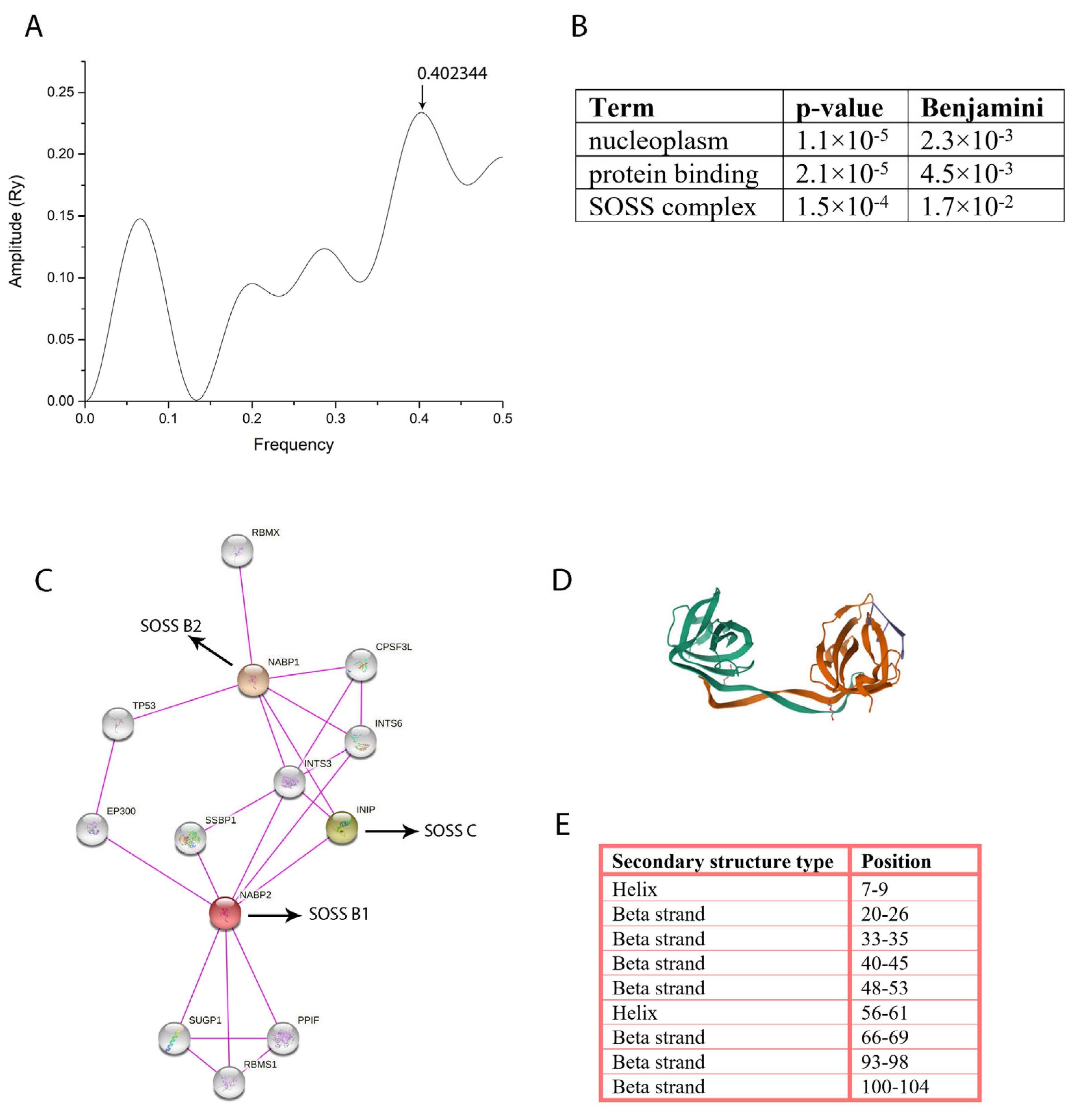
The sequencing of the HeLa cell’s 9462 baseline proteins was screened for potential interactions with the Pd(II) complex using cross-spectral analysis [14]. An IS (information spectrum) of the Pd(II) complex generated by the ISM showed a dominant peak at a frequency of 0.402 (Figure 1A), which was used for further cross-spectral analysis [13,15].
Cross-spectral analysis identified 106 proteins that were subsequently analyzed using the bioinformatics resource DAVID for gene enrichment analysis to determine enriched pathways and GO terms [16]. The Functional Annotation Chart revealed that the GO terms with the highest statistical significance were nucleoplasm and SOSS complex (Figure 1B, p-value after Benjamini–Hochberg correction < 0.05). The nucleoplasm contains a substantial portion of proteins organized as nuclear bodies, which are involved in the regulation of chromosome structure, DNA damage repairs, RNA processing, and epigenetic gene regulation. SOSS protein complex is involved in maintaining genomic stability [17]. Three identified proteins from the SOSS complex are SOSS C (gene name: INIP), SOSS B1 (gene name: NAPB2), and SOSS B2 (gene name: NAPB1). Figure 1C shows the interaction network of these proteins. Protein SOSS B1, encoded by the NAPB2 gene, consists predominantly of β-secondary structures organized into a curved beta-barrel (Figure 1D) [18]. Detailed inspection of the secondary structure of SOSS B1 showed the presence of two helices and seven beta strands (Figure 1E).
Figure 1. Characterization of the [Pd(dach)Cl2] and bioinformatics analysis. (A) The IS of [Pd(dach)Cl2] complex where the arrow indicates the peak position for potential interactions with HeLa cell proteins. Functional Annotation Chart (B) showing the Gene Ontology (GO) terms with the highest statistical significance (p-value after Benjamini correction < 0.05). Interaction network (C) of NAPB1, INIP, and NAPB2, retrieved from the STRING database, including only experimentally verified interactions. NABP1 (orange), NABP2 (red), and INIP (green) correspond to SOSS complex subunits B2, B1, and C, respectively. Crystal structure and secondary structure distribution of SOSS complex subunit B1 are shown in (D) and (E), respectively [18,19].
Inorganics 13 00215 g001

2.2. Differentially Abundant Proteins as a Result of [Pd(dach)Cl2] Treatment

A proteomic analysis was performed to identify target-specific proteins unique to the effects of the [Pd(dach)Cl2] complex in the HeLa cervical carcinoma cell line.
The 1384 identified proteins by comparative proteomics analysis were filtered to remove reverse sequences and proteins identified on one peptide, as well as yeast alcohol dehydrogenase (ADH). The final dataset contained 1255 proteins identified on two or more peptides with 121 proteins that had statistically significant differences in abundance (ANOVA ≤ 0.05) between [Pd(dach)Cl2]- treated HeLa cells and controls (Supplementary Table S1). From the 121 differentially abundant proteins (DAPs), 57 showed increased abundance and 64 showed decreased abundance in [Pd(dach)Cl2]- treated HeLa cells compared to controls. The differential expression is illustrated by volcano plot and hierarchical clustering (Figure 2A,B).
The most prominent biological process associated with the DAPs was RNA splicing (46%), followed by protein-containing complex organization, protein–RNA complex organization, protein folding chaperone, and cytoplasmic translation (Figure 2C). In terms of the DAP’s molecular functions, the majority, or 63%, had ATP hydrolysis activity, while the remaining functions involved different types of RNA and protein binding (Figure 2D). DAPs were mainly intracellular, with 25% located within the intracellular organelle lumen, 23% located within the cytosolic ribosome, 11% in the spliceosomal complex, 12% in different organelles, and 6% as free in the cytosol (Figure 2E). Only 17% of the DAPs were part of the extracellular exosome.

2.3. Altered Pathways in HeLa Cells Due to [Pd(dach)Cl2] Treatment

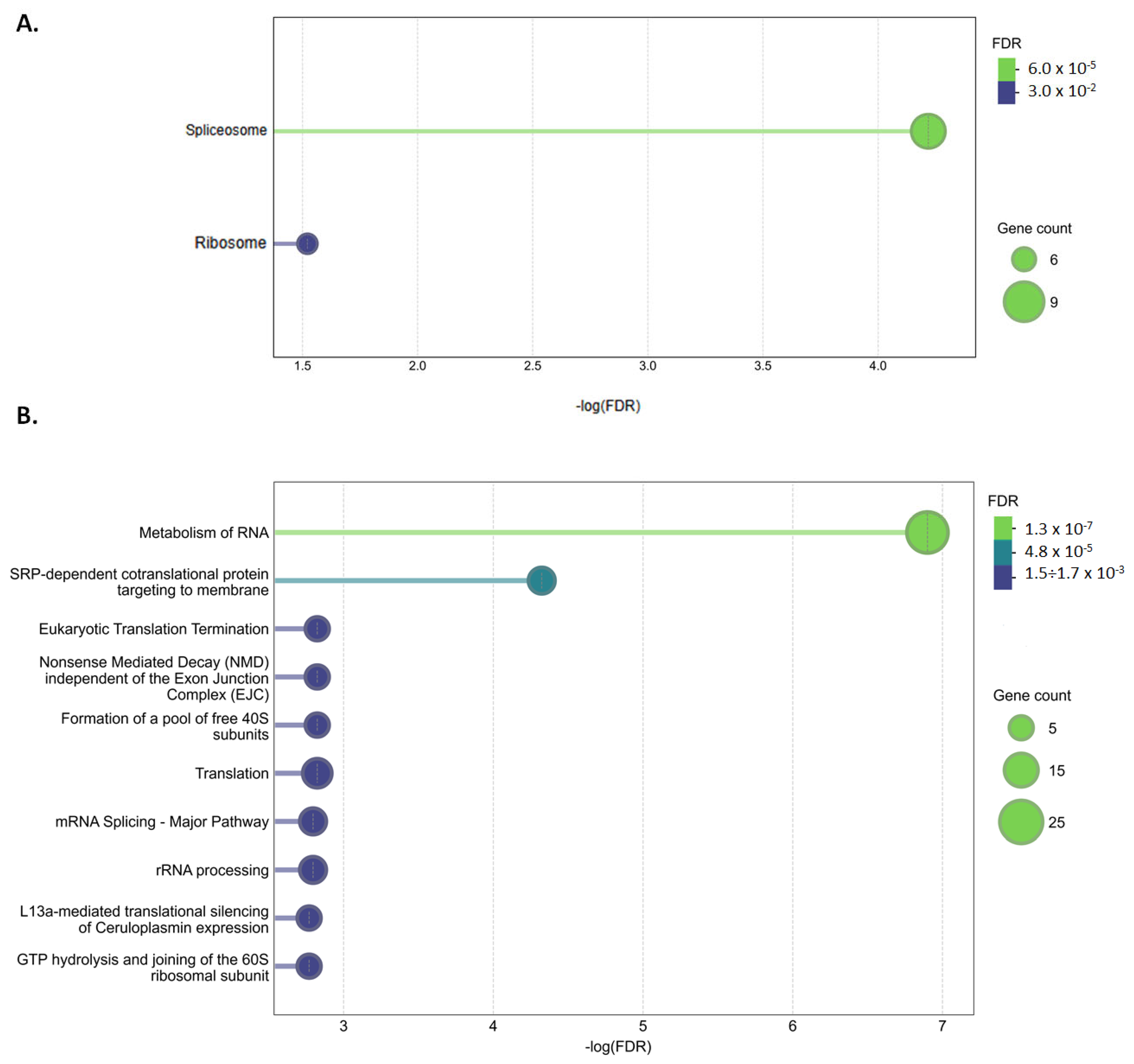
Enrichment analysis of canonical pathways using the KEGG database showed two associated pathways: Spliceosome (hsa03040) and Ribosome (hsa03010) (Figure 3A). Analysis with the Reactome Pathways database showed more than 20 significantly associated pathways, of which the highest significance showed metabolism of RNA, SRP-dependent co-translational protein targeting to membrane, translation, eukaryotic translation termination, Nonsense Mediated Decay (NMD) independent of the Exon Junction Complex (EJC), formation of a pool of free 40 S subunits, mRNA Splicing—Major Pathway, and rRNA processing (Figure 3B).
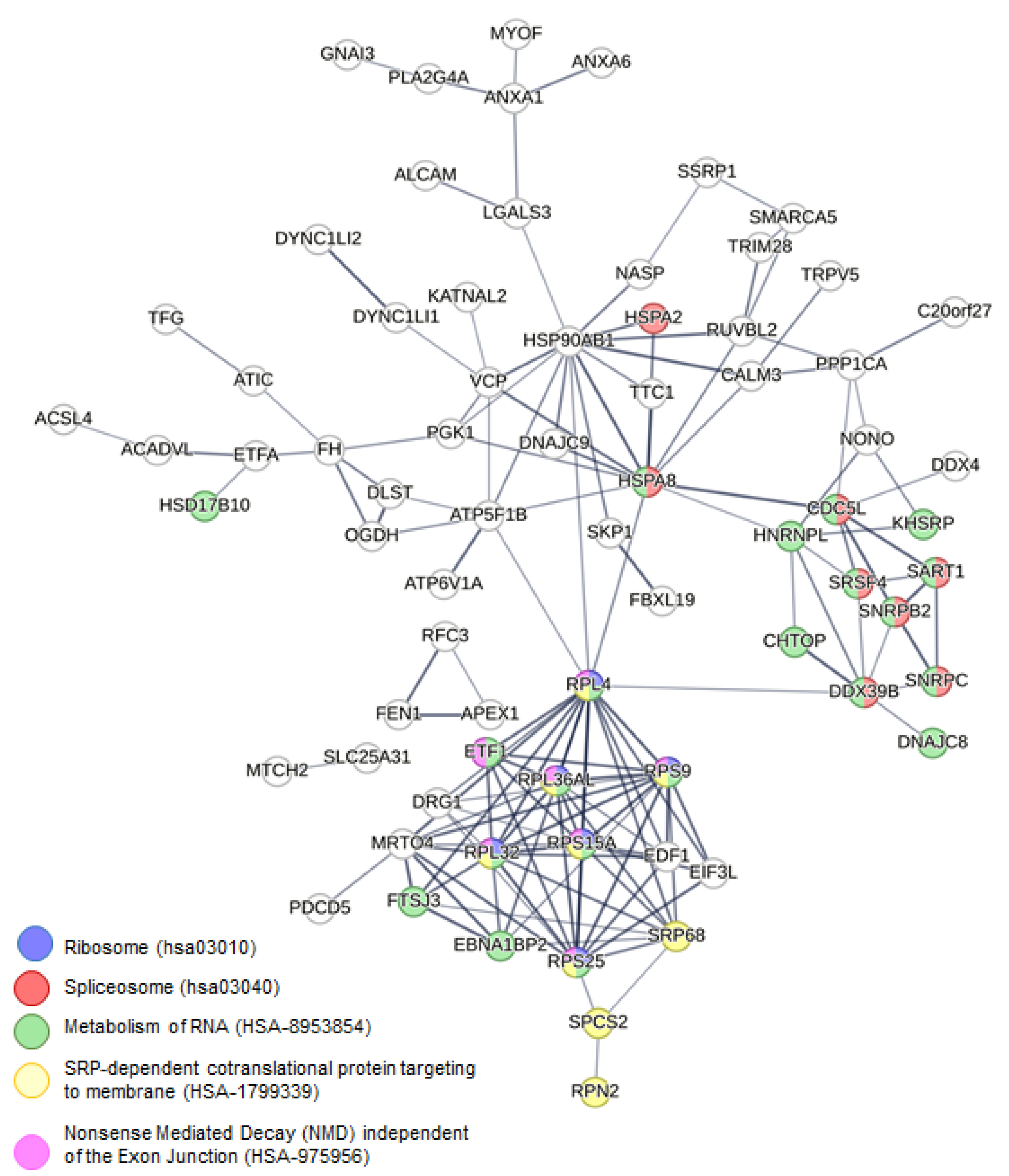
To further investigate the functional significance of DAPs, we constructed a protein–protein interaction network (Figure 4). This network showed that DAPs have significantly increased interactions among themselves compared to the same size and degree of distribution random set of proteins (PPI enrichment p-value = 1.56 × 10⁻⁹). Using this network, we have identified key hub genes using the Cytoscape plug-in cytoHubba v0.1 (Figure 5). The top 25 hub genes were ranked based on the Maximal Clique Centrality (MCC) approach, which prioritizes proteins with high connectivity within the network. As shown in Figure 5, the hub genes are color-coded from highly essential (red) to crucial (yellow), reflecting their potential regulatory importance. The group of highly essential proteins constitutes ribosomal proteins (RPL4, RPS9, RPL32, RPL36AL, RPS15A, RPS25), translational and transcriptional factors (ETF1, EDF1, DRG1), components of the ribosome assembly machinery (MRTO4), components of the signal recognition complexes that target secretory proteins to the rough endoplasmic reticulum membrane (SPCS2, SRP68) and several heat shock proteins involved in correct folding of proteins and degradation pathways (HSPA8), regulation of proteins involved in cell cycle control and signal transduction (HSP90AB1), and ATPases (ATP5F1B, VCP). These hub genes may serve as critical mediators of the observed cellular response and represent potential targets for further investigation.

3. Discussion

The potential protein targets of [Pd(dach)Cl2] in HeLa cells, as predicted using the information spectrum of the Pd(II) complex and cross-spectral analysis, were identified as the nucleoplasm and SOSS complex. The nucleoplasm is a complex fluid that contains RNA and proteins, and substantial fraction of nuclear bodies, namely nucleoli, speckles, and Cajal bodies. Many processes and mechanisms that undergo in the nucleoplasm remain to be revealed; however, nucleoli and speckles have been verified to represent active sites for ribosomal synthesis and RNA splicing, respectively [20]. Therefore, we might speculate that [Pd(dach)Cl2] targets proteins involved in ribosomal biogenesis and RNA splicing in HeLa cells. The computational method also suggested that the SOSS protein complex (Sensor of Single-stranded DNA) could be a potential protein target of the studied Pd(II) complex. The interaction network of SOSS complex predicted as Pd(II) complex interactors includes TP53 (tumor suppressor protein, p53) and EP300 (histone acetyltransferase, p300), which are involved in the cellular response to DNA damage and apoptosis [21,22]. In particular, p53 has been considered to be one of the classical types of tumor suppressors, also known as the “guardian of the genome”, and its inactivation leads to tumor growth and metastasis [23]. Changes in the p53 signaling pathway are one of the possible mechanisms by which Pd(II) complexes induce the inhibition of cell migration [24], significantly affecting HeLa cell viability. In addition to having a role in maintaining genomic stability and responds to DNA double-strand breaks [25], SOSS complex also plays a role in RNA –DNA interactions by promoting RNA degradation and interacting with the RNA polymerase II enzyme, which is crucial in the transcription process [26].
The comprehensive analysis of the proteomic alterations in HeLa cells resulting from the [Pd(dach)Cl2] treatment showed a strong potential for elucidating its cytotoxic effect on cancer cells. The analysis of the significantly enriched GO annotations of 121 proteins with altered abundance revealed a strong association with cytoplasmic translation, with a particular point toward RNA splicing and protein–RNA complex organization. Furthermore, KEGG pathway enrichment analysis revealed that DAPs play a significant role in the “Spliceosome” and “Ribosome” canonical pathways.
The spliceosome is a ribonucleoprotein complex in charge for RNA splicing, creating a continuous mRNA sequence that can be translated into protein. This complex ribonucleoprotein structure is essential for the accurate and efficient production of mature mRNA in eukaryotic cells. Disruptions in this pathway can lead to aberrant splicing, potentially resulting in the production of dysfunctional or non-functional proteins. Functional link between splicing anomalies and the evolution of cancer is well observed by a number of studies [27]. In vivo inhibition of the spliceosome is shown to impair survival and lead to metastatic affinity of MYC-dependent breast cancers [28], indicating that components of the spliceosome may be therapeutic targets. A recent study that analyzed the altered pathways in HeLa cells based on gene, protein, and phosphoprotein expression [29] revealed many spliceosome genes as overexpressed in HeLa cells, further confirming that overactivation of the spliceosome could play a significant role in the development, progression, and maintenance of cancer.
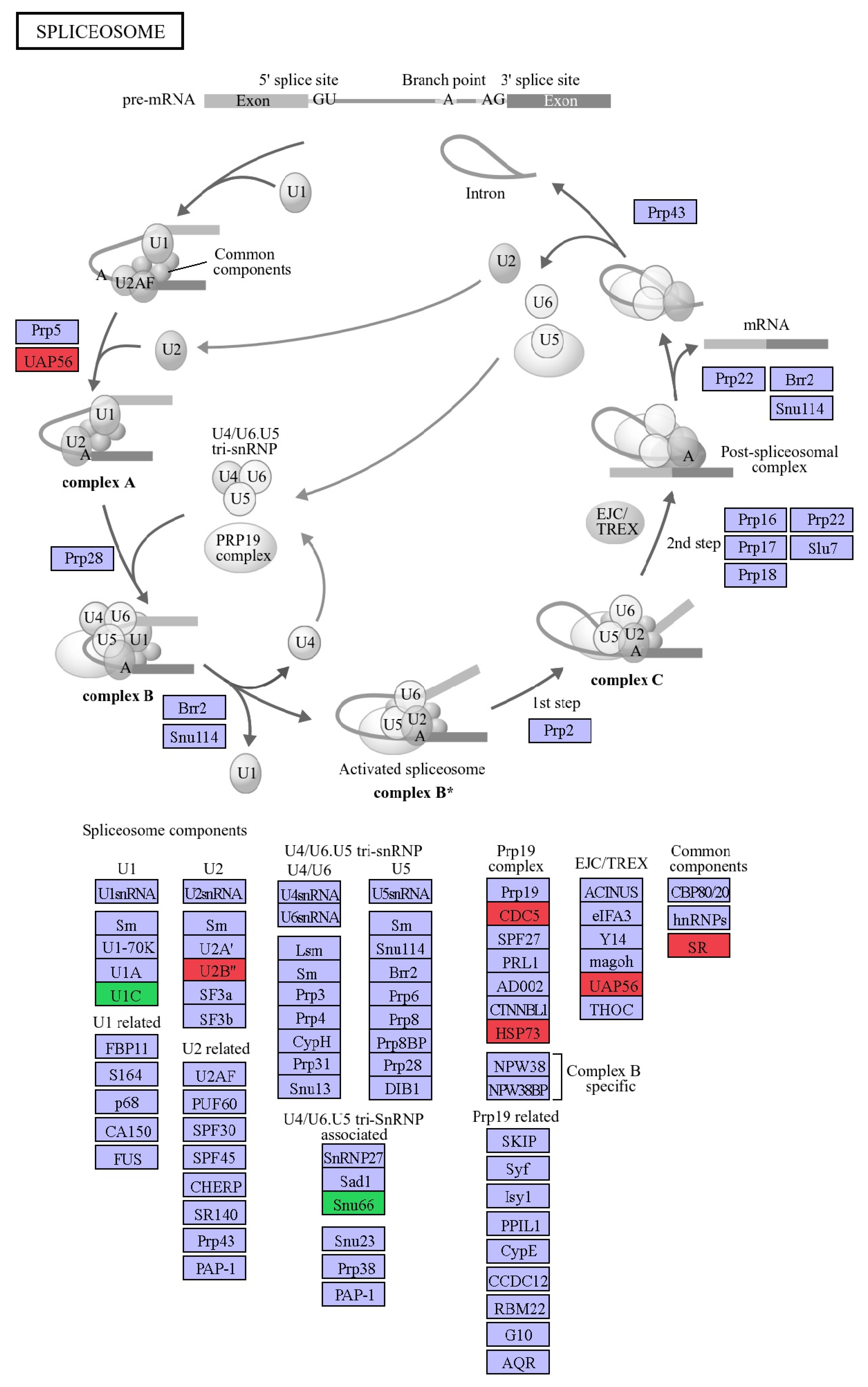
Our comparative proteomics approach identified nine DAPs that are part of the spliceosome, of which five up-regulated (HSPA8 (HSP73), SNRPB2 (U2B″), DDX39B (UAP56), CDC52 (CDC5), SRSF4 (SR)) and four down-regulated (SNRPC (U1C), SART1 (Snu66), RBMXL3, HSPA2) in [Pd(dach)Cl2]-treated HeLa cells compared to controls (Figure 6). For some of the up-regulated proteins, there is extensive data about their involvement in tumorigenesis. To name a few, the up-regulation of Heat shock 70 kDa protein 8 (HSPA8) is closely related to tumorigenesis and tumor progression in various cancer types [30,31]. SNRPB2, a component of the U2 snRNP, is implicated in various cancers, as it is significantly up-regulated and associated with a poor prognosis [32]. DExD-box helicase 39B (DDX39B), which participates in many steps of RNA metabolism beyond mRNA splicing [33], is highly correlated with tumor development and progression in breast [34], colorectal [35], prostate [36], and renal cancers [37] and melanoma [38].
Of particular interest for elucidating the effects of [Pd(dach)Cl2] on HeLa cells are spliceosome proteins that were down-regulated or repressed as a result of the treatment. One of these is RBMXL3, belonging to the U12-type spliceosomal complex that processes U12-type introns. The U12-type introns are rare (comprising <0.5% of introns) and processed by a specialized minor spliceosome distinct from the major (U2-type) spliceosome [39]. These introns are often spliced less efficiently, and disruptions in their processing can limit gene expression and impact cellular processes unrelated to the direct function of the host gene [40]. The reduced expression of RBMXL3 may contribute to widespread post-transcriptional changes following [Pd(dach)Cl2] exposure and could exert a potential therapeutic effect against cancer cells, which are worth further investigation.
We have also observed altered levels of HSPA2, the spliceosome-interacting chaperone protein, which has been previously implicated as a cancer biomarker across diverse tumor types [16]. It was among a small set of genes (alongside SNRPB and LSM7) used to build machine-learning models for pan-cancer classification, reflecting its broad relevance in spliceosomal integrity and possibly tumor cell vulnerability [41]. The lowered expression of HSPA2 in our data suggests that [Pd(dach)Cl2] may interfere not only with the core spliceosome but also with auxiliary proteins critical for its dynamic assembly and function. Such disruption can shift the precise stoichiometry required for standard spliceosome assembly and function in HeLa cells, modifying the proportion of isoforms [29]. Dysregulated splicing can produce protein isoforms with altered localization, function, or stability—some of which may promote oncogenesis, resistance to apoptosis, or cell cycle progression [42]. Some of these changes might be responsible for the observed cytotoxic effect of [Pd(dach)Cl2] on cancer cells [43].
Ribosome is the core molecular machinery that governs protein synthesis and is closely linked to cell activation and proliferation. It has been well established that elevated protein synthesis and up-regulated ribosome biogenesis are characteristic hallmarks of cancer cells [44,45]. Targeting ribosome biogenesis in various cancer types has been an ongoing activity, both in improving the specificity of existing drugs and in identifying new potent compounds with enhanced pharmacological properties [46]. Moreover, there is increasing evidence about the relationship between the expression level of certain ribosomal proteins and cancer progression and differentiation in various cancer types [47]. Comparative proteomics analysis of the [Pd(dach)Cl2] treatment of HeLa cells revealed altered levels of six ribosomal proteins—two up-regulated (RPL4, RPS25) and four down-regulated (RPL32, RPL36AL, RPS15A, RPS9) (Figure 7). One of these, RPL4, i.e., ribosomal protein L4, is vital for ribosome biogenesis and may play a role in transcriptional regulation [48]. Additionally, it is significantly involved in the p53 signaling pathway by direct interaction with MDM2 which is a negative regulator of p53, and suppresses MDM2-mediated p53 ubiquitination and degradation [49]. The overexpression of RPL4 also promotes the binding of MDM2 to RPL5 and RPL11, which further suppress MDM2 in cells [49]. Similarly, it has been demonstrated that ribosomal protein S25 (RPS25) also interacts with MDM2 and inhibits its E3 ligase activity, resulting in the reduction of MDM2-mediated p53 ubiquitination and subsequent stabilization and activation of p53 [50].
On the other hand, overexpression of RPL32 has been observed in lung [51], breast [52], and hepatocellular cancer [52]. High levels of RPL32 were associated with greater metastatic potential and unfavorable outcomes in patients while its silencing inhibits cancer proliferation. Similarly, overexpression of RPS15A and RPS9 has been observed in many cancer types. RPS15A has been implicated in the development and progression of liver, colorectal, and gastric cancers [53,54,55]. Down-regulation of RPS15A has been shown to inhibit cancer cell growth and trigger apoptosis in some cases [53,55]. Overexpression of RPS9 predicted poor prognosis of non-small cell lung cancer patients while its knockdown repressed cell proliferation, metastasis, and induced apoptosis [56]. RPS9 was also up-regulated in osteosarcoma through the activation of the MAPK signaling pathway, while its down-regulation inhibited cell growth in this cancer type [57]. The observed up-regulation of RPL4 and RPS25, which suppresses MDM2-mediated p53 degradation and leads to its activation, as well as the down-regulation of RPL32, RPS15A, and RPS9, which have confirmed role in tumor progression, strongly suggests the potential anti-tumor effect of the [Pd(dach)Cl2] treatment.
Analysis using the Reactome Pathways database, in addition to showing a significant association with “Translation” and “Splicing”, also indicated an alteration in NMD independent of the EJC pathway due to [Pd(dach)Cl2] treatment. This pathway is part of the mRNA surveillance pathway, which represents a cellular mechanism that ensures accurate gene expression by eliminating defective mRNAs and preventing the production of potentially toxic proteins [20]. The mRNA surveillance pathway relies on the ribosome to detect and signal the presence of problematic mRNA transcripts. Overactivation or failure of these pathways could lead to either excessive degradation of functional mRNAs or accumulation of faulty ones, both of which are potentially toxic to the cell.
In our study, we detected significantly altered amounts of four proteins involved in the mRNA surveillance pathway, with DDX39B being up-regulated and PABPC1L, PPP1CA, and ETF1 being down-regulated in the treated group. PABPC1L is a protein that plays a crucial role in cancer development and progression, particularly in the immune evasion and the survival of tumor cells. This protein is being investigated as a potential therapeutic target in various cancers, including renal cell carcinoma [58], colorectal cancer [59], and gastric cancer [60]. PPP1CA, which encodes the alpha catalytic subunit of Protein Phosphatase 1 (PP1), has been implicated in various cancers, and its role can be complex, sometimes acting as an oncogene and at other times as a tumor suppressor. Research suggests it can influence tumor cell growth, migration, invasion, and even apoptosis [61]. ETF1, or eukaryotic translation termination factor 1, is a protein involved in translation termination. Although not directly classified as a cancer gene, research suggests ETF1 overexpression may play a role in cancer development and progression in breast cancer and myeloid malignancies [62,63]. The enrichment of the mRNA surveillance pathway suggests that [Pd(dach)Cl2] disrupts the balance of RNA quality control. In particular, the fact that [Pd(dach)Cl2]-treated HeLa cells exhibit down-regulation of PABPC1L, PPP1CA, and ETF1, which is strongly correlated with cancer progression, points to its possible anti-cancer effects.
However, we should also point out that this study has some limitations. Since this was a proof-of-concept study, the sample size was not adequate to perform multiple testing correction. Therefore, identification of DAPs was based on the unadjusted p-values. Despite this fact, our findings were in line with the existing literature of HeLa cells’ protein and mRNA expression. Moreover, the observed down-regulation of a number of proteins highly correlated with tumor development and progression was consistent with the observed cytotoxic effect of [Pd(dach)Cl2] treatment. Although this confirms the validity of the reported results, further validation in the context of statistically well-powered study is required.

4. Materials and Methods

4.1. Reagents

Human cervical carcinoma (HeLa) cells were acquired from the American TypeCulture Collection (ATCC, Manassas, VA, USA). All chemicals were purchased from Sigma-Aldrich, GmbH (Taufkirchen, Germany). The [Pd(dach)Cl2] was synthesized and characterized in the lab of Prof. Biljana Petrović (Faculty of Natural Sciences, University of Kragujevac, Kragujevac, Serbia) [64].

4.2. Cell Culture

HeLa cells were cultured in DMEM medium at 37 °C and 5% CO2. After being treated with 50 µg/mL [Pd(dach)Cl2] (dissolved in 7% DMSO in PBS), the cells were incubated for 48 h. Control cells were treated with 7% DMSO in PBS for 48 h. [Pd(dach)Cl2]-treated HeLa cells and controls were prepared in five replicates each.

4.3. Sample Preparation

The frozen cells from each biological replicate were resuspended in 100 μL of Lysis buffer (4% SDS, 5 mM MgCl2·6H2O, 10 mM CHAPS, 100 mM NH4HCO3, 50 mM DTT), then lysed by freezing at −80 °C for 15 min, and subsequently thawing for a total of three times. The samples were then sonicated for 20 min in an ice bath to dissolve them further. The protein content was quantified using the Bradford method [65] and stored at −80 °C until use. Samples were prepared for LC-MS/MS using the RapiGest protocol as previously described [66]. The starting protein amount for tryptic digestion per sample was 25 µg protein and the final protein concentration was 200 ng/μL. Internal standard protein, yeast ADH, was added to all samples with a final concentration of 25 fmol/μL.

4.4. LC-MS/MS Acquisition

The proteomics profiling was performed using label-free, data-independent nano-LC-MS/MS acquisition on ACQUITY UPLC® M-Class (Waters Corporation, Milford, MA, USA) coupled with SYNAPT G2-Si High-Definition Mass Spectrometer (Waters Corporation). Data were obtained using the ultra-definition mass spectrometry (UDMSE) mode described in detail elsewhere [67]. The protein load for the UDMSE runs was determined to be 150 ng by testing pool samples (containing an equal amount of each of the ten individual samples), starting from 0.5 to 3.0 µL of sample per run, and processing in ProteinLynx Global SERVER (PLGS, version 3.0.3, Waters Corp.). One test run for each sample was performed for quality assurance and validation of the protein concentration, followed by run at 150 ng. LC and MS parameters were as previously described in detail [68].

4.5. MS Data Processing and Identification

Test runs were processed with PLGS (Waters Corporation) with the following settings: (1) Low-energy (LE) and high-energy (HE) thresholds of 450 counts and 20 counts, respectively; (2) Precursor and fragment ion mass tolerances—auto; (3) One missed cleavages, carbamidomethyl C as a fixed modification, and oxidized M as a variable modification; (4) A minimum of two fragment ion matches per peptide identification and five fragment ion matches per protein identification, with at least one peptide match per protein identification; (5) The protein false discovery rate (FDR) < 1%. The typical RMS error for precursor and product ions were ±5 and ±10 ppm, respectively.
Comparative proteomics analysis was performed by Progenesis QIP version 4.1 (Nonlinear dynamics, Waters Corporation) with the following settings: (1) LE and HE threshold—auto; (2) reference run—auto; (3) normalization—“normalize to all proteins”; (4) digest reagent—trypsin; (5) maximum missed cleavages—one; (6) maximum protein mass—250 kDa; (7) fixed modifications—carbamidomethyl C; (8) variable modification— oxidation M; (9) peptide tolerance—auto; (10) fragment tolerance—auto; (11) FDR < 1%; (12) minimum of two fragment ion matches per peptide identification and five fragment ion matches per protein identification, with at least one peptide match per protein identification; (13) quantification based on non-conflicting peptides; (14) grouping of similar proteins. A combined target-decoy database based on the UniProtKB/Swiss-Prot database with 20,370 proteins (June 2020) was used. The obtained identifications were filtrated to remove peptides ≤ 6 amino acids and a score below 4. Data was exported as a .csv output file for subsequent analysis. The calculated FDR on the whole dataset was 3.2%.

4.6. Proteomics Data Analysis

The criteria for the selection of DAPs were identification based on ≥2 peptides and an ANOVA p-value ≤ 0.05. The enriched GO annotation terms and pathways were retrieved using the Cytoscape plug-in ClueGO [69]. ClueGo selection criteria were the following: Analysis mode—Functional analysis; Organism—Homo sapiens [9606]; Show only pathways with pV ≤ 0.05; GO term selection—GO tree interval—3 to 8 with a minimum of 4 genes to be associated with a term, and these genes represent at least 1% from the total number of associated genes; GO pathway selection—minimum of 3 genes to be associated with a pathway, and these genes represent at least 4% from the total number of associated genes; GO term/pathway network connectivity (Kappa Score) = 0.4; Two-sided hypergeometric test and Bonferroni step down with cutoff p ≤ 0.05; Grouping options—YES; Leading Group Term based on—Highest Significance (Kappa score) with min 50% of genes for group merge and min 50% of terms for group merge. STRING analysis [70] settings: (1) complete STRING network; (2) evidence setting; (3) all active interaction sources; (4) confidence score = 0.600; and (5) max number of interactors to show—none (query proteins only). Cytoscape plug-in cytoHubba [71] was used to rank nodes (proteins) in the network by their network features using the MCC topological analysis method.

4.7. Computational Biology Analysis of the [Pd(dach)Cl2] Interaction with HeLa Cellular Proteins

The ISM focused on the cross-spectral analysis of the informational spectra of the Pd(II) complex and proteins expressed in HeLa cells, serving as a baseline. The workflow consisted of the following steps: (1) Each molecule is presented as a series of numbers based on the electron–ion interaction potential (EIIP) [15], and each amino acid in the protein sequence is replaced by its own EIIP value; (2) Numerical sequence was then transformed into an IS by Fourier transform; (3) The spectra of two molecules, which interact potentially, were multiplied to obtain cross-spectrum (CS) results. The approach used for small molecules (ISM-SM) is modified and utilized to generate the IS of the Pd(II) complex [13].
The list of genes expressed in HeLa cells above the baseline (TPM > 2.0) was retrieved from the Expression Atlas [72]. Protein sequences needed for further ISM analysis were obtained from the UniProt database [73]. The potential protein interactors of the Pd (II) complex in HeLa cells were further analyzed by enrichment analysis using DAVID [16].

5. Conclusions

In conclusion, the significant enrichment of the ribosome as the core molecular machinery governing protein synthesis, along with the spliceosome and the mRNA surveillance pathways in HeLa cervical cancer cells treated with the [Pd(dach)Cl2] complex, suggests a broad disruption in post-transcriptional gene regulation mechanisms. These pathways are relevant as potential targets, as they play essential roles in RNA processing and quality control. The findings presented in this work indicate that the [Pd(dach)Cl2] complex may exert its effects by interfering with RNA metabolism, which might be accompanied with alterations of the expression of key regulatory proteins involved in cell survival and proliferation. Moreover, by using a combination of proteomics analysis and ISM, we predicted the involvement of specific ribosomal proteins in p53 stabilization and activation, which needs to be further experimentally documented. This hypothesis, as well as the confirmed sensitivity of cancer cells to spliceosomal stress, further underlines the therapeutic relevance of these pathways and provides mechanistic insights into the anti-cancer potential of [Pd(dach)Cl2].

Supplementary Materials

The following supporting information can be downloaded at: https://www.mdpi.com/article/10.3390/inorganics13070215/s1, Table S1: List of identified proteins from [Pd(dach)Cl2]-treated HeLa cells and untreated HeLa cells (control group) based on ≥2 peptides.

Author Contributions

Conceptualization, M.P. and K.D.; methodology, K.D., V.R., B.G., and M.S. (Milan Senćanski); formal analysis, K.D. and V.R.; investigation, V.R., K.D., M.D.N., and J.Ž.; writing—original draft preparation, V.R., K.D., M.P. and M.S. (Milutin Stepić); writing—review and editing, M.S. (Milutin Stepić), J.Ž., M.D.N., M.S. (Milan Senćanski), and B.G.; funding acquisition, M.P. and K.D. All authors have read and agreed to the published version of the manuscript.

Funding

The research work was funded and APC paid by the Ministry of Ministry of Science, Technological Development, and Innovation of the Republic of Serbia (Grant No. 451-03-136/2025-03/200017). Mobility scheme to support joint experiments was supported by the Western Balkans Fund Move Grants (PROMETHEAN I and PROMETHEAN II, MO-1-041 and MO-5-084, respectively).

Institutional Review Board Statement

Not applicable.

Informed Consent Statement

Not applicable.

Data Availability Statement

The original contributions presented in this study are included in the article/Supplementary Materials. Further inquiries can be directed to the corresponding author(s).

Acknowledgments

The authors are thankful to Lela Korićanac (Department of Molecular Biology and Endocrinology, VINČA Institute of Nuclear Sciences, National Institute of the Republic of Serbia, University of Belgrade, Serbia) for her assistance in the cell experiments and Iva Popović (COHERENCE-Centre, Department of Atomic Physics, VINČA Institute of Nuclear Sciences, National Institute of the Republic of Serbia, University of Belgrade, Serbia) for her help in the manuscript preparation.

Conflicts of Interest

The authors declare no conflicts of interest. The funders had no role in the design of the study; in the collection, analyses, or interpretation of data; in the writing of the manuscript; or in the decision to publish the results.

Abbreviations

The following abbreviations are used in this manuscript:
DAPsDifferentially abundant proteins
DNADesoxyribonucleic acid
EJCExon Junction Complex
GOGene Ontology
HSPHeat Shock Protein
ISMInformational spectrum method
LC-MS/MSLiquid Chromatography-Tandem mass spectrometry
MCCMaximal Clique Centrality
NDMNonsense Mediated Decay
PPIProtein–protein interaction
RNARibonucleic Acid
snRNPsSmall Nuclear Ribonucleoproteins
SOSS DNASensor of the single-stranded DNA
TP53Tumor Suppressor Protein p53

References

  1. Fowler, J.R.; Maani, E.V.; Dunton, C.J.; Gasalberti, D.P.; Jack, B.W. Cervical Cancer. In StatPearls; StatPearls Publishing: Treasure Island, FL, USA, 2025. [Google Scholar]
  2. Sung, H.; Ferlay, J.; Siegel, R.L.; Laversanne, M.; Soerjomataram, I.; Jemal, A.; Bray, F. Global Cancer Statistics 2020: GLOBOCAN Estimates of Incidence and Mortality Worldwide for 36 Cancers in 185 Countries. CA Cancer J. Clin. 2021, 71, 209–249. [Google Scholar] [CrossRef]
  3. Arbyn, M.; Weiderpass, E.; Bruni, L.; De Sanjosé, S.; Saraiya, M.; Ferlay, J.; Bray, F. Estimates of incidence and mortality of cervical cancer in 2018: A worldwide analysis. Lancet Glob. Health 2020, 8, e191–e203. [Google Scholar] [CrossRef] [PubMed]
  4. Ralić, V.; Nešić, M.D.; Dučić, T.; Stepić, M.; Korićanac, L.; Davalieva, K.; Petković, M. Analysis of Biomolecular Changes in HeLa Cervical Cancer Cell Line Induced by Interaction with [Pd(dach)Cl2]. Inorganics 2025, 13, 20. [Google Scholar] [CrossRef]
  5. Yodoshi, M.; Okabe, N. Structures and Interaction with DNA of Ternary Palladium(II) Complexes: [Pd(Gly)(X)] (Gly=Glycine; X=2,2′-Bipyridine, 1,10-Phenanthroline and 2,2′-Bi-pyridylamine). Chem. Pharm. Bull. 2008, 56, 908–914. [Google Scholar] [CrossRef]
  6. Gao, E.; Liu, F.; Zhu, M.; Wang, L.; Huang, Y.; Liu, H.; Ma, S.; Shi, Q.; Wang, N. Synthesis, characterization, DNA interaction, and cytotoxicity of novel Pd(II) and Pt(II) complexes. J. Enzyme Inhib. Med. Chem. 2010, 25, 557–564. [Google Scholar] [CrossRef]
  7. Hobart, D.B.; Berg, M.A.G.; Merola, J.S. Bis-glycinato complexes of palladium(II): Synthesis, structural determination, and hydrogen bonding interactions. Inorganica Chim. Acta 2014, 423, 21–30. [Google Scholar] [CrossRef]
  8. Hobart, D.B.; Berg, M.A.G.; Rogers, H.M.; Merola, J.S. Synthesis, Characterization, and Non-Covalent Interactions of Palladium(II)-Amino Acid Complexes. Molecules 2021, 26, 4331. [Google Scholar] [CrossRef]
  9. Hobart, D.B.; Merola, J.S.; Rogers, H.M.; Sahgal, S.; Mitchell, J.; Florio, J.; Merola, J.W. Synthesis, Structure, and Catalytic Reactivity of Pd(II) Complexes of Proline and Proline Homologs. Catalysts 2019, 9, 515. [Google Scholar] [CrossRef]
  10. Wang, Y.; Chiu, J.-F. Proteomic Approaches in Understanding Action Mechanisms of Metal-Based Anticancer Drugs. Met.-Based Drugs 2008, 2008, 716329. [Google Scholar] [CrossRef]
  11. Fotis, C.; Antoranz, A.; Hatziavramidis, D.; Sakellaropoulos, T.; Alexopoulos, L.G. Network-based technologies for early drug discovery. Drug Discov. Today 2018, 23, 626–635. [Google Scholar] [CrossRef]
  12. Nešić, M.D.; Dučić, T.; Gonçalves, M.; Stepić, M.; Algarra, M.; Soto, J.; Gemović, B.; Bandosz, T.J.; Petković, M. Biochemical changes in cancer cells induced by photoactive nanosystem based on carbon dots loaded with Ru-complex. Chem. Biol. Interact. 2022, 360, 109950. [Google Scholar] [CrossRef]
  13. Sencanski, M.; Perovic, V.; Pajovic, S.B.; Adzic, M.; Paessler, S.; Glisic, S. Drug Repurposing for Candidate SARS-CoV-2 Main Protease Inhibitors by a Novel In Silico Method. Molecules 2020, 25, 3830. [Google Scholar] [CrossRef] [PubMed]
  14. Veljkovic, N.; Glisic, S.; Prljic, J.; Perovic, V.; Botta, M.; Veljkovic, V. Discovery of New Therapeutic Targets by the Informational Spectrum Method. Curr. Protein Pept. Sci. 2008, 9, 493–506. [Google Scholar] [CrossRef] [PubMed]
  15. Veljkovic, V. A Theoretical Approach to the Preselection of Carcinogens and Chemical Carcinogenesis; Gordon and Breach Science Publishers: New York, NY, USA, 1980. [Google Scholar]
  16. Sherman, B.T.; Hao, M.; Qiu, J.; Jiao, X.; Baseler, M.W.; Lane, H.C.; Imamichi, T.; Chang, W. DAVID: A web server for functional enrichment analysis and functional annotation of gene lists (2021 update). Nucleic Acids Res. 2022, 50, W216–W221. [Google Scholar] [CrossRef]
  17. Huang, J.; Gong, Z.; Ghosal, G.; Chen, J. SOSS Complexes Participate in the Maintenance of Genomic Stability. Mol. Cell 2009, 35, 384–393. [Google Scholar] [CrossRef]
  18. Ren, W.; Chen, H.; Sun, Q.; Tang, X.; Lim, S.C.; Huang, J.; Song, H. Structural Basis of SOSS1 Complex Assembly and Recognition of ssDNA. Cell Rep. 2014, 6, 982–991. [Google Scholar] [CrossRef]
  19. Li, Y.H.; Gao, Z.Q.; Dong, Y.H. Crystal structure of SSB from homo sapiens: 5d8e. 2016. Available online: https://www.rcsb.org/structure/5D8E (accessed on 1 June 2025).
  20. Zidovska, A. The self-stirred genome: Large-scale chromatin dynamics, its biophysical origins and implications. Curr. Opin. Genet. Dev. 2020, 61, 83–90. [Google Scholar] [CrossRef]
  21. Hanel, W.; Moll, U.M. Links between mutant p53 and genomic instability. J. Cell Biochem. 2012, 113, 433–439. [Google Scholar] [CrossRef] [PubMed]
  22. Yang, H.; Salz, T.; Zajac-Kaye, M.; Liao, D.; Huang, S.; Qiu, Y. Overexpression of histone deacetylases in cancer cells is controlled by interplay of transcription factors and epigenetic modulators. FASEB J. 2014, 28, 4265–4279. [Google Scholar] [CrossRef]
  23. Levine, A.J.; Oren, M. The first 30 years of p53: Growing ever more complex. Nat. Rev. Cancer 2009, 9, 749–758. [Google Scholar] [CrossRef]
  24. Bi, Y.; Kong, P.; Zhang, L.; Cui, H.; Xu, X.; Chang, F.; Yan, T.; Li, J.; Cheng, C.; Song, B.; et al. EP300 as an oncogene correlates with poor prognosis in esophageal squamous carcinoma. J. Cancer 2019, 10, 5413–5426. [Google Scholar] [CrossRef]
  25. Richard, D.J.; Bolderson, E.; Cubeddu, L.; Wadsworth, R.I.M.; Savage, K.; Sharma, G.G.; Nicolette, M.L.; Tsvetanov, S.; McIlwraith, M.J.; Pandita, R.K.; et al. Single-stranded DNA-binding protein hSSB1 is critical for genomic stability. Nature 2008, 453, 677–681. [Google Scholar] [CrossRef]
  26. Long, Q.; Sebesta, M.; Sedova, K.; Haluza, V.; Alagia, A.; Liu, Z.; Stefl, R.; Gullerova, M. The phosphorylated trimeric SOSS1 complex and RNA polymerase II trigger liquid-liquid phase separation at double-strand breaks. Cell Rep. 2023, 42, 113489. [Google Scholar] [CrossRef] [PubMed]
  27. Venables, J.P.; Klinck, R.; Koh, C.; Gervais-Bird, J.; Bramard, A.; Inkel, L.; Durand, M.; Couture, S.; Froehlich, U.; Lapointe, E.; et al. Cancer-associated regulation of alternative splicing. Nat. Struct. Mol. Biol. 2009, 16, 670–676. [Google Scholar] [CrossRef] [PubMed]
  28. Hsu, T.Y.-T.; Simon, L.M.; Neill, N.J.; Marcotte, R.; Sayad, A.; Bland, C.S.; Echeverria, G.V.; Sun, T.; Kurley, S.J.; Tyagi, S.; et al. The spliceosome is a therapeutic vulnerability in MYC-driven cancer. Nature 2015, 525, 384–388. [Google Scholar] [CrossRef]
  29. Higareda-Almaraz, J.C.; Valtierra-Gutiérrez, I.A.; Hernandez-Ortiz, M.; Contreras, S.; Hernandez, E.; Encarnacion, S. Analysis and Prediction of Pathways in HeLa Cells by Integrating Biological Levels of Organization with Systems-Biology Approaches. PLoS ONE 2013, 8, e65433. [Google Scholar] [CrossRef]
  30. Zhang, Y.; Shan, N.; Zhou, W.; Zhang, S. Identification of HSPA8 as a candidate biomarker for endometrial carcinoma by using iTRAQ-based proteomic analysis. OncoTargets Ther. 2016, 2016, 2169–2179. [Google Scholar] [CrossRef]
  31. Li, B.; Ming, H.; Qin, S.; Zhou, L.; Huang, Z.; Jin, P.; Peng, L.; Luo, M.; Zhang, T.; Wang, K.; et al. HSPA8 Activates Wnt/β-Catenin Signaling to Facilitate BRAF V600E Colorectal Cancer Progression by CMA-Mediated CAV1 Degradation. Adv. Sci. 2024, 11, 2306535. [Google Scholar] [CrossRef]
  32. Wu, J.; Lu, F.; Yu, B.; Wang, W.; Ye, X. The oncogenic role of SNRPB in human tumors: A pan-cancer analysis. Front. Mol. Biosci. 2022, 9, 994440. [Google Scholar] [CrossRef]
  33. Szymura, S.J.; Bernal, G.M.; Wu, L.; Zhang, Z.; Crawley, C.D.; Voce, D.J.; Campbell, P.-A.; Ranoa, D.E.; Weichselbaum, R.R.; Yamini, B. DDX39B interacts with the pattern recognition receptor pathway to inhibit NF-κB and sensitize to alkylating chemotherapy. BMC Biol. 2020, 18, 32. [Google Scholar] [CrossRef]
  34. Wang, L.; Wang, Y.; Su, B.; Yu, P.; He, J.; Meng, L.; Xiao, Q.; Sun, J.; Zhou, K.; Xue, Y.; et al. Transcriptome-wide analysis and modelling of prognostic alternative splicing signatures in invasive breast cancer: A prospective clinical study. Sci. Rep. 2020, 10, 16504. [Google Scholar] [CrossRef] [PubMed]
  35. Zhang, H.; He, C.; Guo, X.; Fang, Y.; Lai, Q.; Wang, X.; Pan, X.; Li, H.; Qin, K.; Li, A.; et al. DDX39B contributes to the proliferation of colorectal cancer through direct binding to CDK6/CCND1. Cell Death Discov. 2022, 8, 30. [Google Scholar] [CrossRef] [PubMed]
  36. Nakata, D.; Nakao, S.; Nakayama, K.; Araki, S.; Nakayama, Y.; Aparicio, S.; Hara, T.; Nakanishi, A. The RNA helicase DDX39B and its paralog DDX39A regulate androgen receptor splice variant AR-V7 generation. Biochem. Biophys. Res. Commun. 2017, 483, 271–276. [Google Scholar] [CrossRef] [PubMed]
  37. Meng, T.; Huang, R.; Zeng, Z.; Huang, Z.; Yin, H.; Jiao, C.; Yan, P.; Hu, P.; Zhu, X.; Li, Z.; et al. Identification of Prognostic and Metastatic Alternative Splicing Signatures in Kidney Renal Clear Cell Carcinoma. Front. Bioeng. Biotechnol. 2019, 7, 270. [Google Scholar] [CrossRef]
  38. Walbrecq, G.; Lecha, O.; Gaigneaux, A.; Fougeras, M.R.; Philippidou, D.; Margue, C.; Tetsi Nomigni, M.; Bernardin, F.; Dittmar, G.; Behrmann, I.; et al. Hypoxia-Induced Adaptations of miRNomes and Proteomes in Melanoma Cells and Their Secreted Extracellular Vesicles. Cancers 2020, 12, 692. [Google Scholar] [CrossRef]
  39. Turunen, J.J.; Niemelä, E.H.; Verma, B.; Frilander, M.J. The significant other: Splicing by the minor spliceosome. WIREs RNA 2013, 4, 61–76. [Google Scholar] [CrossRef]
  40. Alioto, T.S. U12DB: A database of orthologous U12-type spliceosomal introns. Nucleic Acids Res. 2007, 35, D110–D115. [Google Scholar] [CrossRef]
  41. Ye, Z.; Bing, A.; Zhao, S.; Yi, S.; Zhan, X. Comprehensive analysis of spliceosome genes and their mutants across 27 cancer types in 9070 patients: Clinically relevant outcomes in the context of 3P medicine. EPMA J. 2022, 13, 335–350. [Google Scholar] [CrossRef]
  42. Peng, Q.; Zhou, Y.; Oyang, L.; Wu, N.; Tang, Y.; Su, M.; Luo, X.; Wang, Y.; Sheng, X.; Ma, J.; et al. Impacts and mechanisms of alternative mRNA splicing in cancer metabolism, immune response, and therapeutics. Mol. Ther. 2022, 30, 1018–1035. [Google Scholar] [CrossRef]
  43. Kitamura, K.; Nimura, K. Regulation of RNA Splicing: Aberrant Splicing Regulation and Therapeutic Targets in Cancer. Cells 2021, 10, 923. [Google Scholar] [CrossRef]
  44. Ruggero, D.; Pandolfi, P.P. Does the ribosome translate cancer? Nat. Rev. Cancer 2003, 3, 179–192. [Google Scholar] [CrossRef] [PubMed]
  45. Van Riggelen, J.; Yetil, A.; Felsher, D.W. MYC as a regulator of ribosome biogenesis and protein synthesis. Nat. Rev. Cancer 2010, 10, 301–309. [Google Scholar] [CrossRef] [PubMed]
  46. Zisi, A.; Bartek, J.; Lindström, M.S. Targeting Ribosome Biogenesis in Cancer: Lessons Learned and Way Forward. Cancers 2022, 14, 2126. [Google Scholar] [CrossRef]
  47. Ramalho, S.; Dopler, A.; Faller, W.J. Ribosome specialization in cancer: A spotlight on ribosomal proteins. NAR Cancer 2024, 6, zcae029. [Google Scholar] [CrossRef] [PubMed]
  48. Trifa, Y.; Privat, I.; Gagnon, J.; Baeza, L.; Lerbs-Mache, S. The Nuclear RPL4 Gene Encodes a Chloroplast Protein That Co-purifies with the T7-like Transcription Complex as Well as Plastid Ribosomes. J. Biol. Chem. 1998, 273, 3980–3985. [Google Scholar] [CrossRef]
  49. He, X.; Li, Y.; Dai, M.-S.; Sun, X.-X. Ribosomal protein L4 is a novel regulator of the MDM2-p53 loop. Oncotarget 2016, 7, 16217–16226. [Google Scholar] [CrossRef]
  50. Zhang, X.; Wang, W.; Wang, H.; Wang, M.-H.; Xu, W.; Zhang, R. Identification of ribosomal protein S25 (RPS25)–MDM2–p53 regulatory feedback loop. Oncogene 2013, 32, 2782–2791. [Google Scholar] [CrossRef]
  51. Xie, J.; Zhang, W.; Liang, X.; Shuai, C.; Zhou, Y.; Pan, H.; Yang, Y.; Han, W. RPL32 Promotes Lung Cancer Progression by Facilitating p53 Degradation. Mol. Ther.—Nucleic Acids 2020, 21, 75–85. [Google Scholar] [CrossRef]
  52. Xu, L.; Wang, L.; Jiang, C.; Zhu, Q.; Chen, R.; Wang, J.; Wang, S. Biological effect of ribosomal protein L32 on human breast cancer cell behavior. Mol. Med. Rep. 2020, 22, 2478–2486. [Google Scholar] [CrossRef]
  53. Xu, M.; Wang, Y.; Chen, L.; Pan, B.; Chen, F.; Fang, Y.; Yu, Z.; Chen, G. Down-regulation of ribosomal protein S15A mRNA with a short hairpin RNA inhibits human hepatic cancer cell growth in vitro. Gene 2014, 536, 84–89. [Google Scholar] [CrossRef]
  54. Chen, J.; Wei, Y.; Feng, Q.; Ren, L.; He, G.; Chang, W.; Zhu, D.; Yi, T.; Lin, Q.; Tang, W.; et al. Ribosomal protein S15A promotes malignant transformation and predicts poor outcome in colorectal cancer through misregulation of p53 signaling pathway. Int. J. Oncol. 2016, 48, 1628–1638. [Google Scholar] [CrossRef] [PubMed]
  55. Shi, D.; Liu, J. RPS15a Silencing Suppresses Cell Proliferation and Migration of Gastric Cancer. Yonsei Med. J. 2018, 59, 1166. [Google Scholar] [CrossRef] [PubMed]
  56. Kong, Y.; Shuangshuang, D.; Liang, X.; Zhou, X. RPS9 promotes the progression of NSCLC via activation Stat3 and Erk signaling pathways. J. Cancer 2022, 13, 1346–1355. [Google Scholar] [CrossRef]
  57. Cheng, D.; Zhu, B.; Li, S.; Yuan, T.; Yang, Q.; Fan, C. Down-regulation of RPS9 Inhibits Osteosarcoma Cell Growth through Inactivation of MAPK Signaling Pathway. J. Cancer 2017, 8, 2720–2728. [Google Scholar] [CrossRef]
  58. Shu, G.; Chen, M.; Liao, W.; Fu, L.; Lin, M.; Gui, C.; Cen, J.; Lu, J.; Chen, Z.; Wei, J.; et al. PABPC1L Induces IDO1 to Promote Tryptophan Metabolism and Immune Suppression in Renal Cell Carcinoma. Cancer Res. 2024, 84, 1659–1679. [Google Scholar] [CrossRef] [PubMed]
  59. Wu, Y.-Q.; Ju, C.-L.J.; Wang, B.-J.; Wang, R.-G. PABPC1L depletion inhibits proliferation and migration via blockage of AKT pathway in human colorectal cancer cells. Oncol. Lett. 2019, 17, 3439–3445. [Google Scholar] [CrossRef]
  60. An, T.; Deng, L.; Wang, Y.; Yang, Z.; Chai, C.; Ouyang, J.; Lu, X.; Zhang, C. The Prognostic Impacts of PABPC1 Expression on Gastric Cancer Patients. Future Oncol. 2021, 17, 4471–4479. [Google Scholar] [CrossRef]
  61. Castro, M.E.; Ferrer, I.; Cascon, A.; Guijarro, M.V.; Lleonart, M.; Cajal, S.R.Y.; Leal, J.F.M.; Robledo, M.; Carnero, A. PPP1CA contributes to the senescence program induced by oncogenic Ras. Carcinogenesis 2007, 29, 491–499. [Google Scholar] [CrossRef]
  62. Hu, A.; Hong, F.; Li, D.; Xie, Q.; Chen, K.; Zhu, L.; He, H. KDM3B-ETF1 fusion gene downregulates LMO2 via the WNT/β-catenin signaling pathway, promoting metastasis of invasive ductal carcinoma. Cancer Gene Ther. 2022, 29, 215–224. [Google Scholar] [CrossRef]
  63. Stoddart, A.; Qian, Z.; Fernald, A.A.; Bergerson, R.J.; Wang, J.; Karrison, T.; Anastasi, J.; Bartom, E.T.; Sarver, A.L.; McNerney, M.E.; et al. Retroviral insertional mutagenesis identifies the del(5q) genes, CXXC5, TIFAB and ETF1, as well as the Wnt pathway, as potential targets in del(5q) myeloid neoplasms. Haematologica 2016, 101, e232–e236. [Google Scholar] [CrossRef]
  64. Petrović, B.; Bugarčić, Ž.D.; Van Eldik, R. Kinetic studies on the reactions of [Pd(dach)(X–Y)] complexes with some DNA constituents. Dalton Trans. 2008, 807–813. [Google Scholar] [CrossRef] [PubMed]
  65. Bradford, M.M. A rapid and sensitive method for the quantitation of microgram quantities of protein utilizing the principle of protein-dye binding. Anal. Biochem. 1976, 72, 248–254. [Google Scholar] [CrossRef] [PubMed]
  66. Davalieva, K.; Rusevski, A.; Velkov, M.; Noveski, P.; Kubelka-Sabit, K.; Filipovski, V.; Plaseski, T.; Dimovski, A.; Plaseska-Karanfilska, D. Comparative proteomics analysis of human FFPE testicular tissues reveals new candidate biomarkers for distinction among azoospermia types and subtypes. J. Proteomics 2022, 267, 104686. [Google Scholar] [CrossRef] [PubMed]
  67. Distler, U.; Kuharev, J.; Navarro, P.; Levin, Y.; Schild, H.; Tenzer, S. Drift time-specific collision energies enable deep-coverage data-independent acquisition proteomics. Nat. Methods 2014, 11, 167–170. [Google Scholar] [CrossRef]
  68. Davalieva, K.; Kiprijanovska, S.; Dimovski, A.; Rosoklija, G.; Dwork, A.J. Comparative evaluation of two methods for LC-MS/MS proteomic analysis of formalin fixed and paraffin embedded tissues. J. Proteomics 2021, 235, 104117. [Google Scholar] [CrossRef]
  69. Bindea, G.; Mlecnik, B.; Hackl, H.; Charoentong, P.; Tosolini, M.; Kirilovsky, A.; Fridman, W.-H.; Pagès, F.; Trajanoski, Z.; Galon, J. ClueGO: A Cytoscape plug-in to decipher functionally grouped gene ontology and pathway annotation networks. Bioinformatics 2009, 25, 1091–1093. [Google Scholar] [CrossRef]
  70. Szklarczyk, D.; Gable, A.L.; Lyon, D.; Junge, A.; Wyder, S.; Huerta-Cepas, J.; Simonovic, M.; Doncheva, N.T.; Morris, J.H.; Bork, P.; et al. STRING v11: Protein–protein association networks with increased coverage, supporting functional discovery in genome-wide experimental datasets. Nucleic Acids Res. 2019, 47, D607–D613. [Google Scholar] [CrossRef]
  71. Chin, C.-H.; Chen, S.-H.; Wu, H.-H.; Ho, C.-W.; Ko, M.-T.; Lin, C.-Y. cytoHubba: Identifying hub objects and sub-networks from complex interactome. BMC Syst. Biol. 2014, 8, S11. [Google Scholar] [CrossRef]
  72. Papatheodorou, I.; Moreno, P.; Manning, J.; Fuentes, A.M.-P.; George, N.; Fexova, S.; Fonseca, N.A.; Füllgrabe, A.; Green, M.; Huang, N.; et al. Expression Atlas update: From tissues to single cells. Nucleic Acids Res. 2019, 48, D77–D83. [Google Scholar] [CrossRef]
  73. The UniProt Consortium. UniProt: A worldwide hub of protein knowledge. Nucleic Acids Res. 2019, 47, D506–D515. [Google Scholar] [CrossRef]
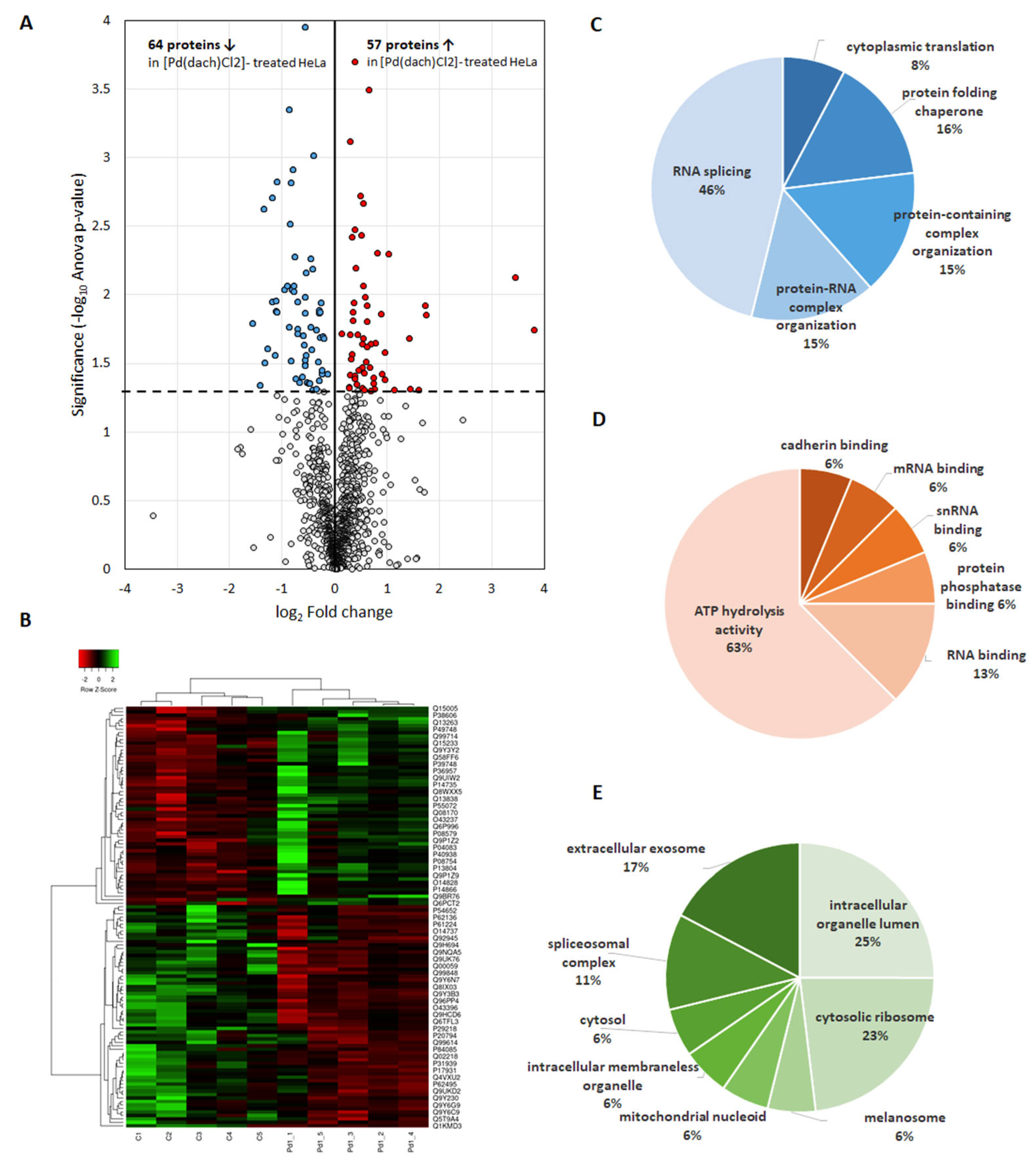
Figure 2. Proteins with altered abundance as a result of [Pd(dach)Cl2] treatment of HeLa cells. (A) Differential expression analysis between [Pd(dach)Cl2]-treated HeLa cells and controls is represented by the volcano plot. Statistically significant altered proteins in [Pd(dach)Cl2]-treated HeLa cells compared to the control (p < 0.05) are above the dashed horizontal line. Proteins with increased abundance are indicated in red and those with decreased abundance in blue. (B) Heatmap of protein expression in [Pd(dach)Cl2]-treated HeLa cells where higher protein abundances are presented in green, and lower ones in red. The samples are shown in columns, and the rows indicate proteins. The clustering method applied was average linkage, and the distance measurement method used was Spearman rank correlation. (CE) Enrichment analysis of GO annotations associated significantly with the differentially abundant proteins: (C) biological processes, (D) molecular functions, and (E) cellular compartment.
Inorganics 13 00215 g002
Figure 3. Enrichment analysis of the cellular pathways related significantly with the DAPs according to (A) KEGG and (B) Reactome Pathways databases. The charts include only terms with statistical significance corrected for multiple testing using the Benjamini–Hochberg procedure (FDR ≤ 0.05).
Inorganics 13 00215 g003
Figure 4. Protein–protein interaction network of the DAPs. The network was obtained using the STRING database. Proteins involved in the top significantly enriched pathways, according to the KEGG and Reactome pathway databases, are colored according to the given legend. The line thickness indicates the strength of data support.
Inorganics 13 00215 g004
Figure 5. Identification of hub genes in the protein–protein interaction network of DAPs using Cytoscape plug-in cytoHubba. The graph represents the top 25 hub genes ranked by the MaxMCC approach, with scores marked from highly essential (red) to crucial (yellow).
Inorganics 13 00215 g005
Figure 6. Graphical representation of the spliceosome pathway based on the KEGG database. The identified proteins from the spliceosome pathway in our study are color-coded: up-regulated proteins are displayed in red and down-regulated in green.
Inorganics 13 00215 g006
Figure 7. Graphical representation of the ribosome pathway based on the KEGG database. Proteins colored with light green are part of the human ribosome. The identified proteins from the ribosome pathway in our study are color-coded: up-regulated proteins are displayed in red and down-regulated in blue.
Inorganics 13 00215 g007
Disclaimer/Publisher’s Note: The statements, opinions and data contained in all publications are solely those of the individual author(s) and contributor(s) and not of MDPI and/or the editor(s). MDPI and/or the editor(s) disclaim responsibility for any injury to people or property resulting from any ideas, methods, instructions or products referred to in the content.