Abstract
Five novel PEPPSI-type palladium(II) complexes, dichloro[1-isopropyl-3-(arylmethyl)-5,6-dimethylbenzimidazolin-2-ylidene]pyridine palladium(II), were synthesized and characterized through nuclear magnetic resonance and Fourier-transform infrared spectroscopy. The DNA- and BSA-binding analyses of PEPPSI-type palladium (II) complexes were performed with UV-Vis spectroscopy by means of the Benesi-Hildebrand method. The results indicated that complex 1b (arylmethyl = 3-methylbenzyl) exhibited the strongest binding constant against DNA, with a value of 5.5 × 10³ M−1, while complex 1d (arylmethyl = 2-chlorobenzyl) exhibited the highest binding affinity for BSA, reaching 2.8 × 10⁴ M−1. In addition, the binding characteristics of DNA and BSA were assessed through the implementation of molecular docking methodologies. These methodologies displayed results that were in accordance with the experimental results. The molecules were also assessed for their ADME properties, with a focus on determining their drug-likeness potential. The five complexes were found to be compatible with the Veber and Egan rules.
1. Introduction
N-Heterocyclic carbenes (NHCs) have demonstrated their status as a highly versatile and indispensable class of ligands in the domain of organometallic chemistry. Their notable σ-donor and moderate π-acceptor properties facilitate the formation of stable complexes with a wide array of transition metals, thereby positioning them at the vanguard of research in catalysis, materials science, and medicinal chemistry [1,2]. Once regarded as mere curiosities, NHCs have evolved into essential building blocks for designing efficient and robust metal complexes. The development of NHC chemistry can be traced back to the seminal contributions of Wanzlick and Öfele in the 1960s. However, it was significantly advanced by isolation of the first stable free carbene by the group of Arduengo in 1991 [3,4,5]. The structural composition of NHCs was characterized by the presence of a divalent carbon atom, which was stabilized by adjacent nitrogen atoms within a heterocyclic ring. This configuration provided substantial electronic and steric stabilization, thereby augmenting the capacity of the ligand to establish robust metal-ligand bonds. While σ-donation from the carbene center was the primary interaction, additional π-backbonding effects also played a role in stabilizing these complexes [6]. A notable advantage of NHC ligands was their high degree of structural tunability. Substituent modifications, particularly those occurring at nitrogen atoms, have been demonstrated to enable precise regulation of electronic properties, steric hindrance, lipophilicity, and overall ligand behavior [7,8,9,10]. This flexibility enabled chemists to tailor NHC ligands for specific applications in both homogeneous catalysis and medicinal chemistry.
In the domain of catalysis, metal-NHC complexes have demonstrated remarkable efficacy in pivotal transformations, including olefin metathesis, hydrogenation, and cross-coupling reactions. Their chemical stability and modularity have also generated significant interest in biomedical research. When combined with biologically relevant metal centers, such as gold, silver, copper, palladium or ruthenium, NHC ligands form complexes that often display enhanced antimicrobial and anticancer properties compared to their non-metallic counterparts [11,12,13,14,15,16]. Guided by these encouraging characteristics, recent endeavors have centered on the development of a diverse array of M-NHC complexes, encompassing mononuclear, multinuclear (di-, tri-, tetranuclear), and pincer-type architectures, with the objective of broadening their catalytic scope. However, many of these complexes exhibited sensitivity to moisture and air, often necessitating strictly anhydrous conditions for synthesis and utilization. In order to address these challenges, Organ and co-workers introduced a significant innovation in 2006 with the development of PEPPSI (Pyridine-Enhanced Precatalyst Preparation, Stabilization, and Initiation) complexes. These palladium-NHC complexes exhibit air- and moisture-stability, comprising a palladium(II) center that was coordinated to an NHC ligand, two anionic ligands (e.g., chloride, bromide, or acetate) and a labile pyridine ligand [17].
The role of deoxyribonucleic acid (DNA) in biological processes was paramount, as it served as the repository for genetic information essential for protein and enzyme synthesis, thereby regulating cellular functions [18]. Furthermore, DNA could undergo irreversible changes through chemical and physical processes, which could result in long- and short-term effects. The utilization of small molecules in the field of DNA-targeted drug development entailed the alteration of gene expression, thereby facilitating the desired therapeutic outcome. The instructions for the synthesis of proteins that regulate metabolism, structure, signaling, and defense in cells were provided by DNA. Consequently, the analysis of small molecule-DNA interactions provided significant insights into the properties that contribute to the development of diseases such as cancer [19,20]. Due to its central role in this field, DNA has become an important target for many small molecules, including anticancer drugs and antibiotics [21,22]. For example, Panicker and Sivaramakrishna conducted a spectroscopic analysis of the DNA-binding properties of terpyridine-nickel(II) complexes [23]. In a separate study, Im, Biswas and co-workers investigated the DNA-binding characteristics of metal-based phenalenyl complexes. The complexes of cobalt, manganese, nickel, aluminum, and iron were examined to determine their bioactivity, with a particular focus on their antioxidant activity. The results revealed interesting findings, particularly for the manganese complex [24]. Recently, manganese carbonyl complexes, whose CO release properties have been subjected to analysis of their DNA binding affinity using experimental and theoretical methods by Üstün and co-workers [25]. In addition, Saygideger and co-workers evaluated the DNA-binding capacity of Pd(II) complexes to enhance comprehension of the antitumor effects of the molecules. The authors measured a binding constant of 4.98 × 10⁵ M−1 with the dichloro-P,N-{[2-(bis(4-methylphenyl)phosphino)phenyl]methylene]-3-(ethan-1-one)-benzenamine} palladium(II) complex [26]. These molecules have the capacity to interact directly with the DNA strand or bind to DNA-associated proteins. The study of such interactions offered significant insights into the design of DNA-targeted therapeutics and enhanced understanding of disease mechanisms. Additionally, it provided critical information on drug efficacy and safety [27].
Albumin, one of the most abundant proteins in mammals, is essential for the transport of diverse endogenous and exogenous molecules and for maintaining the colloid osmotic pressure of blood. Furthermore, it played a pivotal role in pharmacokinetic and toxicokinetic processes by influencing enzymatic activity [28]. Albumin possessed significant physiological relevance due to its interactions with blood cells, vascular components, and extravascular tissues. Furthermore, serum albumin functions as a biomarker for enzymatic, inflammatory, and oxidative processes, and its blood concentration could be modified by the binding of small molecules [29]. The transportation of drug molecules could occur via serum albumin, and the binding of small molecules had the potential to alter their blood concentration [30,31]. In experimental studies, bovine serum albumin (BSA) was frequently utilized as a model protein to assess binding interactions. Moi and co-workers conducted an analysis of the BSA-binding properties of pyrrolidine-based Pd(II) complexes. This analysis utilized both in vitro and in silico methods, resulting in the observation of noteworthy Kb values. Concurrently, a theoretical affinity of −7.12 kcal/mol was recorded for the most effective complex [32]. In addition, Dehghanian and co-workers synthesized the acetylacetonatoethylendiaminepalladium(II) nitrate complex and recorded a binding constant of 9.50 × 10⁴ M−1 against BSA using a spectroscopic method [33]. In a recent study, Ojwach and co-workers investigated the binding interactions of carboxamide pyrazine palladium(II) complexes, intending to elucidate the underlying mechanisms of cytotoxic properties of the complexes [34].
In the present article, the synthesis and characterization of five novel PEPPSI-type complexes of the class dichloro[1-isopropyl-3-(arylmethyl)-5,6-dimethylbenzimidazolin-2-yl]pyridine palladium(II) 1a–e were reported (Figure 1). The interactions of these inorganic complexes with DNA and BSA were investigated using the Benesi-Hildebrand approach. Further insights into the binding modes were obtained through molecular docking studies. In addition, the ADME profiles of the optimized complexes were evaluated.
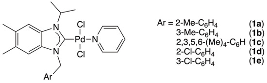
Figure 1.
PEPPSI-type palladium complexes 1a–e studied in the present article.
2. Results and Discussion
2.1. Synthesis and Structural Analysis of PEPPSI-Type Palladium Complexes
The synthesis of the five dichloro[1-isopropyl-3-(arylmethyl)-5,6-dimethylbenzimidazolin-2-yl]pyridine palladium(II) complexes, wherein arylmethyl = 2-methylbenzyl (1a), 3-methylbenzyl (1b), 2,3,5,6-tetramethylbenzyl (1c), 2-chlorobenzyl (1d) or 3-chlorobenzyl (1e), was accomplished through a three-step process from 5,6-dimethylbenzimidazole (2) (Scheme 1).
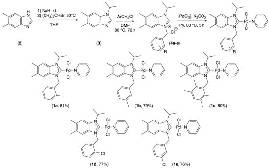
Scheme 1.
Synthesis of PEPPSI-type complexes 1a–e.
The benzimidazolium salts (4a–e) were prepared according to the procedure previously described in our earlier publications [24]. The synthesis began with the N-alkylation of 5,6-dimethylbenzimidazole (2) with isopropyl bromide. This reaction was carried out in tetrahydrofuran (THF) in the presence of NaH at temperatures of 60 °C for a period of 72 h. This reaction yielded the intermediate 1-isopropyl-5,6-dimethylbenzimidazole (3). Subsequently, this compound was treated with various arylmethyl chlorides in N,N-dimethylformamide (DMF) at 80 °C for 72 h to afford the corresponding benzimidazolium salts (4a–e). The selection of arylmethyl chloride was informed by the objective of investigating the impact of a methyl versus chloride substituent and the influence of its position on the aromatic ring in relation to binding with target proteins. The five NHC precursors 4a–e demonstrated adequate air and moisture stability in both solid and solution phases. The structures of the compounds were confirmed through the use of FT-IR, 1H and 13C NMR spectroscopy (see Materials and Methods and Supplementary Materials).
In the NMR spectra of benzimidazolium salts, the NCHN moieties exhibited singlets within the ranges of 11.20–11.87 and 141.14–141.80 ppm in 1H and 13C NMR, respectively. The formation of the salts 4a–e was further confirmed by their FT-IR spectrum, which displayed a CN stretching vibration in the range of 1552–1553 cm−1 (Table 1). These values are consistent with the literature data for analogous benzimidazolium salts [35,36]. An intense band around 3400 cm−1 was visible in the spectra of benzimidazolium salts 4a–e. This phenomenon was attributed to the vibrational modes of H2O associated with the salts. The band disappears once PEPPSI-type complexes are formed [37].

Table 1.
Infrared of CN stretching frequencies and relevant 1H and 13C{1H} NMR data.
The palladium(II) complexes (1a–e), incorporating the previously synthesized benzimidazolin-2-yl moieties, were obtained in satisfactory yields, 76–81%, through the reaction of the benzimidazolium salts 4a–e with palladium(II) chloride ([PdCl2]) in the presence of excess potassium carbonate (K2CO3) in pyridine at 80 °C for 5 h. This synthetic approach was founded on the methodology delineated by Organ [17], and the overarching reaction was illustrated in Scheme 1. The resulting PEPPSI-type Pd(II) complexes 1a–e demonstrated remarkable stability against air, moisture, and light in both solid and solution forms. Furthermore, the compounds exhibited satisfactory solubility in conventional organic solvents, including dimethyl sulfoxide, chloroform and dichloromethane. The structural confirmation of the complexes was achieved through the use of FT-IR and NMR spectroscopy (see Materials and Methods and Supplementary Materials).
The 1H NMR spectra of NHC precursors 4a–e exhibited significant singlets for the acidic proton at approximately 11 ppm. These signals were notably absent in the spectra of the corresponding palladium(II) complexes 1a–e, confirming the deprotonation of the NHC precursors and successful formation of the PEPPSI-type complexes. The 13C{1H} NMR spectra revealed signals in the range of 160.27–161.30 ppm for the characteristic carbenic carbons (NC(Pd)N) (Table 1), which were in accordance with the literature values [35,36].
In addition, the FT-IR analysis demonstrated a shift in the carbon-nitrogen (CN) stretching bands from 1552 to 1557 cm−1 in the benzimidazolium salts 4a–e to 1441–1447 cm−1 in the palladium(II) complex 1a–e. The observed shift is attributable to a weakening of the carbon-nitrogen bond. Subsequent to coordination, the carbon–nitrogen bond transitions into a partial double bond [37].
X-ray analysis of single crystals of the dichloro[1-isopropyl-3-(2-chlorobenzyl)-5,6-dimethylbenzimidazolin-2-yldene]pyridine palladium(II) complex (1d) confirmed the formation of the attempted complexes in an unambiguous manner. The complex 1d crystallized in the monoclinic form with the C2/c space group. The unit cell contains eight complex molecules. The palladium atom adopts square planar geometry with two chloride atoms in a trans configuration, forming a Cl-Pd-Cl angle of 175.04(5)°. The Pd-C carbene and Pd-N pyridine bond lengths were found to be 1.961(4) and 2.107(3) Å, respectively, with a C-Pd-N angle of 177.52(15)°. These values agree with those observed for Pd-PEPPSI complexes [38,39]. The benzimidazole aromatic ring is inclined with respect to the pyridine moiety, as evidenced by a dihedral angle of 48.47°. The 2-chloroenzyl substituent was found to be nearly perpendicular to the benzimidazole ring, with a dihedral angle of 87.36° (Figure 2).
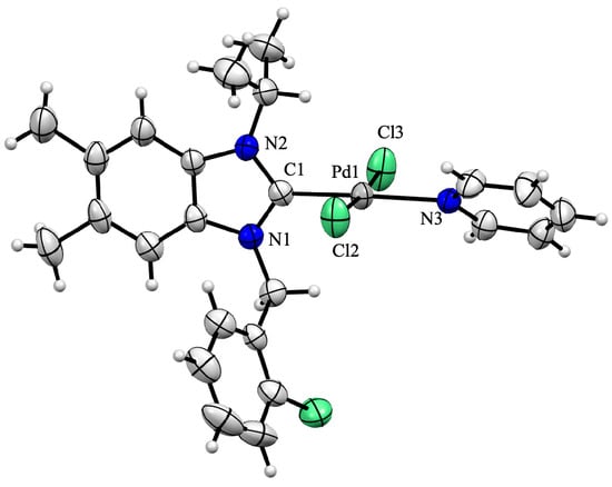
Figure 2.
ORTEP drawing of palladium(II) complex 1d, 50% probability thermal ellipsoids. Important bond lengths (Å) and angles (°): Pd1-C1 1.961(4), Pd1-Cl2 2.2937(11), Pd1-Cl3 2.2918(11), Pd1-N3 2.107(3), C1-N1 1.351(5), C1-N2 1.344(5), C1-Pd1-Cl2 88.83(12), Cl2-Pd1-N3 92.86(9), N3-Pd1-Cl3 91.31(9), Cl3-Pd1-C1 87.09(12), C1-Pd1-N3 177.52(15) and Cl2-Pd1-Cl3 175.04(5). green = chloride; blue = nitrogen and grey = palladium.
2.2. DNA-Binding Analysis
The stability of PEPPSI-type complexes was confirmed prior to DNA-binding evaluation with UV-Vis spectroscopy. The stabilities of the complexes in DMSO were examined for 10 mM solutions over a period of five days, with samples collected at one-day intervals. In contrast, the stabilities of the complexes in a saline environment were analyzed for a duration of 120 min, with samples collected at 10 min intervals. In both tests, no significant shifts were detected for the complexes except for minor fluctuations. The complete set of UV-Vis spectra is presented in Figures S25–S35.
In the present study, an analysis was conducted of the DNA-binding properties of five PEPPSI-type palladium(II) complexes 1a–e. The Benesi-Hildebrand method was utilized, and the following equation was employed [40]:
In the equation, A, A0 and AC represent the absorbance of PEPPSI-type complexes in the presence of varying DNA concentrations, in the absence of DNA, and in the PEPPSI-DNA complex, respectively. The binding constant (Kd) was determined through the calculation of the slope and intercept of the plot of 1/(A − A0) versus 1/[M], where [M] is the DNA concentration. The binding constant was then calculated as the ratio of the intercept to the slope [41].
The higher binding constant was determined to be 5.5 × 10³ M−1 for complex 1b (Figure 3). Furthermore, the binding constants of complexes 1a, 1c, 1d and 1e were determined to be 4.2 × 10³ M−1, 3.8 × 10³ M−1, 3.4 × 10³ M−1 and 4.2 × 10³ M−1, respectively (Figure S36–S39). The larger binding constant indicated a more robust interaction between DNA and the inorganic molecule. The results obtained demonstrate unequivocally that the presence of a substituent in the meta position on the benzyl substituent enhanced the interaction between the palladium complex and DNA (complexes 1a or 1d versus complexes 1b or 1e, respectively). Moreover, this interaction was more favorable when this substituent was a methyl group (complex 1b) than when it was a chlorine atom (complex 1e).
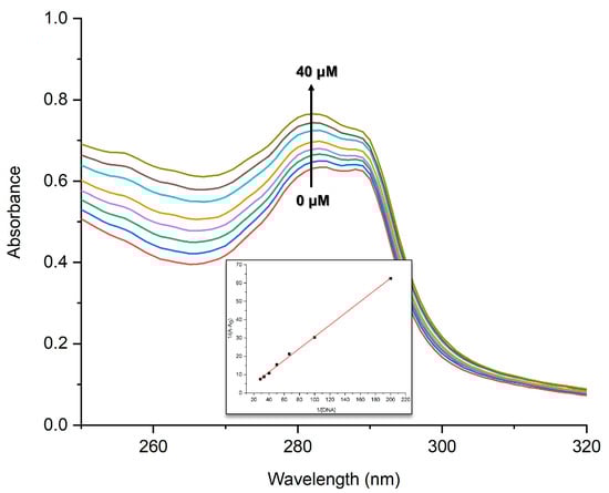
Figure 3.
UV-Vis Spectra of complex 1b (100 µM) with the addition of DNA (0–40 µM); inset: the plot of 1/[DNA] vs. 1/(A − A0).
Le, Zheng and co-workers recorded a relatively weak binding affinity of 2.4 × 10³ M−1 for the cationic [Cu(HPB)(L-Ala)]+ complex (HPB = 2(2-pyridyl)benzimidazole and L-Ala = L-alaninate) [42], while Dunbar, Turro and co-workers measured a binding affinity of 4.6 × 10² M−1 for the dinuclear [Rh2(O2CCH3)4(OH2)2] complex [43]. For their part, Kondaiah, Chakravarty and co-workers reported a binding constant of 1.5 × 10⁴ M−1 for the cis-[Pt(NH3)2(L)Cl](NO3) complex (L = imidazole containing a diiodo-BODIPY substituent) [44]. Conversely, Jagadeesh and co-workers reported binding affinities for copper(II), palladium(II) and zinc(II) complexes with thiosemicarbazone-type ligand ranged from 1.83 × 10⁴ to 1.45 × 10⁵ M−1 [45].
The DNA interactions of the molecules were also investigated by means of a molecular docking method (Figure 4). It was observed that all the palladium(II) complexes interacted with the same residue as Thy7, Thy8, Cyt9, Gua10, and Cyt11. The maximum binding affinity, −5.04 kcal/mol, was calculated for complex 1b, which experimentally exhibited the highest binding constant. The binding affinities of complexes 1a, 1c, 1d and 1e were determined to be −4.56, −4.51, −4.61, and −4.76 kcal/mol, respectively.
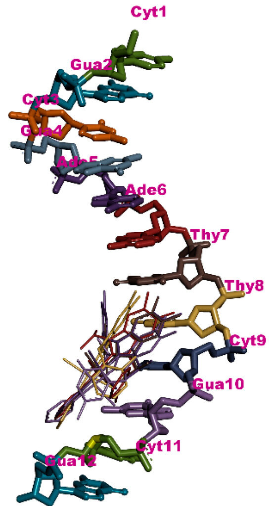
Figure 4.
Interaction residues of the molecules against DNA according to molecular docking results.
2.3. BSA-Binding Analysis
The stability of PEPPSI-type complexes was confirmed before BSA-binding evaluation with UV-Vis spectroscopy in both DMSO and saline environments. In both tests, no significant shifts were detected for the complexes except for minor fluctuations, as presented in Figures S25–S35.
In this study, the binding properties of the palladium(II) complexes 1a–e to BSA were analyzed by means of the Benesi-Hildebrand method, as outlined above. For the purpose of BSA-binding evaluations, the [M] represented the concentration values of the PEPPSI-type complexes. The results indicated that complex 1d exhibited the strongest binding affinity, with a value of 2.8 × 10⁴ M−1 (Figure 5). In contrast, the binding constants of complexes 1a, 1b, 1c and 1e were recorded at 1.8 × 10⁴ M−1, 1.9 × 10⁴ M−1, 1.6 × 10⁴ M−1, and 2.3 × 10⁴ M−1, respectively (see Supplementary Materials). The results of this study demonstrated the positive role of the chlorine atom in the interaction between the complexes and BSA. Indeed, the complexes whose ligands were substituted by chloride (complexes 1d and 1e) exhibited a higher binding affinity than the complexes substituted by a methyl group (complexes 1a and 1b).
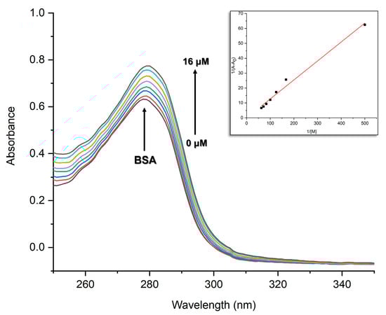
Figure 5.
UV-Vis Spectra of BSA (15 µM) with the addition of complex 1d (0–16 µM); inset: the plot of 1/[1d] vs. 1/(A − A0).
The BSA-binding properties of the molecules were also analyzed by molecular docking method. The most optimal binding affinity was ascertained for complex 1d, with a calculated value of −8.02 kcal/mol. The binding affinity of complex 1e was determined to be −7.97 kcal/mol. Complexes 1a, 1b and 1c exhibited binding affinities of −7.79, −7.79 and −7.67 kcal/mol, respectively. These results were consistent with the experimental findings. It was observed that all the inorganic complexes interacted with the same residue of the macromolecule, albeit with different orientations. The BSA was traditionally divided into three domains, with each domain further subdivided into the A and B subdomains [46]. Furthermore, Wade and co-workers delineated two drug binding sites (I and II) at 2A and 3A, respectively [47]. As illustrated in Figure 6, the PEPPSI-type complexes under investigation exhibited an interaction with drug site II (2A). The complete set of interaction details was presented in Table 2.
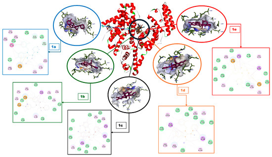
Figure 6.
Interaction residues and interaction details of the complexes 1a–e against BSA according to molecular docking results.
Table 2.
Interaction details and the binding affinities (BA) of the palladium(II) complexes 1a–e.
2.4. ADME Analysis
An analysis of the five PEPPSI-type complexes 1a–e was conducted to assess their absorption, distribution, metabolism, and excretion properties (ADME), which were critical for determining drug-likeness and conducting pharmacokinetic evaluations. The drug-likeness of a molecule was determined by various criteria, including those proposed by Lipinski [48], Ghose [49], Veber [50], Egan [51], Muegge [52] and Bioavailability Score [53]. The molecular weight (MW) of the drugs in question must be less than 500 g∙mol−1, the MlogP must be less than 5, and the number of H-donor and acceptor atoms must be less than 5 and 10, respectively. These molecules were referred to as “Pfizer filters” or “Lipinski’s rules.” It was noteworthy that these molecules were expected to account for more than 90% of drug candidates. In accordance with the Lipinski rules, the studied PEPPSI-type complexes 1a–e were deemed to be non-coherent due to two violations. Additionally, the molecules exhibited violations of the Ghose and Muegge rules (Bayer filter), while the Bioavailability Score (Abbott filter) was determined to be 0.17 for all complexes. Conversely, according to the Veber (Glaxo-Smith Kline filter; TPSA ≤ 140 Å2 and rotatable bonds ≤ 10) (TPSA = total polar surface area) and Egan (Pharmacia filter; MlogP ≤ 5.88 and TPSA ≤ 131.6 Å2) rules, all these molecules could be evaluated as drug candidates. The results of the study were summarized in Table 3.
Table 3.
Some pharmacokinetic and drug-likeness properties of the molecules.
3. Materials and Methods
3.1. General
All compounds were prepared under an argon atmosphere using standard Schlenk line techniques to maintain inert conditions throughout the synthesis. The reagents utilized in the procedures were obtained from Merck (Darmstadt, Germany) and Sigma-Aldrich (Darmstadt, Germany). The melting points were measured in open air using an Electrothermal 9200 apparatus (Vernon Hills, IL, USA) with glass capillaries, and the values are reported without correction. Fourier-transform infrared (FT-IR) spectra were recorded on a Perkin Elmer 100 spectrometer (Shelton, CT, USA) at Catalysis Research and Application Center, İnönü University in Türkiye. Nuclear magnetic resonance (NMR) spectra, encompassing both proton (1H) and carbon (13C) nuclei, were obtained using a Bruker Avance III 400 MHz spectrometer (Billerica, MA, USA). The calibration of the NMR spectra was conducted in accordance with the residual protonated solvent for CDCl3 δ = 7.26 ppm and 77.16 ppm for 1H and 13C{1H} NMR, respectively. The chemical shifts and coupling constants are reported in ppm and Hz, respectively. The preparation of 1-isopropyl-5,6-dimethylbenzimidazole (3), 1-isopropyl-3-(2-methylbenzyl)-5,6-dimethylbenzimidazolium chloride (4a) and 1-isopropyl-3-(2-chlorobenzyl)-5,6-dimethylbenzimidazolium chloride (4d) were conducted in accordance with the established procedures delineated in the extant literature [30].
3.2. General Procedure for the Preparation of Benzimidazolium Salts
The synthesis of benzimidazolium salts was accomplished through a two-step process. The initial step involved the gradual addition of NaH (11 mmol) to a THF solution (20 mL) of 5,6-dimethylbenzimidazole (2; 10 mmol). The solution was stirred at room temperature. After one hour, isopropyl bromide (10.1 mmol) was introduced to the reaction mixture, which was then heated to 60 °C while being continuously stirred for three days. The THF was then removed under reduced pressure. The resulting solid residue was dissolved in CH2Cl2 (50 mL) and the resulting mixture was filtered through a celite pad to remove any insoluble materials. The clear filtrate was evaporated under reduced pressure to yield 1-isopropyl-5,6-dimethylbenzimidazole (3). This intermediate (10 mmol) was subsequently reacted with arylmethyl chloride (10 mmol) in DMF at 80 °C. After a three-day heating period, Et2O (50 mL) was added to the reaction mixture, resulting in the precipitation of a white solid, which was filtered and washed with Et2O (20 mL). Afterwards, dried under vacuum, the benzimidazolium salt was isolated as a white solid
1-Isopropyl-3-(3-methylbenzyl)-5,6-dimethylbenzimidazolium chloride (4b): Yield: 84%; Melting point 215–216 °C; FT-IR ν(CN): 1554 cm−1; 1H NMR (400 MHz, CDCl3) δ = 1.80 (d, 6H, NCH(CH3)2, 3JHH = 6.0 Hz), 2.30 (s, 3H, C6H4(CH3)), 2.35 (s, 3H, C7H3N2(CH3)2), 2.40 (s, 3H, C7H3N2(CH3)2), 4.92 (hept, 1H, NCH(CH3)2, 3JHH = 6.0 Hz), 5.82 (s, 2H, NCH2), 7.10 (d, 1H, arom CH, 3JHH = 7.2 Hz), 7.19–7.24 (m, 2H, arom CH), 7.27–7.28 (m, 2H, arom CH), 7.43 (s, 1H, arom CH), 11.87 (s, 1H, NCHN); 13C{1H} NMR (100 MHz, CDCl3) δ = 20.81 (s, C7H3N2(CH3)2), 21.47 (s, C6H4(CH3)), 22.43 (s, NCH(CH3)2), 51.20 (s, NCH2), 51.69 (NCH(CH3)2), 113.19, 113.58, 125.38, 128.95, 129.12, 129.37, 129.81, 130.21, 133.32, 137.03, 137.19, 139.16 (12s, arom Cs), 141.43 (NCHN) ppm.
1-Isopropyl-3-(2,3,5,6-tetramethylbenzyl)-5,6-dimethylbenzimidazolium chloride (4c): Yield 88%; Melting point 257–258 °C; FT-IR ν(CN): 1552 cm−1; 1H NMR (400 MHz, CDCl3) δ = 1.76 (d, 6H, NCH(CH3)2, 3JHH = 6.4 Hz), 2.20 (s, 3H, C7H3N2(CH3)2), 2.21 (s, 12H, C6H(CH3)4), 2.36 (s, 3H, C7H3N2(CH3)2), 5.02 (hept, 1H, NCH(CH3)2, 3JHH = 6.4 Hz), 5.93 (s, 2H, NCH2), 6.78 (s, 1H, arom CH), 7.02 (s, 1H, arom CH), 7.42 (s, 1H, arom CH), 11.20 (s, 1H, NCHN); 13C{1H} NMR (100 MHz, CDCl3) δ = 16.21 (s, C6H(CH3)4), 20.64 (s, C6H(CH3)4), 20.74 (s, C7H3N2(CH3)2), 20.90 (s, C7H3N2(CH3)2), 22.32 (s, NCH(CH3)2), 48.02 (s, NCH2), 51.74 (NCH(CH3)2), 113.14, 113.91, 128.49, 129.35, 130.44, 133.27, 134.17, 134.87, 136.79, 136.90 (10s, arom Cs), 141.14 (NCHN) ppm.
1-Isopropyl-3-(3-chlorobenzyl)-5,6-dimethylbenzimidazolium chloride (4e): Yield: 82%; Melting point 246–247 °C; FT-IR ν(CN): 1557 cm−1; 1H NMR (400 MHz, CDCl3) δ = 1.78 (d, 6H, NCH(CH3)2, 3JHH = 6.0 Hz), 2.36 (s, 3H, C7H3N2(CH3)2), 2.40 (s, 3H, C7H3N2(CH3)2), 4.89 (hept, 1H, NCH(CH3)2, 3JHH = 6.0 Hz), 5.92 (s, 2H, NCH2), 7.25–7.28 (m, 3H, arom CH), 7.39 (s, 1H, arom CH), 7.45–7.46 (m, 2H, arom CH), 11.86 (s, 1H, NCHN); 13C{1H} NMR (100 MHz, CDCl3) δ = 20.80 (s, C7H3N2(CH3)2), 22.40 (s, NCH(CH3)2), 50.33 (s, NCH2), 51.72 (NCH(CH3)2), 113.26, 113.35, 126.74, 128.12, 129.24, 129.34, 130.04, 130.72, 134.96, 135.47, 137.33, 127.55 (12s, arom Cs), 141.31 (NCHN) ppm.
3.3. General Procedure for the Preparation of PEPPSI-Type Complexes
PEPPSI-type complexes (1a–e) were prepared under an argon atmosphere in accordance with previously reported methods [55]. A suspension of benzimidazolium salt (4a–e; 1 mmol), K2CO3 (5 mmol) and [PdCl2] (1.1 mmol) was stirred in pyridine (5 mL) at 80 °C for five hours. Following a period of cooling to room temperature, the pyridine was extracted under vacuum conditions. The solid residue was dissolved in CH2Cl2 (10 mL) and the mixture was filtered through Celite. Subsequent to the evaporation of the solvent, the crude solid was purified by flash chromatography (CH2Cl2 as eluent) and recrystallized from a CH2Cl2/pentane mixture, yielding the desired yellow complexes 1a–e as microcrystalline powders. However, these powders were unsuitable for X-ray diffraction study.
Dichloro[1-isopropyl-3-(2-methylbenzyl)-5,6-dimethylbenzimidazolin-2-yldene]pyridine palladium(II) (1a): Yield: 81%; Melting point 230–231 °C; FT-IR ν(CN): 1447 cm−1; 1H NMR (400 MHz, CDCl3) δ = 1.84 (d, 6H, NCH(CH3)2, 3JHH = 6.8 Hz), 2.21 (s, 3H, C6H4(CH3)), 2.34 (s, 3H, C7H2N2(CH3)2), 2.51 (s, 3H, C7H2N2(CH3)2), 6.08 (s, 2H, NCH2), 6.42 (hept., 1H, NCH(CH3)2, 3JHH = 6.8 Hz), 6.72 (s, 1H, arom CH of C7H2N2(CH3)2), 7.10 (t, 1H, arom CH, 3JHH = 7.6 Hz), 7.18–7.25 (m, 3H, arom CH), 7.31–7.34 (m, 2H, arom CH), 7.36 (s, 1H, arom CH of C7H2N2(CH3)2), 7.75 (tt, 1H, arom CH, 3JHH = 7.6 Hz and 4JHH = 1.6 Hz), 8.97–8.99 (m, 2H, arom CH); 13C{1H} NMR (100 MHz, CDCl3) δ = 19.87 (s, C7H2N2(CH3)2), 20.35 (s, C7H2N2(CH3)2), 20.55 (s, C6H4(CH3)), 21.36 (s, NCH(CH3)2), 49.96 (s, NCH2), 54.69 (s, NCH(CH3)2), 111.80, 112.83, 124.49, 126.55, 127.83, 128.08, 130.36, 131.25, 131.96, 132.23, 133.33, 134.18, 135.33, 138.12, 151.38 (15 s, arom Cs), 161.04 (NC(Pd)N) ppm. Anal. Calcd. for C25H29Cl2N3Pd: C, 54.71; H, 5.33; N, 7.66. Found C: 54.60; H: 5.26; N: 7.57.
Dichloro[1-isopropyl-3-(3-methylbenzyl)-5,6-dimethylbenzimidazolin-2-yldene]pyridine palladium(II) (1b): Yield: 79%; Melting point 160–161 °C; FT-IR ν(CN): 1447 cm−1; 1H NMR (400 MHz, CDCl3) δ = 1.83 (d, 6H, NCH(CH3)2, 3JHH = 6.8 Hz), 2.22 (s, 3H, C6H4(CH3)), 2.33 (s, 6H, C7H2N2(CH3)2), 6.09 (s, 2H, NCH2), 6.41 (hept, 1H, NCH(CH3)2, 3JHH = 6.8 Hz), 6.88 (s, 1H, arom CH of C7H2N2(CH3)2), 7.10 (d, 1H, arom CH, 3JHH = 7.6 Hz), 7.24 (t, 1H, arom CH, 3JHH = 7.6 Hz), 7.33–7.37 (m, 4H, arom CH), 7.46 (s, 1H, arom CH of C7H2N2(CH3)2), 7.76 (tt, 1H arom CH, 3JHH = 7.6 Hz and 4JHH = 1.6 Hz), 9.03 (dt, 2H, arom CH, 3JHH = 5.2 Hz and 4JHH = 1.6 Hz); 13C{1H} (100 MHz, CDCl3) NMR δ = 20.37 (s, C7H2N2(CH3)2), 20.56 (s, C7H2N2(CH3)2), 21.38 (s, NCH(CH3)2), 21.58 (s, C6H4(CH3)), 52.85 (s, NCH2), 54.61 (s, NCH(CH3)2), 112.08, 112.79, 124.56, 125.10, 128.74, 128.81, 128.89, 131.41, 131.89, 132.13, 134.12, 135.40, 138.16, 138.60, 151.44 (15 s, arom Cs), 160.68 (NC(Pd)N) ppm. Anal. Calcd. for C25H29Cl2N3Pd: C, 54.71; H, 5.33; N, 7.66. Found C: 54.84; H: 5.46; N: 7.59.
Dichloro[1-isopropyl-3-(2,3,5,6-tetramethylbenzyl)-5,6-dimethylbenzimidazolin-2-ylidene]pyridine palladium(II) (1c): Yield: 80%; Melting point 272–273 °C; FT-IR ν(CN): 1444 cm−1; 1H NMR (400 MHz, CDCl3) δ = 1.81 (d, 6H, NCH(CH3)2, 3JHH = 7.2 Hz), 2.10 (s, 3H, C7H2N2(CH3)2), 2.24 (s, 6H, C6H(CH3)4), 2.24 (s, 6H, C6H(CH3)4), 2.30 (s, 3H, C7H2N2(CH3)2), 6.09 (s, 2H, NCH2), 6.28 (s, 1H, arom CH of C6H(CH3)4), 6.42 (hept, 1H, NCH(CH3)2, 3JHH = 7.2 Hz), 7.08 (s, 1H, arom CH of C7H2N2(CH3)2), 7.30 (s, 1H, arom CH of C7H2N2(CH3)2), 7.32-7.36 (m, 2H, arom CH), 7.76 (tt, 1H arom CH, 3JHH = 7.6 Hz and 4JHH = 1.6 Hz), 8.94 (dt, 2H, arom CH, 3JHH = 4.8 Hz and 4JHH = 1.6 Hz); 13C{1H} (100 MHz, CDCl3) NMR δ = 16.64 (s, C6H(CH3)4), 20.45 (s, C7H2N2(CH3)2), 20.52 (s, C7H2N2(CH3)2), 20.72 (s, C6H(CH3)4), 21.28 (s, NCH(CH3)2), 49.83 (s, NCH2), 54.78 (s, NCH(CH3)2), 112.09, 112.48, 124.46, 131.02, 131.15, 131.34, 131.68, 132.38, 134.30, 134.68, 135.30, 138.04, 151.29 (13 s, arom Cs), 160.27 (NC(Pd)N) ppm. Anal. Calcd. for C28H35Cl2N3Pd: C, 56.91; H, 5.97; N, 7.11. Found C: 56.85; H: 5.90; N: 7.08.
Dichloro[1-isopropyl-3-(2-chlorobenzyl)-5,6-dimethylbenzimidazolin-2-yldene]pyridine palladium(II) (1d): Yield: 77%; Melting point 218–219 °C; FT-IR ν(CN): 1441 cm−1; 1H NMR (400 MHz, CDCl3) δ = 1.85 (d, 6H, NCH(CH3)2, 3JHH = 7.2 Hz), 2.10 (s, 3H, C7H2N2(CH3)2), 2.34 (s, 3H, C7H2N2(CH3)2), 6.25 (s, 2H, NCH2), 6.41 (hept, 1H, NCH(CH3)2, 3JHH = 7.2 Hz), 6.86 (s, 1H, arom CH of C7H2N2(CH3)2), 7.15 (t, 1H arom CH of C6H4Cl, 3JHH = 7.6 Hz), 7.23 (td, 1H arom CH of C6H4Cl, 3JHH = 7.6 Hz and 4JHH = 1.2 Hz), 7.32–7.35 (m, 2H, arom CH), 7.36 (s, 1H, arom CH of C7H2N2(CH3)2), 7.40 (d, 1H arom CH of C6H4Cl, 3JHH = 7.6 Hz), 7.45 (dd, 1H arom CH of C6H4Cl, 3JHH = 7.6 Hz and 4JHH = 1.2 Hz), 7.75 (tt, 1H arom CH, 3JHH = 7.6 Hz and 4JHH = 1.6 Hz), 9.00 (dt, 2H, arom CH, 3JHH = 5.2 Hz and 4JHH = 1.6 Hz); 13C{1H} (100 MHz, CDCl3) NMR δ = 20.34 (s, C7H2N2(CH3)2), 20.56 (s, C7H2N2(CH3)2), 21.35 (s, NCH(CH3)2), 49.21 (s, NCH2), 54.74 (s, NCH(CH3)2), 111.70, 112.87, 124.55, 127.45, 129.21, 129.45, 129.84, 131.22, 132.21, 132.51, 132.63, 132.93, 133.90, 138.19, 151.39 (15s, arom Cs), 161.30 (NC(Pd)N) ppm. Anal. Calcd. for C24H26Cl3N3Pd: C, 50.64; H, 4.60; N, 7.38. Found C: 50.57; H: 4.52; N: 7.29.
Dichloro[1-isopropyl-3-(3-chlorobenzyl)-5,6-dimethylbenzimidazolin-2-ylidene] pyridine palladium(II) (1e): Yield: 76%; Melting point 159–160 °C; FT-IR ν(CN): 1444 cm−1; 1H NMR (400 MHz, CDCl3) δ = 1.84 (d, 6H, NCH(CH3)2, 3JHH = 6.8 Hz), 2.24 (s, 3H, C7H2N2(CH3)2), 2.34 (s, 3H, C7H2N2(CH3)2), 6.09 (s, 2H, NCH2), 6.38 (hept, 1H, NCH(CH3)2, 3JHH = 6.8 Hz), 6.85 (s, 1H, arom CH of C7H2N2(CH3)2), 7.27–7.29 (m, 2H arom CH), 7.33–7.37 (m, 3H arom CH), 7.41–7.44 (m, 1H, arom CH), 7.63 (s, 1H, arom CH of C7H2N2(CH3)2), 7.76 (tt, 1H arom CH, 3JHH = 7.6 Hz and 4JHH = 1.6 Hz), 9.02 (dt, 2H, arom CH, 3JHH = 5.2 Hz and 4JHH = 1.4 Hz); 13C{1H} (100 MHz, CDCl3) NMR δ = 20.35 (s, C7H2N2(CH3)2), 20.55 (s, C7H2N2(CH3)2), 21.34 (s, NCH(CH3)2), 52.10 (s, NCH2), 54.70 (s, NCH(CH3)2), 111.72, 112.93, 124.59, 126.21, 128.13, 128.40, 130.26, 131.37, 132.19, 132.44, 133.86, 134.72, 137.54, 138.22, 151.38 (15 s, arom Cs), 161.16 (NC(Pd)N) ppm. Anal. Calcd. for C24H26Cl3N3Pd: C, 50.64; H, 4.60; N, 7.38. Found C: 50.68; H: 4.69; N: 7.31.
3.4. X-Ray Crystal Structure Analysis
Single crystals of palladium(II) complex 1d, suitable for X-ray analysis, were obtained by slow diffusion of Et2O into a CH2Cl2 solution of the complex. The sample was studied on Bruker D8 VENTURE PHOTON 100 CMOS (Billerica, MA, USA) using Mo-Kα radiation (λ = 0.71073 Å) at T = 298.15 K. The structures were solved with SHELXT-2014/5 [56], which revealed the non-hydrogen atoms of the molecule. After anisotropic refinement, all of the hydrogen atoms were found with a Fourier difference map. The structure was refined with SHELXL-2013/4 [57] by the full-matrix least-square techniques (use of F square magnitude; x, y, z, βij for C, Cl, N and Pd atoms; x, y, z in riding mode for H atoms) (Table 4). CCDC contains the supplementary crystallographic data for the structures. The data can be obtained free of charge from the Cambridge Crystallographic Data Centre via www.ccdc.cam.ac.uk/structures, accessed on 24 November 2025.
Table 4.
Crystal data and structure refinement parameters for the ruthenium complex 1d.
Table 4.
Crystal data and structure refinement parameters for the ruthenium complex 1d.
| CCDC depository | 2,504,152 | chemical formula | C24H26Cl3N3Pd | |
| color/shape | yellow/block | formula weight (g∙mol−1) | 569.23 | |
| crystal system | monoclinic | space group | C2/c | |
| unit cell parameters | a (Å) | 25.0789 (11) | volume (Å3) | 5351.3 (4) |
| b (Å) | 13.4807 (6) | Z | 8 | |
| c (Å) | 15.9890 (8) | D (g cm−3) | 1.413 | |
| α (°) | 90 | μ (mm−1) | 1.008 | |
| β (°) | 98.130 (2) | Tmin, Tmax | 0.6281/0.7454 | |
| γ (°) | 90 | F (000) | 2304 | |
| crystal size (mm) | 0.203 × 0.104 × 0.1 | index ranges | −31 ≤ h ≤ 31 | |
| θ range for data collection (°) | 2.998 ≤ θ ≤ 26.610 | −17 ≤ k ≤ 17 | ||
| reflections collected | 37690 | −20 ≤ l ≤ 20 | ||
| data/restraints/parameters | 5680/0/285 | goodness-of-fit on F2 | 1.010 | |
| final R indices (I > 2.0 σ(I)) | R1 = 0.0427 | R indices (all data) | R1 = 0.0813 | |
| wR2 = 0.0783 | wR2 = 0.0961 | |||
| Δρmax, Δρmin (e Å−3) | 0.949, −0.684 | |||
3.5. Stability Test
The solution of each PEPPSI-type complex (10 mM, 1 mL) was prepared in DMSO and the UV-Vis spectra were recorded for all solutions with one-day intervals for five days. The solutions were stored in closed quartz cuvettes under dark conditions during the day. Furthermore, the stability of the complexes in a saline environment was confirmed by monitoring the absorbance spectra of the complexes in the presence of NaCl (0.15 M, 0.05 mL) over a period of 120 min, with measurements taken at 10 min intervals [58].
3.6. DNA Binding Analysis
The palladium(II) complexes were analyzed for their DNA-binding properties using the Benesi-Hildebrand method with a UV-Vis spectrophotometer. The stock solutions of the complexes were prepared in DMSO, while the CT-DNA stock solutions were freshly prepared in Tris-HCl buffer solution. For each DNA stock solution, the ratios of A260/A280 were adjusted to a value greater than 1.8 and the concentrations of the solutions were determined with the absorbance values at 260 nm with 6600 M−1cm−1 (per nucleotide). Each molecule was exposed to DNA solutions of varying concentrations (0, 5, 10, 15, 20, 25, 30, 35 and 40 µM) for a duration of 30 min at room temperature. The alterations were subsequently determined within the range of 200–500 nm. The binding constants were calculated for identical maximum of each complex. We performed the DNA-binding experiments twice and utilized the results with the best R2 values (complex 1a: 0.9889, complex 1b: 0.9919, complex 1c: 0.9986, complex 1d: 0.9863 and complex 1e: 0.9981).
3.7. BSA Binding Analysis
The palladium(II) complexes were analyzed for their binding properties with respect to BSA (Bovine Serum Albumin) using the Benesi-Hildebrand method, a technique that employs UV-Vis spectroscopy.
The stock solution of BSA in PBS (pH: 7.4) was prepared and the concentration of the solution was subsequently adjusted based on the extinction coefficient of 43,824 M−1cm−1 at 280 nm. The final amount of 15 µM BSA was incubated with different concentrations (0, 2, 4, 6, 8, 10, 12, 14 and 16 µM) of each complex at room temperature for 30 min in a 1.5 mL quartz cuvette. The absorbances of the identical maximum of BSA were recorded for the purpose of analyzing the binding process. This analysis was conducted using a Shimadzu UV-1800 spectrophotometer (Tokyo, Japan), with measurements taken between 200 and 500 nanometers. We performed the DSA-binding experiments twice and utilized the results with the best R2 values (complex 1a: 0.9872, complex 1b: 0.9970, complex 1c: 0.9951, complex 1d: 0.9878 and complex 1e: 0.9978).
3.8. Molecular Docking Method
The BSA- and DNA-binding details of the optimized molecules were analyzed by molecular docking methods with AutoDockTools 4.2 [59]. The optimization of the palladium(II) complexes was performed with the ORCA package version 4.0 [60] by def2-SVP basis sets of BP86 functionals [61]. The crystal structures of the BSA (pdb:4f5s) [62] and DNA (pdb:1bna) [63] were obtained from the RCSB Protein Data Bank (https://www.rcsb.org/ accessed on 12 September 2025). The biomacromolecules were first recorded in pdbqt format which were used as rigid molecules during performances. The polar hydrogens and Kollman charges were evaluated, and the water molecules were removed from the target molecules before the processes in which Lamarckian Genetic Algorithms (150) were taken into account [64,65]. The PEPPSI-type complexes were also recorded as pdbqt with Gasteiger charges, and the docking performances were initiated from the random positions of these small complexes [66]. The illustrations depicting the results were created using Discovery Studio 4.1.0.
3.9. ADME Analysis
The pharmacokinetic and drug-likeness capacities of the complexes 1a–e were evaluated according to their ADME properties with SwissADME [54]. The Simplified Molecular Input Line Entry System (SMILES) of PEPPSI-type complexes was collected from Discovery Studio 4.1.0.
4. Conclusions
In summary, the present article described the synthesis of five dichloro[1-isopropyl-3-(arylmethyl)-5,6-dimethylbenzimidazolin-2-yl]pyridine palladium(II) complexes. These PEPPSI-type complexes were obtained with isolated yields ranging from 76 to 81% from the corresponding benzimidazolium salts using palladium(II) chloride as the metal source in the presence of potassium carbonate in pyridine at 80 °C. A combination of analytical techniques, including FT-IR and NMR spectroscopy, was employed to characterize these palladium(II) complexes.
The DNA- and BSA-binding analyses of the five palladium(II) complexes were performed by means of the Benesi-Hildebrand method. The results indicated that the inorganic complexes exhibited satisfactory DNA- and BSA-binding properties. Among the molecules, dichloro[1-isopropyl-3-(3-methylbenzyl)-5,6-dimethylbenzimidazolin-2-yldene]pyridine palladium(II) exhibited optimal binding properties against DNA (5.5 × 10³ M−1), while dichloro[1-isopropyl-3-(2-chlorobenzyl)-5,6-dimethylbenzimidazolin-2-yldene]pyridine palladium(II) demonstrated the strongest affinity for BSA (2.8 × 10⁴ M−1). The experimental results were consistent with the molecular docking results. Furthermore, all molecules exhibited coherence in accordance with the Veber and Egan rules. In subsequent studies, the analysis of novel molecules with diverse substitutions will be conducted to ascertain their potential for interactions with DNA and BSA.
Supplementary Materials
The following supporting information can be downloaded at: https://www.mdpi.com/article/10.3390/inorganics13120391/s1, Characteristic data of 1-isopropyl-3-(3-methylbenzyl)-5,6-dimethylbenzimidazolium (4b) with Figure S1: FT-IR spectrum, Figure S2: 1H NMR spectrum and Figure S3: 13C{1H} NMR spectrum; 1-isopropyl-3-(2,3,5,6-tetramethylbenzyl)-5,6-dimethylbenzimidazolium chloride (4c) with Figure S4: FT-IR spectrum, Figure S5: 1H NMR spectrum and Figure S6: 13C{1H} NMR spectrum; 1-isopropyl-3-(3-chlorobenzyl)-5,6-dimethylbenzimidazolium chloride (4e) with Figure S7: FT-IR spectrum, Figure S8: 1H NMR spectrum and Figure S9: 13C{1H} NMR spectrum; dichloro[1-isopropyl-3-(2-methylbenzyl)-5,6-dimethylbenzimidazolin-2-ylidene]pyridine palladium(II) (1a) with Figure S10: FT-IR spectrum, Figure S11: 1H NMR spectrum and Figure S12: 13C{1H} NMR spectrum; dichloro[1-isopropyl-3-(3-methylbenzyl)-5,6-dimethylbenzimidazolin-2-ylidene]pyridine palladium(II) (1b) with Figure S13: FT-IR spectrum, Figure S14: 1H NMR spectrum and Figure S15: 13C{1H} NMR spectrum; dichloro[1-isopropyl-3-(2,3,5,6-tetramethylbenzyl)-5,6-dimethylbenzimidazolin-2-ylidene]pyridine palladium(II) (1c) with Figure S16: FT-IR spectrum, Figure S17: 1H NMR spectrum and Figure S18: 13C{1H} NMR spectrum; dichloro[1-isopropyl-3-(2-chlorobenzyl)-5,6-dimethylbenzimidazolin-2-ylidene]pyridine palladium(II) (1d) with Figure S19: FT-IR spectrum, Figure S20: 1H NMR spectrum and Figure S21: 13C{1H} NMR spectrum; dichloro[1-isopropyl-3-(3-chlorobenzyl)-5,6-dimethylbenzimidazolin-2-ylidene]pyridine palladium(II) (1e) with Figure S22: FT-IR spectrum, Figure S23: 1H NMR spectrum and Figure S24: 13C{1H} NMR spectrum; DNA-binding with Figure S25: UV-Vis Spectra of complex 1a; Figure S26: UV-Vis Spectra of complex 1b; Figure S27: UV-Vis Spectra of complex 1c; Figure S28: UV-Vis Spectra of complex 1d; Figure S29: UV-Vis Spectra of complex 1e; BSA-binding with Figure S30: UV-Vis Spectra of complex 1a; Figure S31: UV-Vis Spectra of complex 1b; Figure S32: UV-Vis Spectra of complex 1c; Figure S33: UV-Vis Spectra of complex 1d; Figure S34: UV-Vis Spectra of complex 1e; Figure S35: UV-Vis Spectra of complex 1a with the addition of DNA; Figure S36: UV-Vis Spectra of complex 1b with the addition of DNA; Figure S37: UV-Vis Spectra of complex 1c with the addition of DNA; Figure S38: UV-Vis Spectra of complex 1d with the addition of DNA; Figure S39: UV-Vis Spectra of complex 1e with the addition of DNA; Figure S40: UV-Vis Spectra of BSA with the addition of complex 1a; Figure S41: UV-Vis Spectra of BSA with the addition of complex 1b; Figure S42: UV-Vis Spectra of BSA with the addition of complex 1c; Figure S43: UV-Vis Spectra of BSA with the addition of complex 1d; Figure S44: UV-Vis Spectra of BSA with the addition of complex1e.
Author Contributions
Conceptualization, E.Ü., N.Ş. and D.S.; methodology, E.Ü., N.Ş. and D.S.; software, E.Ü.; validation, E.Ü., N.Ş. and D.S.; formal analysis, E.Ü., N.Ş. and D.S.; investigation, E.Ü. and N.Ş.; resources, E.Ü. and N.Ş.; data curation, E.Ü., N.Ş. and D.S.; writing—original draft preparation, E.Ü. and N.Ş.; writing—review and editing, D.S. All authors have read and agreed to the published version of the manuscript.
Funding
This research received no external funding.
Institutional Review Board Statement
Not applicable.
Informed Consent Statement
Not applicable.
Data Availability Statement
The original contributions presented in the study are included in the article/Supplementary Materials, further inquiries can be directed to the corresponding authors.
Conflicts of Interest
The authors declare no conflicts of interest.
References
- Nolan, S.P. N-Heterocyclic Carbenes: Effective Tools for Organometallic Synthesis; Wiley-VCH Verlag GmbH & Co.: Weinheim, Germany, 2014. [Google Scholar]
- Díez-González, S. N-Heterocyclic Carbenes: From Laboratory to Curiosities to Efficient Synthetic Tools, 2nd ed.; The Royal Society of Chemistry: Cambridge, UK, 2017. [Google Scholar]
- Wanzlick, H.-W.; Schönherr, H.-J. Direct synthesis of a mercury salt-carbene complex. Angew. Chem. Int. Ed. 1968, 7, 141–142. [Google Scholar] [CrossRef]
- Öfele, K. 1,3-Dimethyl-4-imidazolinyliden-(2)-pentacarbonylchrom ein neuer Übergangsmetall-carben-komplex. J. Organomet. Chem. 1968, 12, 42–43. [Google Scholar] [CrossRef]
- Arduengo, A.J., III; Harlow, R.L.; Kline, M. A stable crystalline carbene. J. Am. Chem. Soc. 1991, 113, 361–363. [Google Scholar] [CrossRef]
- Herrmann, W.A.; Köcher, C. N-Heterocyclic carbenes. Angew. Chem. Int. Ed. 1997, 36, 2162–2187. [Google Scholar] [CrossRef]
- Liu, W.; Gust, R. Metal N-heterocyclic carbene complexes as potential antitumor metallodrugs. Chem. Soc. Rev. 2013, 42, 755–773. [Google Scholar] [CrossRef]
- Naz, N.; Saqib, S.; Ashraf, R.; Majeed, M.I.; Iqbal, M.A. Synthesis of new organoselenium compounds: Characterization and biological studies. Maced. J. Chem. Chem. Eng. 2020, 39, 1–10. [Google Scholar] [CrossRef]
- Danopoulos, A.A.; Simler, T.; Braunstein, P. N-Heterocyclic carbene complexes of copper, nickel, and cobalt. Chem. Rev. 2019, 119, 3730–3961. [Google Scholar] [CrossRef]
- Gök, Y.; Akkoc, S.; Çelikal, Ö.Ö.; Özdemir, İ.; Günal, S. In vitro antimicrobial studies of naphthalen-1-ylmethyl substituted silver N-heterocyclic carbene complexes. Arab. J. Chem. 2019, 12, 2513–2518. [Google Scholar] [CrossRef]
- Bian, M.; Fan, R.; Zhao, S.; Liu, W. Targeting the thioredoxin system as a strategy for cancer therapy: Miniperspective. J. Med. Chem. 2019, 62, 7309–7321. [Google Scholar]
- Choo, K.B.; Mah, W.L.; Lee, S.M.; Lee, W.L.; Cheow, Y.L. Palladium complexes of bidentate pyridine N-heterocyclic carbenes: Optical resolution, antimicrobial and cytotoxicity studies. Appl. Organomet. Chem. 2018, 32, e4377. [Google Scholar]
- Catalano, A.; Mariconda, A.; Sinicropi, M.S.; Ceramella, J.; Iacopetta, D.; Saturnino, C.; Longo, P. Biological activities of ruthenium NHC complexes: An update. Antibiotics 2023, 12, 365. [Google Scholar] [CrossRef] [PubMed]
- Esarev, I.V.; Wu, C.; Kirsanova, A.A.; Türck, S.; Lippmann, P.; Jones, P.G.; Babak, M.V.; Ott, I. Silver N-heterocyclic biscarbene complexes: Potent inhibitors of thioredoxin reductase with anticancer activity in vitro and in vivo. Chem. Asian J. 2025, 20, e202401672. [Google Scholar] [CrossRef] [PubMed]
- Touj, N.; Chakchouk-Mtibaa, A.; Mansour, L.; Harrath, A.H.; Al-Tamimi, J.; Mellouli, L.; Özdemir, İ.; Yasar, S.; Hamdi, N. Synthesis, spectroscopic properties and biological activity of new Cu(I) N-Heterocyclic carbene complexes. J. Mol. Struct. 2019, 1181, 209–219. [Google Scholar] [CrossRef]
- Estrada-Ortiz, N.; Guarra, F.; de Graaf, I.A.M.; Marchetti, L.; de Jager, M.H.; Groothuis, G.M.M.; Gabbiani, C.; Casini, A. Anticancer gold N-heterocyclic carbene complexes: A comparative in vitro and ex vivo study. ChemMedChem 2017, 12, 1429–1435. [Google Scholar] [CrossRef]
- O’Brien, C.J.; Kantchev, E.A.B.; Valente, C.; Hadei, N.; Chass, G.A.; Lough, A.; Hopkinson, A.C.; Organ, M.G. Easily prepared air-and moisture-stable Pd-NHC (NHC = N-heterocyclic carbene) complexes: A reliable, user-friendly, highly active palladium precatalyst for the Suzuki-Miyaura reaction. Chem. Eur. J. 2006, 12, 4743–4748. [Google Scholar] [CrossRef]
- Travers, A.; Muskhelishvili, G. DNA structure and function. FEBS J. 2015, 282, 2279–2295. [Google Scholar] [CrossRef] [PubMed]
- Patil, V.M.; Gupta, S.P.; Masand, N.; Balasubramanian, K. Experimental and computational models to understand protein-ligand, metal-ligand and metal-DNA interactions pertinent to targeted cancer and other therapies. Eur. J. Med. Chem. Rep. 2024, 10, 100133. [Google Scholar] [CrossRef]
- Jevtovic, V.; Golubović, L.; Alshammari, B.; Alshammari, M.R.; Rajeh, S.Y.; Alreshidi, M.A.; Alshammari, O.A.O.; Rakić, A.; Dimić, D. Crystal structure, theoretical analysis, and protein/DNA binding activity of iron (III) complex containing differently protonated pyridoxal–S-methyl-isothiosemicarbazone ligands. Int. J. Mol. Sci. 2024, 25, 7058. [Google Scholar] [CrossRef]
- Palchaudhuri, R.; Hergenrother, P.J. DNA as a target for anticancer compounds: Methods to determine the mode of binding and the mechanism of action. Curr. Opin. Biotechnol. 2007, 18, 497–503. [Google Scholar] [CrossRef]
- Lu, X.; Wang, L.; Liu, H.; Wang, R.; Chen, J. Studies on the interaction between antibiotics and DNA. Talanta 2007, 73, 444–450. [Google Scholar] [CrossRef] [PubMed]
- Panicker, R.R.; Sivaramakrishna, A. Studies on synthesis and influence of sterically driven Ni(II)-terpyridine (NNN) complexes on BSA/DNA binding and anticancer activity. J. Inorg. Biochem. 2024, 257, 112553. [Google Scholar] [CrossRef]
- Sen, S.; Chowdhury, N.; Kim, T.-W.; Paul, M.; Debnath, D.; Jeon, S.; Bagchi, A.; Im, J.; Biswas, G. Anticancer, antibacterial, antioxidant, and DNA-binding study of metal-phenalenyl complexes. Bioinorg. Chem. Appl. 2022, 2022, 8453159. [Google Scholar] [CrossRef]
- Önbaş, S.C.; Serdaroğlu, G.; Şahin, N.; Üstün, E.; Özdemir, İ. Synthesis, characterization, computational evaluation, CO-releasing properties, and molecular docking interactions of new [Mn(CO)3(bpy)L] PF6- type molecules. ACS Omega 2025, 10, 30798–30814. [Google Scholar] [CrossRef] [PubMed]
- Saygıdeğer Demir, B.; İnce, S.; Yilmaz, M.K.; Sezan, A.; Derinöz, E.; Taskin-Tok, T.; Saygideger, Y. DNA binding and anticancer properties of new Pd(II)-phosphorus Schiff base metal complexes. Pharmaceutics 2022, 14, 2409. [Google Scholar] [CrossRef]
- Watanabe, K.; Seki, N. Biology and development of DNA-targeted drugs, focusing on synthetic lethality, DNA repair, and epigenetic modifications for cancer: A review. Int. J. Mol. Sci. 2024, 25, 752. [Google Scholar] [CrossRef]
- Sleep, D. Albumin and its application in drug delivery. Expert Opin. Drug Deliv. 2015, 12, 793–812. [Google Scholar]
- Quinlan, G.J.; Martin, G.S.; Evans, T.W. Albumin: Biochemical properties and therapeutic potential. Hepatology 2005, 41, 1211–1219. [Google Scholar]
- Üstün, E.; Şahin, N. Insight into the effect of Ca2+, Mg2+ and Zn2+ on serum albumin interaction of benzimidazole-type new isopropyl substituted N-heterocyclic carbene molecules. J. Photochem. Photobiol. A Chem. 2024, 447, 115282. [Google Scholar] [CrossRef]
- Krishnan, D.; Sheela, A. A review on DNA/BSA binding and cytotoxic properties of multinuclear Schiff’s base complexes. Results Chem. 2023, 5, 100732. [Google Scholar] [CrossRef]
- Tarai, S.K.; Pan, A.; Biswas, P.; Bhaduri, R.; Mandal, S.; Paul, A.; Baitalik, S.; Bhattacharjee, A.; Moi, S.C. Anticancer behavior of pyrrolidine-based palladium(II) complexes and biophysical approach on their DNA, BSA binding activity, molecular docking, and DFT study. Langmuir 2023, 39, 10947–10964. [Google Scholar] [CrossRef] [PubMed]
- Tabatabai, A.S.D.; Dehghanian, E.; Mansouri-Torshizi, H.; Feizi-Dehnayebi, M. Computational and experimental examinations of new antitumor palladium(II) complex: CT-DNA-/BSA-binding, in silico prediction, DFT perspective, docking, molecular dynamics simulation and ONIOM. J. Biomol. Struct. Dyn. 2024, 42, 5447–5469. [Google Scholar] [CrossRef] [PubMed]
- Mvelase, S.T.; Benson, S.O.; Omondi, R.O.; Kumah, R.T.; Fatokun, A.A.; Ojwach, S.O. Structural, DNA/BSA binding interactions and cytotoxicity studies of carboxamide (pyridyl)pyrazine palladium (II) complexes. J. Mol. Struct. 2025, 1322, 140267. [Google Scholar]
- Ulu, Ö.D.; Serin, S.; Özdemir, İ. NHC-based Pd–PEPPSI complexes: Synthesis, characterization, DFT studies and catalytic activity in Suzuki-Miyaura cross coupling. J. Indian Chem. Soc. 2025, 102, 101689. [Google Scholar] [CrossRef]
- Atakol, A.; Yiğit, B.; Akdan, H.; Evren, E.; Celepci, D.B.; Yiğit, M.; Aygün, M.; Özdemir, İ. PEPPSI-type N-heterocyclic carbene palladium(II) complexes as catalysts in the direct C5 arylation of furan and thiophene. Inorg. Chimica Acta 2025, 580, 122612. [Google Scholar] [CrossRef]
- Yamada, T.; Tominari, Y.; Tanaka, S.; Mizuno, M. Infrared spectroscopy of ionic liquids consisting of imidazolium cations with different alkyl chain lengths and various halogen or molecular anions with and without a small amount of water. J. Phys. Chem. B 2017, 121, 3121–3129. [Google Scholar] [CrossRef] [PubMed]
- Şahin, N.; Özdemir, İ.; Sémeril, D. Palladium-catalyzed cross-coupling reaction via C-H activation of furanyl and thiofuranyl substrates. Inorganics 2024, 12, 175. [Google Scholar] [CrossRef]
- Şahin, N.; Zengin, S.; Özdemir, İ.; Sémeril, D. C-H activation of furanyl and thiofuranyl substrates catalyzed by trans-dichloro [1-cinnamyl-3-arylmethyl-benzimidazol-2-yliden]pyridine palladium(II) complexes. Polyhedron 2024, 261, 117144. [Google Scholar] [CrossRef]
- Benesi, H.A.; Hildebrand, J.H. A spectrophotometric investigation of the interaction of iodine with aromatic hydrocarbons. J. Am. Chem. Soc. 1949, 71, 2703–2707. [Google Scholar] [CrossRef]
- Düşünceli, S.D.; Kaloğlu, M.; Şahin, O.; Üstün, E. Synthesis, characterization, crystal structure, and BSA-binding properties with the presence of Ca2+, Mg2+, and Zn2+ ions by N(7)-substituted theophyllines. ChemistrySelect 2025, 10, e01358. [Google Scholar] [CrossRef]
- Hu, W.; Deng, S.; Huang, J.; Lu, Y.; Le, X.; Zheng, W. Intercalative interaction of asymmetric copper(II) complex with DNA: Experimental, molecular docking, molecular dynamics and TDDFT studies. J. Inorg. Biochem. 2013, 127, 90–98. [Google Scholar] [CrossRef]
- Sorasaenee, K.; Fu, P.K.-L.; Angeles-Boza, A.M.; Dunbar, K.R.; Turro, C. Inhibition of transcription in vitro by anticancer active dirhodium(II) complexes. Inorg. Chem. 2003, 42, 1267–1271. [Google Scholar] [CrossRef]
- Raza, M.K.; Gautam, S.; Garai, A.; Mitra, K.; Kondaiah, P.; Chakravarty, A.R. Monofunctional BODIPY-appended imidazoplatin for cellular imaging and mitochondria-targeted photocytotoxicity. Inorg. Chem. 2017, 56, 11019–11029. [Google Scholar] [CrossRef]
- Lavanya, M.; Haribabu, J.; Ramaiah, K.; Yadav, C.S.; Chitumalla, R.K.; Jang, J.; Karvembu, R.; Reddy, A.V.; Jagadeesh, M. 2′-Thiophenecarboxaldehyde derived thiosemicarbazone metal complexes of copper(II), palladium(II) and zinc(II) ions: Synthesis, spectroscopic characterization, anticancer activity and DNA binding studies. Inorg. Chim. Acta 2021, 524, 120440. [Google Scholar] [CrossRef]
- Ali, M.S.; Muthukumaran, J.; Jain, M.; Santos-Silva, T.; Al-Lohedan, H.A.; Al-Shuail, N.S. Molecular interactions of cefoperazone with bovine serum albumin: Extensive experimental and computational investigations. J. Mol. Liq. 2021, 337, 116354. [Google Scholar] [CrossRef]
- Sudlow, G.; Birkett, D.J.; Wade, D.N. The characterization of two specific drug binding sites on human serum albumin. Mol. Pharmacol. 1975, 11, 824–832. [Google Scholar] [CrossRef] [PubMed]
- Lipinski, C.A. Drug-like properties and the causes of poor solubility and poor permeability. J. Pharmacol. Toxicol. Methods 2000, 44, 235–249. [Google Scholar] [CrossRef]
- Ghose, A.K.; Viswanadhan, V.N.; Wendoloski, J.J. A knowledge-based approach in designing combinatorial or medicinal chemistry libraries for drug discovery. 1. A qualitative and quantitative characterization of known drug databases. J. Comb. Chem. 1999, 1, 55–68. [Google Scholar] [PubMed]
- Veber, D.F.; Johnson, S.R.; Cheng, H.Y.; Smith, B.R.; Ward, K.W.; Kopple, K.D. Molecular properties that influence the oral bioavailability of drug candidates. J. Med. Chem. 2002, 45, 2615–2623. [Google Scholar] [CrossRef]
- Egan, W.J.; Merz, K.M.; Baldwin, J.J. Prediction of drug absorption using multivariate statistics. J. Med. Chem. 2000, 43, 3867–3877. [Google Scholar] [CrossRef]
- Muegge, I.; Heald, S.L.; Brittelli, D. Simple selection criteria for drug-like chemical matter. J. Med. Chem. 2001, 44, 1841–1846. [Google Scholar] [CrossRef] [PubMed]
- Martin, Y.C. A bioavailability score. J. Med. Chem. 2005, 48, 3164–3170. [Google Scholar] [CrossRef]
- Daina, A.; Michielin, O.; Zoete, V. SwissADME: A free web tool to evaluate pharmacokinetics, drug-likeness and medicinal chemistry friendliness of small molecules. Sci. Rep. 2017, 7, 42717. [Google Scholar] [CrossRef]
- Şahin, N. Palladium-N-heterocyclic carbene complexes: Synthesis, characterization and catalytic application of C-H activation for carboxaldehyde derivatives. ChemistrySelect 2024, 9, e202403032. [Google Scholar] [CrossRef]
- heldrick, G.M. SHELXT—Integrated space-group and crystal structure determination. Acta Crystallogr. Sect. A 2015, A71, 3–8. [Google Scholar] [CrossRef] [PubMed]
- Sheldrick, G.M. Crystal structure refinement with SHELXL. Struct. Chem. 2015, 71, 3–8. [Google Scholar]
- Jimenez, J.; Chakraborty, I.; Del Cid, A.M.; Mascharak, P.K. Five-and six-coordinated silver(I) complexes derived from 2,6-(pyridyl)iminodiadamantanes: Sustained release of bioactive silver toward bacterial eradication. Inorg. Chem. 2017, 56, 4784–4787. [Google Scholar] [CrossRef] [PubMed]
- Morris, G.M.; Huey, R.; Lindstrom, W.; Sanner, M.F.; Belew, R.K.; Goodsell, D.S.; Olson, A.J. AutoDock4 and AutoDockTools4: Automated docking with selective receptor flexibility. J. Comput. Chem. 2009, 30, 2785–2791. [Google Scholar] [CrossRef]
- Neese, F. Software update: The ORCA program system, version 4.0. WIREs Comput. Mol. Sci. 2018, 8, e1327. [Google Scholar] [CrossRef]
- Neese, F.; Wennmohs, F.; Becker, U.; Riplinger, C. The ORCA quantum chemistry program package. J. Chem. Phys. 2020, 152, 224108. [Google Scholar] [CrossRef]
- Bujacz, A. Structures of bovine, equine and leporine serum albumin. Acta Crystallogr. D Struct. Biol. 2012, D68, 1278–1289. [Google Scholar] [CrossRef]
- Drew, H.R.; Wing, R.M.; Takano, T.; Broka, C.; Tanaka, S.; Itakura, K.; Dickerson, R.E. Structure of a B-DNA dodecamer: Conformation and dynamics. Proc. Natl. Acad. Sci. USA 1981, 78, 2179–2183. [Google Scholar] [CrossRef] [PubMed]
- Forli, S.; Huey, R.; Pique, M.E.; Sanner, M.F.; Goodsell, D.S.; Olson, A.J. Computational protein-ligand docking and virtual drug screening with the AutoDock suite. Nat. Protoc. 2016, 11, 905–919. [Google Scholar] [CrossRef]
- Serdaroğlu, G.; Uludag, N.; Üstün, E. Efficient synthesis of chromeno [2,3-b]pyridine derivatives using Zn(OTf)2 as a catalyst: DFT computations, molecular docking and ADME studies. J. Mol. Liq. 2023, 375, 121364. [Google Scholar] [CrossRef]
- Hkiri, S.; Coşkun, K.A.; Üstün, E.; Samarat, A.; Tutar, Y.; Şahin, N.; Sémeril, D. Silver(I) complexes based on oxadiazole-functionalized α-aminophosphonate: Synthesis, structural study, and biological activities. Molecules 2022, 27, 8131. [Google Scholar] [CrossRef] [PubMed]


Disclaimer/Publisher’s Note: The statements, opinions and data contained in all publications are solely those of the individual author(s) and contributor(s) and not of MDPI and/or the editor(s). MDPI and/or the editor(s) disclaim responsibility for any injury to people or property resulting from any ideas, methods, instructions or products referred to in the content. |
© 2025 by the authors. Licensee MDPI, Basel, Switzerland. This article is an open access article distributed under the terms and conditions of the Creative Commons Attribution (CC BY) license (https://creativecommons.org/licenses/by/4.0/).