Article

Ultra-Rapid Synthesis of Co3O4 Nanostructures with Tunable Morphology via Nickel-Assisted Anodization

by
Leydi Julieta Cardenas Flechas
1,2,
Jorge Bautista-Ruiz
3,
Paulo Tarso Cavalcante Freire
4,
Elaine Cristina Paris
5 and
Miryam Rincón Joya
6,*
1
Departamento de Ingeniería, Mecánica y Mecatrónica, Universidad Nacional de Colombia, Carrera 45 N 26-85, Bogotá 111321, Colombia
2
Departamento de Ingeniería, Mecánica y Mecatrónica, Fundación Universidad de América, Bogotá 111321, Colombia
3
Departamento de Ingeniería, Investigación de Materiales Cerámicos, Universidad Francisco de Paula Santander, San José de Cúcuta 540003, Colombia
4
Departamento de Física, Universidade Federal do Ceará, Fortaleza 60455-760, CE, Brazil
5
Embrapa Instrumentação, Rua XV de Novembro, 1452, São Carlos 13560-970, SP, Brazil
6
Departamento de Física, Universidad Nacional de Colombia, Carrera 45 N 26-85, Bogotá 11001, Colombia
*
Author to whom correspondence should be addressed.
Inorganics 2025, 13(11), 350; https://doi.org/10.3390/inorganics13110350
Submission received: 7 October 2025 / Revised: 20 October 2025 / Accepted: 24 October 2025 / Published: 26 October 2025

Abstract

Various morphologies of cobalt oxide Co3O4 films on cobalt (Co) foils were obtained via anodization followed by a thermal treatment at 350 °C. This study introduces a rapid and cost-effective synthesis route, achieving well-defined spinel structures in only 30 min. The novelty of this work lies in exploring nickel (Ni) as a morphological modifier in the anodization electrolyte. FESEM analysis revealed that, while anodization without Ni produced nanoflake structures, the inclusion of Ni transformed the morphology into larger cubic crystals and rice grain–shaped nanoparticles. XPS confirmed the presence of oxygen vacancies during phase formation, TEM showed spinel grains smaller than 20 nm, and Raman spectroscopy exhibited characteristic peak shifts influenced by both anodization and Ni addition. These results demonstrate that Ni not only accelerates the formation of spinel C o 3 O 4 but also plays a decisive role in tailoring morphology, highlighting the efficiency and novelty of this approach.

1. Introduction

Addressing the global challenges of energy production and storage is a pressing priority in modern society. Although fossil fuels still dominate the global energy mix—representing about 80% of total consumption in 2020—renewable energy sources accounted for only 11%, underscoring the urgent need for cleaner and more sustainable alternatives [1,2]. Among renewable sources, solar energy stands out due to its versatility for electricity generation and hydrogen production, particularly through water splitting processes [3]. In this context, hydrogen has emerged as an attractive energy carrier due to its high energy density, eco-friendly profile, and the potential to replace fossil fuels in a wide range of applications [3,4,5,6,7,8,9,10,11,12,13].
Electrochemical systems such as supercapacitors and electrocatalysts for the hydrogen evolution reaction (HER) and oxygen evolution reaction (OER) have gained considerable attention due to their efficiency, durability, and cost-effectiveness [14,15,16,17,18,19]. The performance of these devices strongly depends on the physicochemical properties of the electrode materials. Various transition-metal oxides—including FeOOH, FeCo2O4, CoNi2O4, Ni3S4/Co3S4, and CoNi2S4—have been explored, yet the relationship between nanostructural morphology and electrochemical capacitance in cobalt oxide/hydroxide systems remains insufficiently addressed [15,20,21,22,23,24].
Cobalt oxide (Co3O4), in particular, is a highly promising material due to its versatile redox chemistry, favorable conductivity, and chemical stability under alkaline conditions [25]. However, the synthesis of Co3O4 with controlled morphology remains a challenge, especially when scalable and cost-effective routes are considered. Among the different synthesis methods, anodization offers an attractive approach to directly fabricating ordered nanostructured oxides from metallic substrates. Although Schmuki and colleagues have successfully reported the growth of nanoporous cobalt oxide films on cobalt substrates through anodization [26], most studies have focused on long processing times (typically several hours), which limits the practical applicability of the method. Furthermore, the critical role of subsequent low-temperature thermal treatment in stabilizing the crystalline Co3O4 phase is often overlooked in the introductory sections of related works.
Bimetallic oxides such as CoMoO4 have also been studied for HER catalysis due to their high conductivity and chemical resistance. Nevertheless, their electrocatalytic activity is often hindered by charge-transfer resistance at the electrode/electrolyte interface [27,28,29,30,31]. Nickel foam (NF), a widely used conductive self-supporting substrate, has been employed to improve charge transport, and Co3O4/NF hybrids have shown enhanced activity [32,33]. Despite these advances, long-term stability remains a concern in many of these systems, particularly when nitrogen-doping or complex nanostructuring methods are applied [34,35,36]. In addition, recent studies have focused on core-shell nanostructures, such as ZnFe2O4/ZnO, which have shown significant improvements in photoelectrochemical performance, particularly under visible light. These materials offer enhanced efficiency in solar energy conversion, which is critical for the development of more sustainable and efficient solar technologies [37]. This work complements the growing body of research on advanced nanomaterials for energy conversion and storage, particularly in systems that utilize visible light to drive electrochemical reactions.
In this work, we focus on the rapid synthesis of Co3O4 micro/nanostructures on cobalt sheets by anodization followed by a controlled low-temperature thermal treatment. Our study systematically explores the influence of anodizing conditions—such as applied voltage, electrolyte composition, water and fluoride content, processing time, and the introduction of nickel (Ni) as a morphological modifier—on the resulting structural, vibrational, and electrochemical properties. By condensing the processing time from hours to only 30 min and investigating the role of Ni additives, we provide new insights into the optimization of anodization parameters for tailoring Co3O4 electrodes in clean energy technologies.

2. Results

2.1. Structure Characterization

In Experiment 1, the diffractogram exhibits intense diffraction peaks assigned to the spinel Co3O4 phase (JCPDS card No. 65-3103), along with a peak corresponding to metallic Co (ICDD 15-0806) [38]. However, in the other samples, both phases coexist, with the Co3O4 peaks decreasing in intensity while the Co peaks increase. After a 30 min thermal treatment at 350 °C in air, the anodized cobalt layer shows clear structural evolution, as evidenced by the XRD patterns (Figure 1). In samples 2 and 4, which contain nickel, the diffraction peaks corresponding to Co decrease in intensity, and the Co3O4 peaks broaden slightly, indicating a progressive oxidation of cobalt toward the stable spinel phase. The relative peak intensities and the observed broadening suggest that the crystallite size of Co3O4 is in the nanometric range, consistent with a surface-controlled transformation. This result confirms that a simple and direct thermal treatment is sufficient to convert the anodized Co layer into an ordered porous oxide layer, predominantly composed of Co3O4. Utilizing the Scherrer equation [39], the crystallite size was determined to be approximately in the range of 30 to 32 nm.
The anodization process itself, as reported in the literature [40], requires a delicate balance in the O2− generation rate to favor the growth of an ordered porous structure. Parameters such as applied voltage, water content, fluorine concentration, and electrolyte temperature play a decisive role. In this study, particular attention was given to the anodizing voltage, which strongly influenced the formation of hierarchical interconnected nanoflakes and the anisotropic surface morphology. The resulting Co3O4 crystallizes in the typical cubic spinel structure, with oxygen atoms in a close-packed arrangement and cobalt ions distributed in tetrahedral and octahedral sites. This well-defined crystallographic configuration explains the versatile properties of Co3O4, making it highly attractive for catalytic, magnetic, and electrochemical applications. Furthermore, as a p-type antiferromagnetic semiconductor, nanostructured Co3O4 holds promise for use in energy storage and conversion devices [41]. The predominance of metallic Co reflections is expected, as the anodized oxide layer is much thinner than the bulk Co substrate. Due to the X-ray penetration depth and larger diffracting volume of the substrate, its peaks naturally prevail over those of the surface oxide.
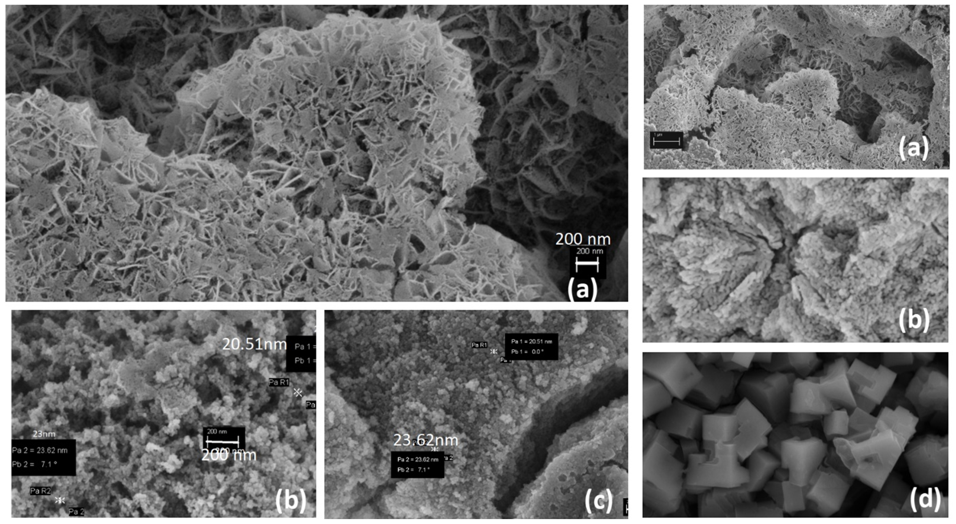
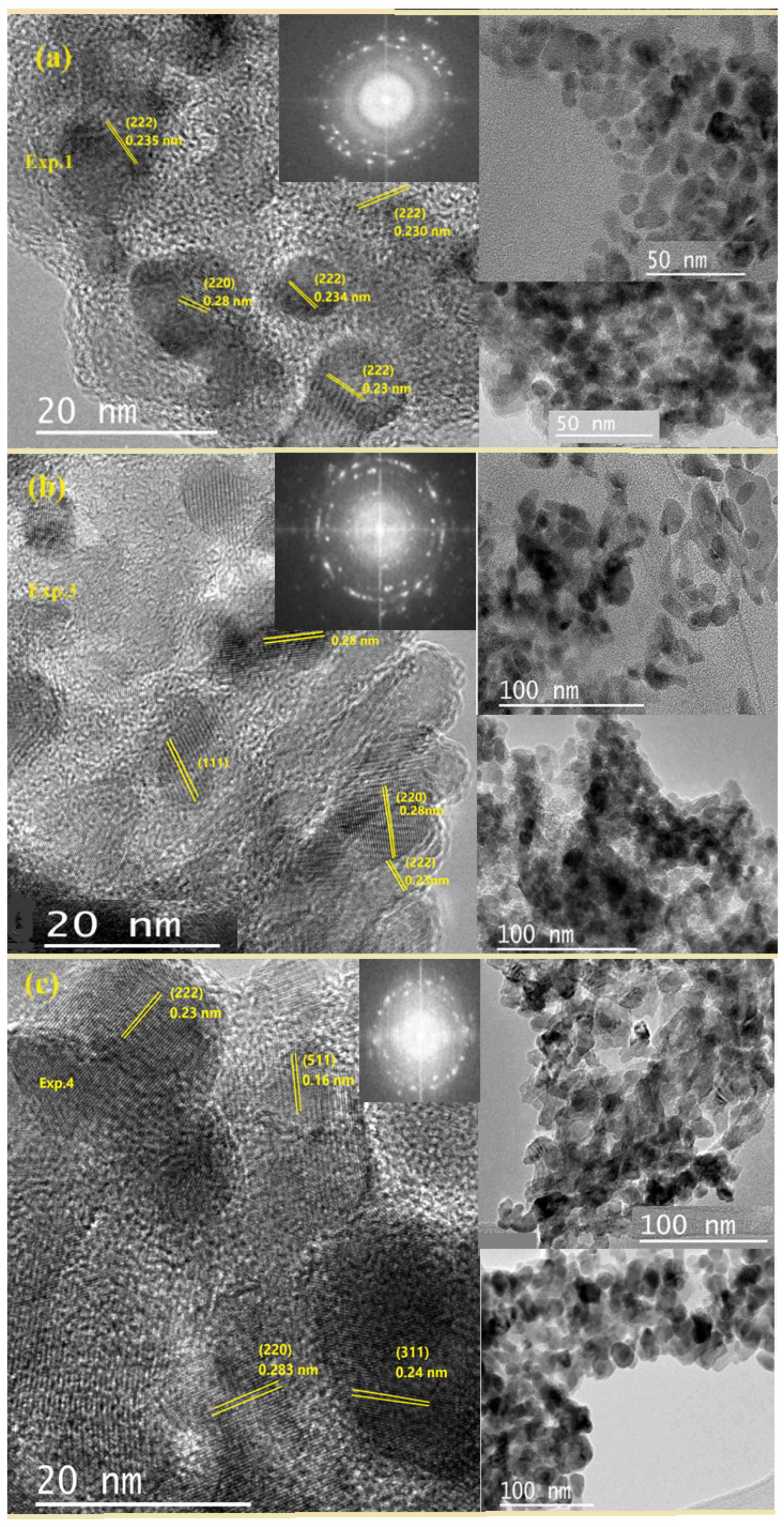
FESEM images (Figure 2 and Figure 3) show that the anodization conditions strongly influence the surface morphology of the oxide layers. Exp. 1 (30 V, no Ni) produced flower-like lamellae with average sizes of ∼200 nm, while Exp. 2 (30 V, Ni + Gly) yielded well-defined rice-like nanoparticles of ∼23 nm. Exp. 3 (30 V, no Ni, Gly) resulted in irregular aggregates with structural cracks, and Exp. 4 (40 V, Ni + Gly) generated micrometer-sized cubic assemblies (∼4 µm). These cubic structures correspond to aggregates of nanocrystals, in agreement with the crystallite sizes of 10–20 nm determined by TEM.
Based on the analysis of Figure 2a, the anodized samples prepared within the 30 V range exhibited excessively large interflake spacing, which could act as favorable channels for ion diffusion through the porous layer. In agreement with previous studies [42], the optimal anodization voltage was found to be 30 V, producing a thicker porous layer with uniformly distributed nanocups. This morphology enhanced chemical accessibility by regulating ion diffusion through the nanoflakes, while simultaneously providing high electrochemical activity, making it suitable for supercapacitor applications [1]. In our study, this morphology was reproduced in Exp. 1, but with a significant improvement in processing time: whereas the literature reported similar results after 7 h, under the conditions listed in Table 1 we achieved it in only 30 min. Moreover, the average flake size decreased from 460 nm (reported in the literature) to 200 nm in our samples.
The samples exhibited markedly different morphologies depending on the anodization parameters, including nanoparticle size, which varied with the processing conditions. For example, in Exp. 4, carried out at 40 V for 20 min, small particles were released into the electrolyte, indicating that the exposure time cannot be prolonged under these conditions [1]. At voltages above 40 V, partial disintegration of the sample was observed and the formation of cubes. As shown in Figure 2, the addition of nickel to the electrolyte produced two distinct morphologies: irregularly shaped particles with an average size of ∼20 nm. In contrast, the nickel-free material displayed flower-like formations composed of pointed lamellae with sizes close to 200 nm. In the case of the sample shown in Figure 2c (Exp. 3), the surface also exhibited irregularly shaped particles, agglomerated nanoparticles and the presence of cracks. The anodization behavior in Exp. 4 suggests that the combination of higher voltage and the presence of glycerol critically contributed to the temperature increase, requiring the anodization time to be limited to 20 min. By comparison, Exp. 0 and Exp. 2 maintained stable temperatures throughout the process. Notably, Exp. 2 also contained glycerol but was anodized at a lower voltage (30 V). This comparison indicates that voltages above 30 V cause significant heating of the sample within a short time. Moreover, since Exp. 0 (nickel-free) exhibited similar behavior to Exp. 2, it can be inferred that the presence of nickel does not substantially affect anodization time; rather, the applied voltage is the dominant factor governing temperature rise during the process.
The characterization results confirm the successful deposition of Ni on the Co3O4/Ni surface and reveal modifications in the underlying Co3O4 layers, as evidenced by the distinct morphologies observed in the FESEM images (Figure 2 and Figure 3). Electrochemical anodization is considered one of the most effective strategies for producing nanostructured metal oxides [1,43]. In both systems, the oxide layer thickness increased with anodization time; however, after several hours, dissolution (manifested as a rise in current) occurred, leading to a maximum layer thickness followed by sample degradation and partial disintegration. This behavior is associated with changes in current density or electrolyte composition, and during prolonged anodization a substantial portion of the surface detached. As shown in Figure 3, a magnification of Figure 2, the samples display uniform structures with varied morphologies, including interconnected hierarchical nanolayers, rice-like grains, deformed plates, and cubic particles. These morphological transformations were induced by adjusting three key parameters: the applied voltage, the electrolyte composition, and the incorporation of Ni. When Ni was introduced into the electrolyte, it underwent incorporation into the lattice, resulting in the formation of only the Co3O4 phase.
Figure 3 shows micrographs of cobalt oxide samples anodized under varying experimental conditions, highlighting the strong influence of anodization parameters on morphology. Exp. 1 (30 V, 30 min, without Gly or NH4F) produced characteristic cobalt oxide nanoflakes (∼200 nm). Exp. 2 (30 V, with Gly and Ni) maintained a stable temperature and yielded well-defined rice-like nanoparticles (∼20 nm), suggesting that Ni incorporation drives this morphological transformation. Exp. 3 (30 V, with Gly but without Ni) formed irregular nanoparticles (∼200 nm) under stable temperature. In Figure 2, smaller nanoparticles can be observed in Exp. 3, suggesting that, depending on the region, the sample presents variations, indicating a lack of uniformity in its structure. Exp. 4 (40 V, with Gly and Ni) experienced overheating, limiting anodization to 20 min and producing large cubic particles (∼4 μm), while TEM analysis revealed that the nanocrystalline Co3O4 grains remained below 20 nm.
The elemental composition of the samples is presented in Table 1, obtained through EDS analysis. The presence of Co, O, C, and Ni is observed in varying proportions, although the exact concentrations are difficult to quantify due to the limitations of the method. Carbon is present in all samples, likely due to the measurement being performed on a carbon tape, which may have influenced the accuracy of the carbon readings.

2.2. Surface Chemical Composition

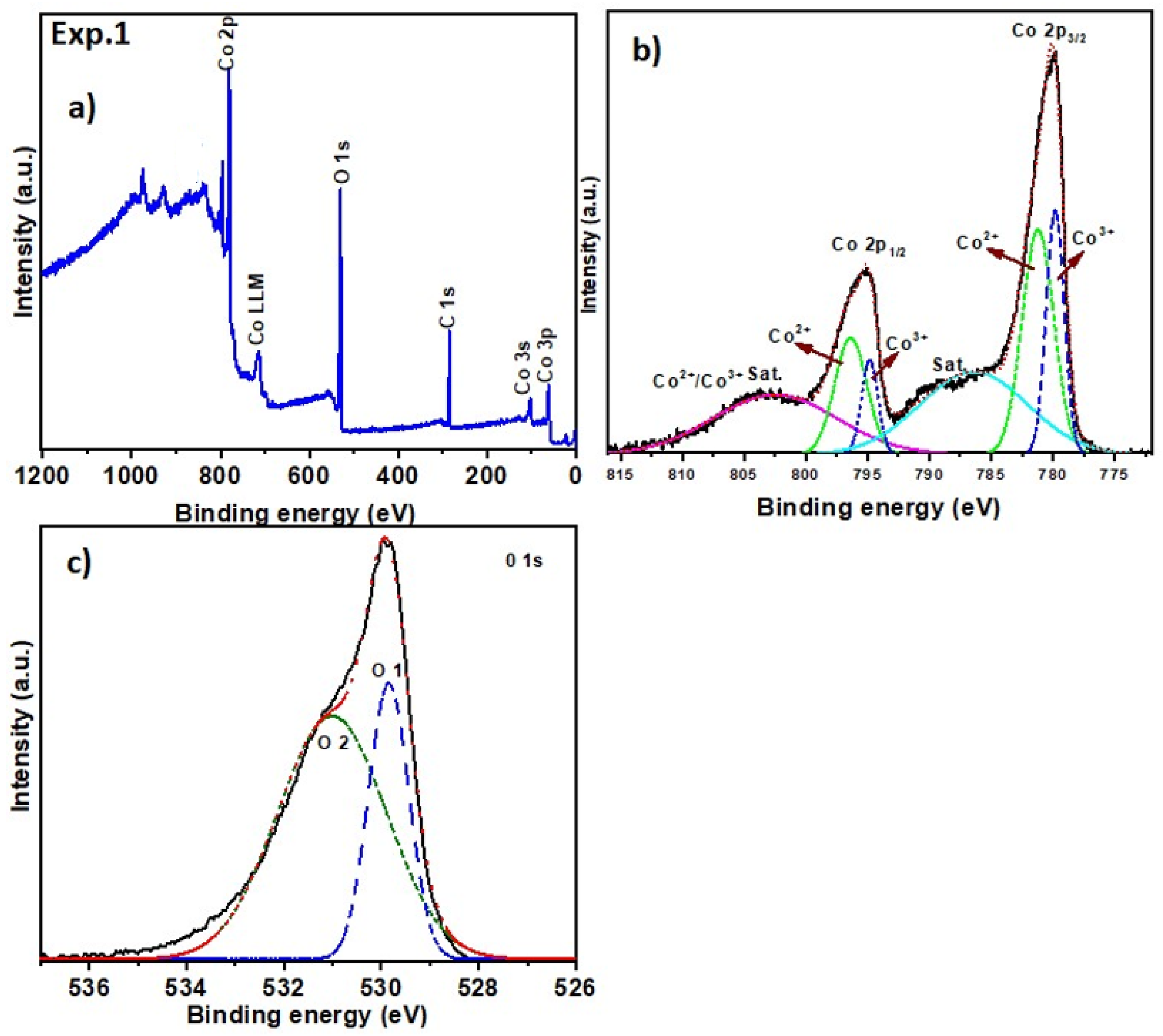
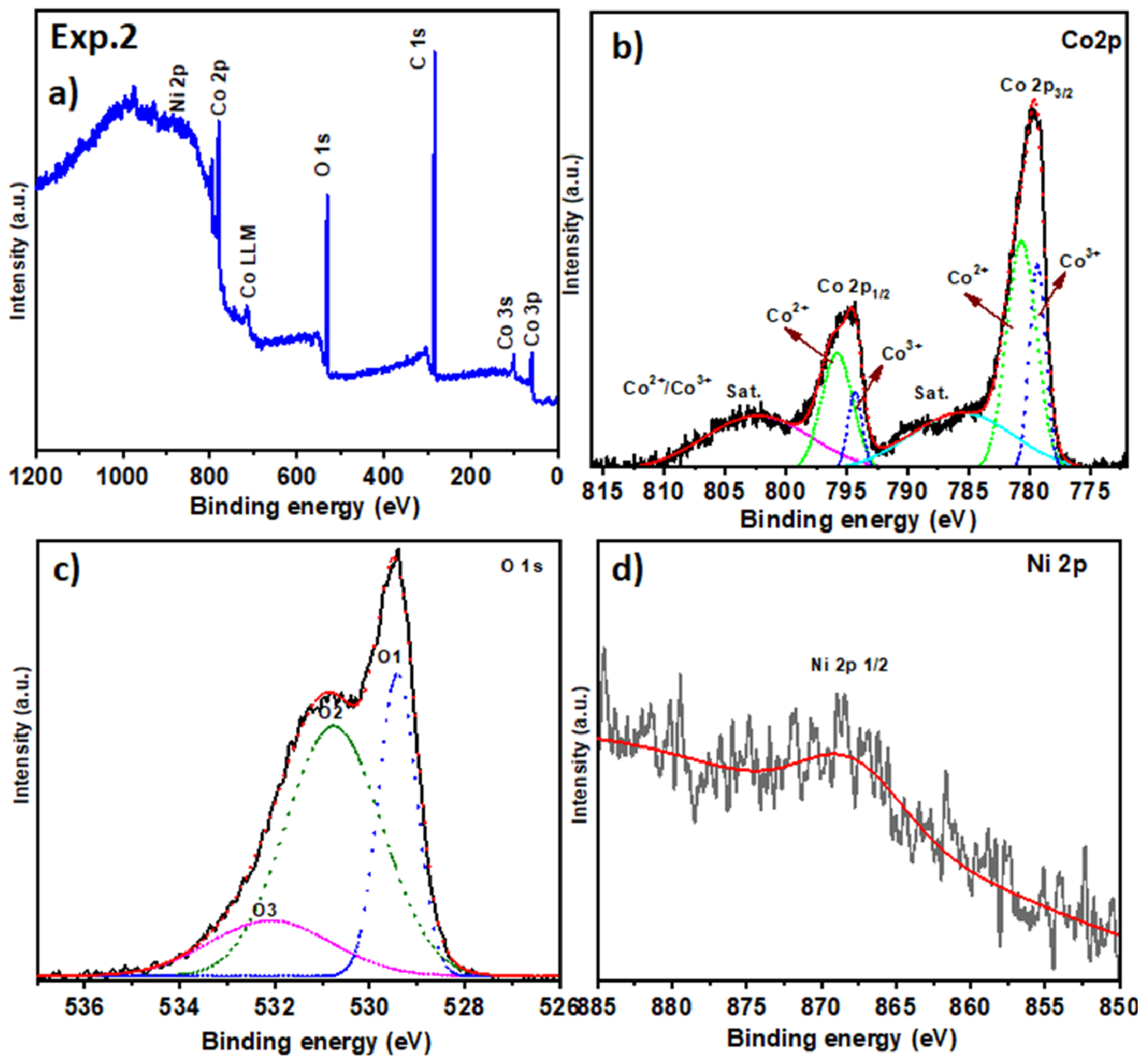
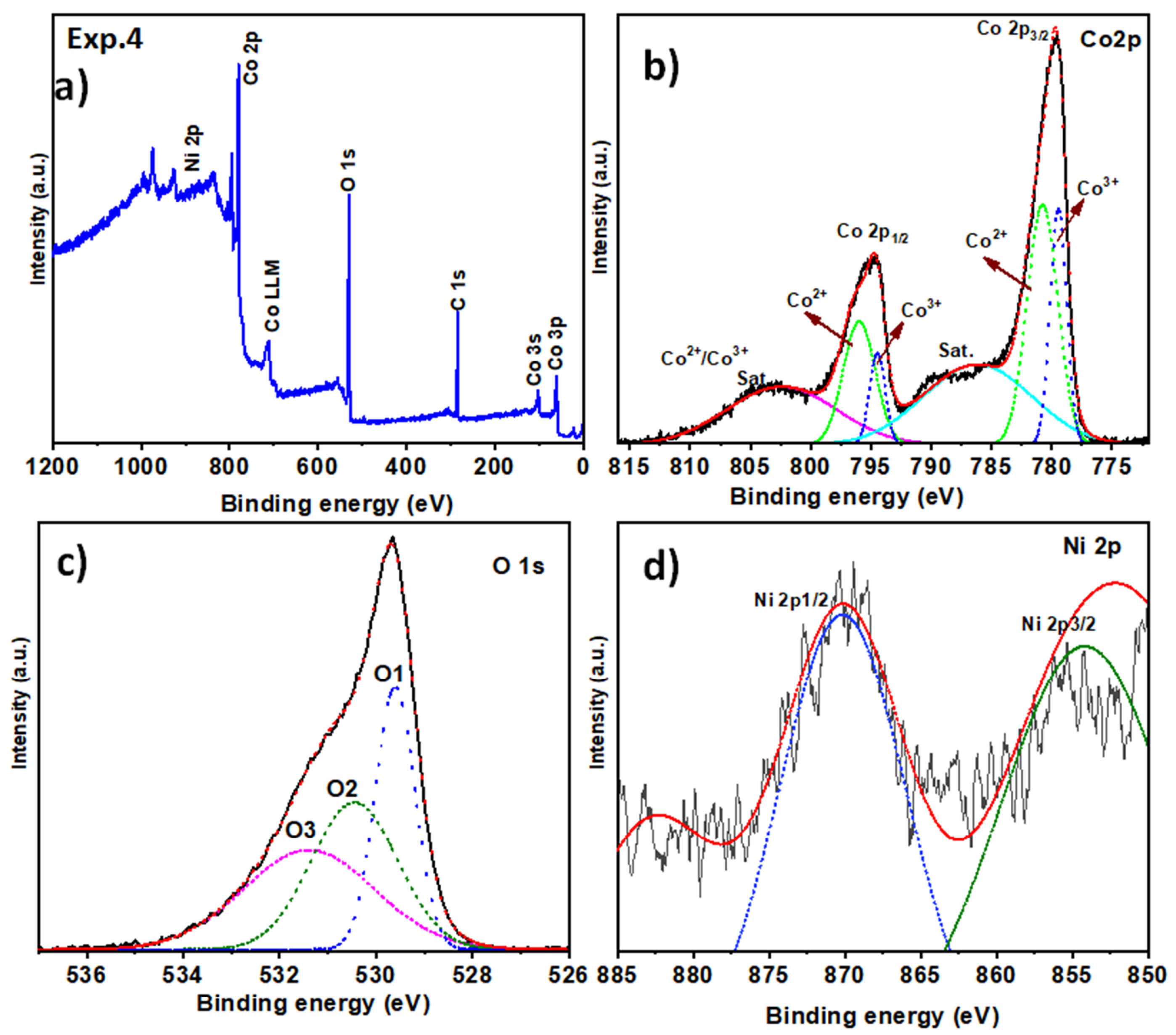
XPS investigations were conducted to examine the chemical composition and valence states present in the Co3O4 structures. The survey spectra of the Co3O4 samples are presented in Figure 4, Figure 5 and Figure 6, confirming the presence of Co, O, and Ni (for the samples containing nickel). The Co 2p spectra (Figure 4b) display two spin-orbit doublets, corresponding to Co2+ and Co3+. The fitted peaks at 779 and 790 eV are assigned to Co 2p3/2, while those at 795 and 797 eV correspond to Co 2p 1 / 2 [44]. The O 1s spectra (Figure 4c) exhibit peaks at 529 and 531 eV, which can be attributed to lattice oxygen and surface hydroxyl species, respectively. In the case of the sample without nickel addition, no Ni 2p signals are observed, consistent with the expected composition. For nickel-containing samples, Ni 2p spectra (Figure 5d and Figure 6d) reveal the presence of Ni in the oxidized state (Ni2+ or Ni3+), supporting its role in modifying the surface chemistry and morphology of the Co3O4 structures.
XPS measurements were performed to validate the valence states of Co3O4 samples and examine surface modifications following calcination. As shown in Figure 4b, the Co 2p spectrum of the Co3O4 polyhedra displays peaks at 780.4 eV (Co 2p 3 / 2 ) and 795.2 eV (Co 2p 1 / 2 ). The Co 2p spectrum was deconvoluted into six peaks, representing three spin-orbit doublets: the peaks at 781 and 797 eV are attributed to Co2+, whereas those at 780 and 795 eV correspond to Co3+. The O 1s spectrum (Figure 4c) shows two contributions at 530 eV and 531 eV, corresponding to lattice oxygen (Co–O) and surface hydroxyl groups, respectively [45]. This sample does not contain Ni, consistent with the absence of Ni 2p signals.
In Figure 5, the XPS analysis of Exp. 2, in which glycerol and Ni were added to the anodization electrolyte, shows Co 2p binding energies similar to those in Figure 4. Two additional satellite peaks associated with Co2+ suggest partial reduction in Co3+ to Co2+, likely generating oxygen vacancies, in agreement with previous studies [45,46,47,48]. The survey spectrum (Figure 5b) also indicates the presence of carbon species. A shift to lower binding energies for Co 2p suggests a change in the electron density around Co atoms. Ni 2p3/2 spectra (Figure 5d) show a weak signal around 867 eV, consistent with metallic Ni or low-valence Ni species. Despite anodization at a low voltage (30 V) and a 30 min duration, sufficient for Co3O4 formation, non-uniform nanoparticle structures resembling rice grains were observed on the surface.
Figure 6 presents the XPS analysis of Exp. 4, highlighting the anodization process and the transition from Co to Co3O4. The Co 2p spectrum (Figure 7b) exhibits the characteristic spin-orbit splitting into 2p 3 / 2 and 2p 1 / 2 . Deconvolution of the Co 2p 3 / 2 peak into Co2+ and Co3+ components reveals a Co3+/Co2+ ratio deviating from the ideal 2:1 stoichiometry, indicative of oxygen vacancy formation, as previously reported for spinel oxides subjected to high-temperature treatments [47]. The O 1s spectrum shows three peaks at 529.5, 530.5, and 531.8 eV, corresponding to lattice oxygen, hydroxyl species, and oxygen vacancies or adsorbed water, respectively. Ni signals appear with higher intensity than in Exp. 2, with Ni 2p 3 / 2 at 855 eV and Ni 2p1/2 at 870 eV, confirming Ni incorporation into the oxide matrix. Morphological analysis indicates the formation of large cube-shaped particles, which may limit practical applications due to their size.
The data summarized in Table 2 compile the XPS binding energies (BE) for Co 2p, O 1s, and Ni 2p core levels, along with their chemical state assignments and comparison to reported literature ranges. A progressive evolution in surface chemistry is evident across Exp. 1, Exp. 2, and Exp. 4, particularly regarding oxygen vacancy formation. While Exp. 1 displays characteristic signatures of Co2+/Co3+ and lattice oxygen, the O 1s spectra in Exp. 2 and Exp. 4 reveal a third contribution at higher binding energies (531.8 eV in Exp. 4), attributed to oxygen vacancies or adsorbed species. Such vacancies have been correlated with enhanced catalytic activity and improved electronic conductivity in spinel-type oxides [47,48,49,50], indicating a functional improvement induced by Ni incorporation. The increased intensity of the Ni 2p3/2 peak in Exp. 4, along with the altered Co3+/Co2+ ratio, supports this interpretation. Notably, the observed shift to lower binding energy in Co 2p in Exp. 2 suggests local electronic redistribution, potentially linked to anionic defect formation. These types of structural and electronic modifications are critical for optimizing transition metal oxides in advanced electrochemical applications [48,50].
Figure 7 shows TEM images of Co3O4 obtained after a 30 min thermal treatment at 350 °C in air, applied to the anodized nanoporous layer. Analysis indicates that Co3O4 has a cubic spinel structure (space group Fd 3 ¯ m, lattice parameter a = 8.083 Å), consistent with XRD results [51], and reveals the crystallographic orientation along the [111], [220], [222], and [311] zone axes [52]. TEM analysis indicates semi-spherical, nanocrystalline particles with grain sizes ranging from 10 to 20 nm and an average size of approximately 20 nm, in agreement with the SAED observations. A comparative size distribution (Figure 7) shows a slight increase in average grain size for sample Exp. 4. Such nanostructural features, combined with dopants and thermal treatment, are known to significantly affect conductivity and activation energy in cobalt oxide systems.
Figure 8a shows the Raman spectra of Co3O4 obtained from experiments Exp. 0 to Exp. 4. The primary vibrational modes of the spinel phase were identified at approximately 197 cm−1 (F 2 g 1 ), 489 cm−1 (Eg), 533 cm−1 (F 2 g 2 ), 633 cm−1 (F 2 g 3 ), and 702 cm−1 (A 1 g ).
For Exp. 0, the A 1 g mode is centered at 702 cm−1 with a FWHM of 15 cm−1, while F 2 g 1 appears at 197 cm−1 with a FWHM of 8 cm−1. In Exp. 2 and Exp. 4, where Ni was introduced into the electrolyte, a redshift of the A1g mode to 698 cm−1 and 694 cm−1, respectively, was observed, accompanied by an increase in FWHM, indicating lattice distortion and defect formation. These shifts are comparable to or slightly smaller than the peak shifts induced by changes in anodization voltage and electrolyte conditions (Exp. 1 and Exp. 3), demonstrating that both the nickel incorporation and the anodizing parameters contribute to structural modifications [53].
Figure 8b shows a detailed view of the A 1 g mode, highlighting the progressive redshift and peak broadening across the samples. The broadening of F 2 g 1 and A1g modes indicates decreased crystallinity and the presence of oxygen vacancies, in agreement with XPS analysis (Section 2.2), which detected a lower Co3+/Co2+ ratio and enhanced oxygen vacancy concentration in Exp. 4. Additionally, shifts in Eg and F 2 g 3 modes suggest distortions in tetrahedral Co2+-O (CoO4) and octahedral Co3+-O (CoO6) bonds, consistent with partial substitution of Co3+ by Ni2+. The Raman results clearly show that nickel incorporation, together with the anodization parameters, causes lattice distortions, alters crystallinity, and generates defects, thus providing a detailed view of the structural changes in Co3O4 [54].
Compared to nickel-free Co3O4 [55], the A 1 g mode shows a clear redshift, while the F 2 g 1 mode exhibits peak broadening, as observed in Figure 8. These changes indicate that nickel incorporation primarily substitutes octahedrally coordinated cobalt ions, altering the symmetry of the (CoO6) units [56]. The red or blue shifts in the Raman peaks can be attributed to lattice distortions caused by the introduction of different metal atoms into the Co3O4 matrix, which also promotes the formation of oxygen vacancies. Nickel preferentially occupies octahedral sites, whereas cobalt is distributed in both octahedral and tetrahedral positions. Replacing cobalt with nickel can generate catalytically active sites and modify the local charge distribution at cobalt sites. In addition, the substitution of atoms with different radii increases lattice strain, further enhancing the concentration of oxygen vacancies [54].
The systematic redshift of the A 1 g Raman peak from 702 to 694 cm−1, accompanied by a modest FWHM broadening, reflects subtle lattice distortions and enhanced phonon scattering in Co3O4. This trend evidences the impact of local strain and defect formation on the vibrational dynamics of the octahedral Co–O framework, revealing a delicate interplay between structural perturbations and spinel lattice stability (Table 3).

3. Materials and Methods

3.1. Anodization Process

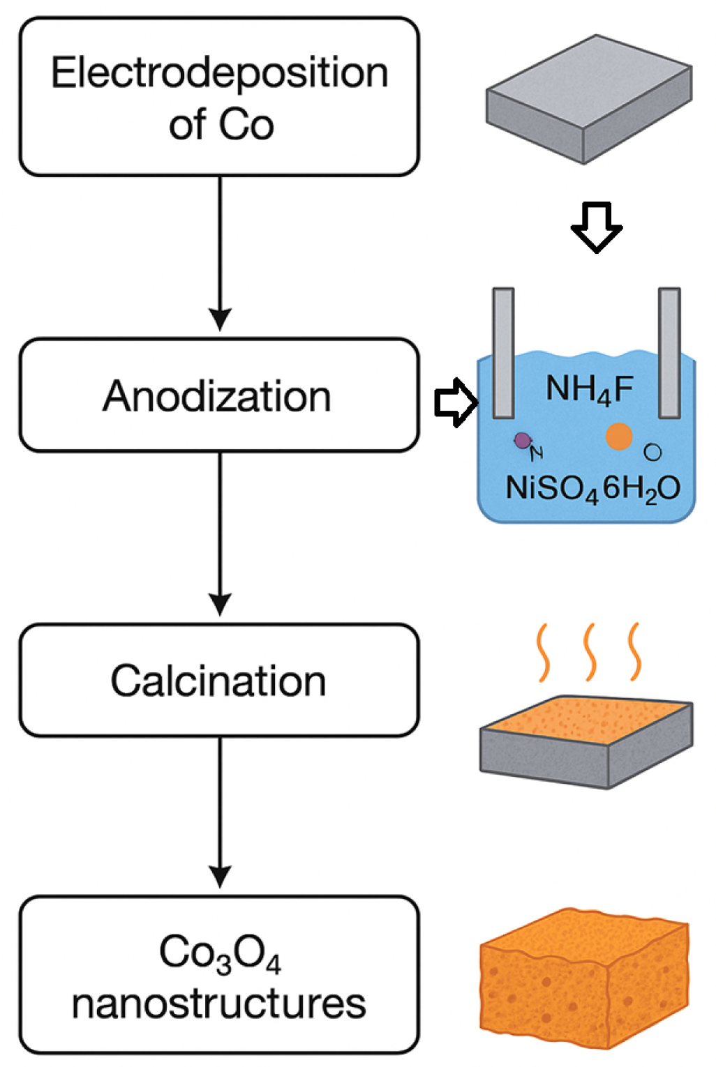
Porous cobalt oxide nanostructures were fabricated by anodizing electrodeposited cobalt films under different experimental conditions, varying the applied voltage, electrolyte composition, and anodization time. The electrolyte consisted of ethylene glycol (EG), glycerol (GL), deionized water (H2O), ammonium fluoride (NH4F), and nickel sulfate hexahydrate (NiSO4·6H2O), with the precise amounts summarized in Table 4. Voltages were adjusted between 30 and 40 V, based on prior reports [42,43,57]. The anodization duration was carefully optimized to prevent electrolyte overheating, with shorter times used in specific cases depending on the applied voltage and electrolyte composition. Selected tests included the addition of 1 g of NiSO4·6H2O to investigate the effect of Ni on morphology and capacitance.

3.2. Post-Anodization Thermal Treatment

Following anodization, all samples were subjected to a thermal annealing step to achieve complete transformation into the Co3O4 phase. The calcination process was carried out in air at 350 °C for 30 min in a muffle furnace, ensuring uniform crystallization of the anodized layers. This post-anodization treatment is essential for stabilizing the spinel structure of Co3O4, Figure 9.

3.3. Characterization

The structural and morphological characterization of the samples was performed using the following techniques:
X-ray diffraction (XRD): Measurements were performed on a PANalytical X’Pert Pro diffractometer with Cu K α radiation ( λ = 1.5406 Å), operated in Bragg–Brentano geometry. Data were collected over a 2 θ range of 10°–90° with a step size of 0.0263°. Raman spectroscopy. Raman spectra were collected using a Jobin Yvon T64000 triple-grating spectrometer operating in subtractive mode, equipped with an N2-cooled CCD detector. Excitation was provided by the 514 nm line of an Ar+ ion laser, focused through an Olympus microscope objective with a 20.5 mm focal distance and a numerical aperture of 0.35. Each spectrum represents the average of five accumulations with an integration time of 60 s. X-ray photoelectron spectroscopy (XPS): XPS was performed in an ultra-high-vacuum (UHV) chamber (∼10−9 Pa) using a Scienta Omicron ESCA+ spectrometer (FESEM: JEOL JSM-6701F, JEOL Ltd., Tokyo, Japan). with a monochromatic Al K α source (h ν = 1486.6 eV). High-resolution spectra were deconvoluted using SDP software (version 4.1) developed by Scienta Omicron, Uppsal, Sweden, applying an 80% Gaussian–Lorentzian peak fitting. Transmission electron microscopy (TEM): TEM, HRTEM, and SAED analyses were carried out using a Tecnai F20 Super-Twin TMP (FEI Company, Hillsboro, OR, USA), microscope operated at 200 kV. Sample preparation involved gently scraping the anodized films from the Co substrate and dispersing them in ethanol, followed by deposition onto carbon-coated copper grids. Field emission scanning electron microscopy (FESEM): Surface morphology and nanoparticle distribution were examined with a JEOL JSM-6701F microscope (Tokyo, Japan) operating at 7 kV.

4. Conclusions

This study presents a fast and efficient method for the synthesis of Co3O4 nanostructures via direct anodization of cobalt foils, followed by low-temperature heat treatment. Unlike previous reports, where similar structures required several hours to form, our approach achieves well-defined spinel morphologies in just 30 min. The systematic addition of nickel to the electrolyte was explored as a morphological modifier for the first time, revealing a significant influence on the surface structure. FESEM analysis showed that the morphology could be tuned from nanoflakes and rice-like grains to cubic micrometric aggregates. The formation of cubic structures at high voltage required limiting the anodization time to 20 min due to thermal instability induced by the combined effect of voltage and nickel addition. TEM analysis confirmed that the internal nanocrystalline size (10–20 nm) was preserved across all conditions. XPS and Raman spectroscopy validated the surface composition and provided insight into the doping mechanism: nickel predominantly substitutes octahedrally coordinated cobalt ions, altering the spinel symmetry, while oxygen vacancies were evidenced by deviations in the Co 3 + / Co 2 + ratio and shifts in Raman vibrational modes. Overall, these results demonstrate controllable morphology and composition, offering a versatile synthetic route for potential applications in catalysis, sensors, and energy storage, without overstating novelty regarding the use of cobalt foils.

Author Contributions

M.R.J.: project conception, writing, and editing. L.J.C.F.: experimental section, graphics, and editing. J.B.-R.: anodizing experimental section, writing, and editing. P.T.C.F.: characterization and editing. E.C.P.: characterization and editing. All authors have read and agreed to the published version of the manuscript.

Funding

This research received no external funding.

Institutional Review Board Statement

Not applicable.

Informed Consent Statement

Not applicable.

Data Availability Statement

The original contributions presented in this study are included in the article. Further inquiries can be directed to the corresponding author.

Acknowledgments

We gratefully acknowledge the support provided by the UNAL Colombia–EMBRAPA Brazil internationalization project.

Conflicts of Interest

The authors declare that they have no conflicts of interest that could influence the results or interpretations presented in this article.

References

  1. Qorbani, M.; Naseri, N.; Moshfegh, A.Z. Hierarchical Co3O4/Co(OH)2 Nanoflakes as a Supercapacitor Electrode: Experimental and Semi-Empirical Model. ACS Appl. Mater. Interfaces 2015, 7, 11172–11179. [Google Scholar] [CrossRef]
  2. International Energy Agency (IEA). World Energy Outlook 2021; International Energy Agency: Paris, France, 2021; Available online: https://www.iea.org/reports/world-energy-outlook-2021 (accessed on 23 October 2025).
  3. Statista Research Department. Percentage Distribution of Global Energy Consumption in 2019, by Energy Type; Statista: Hamburg, Germany, 2021; Available online: https://es.statista.com/estadisticas/633695/peso-de-los-diferentes-tipos-de-energias-en-el-consumo-energetico-mundial/statisticContainer (accessed on 23 October 2025).
  4. Ling, C.; Shi, L.; Ouyang, Y.; Chen, Q.; Wang, J. Transition Metal-Promoted V2CO2 (MXenes): A New and Highly Active Catalyst for Hydrogen Evolution Reaction. Adv. Sci. 2016, 3, 1600180. [Google Scholar] [CrossRef]
  5. Sheng, W.; Song, Y.; Dou, M.; Ji, J.; Wang, F. Constructing 1D Hierarchical Heterostructures of MoS2/In2S3 Nanosheets on CdS Nanorod Arrays for Enhanced Photoelectrocatalytic H2 Evolution. Appl. Surf. Sci. 2018, 436, 613–623. [Google Scholar] [CrossRef]
  6. United Nations. Transforming Our World: The 2030 Agenda for Sustainable Development (A/RES/70/1); United Nations: New York, NY, USA, 2015; Available online: https://sdgs.un.org/2030agenda (accessed on 23 October 2025).
  7. Liu, Y.; Jia, Y.; Jia, H.; Gao, L. Recent Development on the Synthesis Strategies and Mechanisms of Co3O4-Based Electrocatalysts for Oxygen Evolution Reaction: A Review. Molecules 2025, 30, 3238. [Google Scholar] [CrossRef] [PubMed]
  8. Wagh, K.S.; Mane, S.M.; Teli, A.M.; Shin, J.C.; Lee, J. Recent Advancements in Co3O4-Based Composites for Enhanced Electrocatalytic Water Splitting. Micromachines 2024, 15, 1450. [Google Scholar] [CrossRef]
  9. Mondal, S.; Mondal, A.K.; Chintala, V.; Tauseef, S.M.; Kumar, S.; Pandey, J.K. Thermochemical Pyrolysis of Biomass Using Solar Energy for Efficient Biofuel Production: A Review. Biofuels 2021, 12, 125–134. [Google Scholar] [CrossRef]
  10. Sebestyén, V. Environmental Impact Networks of Renewable Energy Power Plants. Renew. Sustain. Energy Rev. 2021, 151, 111626. [Google Scholar] [CrossRef]
  11. El Haj Assad, M.; Zubayda, S.; Khuwaileh, B.; Hmida, A.; Al-Shabi, M. Geothermal Energy as Power Producer. Proc. SPIE 2021, 11722, 117220V. [Google Scholar] [CrossRef]
  12. Hoang, A.T.; Nizetic, S.; Ong, H.C.; Chong, C.T.; Atabani, A.E.; Pham, V.V. Acid-Based Lignocellulosic Biomass Biorefinery for Bioenergy Production: Advantages, Application Constraints, and Perspectives. J. Environ. Manag. 2021, 296, 113194. [Google Scholar] [CrossRef]
  13. Aguilar Vargas, S.; Telles Esteves, G.R.; Medina Maçaira, P.; Quaresma Bastos, B.; Cyrino Oliveira, F.L.; Castro Souza, R. Wind Power Generation: A Review and a Research Agenda. J. Clean. Prod. 2019, 218, 850–870. [Google Scholar] [CrossRef]
  14. Worku, A.K.; Asfaw, A.A.; Ayele, D.W. Engineering of Co3O4 Electrodes via Ni and Cu-Doping for Supercapacitor Applications. Front. Chem. 2024, 12, 1357127. [Google Scholar] [CrossRef]
  15. Shinde, S.R.; Ruban, K. Advances in Cobalt Oxide-Based Supercapacitors: Recent Strategies and Performance Enhancement. ChemistrySelect 2025, 10, e01497. [Google Scholar] [CrossRef]
  16. Yang, J.; Shin, H.S. Recent Advances in Layered Transition Metal Dichalcogenides for Hydrogen Evolution Reaction. J. Mater. Chem. A 2014, 2, 5979–5985. [Google Scholar] [CrossRef]
  17. Zhang, N.; Wang, Y.; Hao, Y.-C.; Ni, Y.-M.; Su, X.; Yin, A.-X.; Hu, C.-W. Ultrathin Cobalt Oxide Nanostructures with Morphology-Dependent Electrocatalytic Oxygen Evolution Activity. Nanoscale 2018, 10, 20313–20320. [Google Scholar] [CrossRef]
  18. Zou, X.; Zhang, Y. Noble Metal-Free Hydrogen Evolution Catalysts for Water Splitting. Chem. Soc. Rev. 2015, 44, 5148–5180. [Google Scholar] [CrossRef]
  19. Wang, W.; Tian, Y.; Song, Y.; Ji, J.; Wang, F. Phase Controlled Synthesis and the Phase Dependent Photo- and Electrocatalysis of CdS@CoMo2S4/MoS2 Catalyst for HER. Int. J. Hydrogen Energy 2019, 44, 19890–19899. [Google Scholar] [CrossRef]
  20. Zhao, J.; Ren, X.; Ma, H.; Sun, X.; Zhang, Y.; Yan, T.; Wei, Q.; Wu, D. Synthesis of Self-Supported Amorphous CoMoO4 Nanowire Array for Highly Efficient Hydrogen Evolution Reaction. ACS Sustain. Chem. Eng. 2017, 5, 10093–10098. [Google Scholar] [CrossRef]
  21. Liu, Z.; Wang, J.; Zhan, C.; Yu, J.; Cao, Y.; Tu, J.; Shi, C. Phosphide–Oxide Honeycomb-Like Heterostructure CoP@CoMoOV/CC for Enhanced Hydrogen Evolution Reaction in Alkaline Solution. J. Mater. Sci. Technol. 2020, 46, 177–184. [Google Scholar] [CrossRef]
  22. Li, Z.; Zheng, M.; Zhao, X.; Yang, J.; Fan, W. Synergistic Engineering of Architecture and Composition in NixCo1−xMoO4–CoMoO4 Nanobrush Arrays towards Efficient Overall Water Splitting Electrocatalysis. Nanoscale 2019, 11, 22820–22831. [Google Scholar] [CrossRef]
  23. Gong, Y.; Yang, Z.; Lin, Y.; Wang, J.; Pan, H.; Xu, Z. Hierarchical Heterostructure NiCo2O4@CoMoO4/NF as an Efficient Bifunctional Electrocatalyst for Overall Water Splitting. J. Mater. Chem. A 2018, 6, 16950–16958. [Google Scholar] [CrossRef]
  24. Lu, W.; Song, Y.; Dou, M.; Ji, J.; Wang, F. Ni3S2@MoO3 Core/Shell Arrays on Ni Foam Modified with Ultrathin CdS Layer as a Superior Electrocatalyst for Hydrogen Evolution Reaction. Chem. Commun. 2018, 54, 646–649. [Google Scholar] [CrossRef]
  25. Lee, C.-Y.; Lee, K.; Schmuki, P. Anodic Formation of Self-Organized Cobalt Oxide Nanoporous Layers. Angew. Chem. Int. Ed. 2013, 52, 2077–2081. [Google Scholar] [CrossRef]
  26. Chondath, S.K.; Bansal, L.; Sahu, B.; Kumar, R. Dopant-Induced Defect Engineering in Transition Metal Oxide/Chalcogenide-Based Electrodes for High-Performance Supercapacitors: A Critical Review. ACS Appl. Energy Mater. 2025, 8, 8680–8709. [Google Scholar] [CrossRef]
  27. Jana, J.; Bhamu, K.C.; Ngo, Y.-L.T.; Kang, S.G.; Chung, J.S.; Hur, S.H. Designing a Bimetallic Transition Metal Oxide/Hydroxide Composite for Effective Electrocatalytic Oxygen Evolution Reaction. Appl. Surf. Sci. 2021, 562, 150253. [Google Scholar] [CrossRef]
  28. Pei, Z.; Xu, L.; Xu, W. Hierarchical Honeycomb-Like Co3O4 Pores Coating on CoMoO4 Nanosheets as Bifunctional Efficient Electrocatalysts for Overall Water Splitting. Appl. Surf. Sci. 2018, 433, 256–263. [Google Scholar] [CrossRef]
  29. Yan, Q.; Yang, X.; Wei, T.; Wu, W.; Yan, P.; Zeng, L.; Zhu, R.; Cheng, K.; Ye, K.; Zhu, K.; et al. Self-Supported Cobalt–Molybdenum Oxide Nanosheet Clusters as Efficient Electrocatalysts for Hydrogen Evolution Reaction. Int. J. Hydrogen Energy 2019, 44, 21220–21228. [Google Scholar] [CrossRef]
  30. Du, X.; Su, H.; Zhang, X. 3D Hierarchical Co3O4@Co3S4 Nanoarrays as Anode and Cathode Materials for Oxygen Evolution Reaction and Hydrogen Evolution Reaction. Dalton Trans. 2018, 47, 16305–16312. [Google Scholar] [CrossRef]
  31. Zhang, H.; Zhang, J.; Li, Y.; Jiang, H.; Jiang, H.; Li, C. Continuous Oxygen Vacancy Engineering of the Co3O4 Layer for an Enhanced Alkaline Electrocatalytic Hydrogen Evolution Reaction. J. Mater. Chem. A 2019, 7, 12240–12247. [Google Scholar] [CrossRef]
  32. Qu, C.; Cao, J.; Chen, Y.; Wei, M.; Fan, H.; Liu, X.; Li, X.; Wu, Q.; Feng, B.; Yang, L. In-Situ Growth of Hierarchical Trifunctional Co4S3/Ni3S2@MoS2 Core–Shell Nanosheet Array on Nickel Foam for Overall Water Splitting and Supercapacitor. Int. J. Hydrogen Energy 2023, 48, 648–661. [Google Scholar] [CrossRef]
  33. Xie, W.; Yu, T.; Ou, Z.; Zhang, J.; Li, R.; Song, S.; Wang, Y. Self-Supporting Clusters Constituted of Nitrogen-Doped CoMoO4 Nanosheets for Efficiently Catalyzing the Hydrogen Evolution Reaction in Alkaline Media. ACS Sustain. Chem. Eng. 2020, 8, 9070–9078. [Google Scholar] [CrossRef]
  34. Lu, W.; Song, Y.; Dou, M.; Ji, J.; Wang, F. Self-Supported Ni3S2@MoS2 Core/Shell Nanorod Arrays via Decoration with CoS as a Highly Active and Efficient Electrocatalyst for Hydrogen Evolution and Oxygen Evolution Reactions. Int. J. Hydrogen Energy 2018, 43, 8794–8804. [Google Scholar] [CrossRef]
  35. Xu, J.; Gao, L.; Cao, J.; Wang, W.; Chen, Z. Preparation and Electrochemical Capacitance of Cobalt Oxide (Co3O4) Nanotubes as Supercapacitor Material. Electrochim. Acta 2010, 56, 732–736. [Google Scholar] [CrossRef]
  36. Xiang, R.; Duan, Y.; Peng, L.; Wang, Y.; Tong, C.; Zhang, L.; Wei, Z. Three-Dimensional Core@Shell Co@CoMoO4 Nanowire Arrays as Efficient Alkaline Hydrogen Evolution Electrocatalysts. Appl. Catal. B Environ. 2019, 246, 41–49. [Google Scholar] [CrossRef]
  37. Lys, A.; Zabolotnii, V.; Čaplovičová, M.; Tepliakova, I.; Berzins, A.; Sahul, M.; Čaplovič, Ľ.; Pogrebnjak, A.; Iatsunskyi, I.; Viter, R. Core–Shell Nanofibers of ZnFe2O4/ZnO for Enhanced Visible-Light Photoelectrochemical Performance. J. Alloys Compd. 2024, 984, 173885. [Google Scholar] [CrossRef]
  38. Su, D.; Dou, S.; Wang, G. Single Crystalline Co3O4 Nanocrystals Exposed with Different Crystal Planes for Li–O2 Batteries. Sci. Rep. 2014, 4, 5767. [Google Scholar] [CrossRef]
  39. Gorimbo, J.; Chikati, R.; Khangale, P.; Beas, I.N.; Mguni, L.L.; Nkazi, D. Debunking the Impact of Crystallite/Particle Size in Cobalt-Based Fischer–Tropsch Synthesis. Chem. Eng. Commun. 2024, 211, 1262–1287. [Google Scholar] [CrossRef]
  40. Li, Y.; Keith, K.; Chopra, N. Structural and morphological evolution of free-standing Co3O4 nanowires via water vapor-assisted thermal oxidation of Co foil. J. Alloys Compd. 2017, 703, 414–423. [Google Scholar] [CrossRef]
  41. Abid, H.N.; Nayef, U.M.; Mutlak, F.A.H. Preparation and Characterization of Co3O4 Nanoparticles on Porous Silicon for Humidity Sensor by Photoluminescence. Optik 2019, 178, 379–383. [Google Scholar] [CrossRef]
  42. Mateus, H.M.; Bautista-Ruiz, J.; Barba-Ortega, J.; Rincón Joya, M. Formation of Titanium Oxide Nanotube Arrays by Controlling H2O and Time through Anodic Oxidation. Rasayan J. Chem. 2019, 12, 1304–1314. [Google Scholar] [CrossRef]
  43. Raja, K.S.; Misra, M.; Paramguru, K. Formation of Self-Ordered Nano-Tubular Structure of Anodic Oxide Layer on Titanium. Electrochim. Acta 2005, 51, 154–165. [Google Scholar] [CrossRef]
  44. Kaviyarasu, K.; Raja, A.; Devarajan, P.A. Structural Elucidation and Spectral Characterizations of Co3O4 Nanoflakes. Spectrochim. Acta Part A Mol. Biomol. Spectrosc. 2013, 114, 586–591. [Google Scholar] [CrossRef]
  45. Tüysüz, H.; Liu, Y.; Weidenthaler, C.; Schüth, F. Pseudomorphic Transformation of Highly Ordered Mesoporous Co3O4 to CoO via Reduction with Glycerol. J. Am. Chem. Soc. 2008, 130, 14108–14110. [Google Scholar] [CrossRef] [PubMed]
  46. Li, J.; Tang, S.; Lu, L.; Zeng, H.C. Preparation of Nanocomposites of Metals, Metal Oxides, and Carbon Nanotubes via Self-Assembly. J. Am. Chem. Soc. 2007, 129, 9401–9409. [Google Scholar] [CrossRef]
  47. Zhang, S.-Y.; Li, T.-T.; Zhu, H.-L.; Zheng, Y.-Q. Co3O4 Polyhedrons with Enhanced Electric Conductivity as Efficient Water Oxidation Electrocatalysts in Alkaline Medium. J. Mater. Sci. 2018, 53, 4323–4333. [Google Scholar] [CrossRef]
  48. Cárdenas, L.J.; Parra, C.A.; Cuervo, J.A.; Chiquito, A.J.; Moreno, L.C.; Rodríguez, J.E.; Rincón Joya, M. Electrical, Magnetic, and Sensing Properties of Acetone in Co3−xNixO4–rGO Samples. ACS Appl. Electron. Mater. 2024, 6, 4682–4697. [Google Scholar] [CrossRef]
  49. Babar, P.; Mahmood, J.; Maligal-Ganesh, R.V.; Kim, S.-J.; Xue, Z.; Yavuz, C.T. Electronic Structure Engineering for Electrochemical Water Oxidation. J. Mater. Chem. A 2022, 10, 20218–20241. [Google Scholar] [CrossRef]
  50. Zu, D.; Wang, H.; Lin, S.; Ou, G.; Wei, H.; Sun, S.; Wu, H. Oxygen-Deficient Metal Oxides: Synthesis Routes and Applications in Energy and Environment. Nano Res. 2019, 12, 2150–2163. [Google Scholar] [CrossRef]
  51. Vladimirova, S.; Krivetskiy, V.; Rumyantseva, M.; Gaskov, A.; Mordvinova, N.; Lebedev, O.; Martyshov, M.; Forsh, P. Co3O4 as p-Type Material for CO Sensing in Humid Air. Sensors 2017, 17, 2216. [Google Scholar] [CrossRef]
  52. Yin, H.; Zhu, J.; Chen, J.; Gong, J.; Nie, Q. PEG-Templated Assembling of Co3O4 Nanosheets with Nanoparticles for Enhanced Sensitive Non-Enzymatic Glucose Sensing Performance. J. Mater. Sci. Mater. Electron. 2018, 29, 17467–17475. [Google Scholar] [CrossRef]
  53. Rivas-Murias, B.; Salgueiriño, V. Thermodynamic CoO–Co3O4 Crossover Using Raman Spectroscopy in Magnetic Octahedron-Shaped Nanocrystals. J. Raman Spectrosc. 2017, 48, 837–841. [Google Scholar] [CrossRef]
  54. Luo, W.; Tian, H.; Li, Q.; Meng, G.; Chang, Z.; Chen, C.; Shen, R.; Yu, X.; Zhu, L.; Kong, F.; et al. Controllable Electron Distribution Reconstruction of Spinel NiCo2O4 Boosting Glycerol Oxidation at Elevated Current Density. Adv. Funct. Mater. 2024, 34, 2306995. [Google Scholar] [CrossRef]
  55. Cardenas-Flechas, L.J.; Freire, P.T.C.; Paris, E.C.; Moreno, L.C.; Joya, M.R. Temperature-Induced Structural Phase Transformation in Samples of Co3O4 and Co3−xNixO4 for CoO. Materialia 2021, 18, 101155. [Google Scholar] [CrossRef]
  56. Zhao, M.; Deng, J.; Liu, J.; Li, Y.; Liu, J.; Duan, Z.; Xiong, J.; Zhao, Z.; Wei, Y.; Song, W.; et al. Roles of Surface-Active Oxygen Species on 3DOM Cobalt-Based Spinel Catalysts MxCo3−xO4 (M = Zn and Ni) for NOx-Assisted Soot Oxidation. ACS Catal. 2019, 9, 7548–7567. [Google Scholar] [CrossRef]
  57. Mateus, H.M.; Barba-Ortega, J.; Rincón Joya, M. Comparison of the Growth of TiO2 Nanotubes in Different Solutions. J. Inorg. Organomet. Polym. 2018, 28, 612–623. [Google Scholar] [CrossRef]
Figure 1. Exp. 1 Anodized at 30 V without NH4F, Exp. 2 Anodized at 30 V with Ni, Exp. 3 Anodized at 30 V without Ni, and Exp. 4 Anodized at 40 V with Ni addition.
Inorganics 13 00350 g001
Figure 2. (a) Exp. 1 without nickel addition, (b) Exp. 2 with nickel addition in the electrolyte, (c) Exp. 3 without nickel, and (d) Exp. 4 with nickel addition in the electrolyte.
Inorganics 13 00350 g002
Figure 3. Exp. 1 Flake size: 200 nm without Ni; Exp. 2 Grain size, rice-like morphology, 20 nm with Ni; Exp. 3 characterized by an irregular morphology, size 200 nm without Ni; and Exp. 4 cubical particle size 4 µm with Ni.
Inorganics 13 00350 g003
Figure 4. XPS spectrum of samples in the (a) Exp.1 without the addition of Ni, (b) Co (2p3/2) core levels and (c) O (1s) XPS spectrum of the anodized sample at voltages 30 V.
Inorganics 13 00350 g004
Figure 5. XPS spectrum in (a) Exp. 2 with addition of Ni, (b) Co(2p), (c) O(1S) and (d) Ni(2p).
Inorganics 13 00350 g005
Figure 6. XPS spectrum in (a) Exp. 4 with the addition of Ni, binding energy of (b) Co(2p), (c) O(1S) and (d) Ni(2P).
Inorganics 13 00350 g006
Figure 7. (a) Exp. 1 without the addition of Ni, (b) Exp. 3 without addition of Ni, and (c) Exp. 4 with the addition of Ni.
Inorganics 13 00350 g007
Figure 8. (a) Raman spectra from Exp. 0 to Exp. 4. (b) Amplification of the A 1 g vibrational mode.
Inorganics 13 00350 g008
Figure 9. Illustrative scheme of the anodization process for the samples.
Inorganics 13 00350 g009
Table 1. Elemental composition (wt.%)–EDS analysis.
ElementExp. 1Exp. 2Exp. 3Exp. 4
Co (Cobalt)74.5862.3990.5953.86
O (Oxygen)21.3232.326.9240.04
C (Carbon)4.104.442.505.20
Ni (Nickel)0.000.750.000.90
Total100.00100.00100.00100.00
Table 2. XPS binding energies (BE) and chemical state assignments for Co, O, and Ni in Exp. 1, Exp. 2, and Exp. 4. The table includes comparison with literature values for validation.
SampleCore LevelBE (eV)AssignmentLit. Range (eV)
Exp. 1 (Co3O4)Co 2p3/2780.4Co3+779.6–780.2 [44]
Co 2p3/2781.0Co2+780.5–781.5 [44]
Co 2p1/2795.2Co3+794.5–795.5 [44]
Co 2p1/2797.0Co2+796.5–797.5 [44]
O 1s530.0Lattice O (Co-O)529.6–530.2 [46]
Exp. 2 (Co3O4 with Ni)Co 2p3/2∼780.0Co3+779.6–780.2 [44]
Co 2p3/2∼781.0Co2+780.5–781.5 [44]
Co 2p (Sat.)786Satellite peaks (Co2+)– [47]
Co 2p (shift)Partial reduction in Co2+802.5 [47]
Ni 2p3/2867.0Ni0 or low-valence Ni866.5–867.5 [51]
Exp. 4 (Co3O4 with Ni)Co 2p3/2780.0Co3+779.6–780.2 [44]
Co 2p3/2781.5Co2+780.5–781.5 [44]
Co 2p1/2795.0Co3+794.5–795.5 [44]
Co 2p1/2797.0Co2+796.5–797.5 [44]
O 1s529.5Lattice O (Co-O)529.6–530.2 [46]
O 1s530.5Hydroxyl groups530.8–531.5 [46]
O 1s531.8Oxygen vacancies or adsorbed H2O531.5–532.0 [47]
Ni 2p3/2855.0Ni2+ or Ni3+854.5–855.5 [51]
Ni 2p1/2870.0Ni2+ or Ni3+869.5–870.5 [51]
Table 3. Raman peaks of the A 1 g mode of Co3O4, taking the black curve (Exp. 0) as reference. The peak position ( ν ) and full width at half maximum (FWHM) are reported.
Sampleν
(cm−1)
Δν
(cm−1)
FWHM
(cm−1)
Exp. 0702015
Exp. 3700−214
Exp. 2698−416
Exp. 1696−617
Exp. 4694−818
Table 4. Anodizing conditions: water content (mL), ammonium fluoride (NH4F) (g), nickel sulfate hexahydrate content (g), Ethylene Glycol (mL), Glycerol (mL), voltage (v), and anodizing time (min).
SamplesH2O (mL)NH4F (g)NiSO4·6H2O (g)EG (mL)Gly (mL)V (V)t (min.)
Exp. 00.50.3014503030
Exp. 12.00.0014503030
Exp. 20.50.31145483030
Exp. 32.00.30145483030
Exp. 40.50.31145484020
Disclaimer/Publisher’s Note: The statements, opinions and data contained in all publications are solely those of the individual author(s) and contributor(s) and not of MDPI and/or the editor(s). MDPI and/or the editor(s) disclaim responsibility for any injury to people or property resulting from any ideas, methods, instructions or products referred to in the content.