Article

An Integrative Biosynthetic Approach to Silver Nanoparticles: Optimization Modeling, and Antimicrobial Assessment

1
Nanotechnology Research Unit, Jazan University, P.O. Box 114, Jazan 45142, Saudi Arabia
2
Environment and Nature Research Centre, Jazan University, P.O. Box 114, Jazan 45142, Saudi Arabia
3
Department of Biology, College of Science, Jazan University, P.O. Box 114, Jazan 45142, Saudi Arabia
4
Department of Biology, College of Science, University of Hafr Al Batin, P.O. Box 1803, Hafr Al Batin 31991, Saudi Arabia
5
Department of Biology, Darb University College, P.O. Box 114, Jazan 45142, Saudi Arabia
*
Author to whom correspondence should be addressed.
Inorganics 2025, 13(11), 342; https://doi.org/10.3390/inorganics13110342
Submission received: 12 September 2025 / Revised: 8 October 2025 / Accepted: 14 October 2025 / Published: 22 October 2025

Abstract

Silver nanoparticles (AgNPs) are valued for their antimicrobial properties, but conventional synthesis often involves toxic chemicals. Eco-friendly biosynthesis using silver-tolerant microbes from contaminated sites offers a sustainable alternative. This study biosynthesized and characterized AgNPs using a native Bacillus sp. from contaminated soil in the Jazan region, Saudi Arabia, and developed predictive models for optimizing synthesis and antimicrobial activity. AgNPs were synthesized under optimized conditions (1.0 mM AgNO3, 4.0 mL supernatant, pH 8, 85 °C). Characterization using UV–Vis, SEM, TEM, XRD, and FTIR assessed size, shape, structure, and chemistry. Gaussian and second models evaluated yield and inhibition zones based on AgNP concentration, microorganism type, and MIC. The AgNPs were spherical with diameters of 5–10 nm. The optimal nanoparticle yield occurs when the parameters are at their optimal values; C0 = 1.0 mM, V0 = 4.0 mL, pH0 = 8, T0 = 85 °C. XRD confirmed their crystalline nature, and FTIR showed biomolecular capping agents for stabilization. The Gaussian model accurately predicted synthesis efficiency, validated by 3D plots matching experimental data. The AgNPs showed strong antimicrobial activity against Gram-positive (Bacillus subtilis) (ATCC6051), Staphylococcus aureus (ATCC12600), Gram-negative bacteria Escherichia coli (ATCC11775) and fungi Candida albicans (ATCC10231); with E. coli having the lowest MIC (1.87 μg/mL). The inhibition zone model closely matched observed data. Biosynthesized AgNPs using silver-tolerant Bacillus sp. demonstrated potent antimicrobial effects and provide a green alternative to chemical synthesis. Integrating modeling optimizes biosynthesis and predicts biological performance, supporting future nanobiotechnology and antimicrobial applications.

1. Introduction

Nanotechnology, which has roots in ancient practices and was formalized in the 20th century, focuses on manipulating matter at the nanoscale (1–100 nm) to unlock unique properties that traditional materials cannot exhibit [1]. Among the numerous nanomaterials, silver nanoparticles (AgNPs) have garnered attention due to their outstanding antimicrobial and medicinal properties, making them highly suitable for applications in healthcare, diagnostics, and environmental protection [2]. Their strong biological activity is attributed to their small size, high surface area, and the ability to interact with cellular structures, which has led to their increasing use in drug delivery systems, wound healing, and even cancer therapy [3].
The green synthesis of nanoparticles, which is a more eco-friendly and sustainable alternative to traditional physical and chemical methods, has emerged as a promising approach for the rapid and efficient production of metal nanoparticles like AgNPs [4]. Green synthesis methods utilize biological organisms such as bacteria, fungi, and plants to reduce metal ions into nanoparticles, ensuring that the process is cost-effective, environmentally safe, and biocompatible [5]. Biological agents act as reducing, stabilizing, and capping agents, providing a controlled environment for nanoparticle production, and bacteria, in particular, have shown significant promise in the biosynthesis of silver nanoparticles [6].
Numerous studies have demonstrated the capacity of bacterial strains like Bacillus subtilis, Pseudomonas sp., and Escherichia coli to synthesize AgNPs with potent antimicrobial properties [7]. These bacteria, isolated from various environments, such as industrial effluents, contaminated soils, and even copper mines, can tolerate high concentrations of silver ions and effectively convert them into metallic nanoparticles [8]. For instance, Bacillus megaterium has been widely studied for its ability to produce AgNPs with distinct antimicrobial properties [9]. Research has shown that Bacillus species, including Bacillus subtilis and Bacillus megaterium, can produce silver nanoparticles ranging from 10 to 60 nm in size with spherical morphology, as observed through techniques such as UV–vis spectrophotometry, Transmission Electron Microscopy (TEM), and Scanning Electron Microscopy (SEM) [10]. These nanoparticles have demonstrated strong antibacterial effects against various multidrug-resistant pathogens, including Staphylococcus aureus, Klebsiella pneumoniae, and Escherichia coli, underscoring their potential as novel antimicrobial agents [11]. Fungi also play a significant role in the biosynthesis of AgNPs, with Penicillium fellutanum and Nigrospora sphaerica being commonly used for the production of silver nanoparticles [12]. Fungal synthesis methods offer scalability and higher biomass yields, which make them advantageous for large-scale production [13]. Similarly, plants like clove, cinnamon, and Arabian primrose have been used for the green synthesis of AgNPs, utilizing natural phytochemicals to reduce silver ions and stabilize the nanoparticles [14]. These plant-based methods are often more sustainable due to their rapid growth and eco-friendly nature, providing an effective alternative to chemical synthesis techniques [15]. In recent years, green synthesis has seen applications across various fields, including medical (e.g., wound healing, cancer therapy), environmental (e.g., pollution monitoring), and industrial (e.g., food preservation) sectors [16]. However, despite the growing interest in biosynthesis, existing methods often lack efficient optimization models, which can lead to inconsistent yields and variable antimicrobial properties. To address these issues, it is essential to explore more predictive models that can better optimize synthesis conditions and improve the consistency of AgNP production [17]. In this regard, the application of Gaussian optimization models can help enhance yields and predict optimal synthesis parameters, ensuring reproducible and high-quality nanoparticle production. A study investigates the biosynthesis of AgNPs using Bacillus megaterium, a silver-resistant bacterial strain isolated from a copper mine. This bacterial species is particularly relevant due to its ability to survive and thrive in environments with high concentrations of metals, offering a promising microbial source for AgNP synthesis. The main challenges in bacterial AgNP biosynthesis include strain variability, leading to inconsistent nanoparticle size and yield, and lack of standardized protocols for optimizing synthesis conditions. Variations in growth conditions, culture medium, and pH can influence nanoparticle characteristics. The mechanisms behind silver ion reduction and nanoparticle stabilization are still not fully understood, hindering process control. Additionally, scaling up the biosynthesis process for industrial applications remains a challenge due to the need for large-scale bacterial cultures. Ensuring biological stability of the nanoparticles and reducing aggregation during synthesis are further obstacles. More research is needed to optimize environmental parameters and improve strain selection for consistent, high-yield nanoparticle production.
The study aims to contribute to the understanding of the biosynthesis mechanisms of AgNPs, utilizing both environmentally friendly and efficient methods for nanoparticle production. Furthermore, the study emphasizes predictive modeling for optimizing synthesis parameters and enhancing the yield of AgNPs, with applications in antimicrobial and industrial fields. Recent studies have also highlighted the importance of comprehensive characterization techniques such as UV–vis spectrophotometry, SEM, TEM, X-ray diffraction (XRD), and Fourier Transform Infrared (FTIR) spectroscopy to validate the formation and structural properties of AgNPs. For example, UV–vis spectroscopy provides information on the surface plasmon resonance (SPR) band of AgNPs, typically found at around 430 nm, confirming nanoparticle formation. TEM and SEM allow for the visualization of particle morphology and size distribution, while FTIR spectroscopy identifies the functional groups involved in nanoparticle stabilization. These techniques are essential for characterizing the size, shape, and surface properties of biosynthesized nanoparticles. This study introduces an innovative approach by using native Bacillus sp. isolated from contaminated soils in the Jazan region of Saudi Arabia, adding a unique aspect to AgNP synthesis. This indigenous microbial strain is likely adapted to local environmental conditions, which may result in the production of silver nanoparticles with distinct properties compared to those produced by other bacterial strains. Additionally, the use of this local strain offers a sustainable and region-specific method for AgNP production.
Finally, this work introduces the application of Gaussian optimization models to predict and optimize synthesis parameters for AgNP production, which may serve as a valuable tool for improving the efficiency and consistency of nanoparticle yields. In addition to biosynthesis, this study contributes to bioremediation efforts, as the bacteria utilized for AgNP synthesis simultaneously aid in the remediation of contaminated environments.

2. Results

2.1. Isolation of Silver-Tolerant Bacteria and Gram Stain

Soil and water samples were collected from sewage-treated and industrial zones in the Jazan region, Saudi Arabia. Bacterial colonies were cultured on nutrient agar plates, exhibiting distinct morphological characteristics, white pigmentation, 1–2 mm diameter, and either irregular or entire margins. Selected colonies were further screened on nutrient agar supplemented with 1 mM AgNO3. Among the isolates, a colony from industrial soil developed a brownish halo indicative of silver nitrate reduction and was selected for subsequent analyses (Figure 1A).
Microscopic examination of the selected isolate using the Gram stain technique revealed Gram-positive, elongated rod-shaped bacteria. These characteristics aligned with the morphology typically observed in Bacillus species (Figure 1B).

2.2. Molecular Identification via 16S rRNA

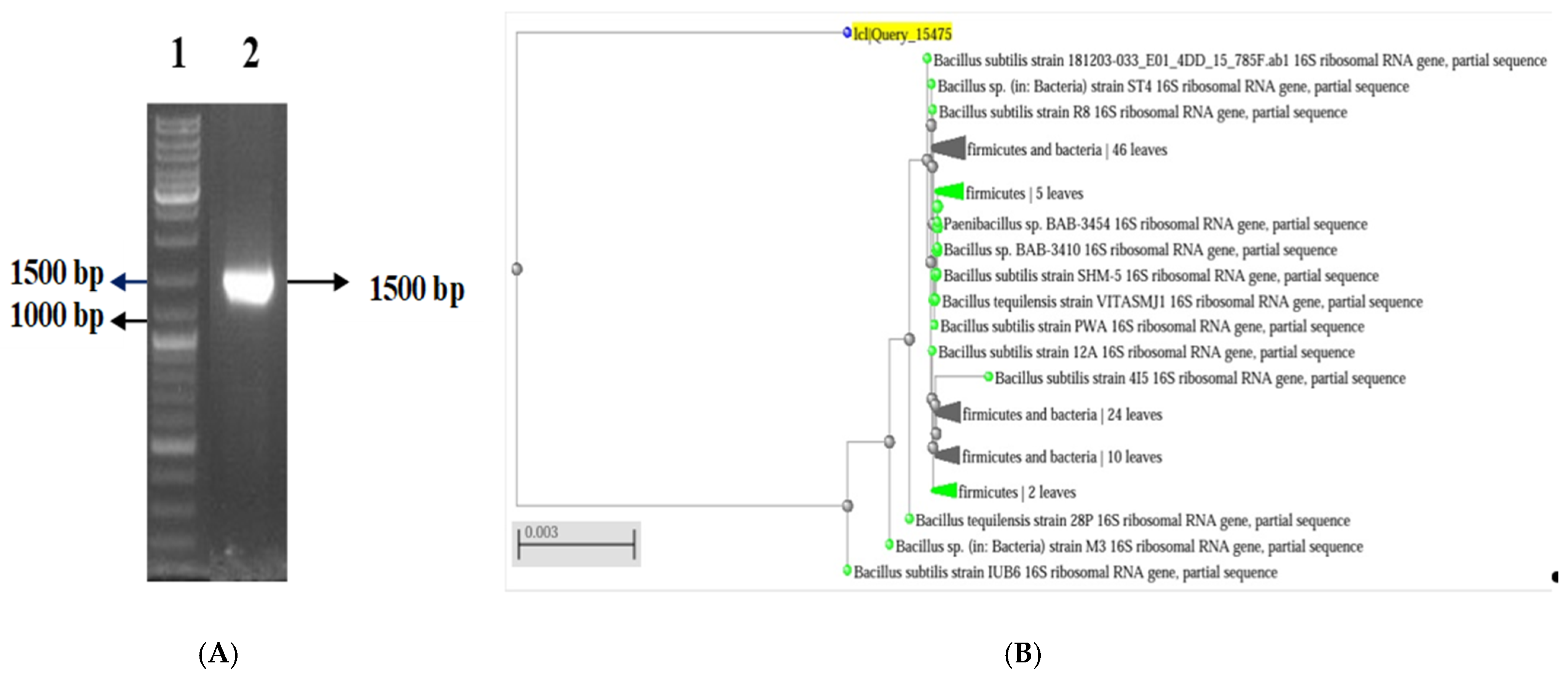
Genomic DNA extracted from the bacterial isolate was subjected to PCR amplification targeting the 16S rRNA gene using universal primers 27F and 1492R. The amplified product, approximately 1500 base pairs in length, was confirmed via gel electrophoresis. Sequencing and BLAST (https://blast.ncbi.nlm.nih.gov/Blast.cgi, accessed on 14 June 2025) analysis of the partial 16S rRNA sequence identified the isolate as Bacillus sp. ST4, exhibiting 97% sequence homology (Figure 2A,B).

2.3. Optimization of AgNPs Synthesis Conditions

A negative control was performed using the cell supernatant without the bacterial strain, and the results indicated that no significant reduction activity occurred in the absence of the biological agents.

2.3.1. Effect of Silver Nitrate Concentration

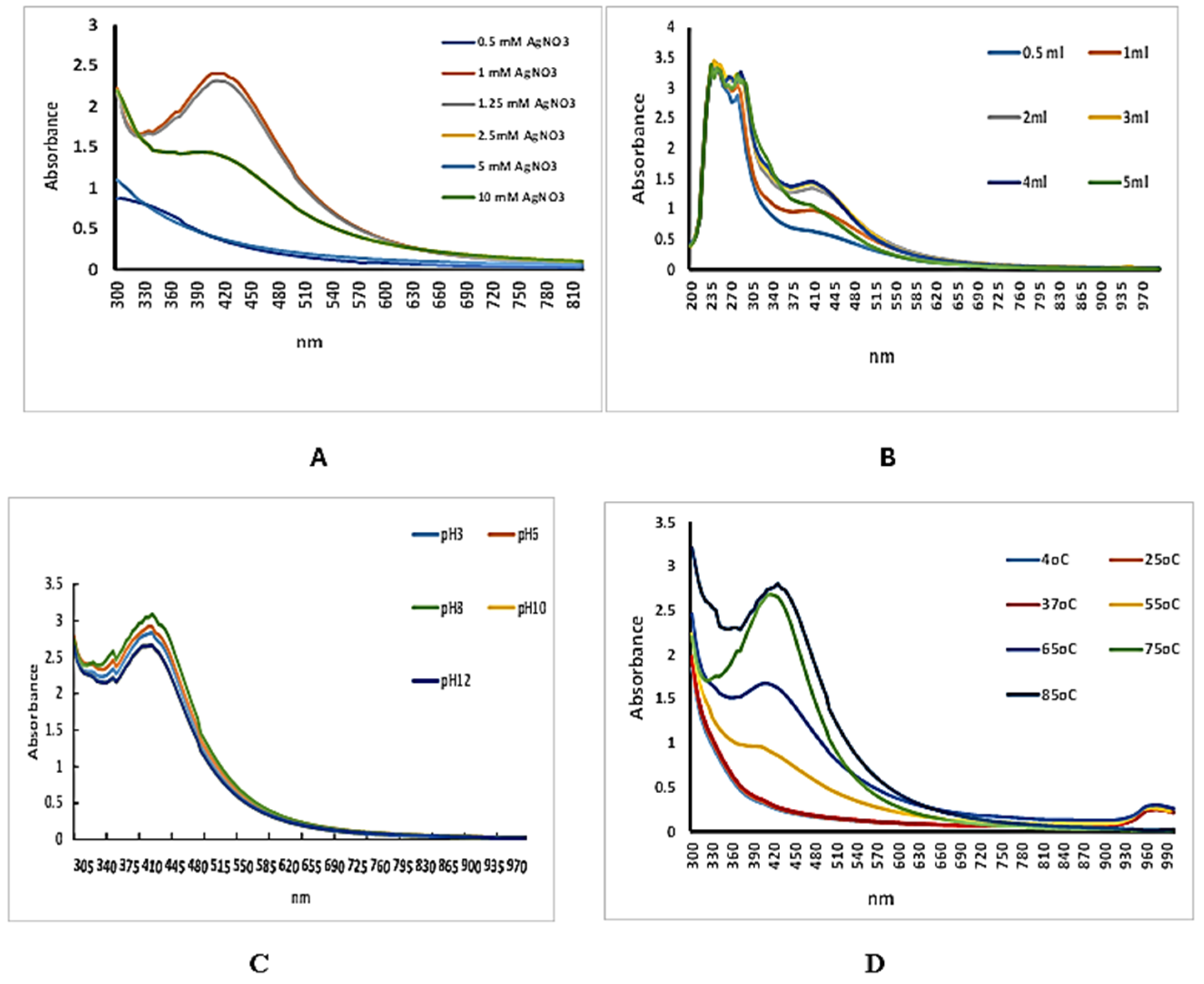
The results indicate that the synthesis of AgNPs is highly influenced by AgNO3 concentration. The maximum absorbance at 425 nm was recorded at a concentration of 1.0 mM AgNO3, suggesting this as the optimal condition for nanoparticle formation. Slightly lower but comparable absorbance was observed at 1.25 mM, indicating sustained nanoparticle production. At concentrations of 0.5 mM, 2.5 mM, 5 mM, and 10 mM, the absorbance values were notably lower, reflecting reduced efficiency in nanoparticle synthesis under those conditions. These findings confirm that 1.0 mM AgNO3 provides the most favorable conditions for the biosynthesis of silver nanoparticles in terms of yield and stability (Figure 3A). AgNO3 concentration significantly affected SPR peak absorbance (one-way ANOVA, p < 0.001); 1.0 mM (and 1.25 mM) produced higher absorbance than 0.5, 2.5, 5, and 10 mM (Tukey, p < 0.05).

2.3.2. Effect of Culture Supernatant Volume

The synthesis of silver nanoparticles (AgNPs) was assessed using varying volumes of bacterial culture supernatant (0.5, 1, 2, 3, 4, and 5 mL) combined with a fixed concentration of 1 mM AgNO3. UV–Visible spectrophotometry was used to monitor nanoparticle formation.
All tested volumes resulted in characteristic surface plasmon resonance (SPR) peaks, indicating the successful formation of AgNPs. The highest absorbance at approximately 425 nm was recorded with 2 and 4 mL of culture supernatant, suggesting optimal nanoparticle synthesis at this volume. Lower absorbance values were observed at 5 mL volumes, compared to the peak observed at 4 mL (Figure 3B). Volume had a significant effect (ANOVA, p < 0.001); 4 mL > 0.5–3 mL and ≈5 mL (Tukey, p < 0.05).
These results suggest that 4 mL of bacterial supernatant was the most effective volume for producing a high yield of silver nanoparticles under the specified conditions.

2.3.3. Effect of pH

The influence of pH on the biosynthesis of silver nanoparticles (AgNPs) was investigated by adjusting the reaction mixture to different pH values (3, 6, 8, 10, and 12). The formation of AgNPs was monitored using UV–Visible spectroscopy. All samples exhibited surface plasmon resonance (SPR) peaks, indicating successful nanoparticle formation. Among the tested conditions, the maximum absorbance at ~425 nm was observed at pH 8, suggesting it as the optimal pH for AgNP synthesis. Slightly lower absorbance values were recorded at pH 6 and pH 10, while more acidic (pH 3) and highly alkaline (pH 12) conditions resulted in reduced peak intensities. These findings demonstrate that pH 8 provides the most favorable environment for the efficient biosynthesis of AgNPs under the experimental conditions used (Figure 3C). pH influenced AgNP yield (ANOVA/Kruskal–Wallis, p < 0.001); pH 8 exceeded pH 3, 6, 10, 12 (post hoc, p < 0.05).

2.3.4. Effect of Temperature

The effect of temperature on the biosynthesis of silver nanoparticles (AgNPs) was assessed at seven different incubation temperatures: 4 °C, 25 °C, 37 °C, 55 °C, 65 °C, 75 °C, and 85 °C. Nanoparticle formation was monitored using UV–Visible spectroscopy. All temperatures tested resulted in the formation of AgNPs, as indicated by the presence of surface plasmon resonance (SPR) peaks. The maximum absorbance at approximately 425 nm was observed at 85 °C, indicating this temperature yielded the highest nanoparticle production. A strong SPR signal was also observed at 75 °C and 65 °C, though slightly lower than at 85 °C. In contrast, lower absorbance values were recorded at 4 °C, 25 °C, 37 °C, and 55 °C, suggesting reduced synthesis efficiency under those conditions. The lowest SPR intensities were seen at 37 °C and 55 °C, reflecting minimal nanoparticle formation.
These results indicate that higher temperatures, particularly 85 °C, significantly enhance the biosynthesis of silver nanoparticles, likely by accelerating the reduction kinetics of silver ions and promoting effective nucleation and growth of nanoparticles (Figure 3D). Temperature significantly affected absorbance (ANOVA, p < 0.001); 85 °C (and 75–65 °C) >4–55 °C (post hoc, p < 0.05).

2.4. Gaussian Modeling and Visualization of AgNP Optimization

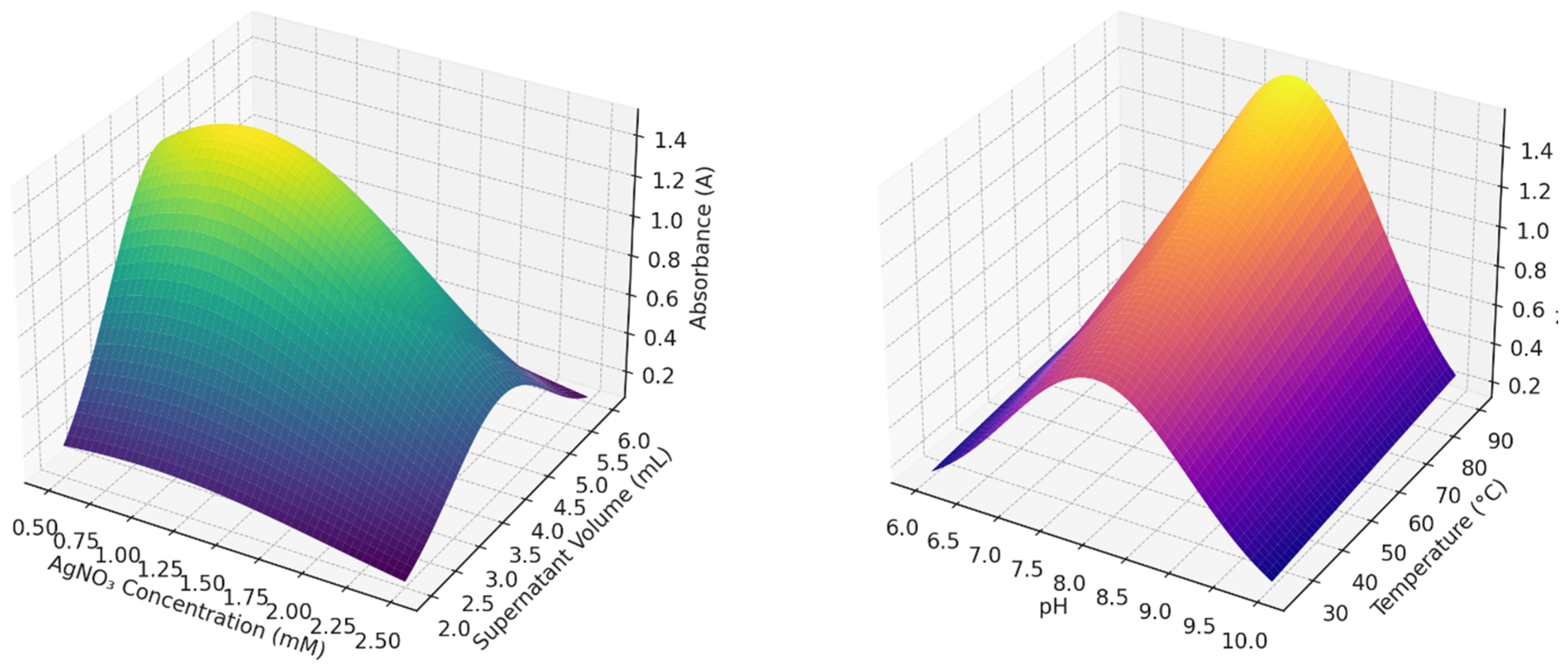
A Gaussian model was developed to describe the influence of four key parameters, AgNO3 concentration, culture supernatant volume, pH, and temperature on the efficiency of silver nanoparticle (AgNP) biosynthesis. The model showed that the highest predicted absorbance, and thus optimal nanoparticle yield, occurs when the following conditions are applied, 1.0 mM AgNO3, 4.0 mL supernatant, pH 8, and temperature 85 °C.
A = K ·   e C 1 2 + V 4 2 + p H 8 2 / σ 2   ·   1 +   ·   T 85 48  
The highest absorbance, and therefore the optimal nanoparticle yield, occurs when the parameters are at their optimal values; C0 = 1.0 mM, V0 = 4.0 mL, pH0 = 8, T0 = 85 °C. The Two 3D surface plots were generated to visualize the model output, The first plot illustrated how absorbance varies with silver nitrate concentration and culture supernatant volume. The second plot demonstrated the effects of pH and temperature on AgNP yield. Both visualizations confirmed the experimental findings, with maximum absorbance centered around the predicted optimal conditions. These results validate the Gaussian optimization Model for AgNP synthesis as a reliable representation of the nanoparticle synthesis response surface (Figure 4).
R2 calculates the explained variance (the numerator) and the total variance (the denominator). A higher R2 value indicates that a larger proportion of the variance is accounted for by the model, and a value close to 1 signifies that the model does a good job predicting the response variable.
Interpretation of R2:
R2 = 1: Perfect prediction model explains all the variance in the data.
R2 = 0: No explanatory power—model fails to explain the variance.
In this study, R2 = 0.98 indicates that 98% of the variance in AgNP yield is explained by the model, meaning the model is highly effective at predicting the outcomes based on the input parameters, while RMSE is computed as the square root of the average of the squared differences between observed and predicted values. Lower RMSE values indicate better model performance, meaning the model’s predictions are closer to the actual observed values; Higher RMSE values indicate poorer performance, suggesting that the predictions deviate significantly from the observed data. In this study, an RMSE value of 0.12 suggests that, on average, the model’s predictions deviate by only 0.12 units from the actual measured values, which indicates a high level of predictive accuracy.

2.5. Characterization of Synthesized AgNPs

2.5.1. Scanning Electron Microscopy (SEM)

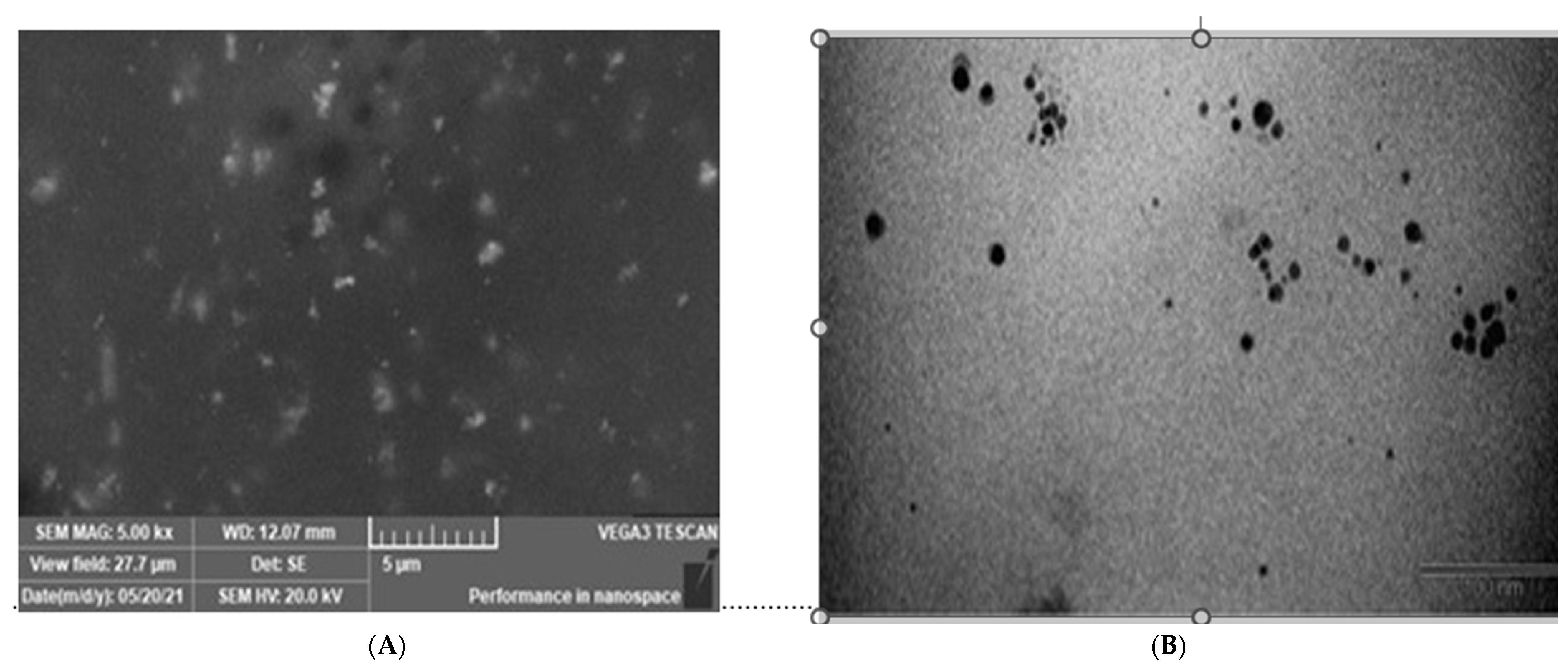
The surface morphology of silver nanoparticles (AgNPs) synthesized by the bacterial culture supernatant was examined using Scanning Electron Microscopy (SEM). The SEM micrograph revealed a relatively uniform distribution of nanoparticles across the field. The AgNPs appeared as discrete, granular structures, with varying degrees of aggregation observable at higher magnifications. The nanoparticles were mostly spherical or quasi-spherical, with sizes in the nanometric scale, indicating successful biosynthesis. The absence of significant clustering in large domains suggests that the biological agents in the culture supernatant acted effectively as capping and stabilizing agents during nanoparticle formation (Figure 5A).

2.5.2. Transmission Electron Microscopy (TEM)

Transmission Electron Microscopy (TEM) was employed to examine the size and morphology of silver nanoparticles (AgNPs) synthesized using the bacterial culture supernatant. The TEM image reveals that the AgNPs were predominantly spherical to irregular in shape, with a relatively narrow size distribution. Most particles appeared well-dispersed, although minor aggregation was observed in some regions. The particle sizes were generally in the nanometer range, estimated to be between 5 and 10 nm (polydisperse), consistent with typical biologically synthesized silver nanoparticles. The clarity of the particles and their uniform contrast suggest successful reduction and stabilization by biomolecules present in the bacterial supernatant (Figure 5B).

2.5.3. X-Ray Diffraction (XRD)

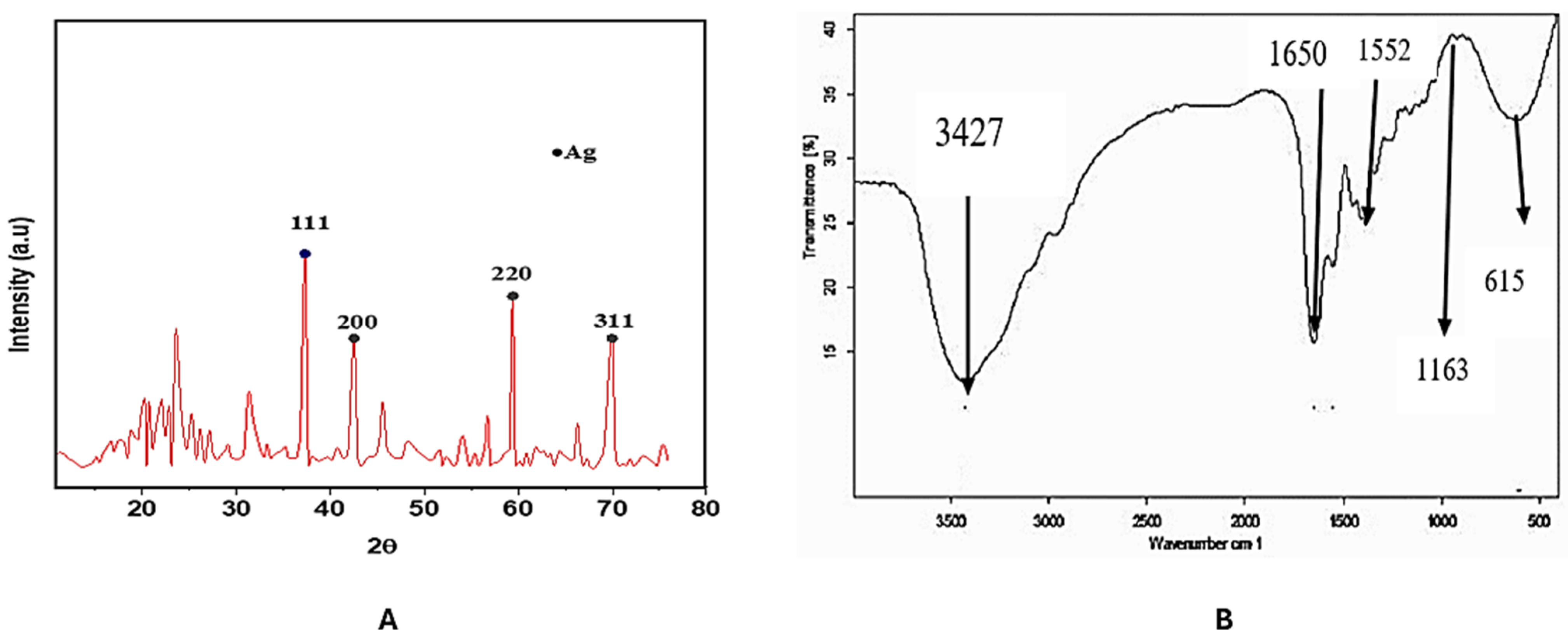
The crystalline structure of the biosynthesized silver nanoparticles (AgNPs) was confirmed by X-ray diffraction (XRD) analysis. The XRD pattern displayed distinct diffraction peaks at 2θ values of approximately 38.1°, 44.3°, 64.5°, and 77.3°, which correspond to the (111), (200), (220), and (311) crystallographic planes of face-centered cubic (fcc) silver, respectively. These values are in close agreement with the standard diffraction data for metallic silver (JCPDS card no. 04-0783), confirming the presence of highly crystalline, pure silver nanoparticles. The intense peak at 38.1° for the (111) plane suggests preferential growth orientation along this plane, which is commonly observed in biosynthesized AgNPs. No significant peaks for impurities were detected, indicating the successful reduction of silver ions and the absence of other crystalline phases (Figure 6A).

2.6. Fourier-Transform Infrared Spectroscopy (FTIR)

FTIR spectroscopy was employed to identify the functional groups involved in the reduction and stabilization of biosynthesized silver nanoparticles. The FTIR spectrum revealed several prominent absorption bands, indicating the presence of various biomolecules on the surface of the AgNPs. A strong, broad peak at 3427 cm−1 corresponds to O–H stretching vibrations, likely from alcohols or phenolic groups, suggesting the involvement of hydroxyl-containing compounds. The sharp band at 1650 cm−1 is attributed to C=O stretching of amide or carbonyl groups, while the peak at 1552 cm−1 corresponds to N–H bending, both of which point to the presence of proteins or peptides in the reaction mixture. Additional bands were observed at 1163 cm−1, indicating C–O–C or C–N stretching, and at 615 cm−1, possibly due to out-of-plane bending vibrations of aromatic compounds. These results confirm that biomolecules from the bacterial culture supernatant such as proteins, amino acids, or polyphenols—acted as both reducing and capping agents during AgNP synthesis (Figure 6B).

2.7. Antimicrobial Activity

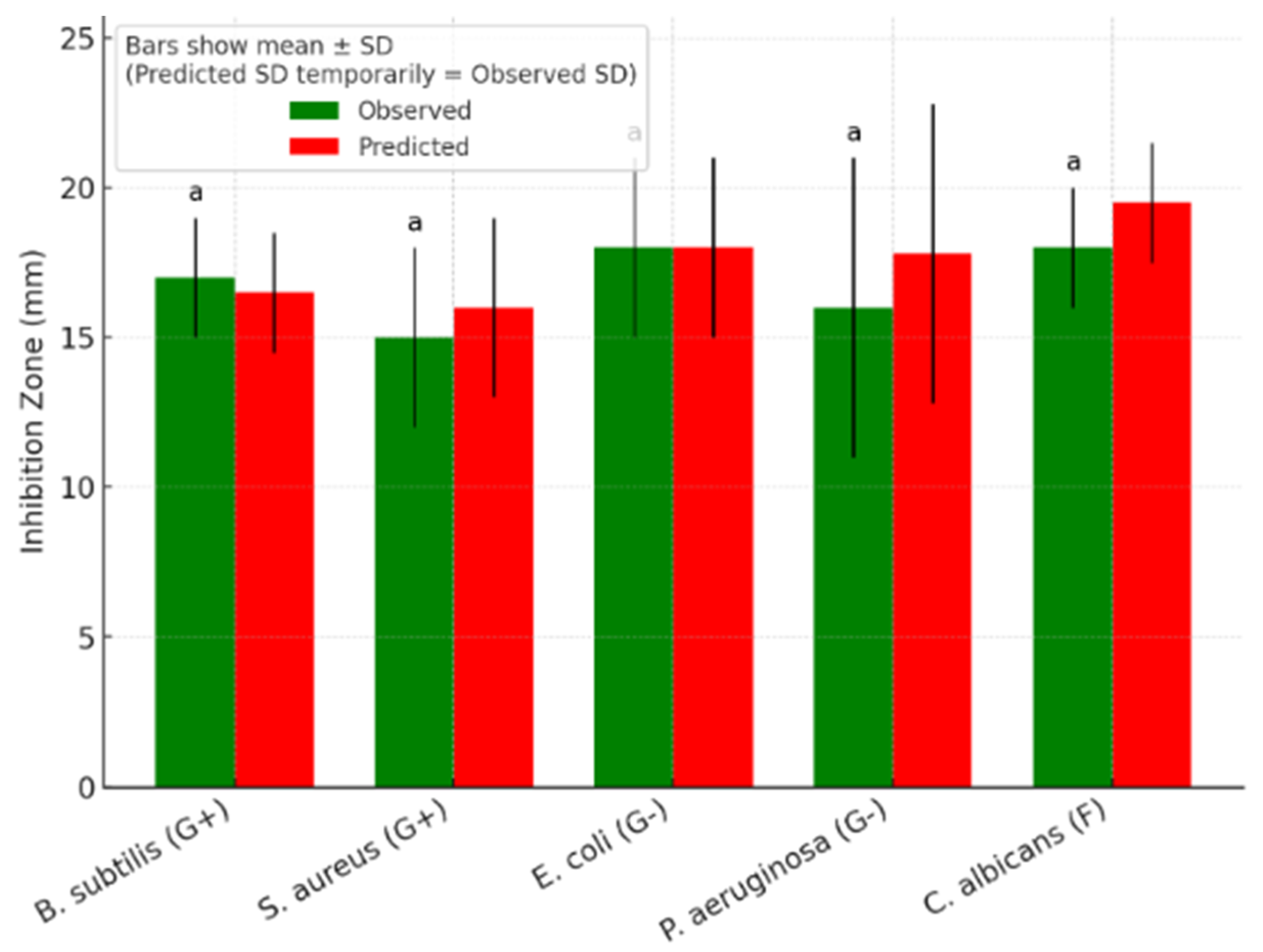
The antimicrobial potential of biosynthesized silver nanoparticles (AgNPs) was evaluated against a panel of pathogenic microorganisms, including both Gram-positive and Gram-negative bacteria, as well as the fungal strain Candida albicans. The AgNPs were tested at a concentration of 30 µg/mL, and their effectiveness was compared to Ampicillin (30 µg/mL) as a control antibiotic for bacterial strains, and Amphotericin B (30 µg/mL) for Candida albicans. The results (Table 1) showed that AgNPs exhibited notable antimicrobial activity against all tested organisms. Among the bacterial strains, Escherichia coli showed the largest zone of inhibition (18 ± 3 mm), followed by Bacillus subtilis (17 ± 2 mm), Pseudomonas aeruginosa (16 ± 5 mm), and Staphylococcus aureus (15 ± 3 mm). For the fungal strain Candida albicans, the AgNPs produced an inhibition zone of 18 ± 2 mm. These results confirm the broad-spectrum antimicrobial efficacy of biosynthesized AgNPs, demonstrating their potential as an alternative or complementary agent to conventional antibiotics in controlling microbial infections.

2.8. Minimum Inhibitory Concentration (MIC)

The results of the MIC assay revealed that Escherichia coli exhibited complete growth inhibition at an AgNP concentration of 1.87 μg/mL, indicating this as the minimum inhibitory concentration. This low MIC value reflects the high antibacterial potency of the biosynthesized silver nanoparticles, even at minimal doses. The data further support the efficacy of AgNPs as a promising antimicrobial agent with potential applications in combating drug-resistant bacterial pathogens.

2.9. Predictive Modeling of Antimicrobial Activity

To evaluate and mathematically describe the antimicrobial effect of the biosynthesized silver nanoparticles (AgNPs), a predictive model was developed using the experimental data obtained from inhibition zone assays and MIC determination. This model estimates the inhibition zone diameter (I) based on the applied AgNP concentration, the MIC value for the respective microorganism, and its microbial classification. Letters use compact-letter display; here all are “a” (no significant differences among organisms at α = 0.05, based on mean ± SD with n ≈ 3) (Figure 7).
Let:
I = Zone of inhibition (mm)
C = AgNP concentration (μg/mL)
T = Pathogen type factor:
T = 1 for Gram-positive
T = 1.1 for Gram-negative
T = 1.2 for fungal (e.g., Candida albicans)
M I C = Minimum inhibitory concentration for the specific organism
We define the inhibition zone as:
I = a · log C M I C · T + b
where
a = scaling coefficient for inhibition sensitivity
b = base inhibition zone contributed by the carrier/stabilizer or media (~5 mm by default)
If we plug in:
C = 30 μg/mL
M I C E. coli = 1.87 μg/mL
T = 1.1 for Gram-negative (E. coli)
Then:
I = a · log 30 1.87 · 1.1 + b
Using your E. coli inhibition zone ≈ 18 mm, we solve for a and b
Let us assume b = 5, then:
18   =   a   ·   log   ( 16.04 )   ·   1.1   +   5   a   ·   2.77   ·   1.1   =   13   a     13   13 3.05 4.26
Final General Model:
I =   4.26 · log   ( C M I C ) · T + 5
where
I = predicted zone of inhibition (mm)
C = AgNP concentration (μg/mL)
M I C = organism-specific MIC (μg/mL)
T     1.0 , 1.1 , 1.2
The model closely aligned with the experimental results across all tested organisms, with minor variations that fall within acceptable experimental error ranges. This strong correlation demonstrates the model’s effectiveness in predicting AgNP antimicrobial performance based on pathogen type and MIC, providing a useful tool for estimating outcomes across different microbial categories.

3. Discussion

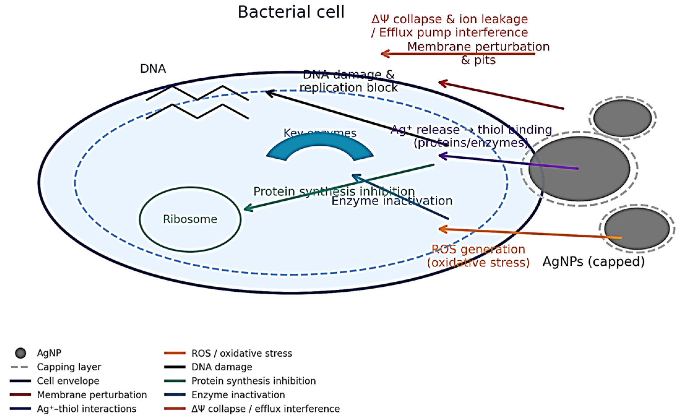
The present study successfully demonstrated the biosynthesis of silver nanoparticles (AgNPs) using a silver-tolerant Bacillus sp. isolated from industrial and sewage-contaminated soils in the Jazan region of Saudi Arabia. The isolate, confirmed via 16S rRNA sequencing with 97% similarity to Bacillus sp. ST4, showed a distinct ability to reduce silver ions, as evidenced by color change and the development of a brown halo in the presence of AgNO3. This finding supports previous research indicating that members of the Bacillus genus possess the enzymatic machinery and metabolic flexibility necessary for extracellular synthesis of metal nanoparticles [18]. Using Bacillus species from contaminated sites for AgNP biosynthesis offers significant environmental benefits, including the bioremediation of polluted areas. These bacteria help degrade contaminants while simultaneously producing valuable nanoparticles, making the process both eco-friendly and sustainable. The antimicrobial activity of silver nanoparticles (AgNPs) is primarily influenced by their size, shape, surface charge, and surface area. Smaller nanoparticles, especially in the 5–10 nm range, exhibit enhanced activity due to their larger surface area, which facilitates greater interaction with microbial cells and silver ion release. Positively charged AgNPs tend to bind more effectively with bacterial cell membranes, causing structural damage and disrupting cellular functions. Additionally, the release of silver ions interferes with cellular processes, such as protein denaturation and DNA damage, contributing to the overall antimicrobial effect observed in both Gram-positive and Gram-negative bacteria [19] (Figure 8).
The optimization of AgNP synthesis conditions revealed that silver nitrate concentration, culture supernatant volume, pH, and temperature significantly influenced nanoparticle yield. The highest nanoparticle production was achieved using 1.0 mM AgNO3, 4 mL of bacterial supernatant, pH 8, and an incubation temperature of 85 °C. These optimal conditions corresponded to maximum absorbance values at ~425 nm, which is characteristic of the surface plasmon resonance (SPR) of AgNPs [20]. Unlike conventional methods, such as linear regression or grid search, the Gaussian process model is particularly well-suited for capturing non-linear relationships between the synthesis parameters (e.g., AgNO3 concentration, pH, temperature) and the nanoparticle yield. This allows for more accurate modeling of the complex, non-linear nature of the biosynthesis process. One key advantage of the Gaussian process model is its ability to quantify uncertainty in predictions. This model provides not only predictions of the outcome but also a measure of the uncertainty associated with those predictions. This feature is crucial for understanding how confident we can be in the model’s output and for guiding future experimental work. The Gaussian process model allows for sensitivity analysis, which helps identify how sensitive the model is to changes in different parameters. This is especially useful in optimization studies where it is essential to understand the influence of each factor on the outcome and to prioritize resources effectively for further experimental work. Notably, the Gaussian Optimization Model developed in this study further validated these results by mathematically predicting the synthesis efficiency across various parameter combinations. In the predictive model, the ‘Pathogen type factor (T)’ is included to account for the differential susceptibility of different microbial categories to silver nanoparticles (AgNPs). The values for each pathogen type were assigned based on general trends observed in the literature regarding the relative sensitivity of Gram-positive, Gram-negative, and fungal microorganisms to antimicrobial agents like silver nanoparticles. For Gram-negative bacteria, a factor of 1.1 was assigned, reflecting the slightly higher resistance these bacteria typically exhibit due to their outer membrane, which can limit the penetration of nanoparticles. For Gram-positive bacteria, the factor was set to 1.0, indicating a more direct interaction between AgNPs and the cell wall, which is generally more permeable than that of Gram-negative bacteria. A factor of 1.2 was applied to fungi, such as Candida albicans, to reflect their unique cellular structures and potentially different mechanisms of nanoparticle uptake and toxicity compared to bacteria.
These factors were derived from empirical studies on the interaction of silver nanoparticles with microbial pathogens, which indicate that Gram-negative bacteria often show greater resistance due to their thicker, more protective cell membranes, while fungi tend to be slightly more susceptible than Gram-negative bacteria but less so than Gram-positive bacteria [21].
Our biosynthesis method for preparing silver nanoparticles (AgNPs) offers several advantages over conventional techniques such as chemical reduction and physical vapor deposition (PVD). It is more sustainable as it utilizes natural biological agents, avoiding toxic chemicals required in chemical reduction and reducing waste compared to the energy-intensive PVD process. Additionally, biosynthesis is cost-effective, requiring fewer resources and no specialized equipment, unlike PVD. It is also more scalable and eco-friendly, with minimal environmental impact. Moreover, the biosynthesis method allows for better control over nanoparticle size and shape due to the influence of biological agents, making it ideal for applications requiring biocompatibility and consistency. The 3D surface visualizations generated from this model aligned closely with experimental data, reinforcing the robustness and predictive strength of the approach. This modeling technique can be valuable in guiding future process design and scaling for AgNP production [22]. Characterization studies confirmed the successful biosynthesis and stability of the nanoparticles. SEM and TEM analysis revealed that the AgNPs were predominantly spherical to quasi-spherical in shape, with a narrow size distribution of 5–10 nm, and were well-dispersed with limited aggregation. These morphological traits are consistent with biologically synthesized nanoparticles reported in the literature [23]. XRD analysis showed strong diffraction peaks at 2θ values corresponding to the (111), (200), (220), and (311) planes of face-centered cubic silver, confirming their high crystallinity and purity [24]. FTIR spectroscopy identified functional groups including hydroxyl, carbonyl, and amine moieties, indicating the involvement of bacterial biomolecules such as proteins, amino acids, and polyphenols in the reduction and capping of AgNPs. This aligns with the widely accepted mechanism of biosynthesis, where microbial metabolites act simultaneously as reducing and stabilizing agents [25]. The antimicrobial assessment of the synthesized AgNPs highlighted their broad-spectrum efficacy. Significant inhibitory effects were observed against both Gram-positive (B. subtilis, S. aureus) and Gram-negative bacteria (E. coli, P. aeruginosa), as well as the fungal strain Candida albicans. Among these, E. coli showed the highest sensitivity, with a mean inhibition zone of 18 mm, and the lowest MIC value of 1.87 μg/mL which is lower than values reported in some studies but comparable to others. For instance, it was reported an MIC of 2 μg/mL for E. coli using AgNPs synthesized via chemical reduction, while observed a MIC of 3 μg/mL for E. coli using AgNPs from a biological synthesis approach. These values are in line with our findings, suggesting that our biosynthesized AgNPs exhibit comparable or superior antimicrobial activity. In contrast, studies such as observed higher MIC values for E. coli, ranging from 5 μg/mL to 10 μg/mL, when AgNPs were synthesized using traditional chemical methods. The differences in MIC values across studies could be attributed to variations in particle size, shape, and surface charge, all of which are known to influence the antimicrobial efficacy of AgNPs. Our approach, utilizing a native Bacillus strain for biosynthesis, may contribute to the enhanced antimicrobial performance observed in this study due to the unique properties imparted by the biological synthesis process. These comparisons highlight the efficacy of our biosynthesized AgNPs and reinforce the potential of green synthesis methods for producing AgNPs with significant antimicrobial activity against common pathogens [26]. These results suggest that AgNPs are particularly effective against Gram-negative pathogens, possibly due to their thinner peptidoglycan layer and increased membrane permeability. The observed antifungal activity also supports prior evidence of silver’s disruptive effects on fungal cell membranes and metabolic enzymes [27]. To complement the empirical findings, a predictive model was developed to estimate inhibition zone diameters based on nanoparticle concentration, microbial classification, and MIC values. The proposed equation,
I = 4.26   ·   log   ( C M I C )   ·   T + 5
produced inhibition zone estimates that closely matched experimental observations across all tested organisms. The model’s logarithmic structure reflects the non-linear relationship between antimicrobial efficacy and nanoparticle dosage, while the microbial factor (T) captures the differential susceptibility of Gram-positive, Gram-negative, and fungal strains. The high degree of correlation between predicted and observed values underscores the reliability of this approach for forecasting antimicrobial activity, potentially aiding in the rational design of AgNP-based therapeutics. As demonstrated by [28], the use of AgNPs in biosensing underscores their broad applicability beyond antimicrobial domains, further emphasizing the importance of optimizing their synthesis for diverse technological applications. This aligns with our study’s aim to optimize AgNP production, ensuring that they can be used efficiently in a wide range of applications [29].
The antimicrobial potential of AgNPs explored in this study aligns with their multifunctional integration into therapeutic nanoplatforms, such as drug delivery systems. As reported by [30,31], AgNPs have shown promise in enhancing the efficiency of drug delivery, owing to their ability to interact with cell membranes and facilitate controlled drug release. This highlights the broader therapeutic potential of AgNPs, beyond their antimicrobial properties, further reinforcing the importance of optimizing their synthesis for diverse applications.

4. Materials and Methods

4.1. Sample Collection

A total of ten soil and water samples were aseptically collected from sewage-treated water sites and industrial zones located in the Jazan region of the Kingdom of Saudi Arabia. The samples were transferred into sterile polyethylene bags, immediately stored on ice, and transported to the laboratory for further processing and microbial analysis [32].

4.2. Isolation of Bacterial Strains

For soil samples, 1 g of each sample was suspended in 9 mL of sterile distilled water and serially diluted up to 10−5. From selected dilutions (10−1, 10−3, 10−5), 1 mL was poured onto nutrient agar plates and incubated at 30 °C for 48 h. Water samples underwent the same serial dilution technique. Following incubation, colonies were examined morphologically and enumerated as colony-forming units (CFU) [33].

4.3. Screening for Silver-Tolerant Bacteria

Isolates were screened for silver nitrate (AgNO3) tolerance by streaking onto nutrient agar plates supplemented with various AgNO3 concentrations (0.5 to 5 mM). The plates were incubated at 30 °C for 24 h. Colonies demonstrating growth at 1 mM AgNO3, especially those showing a characteristic brown pigmentation, were selected and maintained on nutrient agar slants containing the same concentration of silver nitrate, following the method of [34].

4.4. Molecular Identification via 16S rRNA Sequencing

Genomic DNA was extracted from promising bacterial isolates using the phenol-chloroform method. DNA quality and concentration were verified through agarose gel electrophoresis under UV illumination. Amplification of the 16S rRNA gene was carried out using universal primers 27F (5′-AGAGTTTGATCMTGGCTCAG-3′) and 1492R (5′-TACGGYTACCTTGTTACGACTT-3′). PCR reactions (25 μL) included genomic DNA, MilliQ water, Taq polymerase, dNTPs, MgCl2, and primers under the following cycling conditions: initial denaturation at 95 °C for 2 min, followed by 30 cycles (95 °C for 20 s, 55 °C for 60 s, and 72 °C for 2 min), with a final extension at 72 °C for 7 min. Amplified products were purified using the GeneJET™ PCR Purification Kit (Thermo Scientific, Mundelein, IL, USA) and sequenced by GATC Company, Seoul, Republic of Korea. Sequences were analyzed using NCBI BLAST to determine taxonomic identity [35].

4.5. Biosynthesis of Silver Nanoparticles

A fresh bacterial colony was inoculated into nutrient broth and incubated at 30 °C for 48 h with shaking. Cultures were centrifuged at 8000 rpm for 20 min at 4 °C. The cell-free supernatant was collected and mixed with silver nitrate solution to initiate the synthesis of silver nanoparticles; a negative control was performed using the cell supernatant without the bacterial strain based on the methodology of [36].

4.6. Optimization of Silver Nanoparticle Synthesis

4.6.1. AgNO3 Concentration

Various concentrations of AgNO3 (0.5–10 mM) were added to the culture supernatant and incubated for 24 h. Nanoparticle formation was monitored by UV-Visible spectrophotometry within the 200–1000 nm range.

4.6.2. Supernatant Volume

Different volumes of the supernatant (0.5–5 mL) were combined with 1 mM AgNO3 and incubated for 24 h. Color changes and UV-Vis spectra were recorded.

4.6.3. pH Variation

To assess pH effects, AgNO3 solutions adjusted to pH 3, 6, 8, 10, and 11 were used in the reaction. Synthesis was monitored spectrophotometrically.

4.6.4. Temperature Influence

Reactions were conducted at temperatures ranging from 4 °C to 85 °C. The formation of AgNPs was analyzed using UV-Vis spectroscopy [21].

4.7. Gaussian Optimization Model for AgNP Synthesis

To mathematically represent the combined effects of key experimental factors on AgNP biosynthesis, a predictive model referred to as the Gaussian Optimization Model for AgNP Synthesis was formulated using data from optimization trials. This equation was formulated based on the optimization of experimental factors influencing silver nanoparticle synthesis, using Gaussian optimization. The Gaussian model is commonly employed in various scientific fields to describe how changes in experimental factors (such as concentration, pH, volume, and temperature) affect a response (such as nanoparticle yield or absorbance). The equation was formulated by analyzing your experimental data and applying a Gaussian model for optimization, using Design Of Experiments (DOE) (https://www.jmp.com/en/statistics-knowledge-portal/design-of-experiments, accessed on 14 June 2025). This model was applied to our study to predict and optimize the synthesis process. The formula was derived from experimental data using response surface methodology (RSM) and Gaussian functions. We applied statistical techniques (e.g., regression analysis) to fit the model to the experimental results, allowing us to account for the effects of the variables and optimize conditions for the best nanoparticle yield.
The absorbance at 425 nm (used as a proxy for nanoparticle yield) was modeled as a function of silver nitrate, as follows:
A = K ·   e [ C C 0 2 + V V 0 2 + p H p H 0 2 ] / σ 2   ·   1 +   ·   T T 0 48
where
  • A is the absorbance (the outcome we are measuring).
  • K is a constant that normalizes the equation.
  • C is the silver nitrate concentration, and C0 is the optimal silver nitrate concentration.
  • V is the volume of the culture supernatant, and V0 is the optimal volume.
  • pH is the pH of the solution, and pH0 is the optimal pH.
  • σ is the standard deviation (a measure of variability).
  • α is the temperature scaling factor, which adjusts the effect of temperature.
  • T is the temperature of the reaction, and T0 is the optimal temperature.
  • 48 is a constant used to normalize the temperature difference [37].

4.8. Statistical Techniques Applied to Model Validation

In this study, two key statistical techniques R2 (Coefficient of Determination) and RMSE (Root Mean Square Error) were employed to validate the predictive performance of the Gaussian Optimization Model for AgNP biosynthesis. These techniques are essential in evaluating how well the model fits the observed data and provides an accurate prediction of the nanoparticle yield based on experimental conditions.
R 2 = 1 ( y i y ^ i ) 2   ( y i y ¯ ) 2
where
y i = actual observed values; y ^ i = predicted values from the model, while y ¯ mean of the observed values.
R M S E = 1 n i = 1 n ( y i ŷ i ) 2
where n = number of data points; y i = actual observed value; ŷ i = predicted value from the model.

4.9. Characterization of Synthesized AgNPs

4.9.1. UV-Visible Spectroscopy

The reduction of silver ions and formation of nanoparticles was confirmed by measuring absorption spectra between 200 and 1000 nm [38].

4.9.2. Scanning Electron Microscopy (SEM)

Surface morphology was assessed using field emission SEM (JSM-6700, JEOL, Tokyo, Japan) after platinum coating of dried nanoparticle samples mounted on carbon stubs [39].

4.9.3. Transmission Electron Microscopy (TEM)

Size and shape of AgNPs were evaluated using a JEM-1010 JEOL TEM operated at 80 kV. Samples were prepared by placing drops of nanoparticle solution on copper grids and air-drying under infrared light [40].

4.9.4. X-Ray Diffraction (XRD)

Structural crystallinity was examined using a D8 Advance diffractometer (Karlsruhe, Germany) with Co-Kα radiation (20–80°, 0.02°/min scan rate) [41].

4.9.5. Fourier Transform Infrared Spectroscopy (FTIR)

Functional groups involved in nanoparticle stabilization were analyzed using the Agilent Cary 660 FT-IR model, Santa Clara, CA, USA). Dried AgNPs were mixed with KBr and pressed into pellets for spectral analysis within 400–4000 cm−1 [42].

4.10. Antimicrobial Activity Assessment

The antimicrobial efficacy of synthesized AgNPs was evaluated against (Bacillus subtilis) (ATCC6051), Staphylococcus aureus (ATCC12600), Gram-negative bacteria Escherichia coli (ATCC11775) and fungi Candida albicans (ATCC10231); using the agar well diffusion method. Standardized inocula (0.5 McFarland; ~108 CFU/mL) were spread on Mueller-Hinton and Sabouraud agar plates. Wells (6 mm) were loaded with 30 μg/mL AgNPs. Plates were incubated at 35 °C (bacteria) and 30 °C (fungi) for 24–48 h. Zones of inhibition were measured to assess antimicrobial potency [43].

4.11. Minimum Inhibitory Concentration (MIC) Determination

MIC values were determined via broth microdilution. The most sensitive bacterial strain was cultured overnight in Mueller-Hinton broth and adjusted to 0.5 McFarland turbidity. Aliquots of AgNP solutions at various concentrations (30 µg/mL), (15 µg/mL), (7.5 µg/mL), (3.75 µg/mL), and (1.87 µg/mL) were added to microtubes containing bacterial suspensions, respectively. Control tubes received only broth. After 24-h incubation at 37 °C, absorbance was measured at 600 nm. MIC was defined as the lowest AgNP concentration that visibly inhibited bacterial growth [44]. The predictive modeling of antimicrobial activity was driven by the aid of [45].

4.12. Statistical Analysis

All experiments were performed in biological triplicate unless otherwise stated, and data are reported as mean ± SD. For antimicrobial experiments (Table 1), inhibition-zone diameters (mm) were analyzed using a two-way ANOVA with factors Organism (B. subtilis, S. aureus, E. coli, P. aeruginosa, C. albicans) and Agent (AgNPs vs. reference antibiotic/antifungal), including the interaction term [46].

5. Conclusions

This study demonstrates a sustainable and eco-friendly approach for synthesizing silver nanoparticles (AgNPs) through a green biosynthesis method, providing a promising alternative to conventional antimicrobial agents. By integrating green synthesis techniques with predictive modeling tools, such as the Gaussian Optimization Model and antimicrobial activity simulations, this research establishes a framework to enhance nanoparticle production and their subsequent applications. The mathematical modeling and visualization strategies developed herein offer valuable insights for advancing nanotechnology, enabling more efficient and targeted designs for future applications.
Future work should focus on scaling the biosynthesis process from laboratory-scale experiments to industrial production, addressing challenges related to production efficiency and sustainability. In vivo studies are essential to assess the antimicrobial efficacy and potential toxicity of AgNPs, ensuring their safety for broader practical use. Further exploration of diverse microbial strains from various environmental sources could optimize the size, stability, and other characteristics of AgNPs. Additionally, investigating the therapeutic potential of AgNPs in nanomedicine, including their applications in drug delivery systems and wound healing, should be prioritized. Lastly, assessing the environmental impact of large-scale AgNP production, particularly its effects on aquatic ecosystems, will be crucial for ensuring their long-term sustainability and safety. This study contributes significantly to the development of AgNPs with tailored applications in medicine, industry, and environmental protection, supporting the ongoing transition to more sustainable and responsible nanotechnological solutions.

Author Contributions

Conceptualization, E.A. and Y.M.; Methodology, M.S. (Mari Sumayli), I.Y.Y.S., W.A. and E.A.; Software, A.A.A.; Validation, Y.M., S.O.A., A.A. and E.A.; Formal analysis, W.A. and E.A.; Investigation, I.Y.Y.S., Y.M. and S.O.A.; Resources, M.S. (Mukul Sharma) and A.A.A.; Data curation, M.S. (Mari Sumayli); Writing—original draft, E.A., Y.M., S.O.A., W.A., I.Y.Y.S. and A.A.; Writing—review & editing, M.S. (Mukul Sharma) and E.A. All authors have read and agreed to the published version of the manuscript.

Funding

This article is derived from a research grant funded by the Research, and Innovation Authority (RDIA)—Kingdom of Saudi Arabia—with grant (12894-JAZAN-2023-JZU-R-2-1-SE).

Institutional Review Board Statement

Not applicable.

Informed Consent Statement

Not applicable.

Data Availability Statement

All the data are presented within the article.

Acknowledgments

This article is derived from a research grant funded by the Research, and Innovation Authority (RDIA)—Kingdom of Saudi Arabia—with grant (12894-JAZAN-2023-JZU-R-2-1-SE).

Conflicts of Interest

The authors declare no conflict of interest.

References

  1. Durlo, A. 1950–2022: A History of Nanotechnology into Physical and Mathematical Relationship. Ph.D. Thesis, Université de Lille, Lille, France, 2023. [Google Scholar]
  2. Devi, R.S.; Girigoswami, A.; Siddharth, M.; Girigoswami, K. Applications of gold and silver nanoparticles in theranostics. Appl. Biochem. Biotechnol. 2022, 194, 4187–4219. [Google Scholar] [CrossRef]
  3. Sharifi-Rad, M.; Mohanta, Y.K.; Pohl, P.; Nayak, D.; Messaoudi, M. Facile phytosynthesis of gold nanoparticles using Nepeta bodeana Bunge: Evaluation of its therapeutics and potential catalytic activities. J. Photochem. Photobiol. A Chem. 2024, 446, 115150. [Google Scholar] [CrossRef]
  4. Omar, A.H.; Abdelkareem, O.F.; Salama, H.S. Green synthesized metallic nanoparticles and their applications: A review. Sci. J. Fac. Sci. Menoufia Univ. 2024, 28, 15–26. [Google Scholar] [CrossRef]
  5. Snoussi, M.; Lajimi, R.H.; Latif, S.; Hamadou, W.S.; Alreshidi, M.M.; Ashraf, S.A.; Patel, M.; Humaidi, J.; Anouar, E.A.; Kadri, A.; et al. Green synthesis and characterization of silver nanoparticles from Ducrosia flabellifolia Boiss. aqueous extract: Anti-quorum sensing screening and antimicrobial activities against ESKAPE pathogens. Cell. Mol. Biol. 2024, 70, 88–96. [Google Scholar] [CrossRef] [PubMed]
  6. Rodrigues, A.S.; Batista, J.G.S.; Rodrigues, M.Á.V.; Thipe, V.C.; Minarini, L.A.R.; Lopes, P.S.; Lugão, A.B. Advances in silver nanoparticles: A comprehensive review on their potential as antimicrobial agents and their mechanisms of action elucidated by proteomics. Front. Microbiol. 2024, 15, 1440065. [Google Scholar] [CrossRef] [PubMed]
  7. Najitha, B.B. Springer-Verlag and Berlin Heidelberg. Extracellular synthesis of silver nanoparticles using Bacillus megaterium against malarial and dengue vector (Diptera: Culicidae). Parasitol. Res. 2015, 114, 4069–4079. [Google Scholar] [CrossRef]
  8. Abd-Elsalam, K.A. (Ed.) Fungal Cell Factories for Sustainable Nanomaterials Productions and Agricultural Applications; Elsevier: Amsterdam, The Netherlands, 2022. [Google Scholar]
  9. Yassin, M.A.; Elgorban, A.M.; El-Samawaty, A.E.R.M.; Almunqedhi, B.M.A. Biosynthesis of silver nanoparticles using Penicillium verrucosum and analysis of their antifungal activity. Saudi J. Biol. Sci. 2021, 28, 2123–2127. [Google Scholar] [CrossRef]
  10. Saravanan, A.K.V.; Barik, S.K. Rapid biosynthesis of silver nanoparticles from Bacillus megaterium (NCIM 2326) and their antibacterial activity on multi drug resistant clinical pathogens. Colloids Surf. B Biointerfaces 2011, 88, 325–331. [Google Scholar] [CrossRef]
  11. Abada, E.; Mashraqi, A.; Modafer, Y.; Al Abboud, M.A.; El-Shabasy, A. Review: Green synthesis of silver nanoparticles by using plant extracts and their antimicrobial activity. Saudi J. Biol. Sci. 2024, 31, 103877. [Google Scholar] [CrossRef]
  12. Alsamhary, K.A. Eco-friendly synthesis of silver nanoparticles by Bacillus subtilis and their antibacterial activity. Saudi J. Biol. Sci. 2020, 27, 2185–2191. [Google Scholar] [CrossRef]
  13. Dhaka, A.; Mali, S.C.; Sharma, S.; Trivedi, R. A review on biological synthesis of silver nanoparticles and their potential applications. Results Chem. 2023, 6, 101108. [Google Scholar] [CrossRef]
  14. Priyadarshini, S.; Gopinath, V.; Priyadharsshini, N.M.; MubarakAli, D.; Velusamy, P. Synthesis of anisotropic silver nanoparticles using novel strain, Bacillus flexus and its biomedical application. Colloids Surf. B Biointerfaces 2013, 102, 232–237. [Google Scholar] [CrossRef]
  15. Karkaj, O.S.; Salouti, M.; Zanjani, R.S.; Derakhshan, F.K. Extracellular Deposition of Silver Nanoparticles by Bacillus Megaterium. Synth. React. Inorg. Met.-Org. Nano-Met. Chem. 2013, 43, 903–906. [Google Scholar] [CrossRef]
  16. Pokrajac, L.; Abbas, A.; Chrzanowski, W.; Dias, G.M.; Eggleton, B.J.; Maguire, S.; Maine, E.; Malloy, T.; Nathwani, J.; Nazar, L.; et al. Nanotechnology for a sustainable future: Addressing global challenges with the international network for sustainable nanotechnology. ACS Nano 2021, 15, 18608–18623. [Google Scholar] [CrossRef]
  17. Barsola, B.; Saklani, S.; Pathania, D.; Kumari, P.; Sonu, S.; Rustagi, S.; Singh, P.; Raizada, P.; Moon, T.S.; Kaushik, A.; et al. Exploring bio-nanomaterials as antibiotic allies to combat antimicrobial resistance. Biofabrication 2024, 16, 042007. [Google Scholar] [CrossRef]
  18. Al-Asbahi, M.G.S.S.; Al-Ofiry, B.A.; Saad, F.A.A.; Alnehia, A.; Al-Gunaid, M.Q.A. Silver nanoparticles biosynthesis using mixture of Lactobacillus sp. and Bacillus sp. growth and their antibacterial activity. Sci. Rep. 2024, 14, 10224. [Google Scholar] [CrossRef]
  19. Sharifi-Rad, M.; Pohl, P.; Epifano, F.; Álvarez-Suarez, J.M. Green synthesis of silver nanoparticles using Astragalus tribuloides Delile root extract: Characterization, antioxidant, antibacterial, and anti-inflammatory activities. Nanomaterials 2020, 10, 2383. [Google Scholar] [CrossRef]
  20. Kaliappan, K.; Nagarajan, P.; Jayabalan, J.; Pushparaj, H.; Elumalai, S.; Paramanathan, B.; Manickam, V.; Jang, H.T.; Mani, G. Systematic antimicrobial, biofilm, free radical inhibition and tyrosinase inhibition assessments of efficient green silver nanoparticles from the aqueous root extract of Cyphostemma adenocaule (CA). RSC Pharm. 2025, 2, 147–162. [Google Scholar] [CrossRef]
  21. Abada, E.; Habib, F.; Mashraqi, A.; Modafer, Y.; Alsolami, W.; Ismail, K.; Alamri, A.A.; Mashlawi, A.M.; Shater, A.M. Eco-Friendly Synthesis and Characterization of Senna italica-Derived Silver Nanoparticles With Broad-Spectrum Antimicrobial Activity. Int. J. Microbiol. 2025, 2025, 2072594. [Google Scholar] [CrossRef]
  22. Dong, S.; Yu, H.; Poupart, P.; Ho, E.A. Gaussian processes modeling for the prediction of polymeric nanoparticle formulation design to enhance encapsulation efficiency and therapeutic efficacy. Drug Deliv. Transl. Res. 2025, 15, 372–388. [Google Scholar] [CrossRef] [PubMed]
  23. Agnihotri, S.; Mukherji, S.; Mukherji, S. Size-controlled silver nanoparticles synthesized over the range 5–100 nm using the same protocol and their antibacterial efficacy. RSC Adv. 2014, 4, 3974–3983. [Google Scholar] [CrossRef]
  24. Alam, A.; Sadia, S.I.; Shishir, K.H.; Bishwas, R.K.; Ahmed, S.; Al-Reza, S.M.; Jahan, S.A. Crystallinity integration and crystal growth behavior study of preferred oriented (111) cubic silver nanocrystal. Inorg. Chem. Commun. 2025, 173, 113834. [Google Scholar] [CrossRef]
  25. Sharma, N.K.; Vishwakarma, J.; Rai, S.; Alomar, T.S.; AlMasoud, N.; Bhattarai, A. Green route synthesis and characterization techniques of silver nanoparticles and their biological adeptness. ACS Omega 2022, 7, 27004–27020. [Google Scholar] [CrossRef]
  26. El-Bendary, M.A.; Afifi, S.S.; Moharam, M.E.; El-Ola, S.M.A.; Salama, A.; Omara, E.A.; Shaheen, M.N.F.; Hamed, A.A.; Gawdat, N.A. Biosynthesis of silver nanoparticles using isolated Bacillus subtilis: Characterization, antimicrobial activity, cytotoxicity, and their performance as antimicrobial agent for textile materials. Prep. Biochem. Biotechnol. 2021, 51, 54–68. [Google Scholar] [CrossRef]
  27. Matras, E.; Gorczyca, A.; Przemieniecki, S.W.; Oćwieja, M. Surface properties-dependent antifungal activity of silver nanoparticles. Sci. Rep. 2022, 12, 18046. [Google Scholar] [CrossRef] [PubMed]
  28. Ma, Y.; Leng, Y.; Huo, D.; Zhao, D.; Zheng, J.; Zhao, P.; Hou, C. A portable sensor for glucose detection in Huangshui based on blossom-shaped bimetallic organic framework loaded with silver nanoparticles combined with machine learning. Food Chem. 2023, 429, 136850. [Google Scholar] [CrossRef] [PubMed]
  29. Khanal, L.N.; Dhakal, P.P.; Kandel, M.R.; Acharya, D.; Baral, E.R.; Chhetri, K.; Kalauni, S.K. Stem Bark-Mediated Green Synthesis of Silver Nanoparticles from Pyrus pashia: Characterization, Antioxidant, and Antibacterial Properties. Inorganics 2023, 11, 263. [Google Scholar] [CrossRef]
  30. Feng, C.; Wang, Y.; Xu, J.; Zheng, Y.; Zhou, W.; Wang, Y.; Luo, C. Precisely tailoring molecular structure of doxorubicin prodrugs to enable stable nanoassembly, rapid activation, and potent antitumor effect. Pharmaceutics 2024, 16, 1582. [Google Scholar] [CrossRef]
  31. Wang, Y.; Zhai, W.; Yang, L.; Cheng, S.; Cui, W.; Li, J. Establishments and evaluations of post-operative adhesion animal models. Adv. Therap. 2023, 6, 2200297. [Google Scholar] [CrossRef]
  32. Abada, E.; Al-Fifi, Z.; Al-Rajab, A.J.; Mahdhi, M.; Sharma, M. Molecular identification of biological contaminants in different drinking water resources of the Jazan region, Saudi Arabia. J. Water Health 2019, 17, 622–632. [Google Scholar] [CrossRef]
  33. Karthikeyan, S.; Hoti, S.L.; Prasad, N.R. Resveratrol loaded gelatin nanoparticles synergistically inhibit cell cycle progression and constitutive NF-kappaB activation, and induce apoptosis in non-small cell lung cancer cells. Biomed. Pharmacother. 2015, 70, 274–282. [Google Scholar] [CrossRef] [PubMed]
  34. Balachandar, V.; Oh, B.T. Synthesis of silver and gold nanoparticles using cashew nut shell liquid and its antibacterial activity against fish pathogens. Indian J. Microbiol. 2014, 54, 196–202. [Google Scholar] [CrossRef]
  35. Abada, E.; Galal, T.; Ismail, I. Biosynthesis of silver nanoparticles by Nocardiopsis sp.-MW279108 and its antimicrobial activity. J. Basic Microbiol. 2021, 61, 993–1001. [Google Scholar] [CrossRef] [PubMed]
  36. Abada, E.; Galal, T.M.; Alhejely, A.; Mohammad, A.M.; Alruwaili, Y.; Almuhayawi, M.S.; Shater, A.-R.M.; Alruhaili, M.H.; Selim, S. Bio-preparation of CuO@ZnO nanocomposite via spent mushroom substrate and its application against Candida albicans with molecular docking study. BioResources 2024, 19, 6670–6689. [Google Scholar] [CrossRef]
  37. Ubaldo, P.C. Towards Combined Computational and Experimental Studies on Toxicity of Silver Nanoparticles. Ph.D. Thesis, Southern Illinois University at Carbondale, Carbondale, IL, USA, 2015. [Google Scholar]
  38. Fouda, A.; Hassan, S.E.-D.; Saied, E.; Hamza, M.F. Photocatalytic degradation of real textile and tannery effluent using biosynthesized magnesium oxide nanoparticles (MgO-NPs), heavy metal adsorption, phytotoxicity, and antimicrobial activity. J. Environ. Chem. Eng. 2021, 9, 105346. [Google Scholar] [CrossRef]
  39. Ausaj, A.A.; Al-Shmgani, H.S.; Abid, W.B.; Gadallah, A.A.; Mashlawi, A.M.; Khormi, M.A.; Alamri, A.A.; Abada, E. Immobilization of Bromelain on Gold Nanoparticles for Comprehensive Detection of Their Antioxidant, Anti-Angiogenic, and Wound-Healing Potentials. Inorganics 2024, 12, 325. [Google Scholar] [CrossRef]
  40. Ahmed, M.J.; Murtaza, G.; Rashid, F.; Iqbal, J. Eco-friendly green synthesis of silver nanoparticles and their potential applications as antioxidant and anticancer agents. Drug Dev. Ind. Pharm. 2019, 45, 1682–1694. [Google Scholar] [CrossRef]
  41. Asif, M.; Yasmin, R.; Asif, R.; Ambreen, A.; Mustafa, M.; Umbreen, S. Green synthesis of silver nanoparticles (AgNPs), structural characterization, and their antibacterial potential. Dose-Response 2022, 20, 15593258221088709. [Google Scholar] [CrossRef]
  42. Keskin, M.; Kaya, G.; Bayram, S.; Kurek-Górecka, A.; Olczyk, P. Green synthesis, characterization, antioxidant, antibacterial and enzyme inhibition effects of chestnut (Castanea sativa) honey-mediated silver nanoparticles. Molecules 2023, 28, 2762. [Google Scholar] [CrossRef]
  43. Patel, B.; Yadav, V.K.; Desai, R.; Patel, S.; Amari, A.; Choudhary, N.; Osman, H.; Patel, R.; Balram, D.; Lian, K.-Y.; et al. Bacteriogenic synthesis of morphologically diverse silver nanoparticles and their assessment for methyl orange dye removal and antimicrobial activity. PeerJ 2024, 12, e17328. [Google Scholar] [CrossRef] [PubMed]
  44. Da Silva, A.C.F.; Roque, S.M.; Duarte, M.C.T.; Nakazato, G.; Durán, N.; Cogo-Müller, K. Modulation of Staphylococcus aureus biofilm formation through subinhibitory concentrations of biogenic silver nanoparticles and simvastatin. Future Pharmacol. 2024, 4, 3–16. [Google Scholar] [CrossRef]
  45. Jianchao, Z. Modeling MIC and Metal Precipitation with a 1D Reactive Transport Model. Master’s Thesis, Delft University of Thechnology, Delft, The Netherlands, 2015. [Google Scholar]
  46. Zeraatpisheh, A.; Moradi, M.; Arshadi, M.; Taheri, B.; Zahmatkeshan, M.; Dehbashi, F.N.; Esmaeili, F.; Amanzadi, B.; Farshadzadeh, Z. The in vitro evaluation of synergistic effects of ciprofloxacin and berberine hydrochloride against Pseudomonas aeruginosa. BMC Microbiol. 2025, 25, 487. [Google Scholar] [CrossRef] [PubMed]
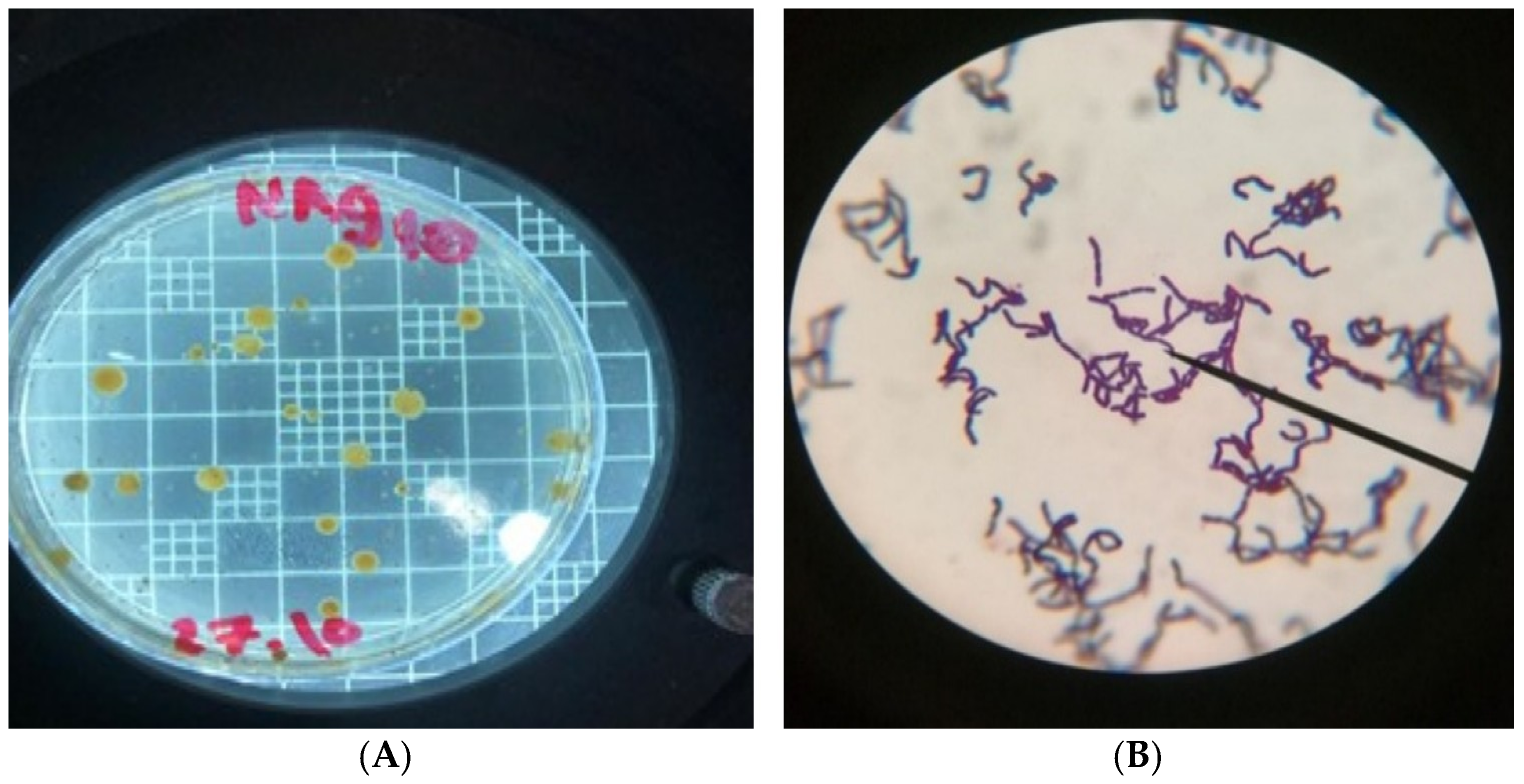
Figure 1. (A): Growth of bacterial isolates on Nutrient agar supplemented with 1 mM AgNO3, 1 (B): The appearance of Gram-positive isolate under the microscope (1000×).
Inorganics 13 00342 g001
Figure 2. (A): PCR product of the amplified partial sequence 16S rRNA region of the Bacillus sp. ST4, (B): Phylogenetic tree analysis of the 16S rRNA gene sequence of the bacterial strain.
Inorganics 13 00342 g002
Figure 3. (A): Effect of AgNO3 concentrations on AgNPs synthesis, (B): Effect of supernatant volume on AgNPs synthesis, (C): Effect of pH on AgNPs synthesis, (D): Effect of Temperatures on AgNPs synthesis.
Inorganics 13 00342 g003
Figure 4. (Left) 3D surface plot showing absorbance (A) as a function of AgNO3 concentration and culture supernatant volume at fixed pH 8 and temperature 85 °C. (Right) 3D surface plot showing absorbance as a function of pH and temperature at fixed AgNO3 concentration (1.0 mM) and culture supernatant volume (4 mL).
Inorganics 13 00342 g004
Figure 5. (A): SEM electron micrograph of theAgNPs biosynthesized by the culture supernatant of the bacterium, (B): TEM electron micrograph of theAgNPs biosynthesized by the culture supernatant of the bacterium.
Inorganics 13 00342 g005
Figure 6. (A): XRD pattern of the silver nanoparticles, (B): Fourier-transform infrared spectroscopy (FTIR) spectrum of biosynthesized silver nanoparticles (AgNPs).
Inorganics 13 00342 g006
Figure 7. A bar chart comparing the observed vs. predicted inhibition zones for AgNPs against different microorganisms.
Inorganics 13 00342 g007
Figure 8. Mechanisms of antimicrobial action of silver nanoparticles (AgNPs) on bacterial cell.
Inorganics 13 00342 g008
Table 1. Antimicrobial activity of biosynthesized silver nanoparticles (AgNPs) against various pathogenic microorganisms.
MicroorganismsInhibition Zone (mm)
AgNPs (30 µg/mL) Antibiotics 30 µg/mL
Bacteria
Bacillus subtilis
(ATCC6051)
17 ± 2 b*Ampicillin
26 ± 3.0 a
Staphylococcus aureus
(ATCC12600)
15 ± 3.0 a21 ± 4.0 a
Escherichia coli
(ATCC11775)
18 ± 3.0 a25 ± 5.0 a
Pseudomonas aeruginosa
(ATCC10145)
16 ± 5.0 b26 ± 3.0 a
Fungi
Candida albicans
(ATCC10231)
18 ± 2.0 aAmphotericin B
21 ± 3.0 a
* Values that share the same letter are not significantly different; values with different letters are significantly different (α = 0.05).
Disclaimer/Publisher’s Note: The statements, opinions and data contained in all publications are solely those of the individual author(s) and contributor(s) and not of MDPI and/or the editor(s). MDPI and/or the editor(s) disclaim responsibility for any injury to people or property resulting from any ideas, methods, instructions or products referred to in the content.