Review

A Comprehensive Physicochemical Characterization of Silver Nanoparticles as a Prerequisite for Their Successful Biomedical Applications

by
Anastasia Ntolia
1,
Theofania Chatzigiannakou
1,
Nikolaos Michailidis
2 and
Amalia Aggeli
1,*
1
Department of Chemical Engineering, Faculty of Engineering, Aristotle University of Thessaloniki, 541 24 Thessaloniki, Greece
2
Department of Mechanical Engineering, Faculty of Engineering, Aristotle University of Thessaloniki, 541 24 Thessaloniki, Greece
*
Author to whom correspondence should be addressed.
Inorganics 2025, 13(10), 341; https://doi.org/10.3390/inorganics13100341
Submission received: 31 August 2025 / Revised: 28 September 2025 / Accepted: 6 October 2025 / Published: 21 October 2025

Abstract

Silver nanoparticles (AgNPs) are highly promising components for the development of innovative biomedical products. However, a critical issue remains the insufficient deep and quantitative understanding of their fundamental physicochemical properties. These properties essentially govern the bioactivity of silver nanoparticles and, consequently, the success of their biomedical applications. Current characterization methods do not fully capture the complex nature of AgNPs, leaving key questions unresolved, such as detailed surface properties, dynamic interactions in real biological environments, long-term changes, and the release of silver ions—all factors that influence the toxicity and performance of the nanoparticles. This gap in knowledge complicates the reproducibility of experiments, comparison of results, and proper evaluation of potential health risks associated with their use. While advanced techniques such as Atomic Force Microscopy (AFM), Inductively Coupled Plasma (ICP) spectroscopy, and X-ray Photoelectron Spectroscopy (XPS) further significantly our understanding, they still do not fully meet all the demands for understanding silver nanoparticles. Specifically, these methods face limitations in monitoring the dynamic and complex interactions of nanoparticles within real biological settings, especially physicochemical properties that are linked to toxicity and also the biological. Therefore, despite their invaluable role, these techniques represent only part of the solution for the thorough understanding and assessment of the biomedical performance of AgNPs, highlighting the need for continued research to ensure their safe and efficient biomedical utilization.

Graphical Abstract

1. Introduction to Silver Nanoparticles (AgNPs)

Silver is a soft transition metal that exhibits superior reflectivity and exceptional electrical and thermal conduction properties among all metals. The medical and therapeutic benefits of silver were known for thousands of years, before it was recognized that microbes cause infections. In ancient times, liquids and food were kept in silver containers to prevent spoilage. In 1884, silver nitrate eye drops were introduced to prevent infections in newborns [1], and silver preparations were developed to treat infections in wounds and burns. Nevertheless, in the 1940s, antibiotics took the place of silver in medical applications. The misuse of antibiotics led to antimicrobial resistance, which is now a global problem, and therefore silver has again attracted much attention. Silver is biologically active in ionic form (Ag+), during its dissolution in aqueous environments. In this form it occurs in silver salts including silver nitrate and silver sulfadiazine, which have often been applied in wound treatment [2]. It is currently applied in antimicrobial dressings for burn care in various forms, incorporated in food packaging to inhibit microbial growth, and integrated into household electronics and appliances. Silver nanoparticles (AgNPs) can be defined as silver-based nanomaterials with at least one dimension in the range of 1–100 nm. They have attracted increasing interest due to their unique physical, chemical and biological properties compared to silver in its bulk or ionic form. In particular, AgNPs exhibit special optical properties, high electrical and thermal conductivity as well as excellent catalytic and antibacterial action [3].
Noble metal nanoparticles have unique optical properties owing to their strong resonance with specific wavelengths within the electromagnetic spectrum, as opposed to the same materials on a macroscopic scale. The conduction electrons in metallic nanoparticles move freely within the particle and, when illuminated, the oscillating electric field causes the electrons to accumulate on one side. This creates an electric dipole with positively and negatively charged sides for each nanoparticle as shown in the following figure (Figure 1). This dipole creates an electric field within the nanoparticle, which acts opposite to incident light, as a force to restore the equilibrium position of electrons. The greater the electron shift, the greater the restoring force.
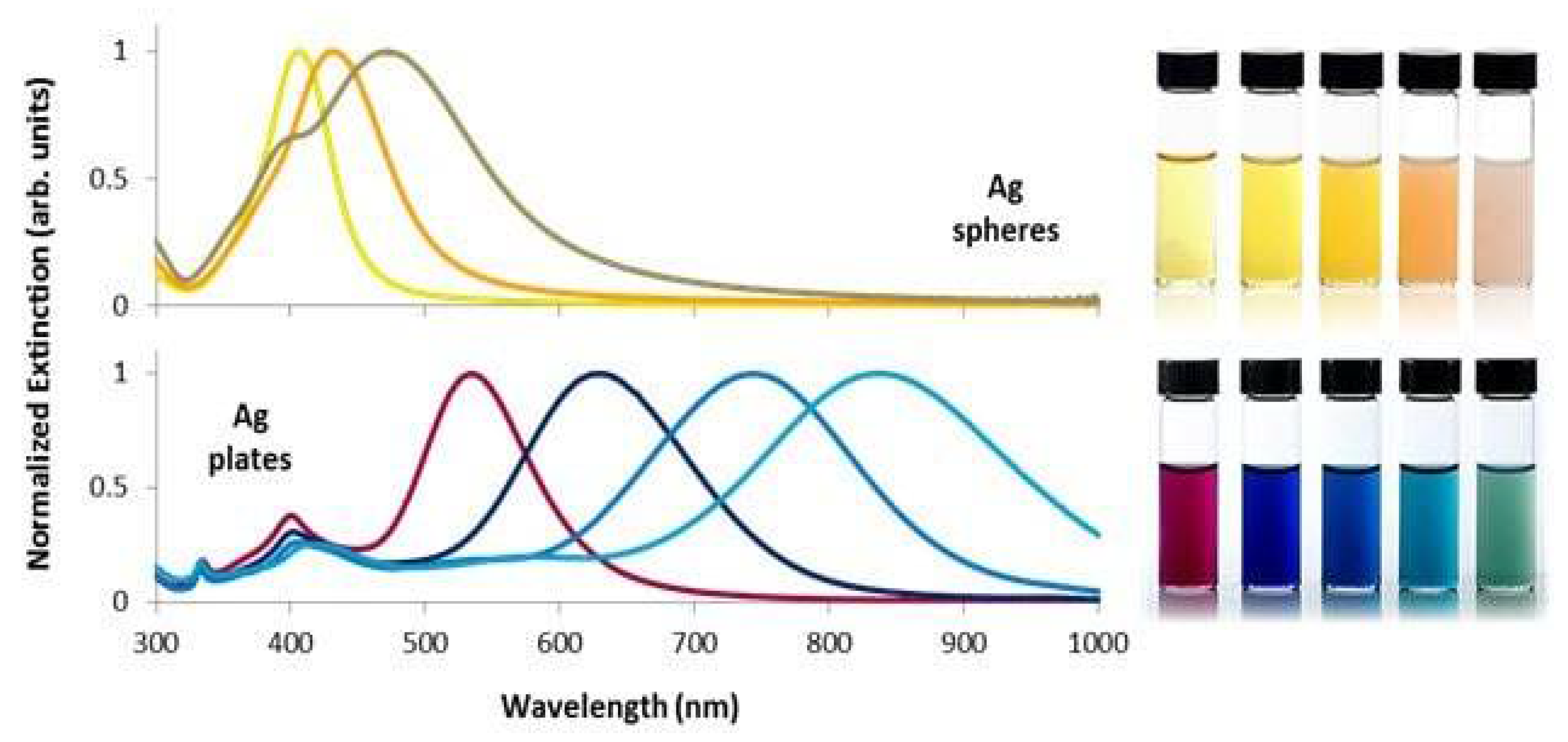
If there is a deviation from the equilibrium position, the electrons begin to oscillate at a certain frequency, called plasmonic frequency, which is influenced by the material, size, and shape of the metal nanoparticle, as well as the dielectric properties of the metal and its environment. Conduction electrons absorb energy from light, resulting in their collective oscillation. The collective motion of electrons is called plasmon. When the incident light is in resonance with the plasmonic frequency of the nanoparticle, then we have maximum energy absorption and the phenomenon is called surface plasmon resonance (SPR). In order to observe this phenomenon, it is necessary to have a uniform electric field and therefore the particle size needs to be significantly smaller than the wavelength of the incident light (380–760 nm), which applies to nanoparticles. This collective absorption by nanoparticles gives nanoparticle dispersion a characteristic color that depends on the material, size and shape of the AgNPs as shown in Figure 2. An increase in the size of AgNPs correlates with a shift to longer wavelengths in the maximum SPR. Thus, changes in the size and the local refractive index near the surface of spherical AgNPs allow the maximum SPR wavelength to be adjusted between 400 nm (violet) and 530 nm (green). Additionally, the shape of nanoparticles affects the number of SPR peaks, and spherical (isotropic) particles exhibit a single SPR band. Nevertheless, anisotropic particles with decreased symmetry demonstrate two or more SPR peaks [5,6,7].
The thermal behavior of nanomaterials is also important. Melting temperature is a critical property for a material, and it has been shown that due to the thermodynamic effect of size, metal nanoparticles exhibit a low melting temperature compared to bulk state. Nanomaterials possess a large surface to volume ratio, and their surface atoms have fewer direct neighboring atoms. Thus, the binding energy per atom is reduced, resulting in a decrease in the melting temperature of the nanoparticles. For example, the melting temperature of 2.5 nm Au nanoparticles is reduced by 407 degrees compared to bulk gold. By reducing the size of nanoparticles, the surface-to-volume ratio increases owning to the improved free energy at their surface the melting temperature decreases. Experiments were performed to assess the melting points of silver nanoparticles with sizes between 4 and 50 nm. It was discovered that by reducing the size of AgNPs, melting happened at lower temperatures [9,10].
In addition, the thermal conductivity of nanoparticles is greater than that of the bulk materials, owing to their higher surface-to-volume ratio. Thus, they possess a greater quantity of electrons for heat transfer; also through microconvection from the Brownian motion of NPs, the thermal conductivity increases. This occurs when solid nanoparticles are dispersed in a liquid such as water, ethylene glycol or oils, creating nanofluids. Nanofluids exhibit enhanced thermal conductivity in relation to fluids without solid nanoparticles. For instance, nanofluids with dispersed 20 nm silver nanoparticles are reported to achieve thermal conductivity values of about 0.73 W/mK at 80 °C, higher than typical values for water (approximately 0.64 W/mK) [10,11].
Nanocatalysis is a rapidly growing field in which nanoparticles are used as catalysts. Nanoparticle catalysts carry novel and improved catalytic properties, including reactivity and selectivity, in comparison with their bulk counterparts. The catalytic behavior of nanoparticles is affected by their size and shape. In particular, with the reduction in size, their catalytic activity increases. Furthermore, the shape of NPs has been found to affect their reactivity and selectivity. It has been found that when using AgNPs to oxidize styrene, the conversion rate with Ag nanocubes shows a 14-fold increase over nanoplates and a 4-fold increase over nanospheres. This may stem from the variation in the relative surface area of catalytically active facets associated with diverse nanoparticle shapes [10].
Magnetic nanoparticles have also been extensively investigated in numerous fields, such as heterogeneous and homogeneous catalysis, biomedicine, magnetic resonance imaging (MRI), magnetic fluids and water disinfection. Silver nanoparticles (AgNPs) generally exhibit weak or no intrinsic magnetism because bulk silver is diamagnetic. However, their magnetic properties at the nanoscale can vary significantly depending on structural features such as crystallinity and defects. Studies show that single-crystalline silver nanoparticles tend to behave as diamagnetic or classical paramagnetic materials, while multi-twinned silver nanoparticles can exhibit paramagnetic or even weak ferromagnetic behavior [10,12].
The physicochemical characteristics of silver nanoparticles affect not only their basic optical, electrical, thermal and magnetic properties and catalytic properties but also their usage in biomedical applications and in bionanotechnology [13]. Various physicochemical techniques are available to accurately characterize silver nanoparticles (AgNPs) in solutions, assessing parameters such as size, shape, concentration, and zeta potential. Functional characterization focuses on evaluating the antimicrobial effectiveness of AgNPs as well as other potential biological effects on cells, tissues, and the organism during application, including their fate, potential accumulation, and pathways of excretion [14]. In fact, development of successful biomedical applications of silver nanoparticles relies on the fundamental prerequisite of full, comprehensive and detailed multifaceted physicochemical characterization of the nanoparticles. The complete physicochemical characterization of silver nanoparticles can be envisaged as a puzzle (Figure 3); currently the application of advanced complementary techniques allows us to add only some pieces in this puzzle; several parts of the puzzle are still missing, waiting for novel approaches in silver nanoparticle characterization to be introduced, in order to complete the full picture of the puzzle [15]. The purpose of this review is to contribute to highlight the importance of physicochemical characterization of silver nanoparticles for their effective biomedical applications, and the gap that exists in this respect at present. In Section 2, the most widely employed complementary techniques for the physicochemical characterization of silver nanoparticles are presented, highlighting their respective advantages and limitations. In addition, several advanced analytical methods are introduced; however, it is crucial to acknowledge that these sophisticated techniques are not without limitations. Challenges such as high operational costs, complexity of instrumentation, time-consuming sample preparation, and limitations in sensitivity or applicability to certain sample types can affect their widespread use. Furthermore, some advanced methods may provide information that is difficult to interpret without complementary techniques, and surface-sensitive methods often require ultra-clean environments and specialized expertise. Then, in Section 3 a link is attempted between the most important biomedical applications of silver nanoparticles and their physicochemical characteristics. The final section, Section 4, provides a comprehensive overview of the conclusions drawn from this review, along with the outlook and potential opportunities within this field.

2. Physicochemical Properties of Silver Nanoparticles Studied by Complementary Characterization Techniques

Following synthesis, rigorous physicochemical characterization of silver nanoparticles is indispensable due to the critical influence of their physicochemical attributes on their useful properties and efficacy in applications. Consequently, comprehensive evaluation of parameters such as particle size, morphology, surface properties, solubility and aggregation state is essential prior to any application. This characterization is typically conducted through a suite of advanced analytical techniques, including Ultraviolet-Visible spectroscopy for examining surface plasmon resonance phenomena, Fourier Transform InfraRed spectroscopy to examine the organic layers on the surface of nanoparticles, Zeta- potential to look at the electrostatic repulsion between nanoparticles, X-ray diffraction for determining crystalline structure and phase composition, dynamic light scattering for measuring hydrodynamic diameter and colloidal stability, scanning electron microscopy for high-resolution surface morphology assessment, and transmission electron microscopy for detailed ultrastructural and dimensional analysis [16]. Some of the main techniques that are currently used for the characterization of silver nanoparticles are summarized below.

2.1. Ultraviolet-Visible Spectroscopy (UV–Vis)

UV–Vis spectroscopy is a highly useful and dependable method for the preliminary analysis of nanoparticles after synthesis. Additionally, it is used to assess the synthesis reaction, kinetics and stability of AgNPs. The UV–Vis spectrophotometer is quick, simple, sensitive, and selective for various types of nanoparticles. The measuring process is quick and it does not require calibration for the characterization of colloidal particles [16]. The typical UV–Vis spectroscopy operates by quantifying the amount of electromagnetic radiation transmitted through a liquid sample over a wavelength range from 180 to 1100 nm. The light intensity upon entering the sample is denoted as I0, whereas the intensity of light exiting on the opposite side is labeled as I. The Beer-Lambert law describes the connection between A (absorbance) in relation to sample concentration C, the sample extinction coefficient ε, and the two intensities [17]: A = ε c l = log I o I = l o g ( 1 T ) , where (A) refers to the quantity of light absorbed by the sample at a certain wavelength, (ε) is the molar absorptivity (L mol−1 cm−1), (l) is the path length through the sample (cm), and (c) is the concentration of the absorbing substance (mol/L). The ratio of I to I0 is referred to as transmittance (T), indicating the amount of light that has gone through a sample [17].
UV–Vis spectroscopy data can be displayed in the form of a graph depicting absorbance, optical density, or transmittance in relation to wavelength. While it is commonly referred to as an absorption spectrum, extinction is actually calculated as total optical interaction including absorption and scattering by the nanoparticles. Absorption tends to prevail in the case of small nanoparticles, while scattering is more prominent for larger NPs. However, for intermediate sizes (40−100 nm), both absorption and scattering can be comparable in magnitude [18].
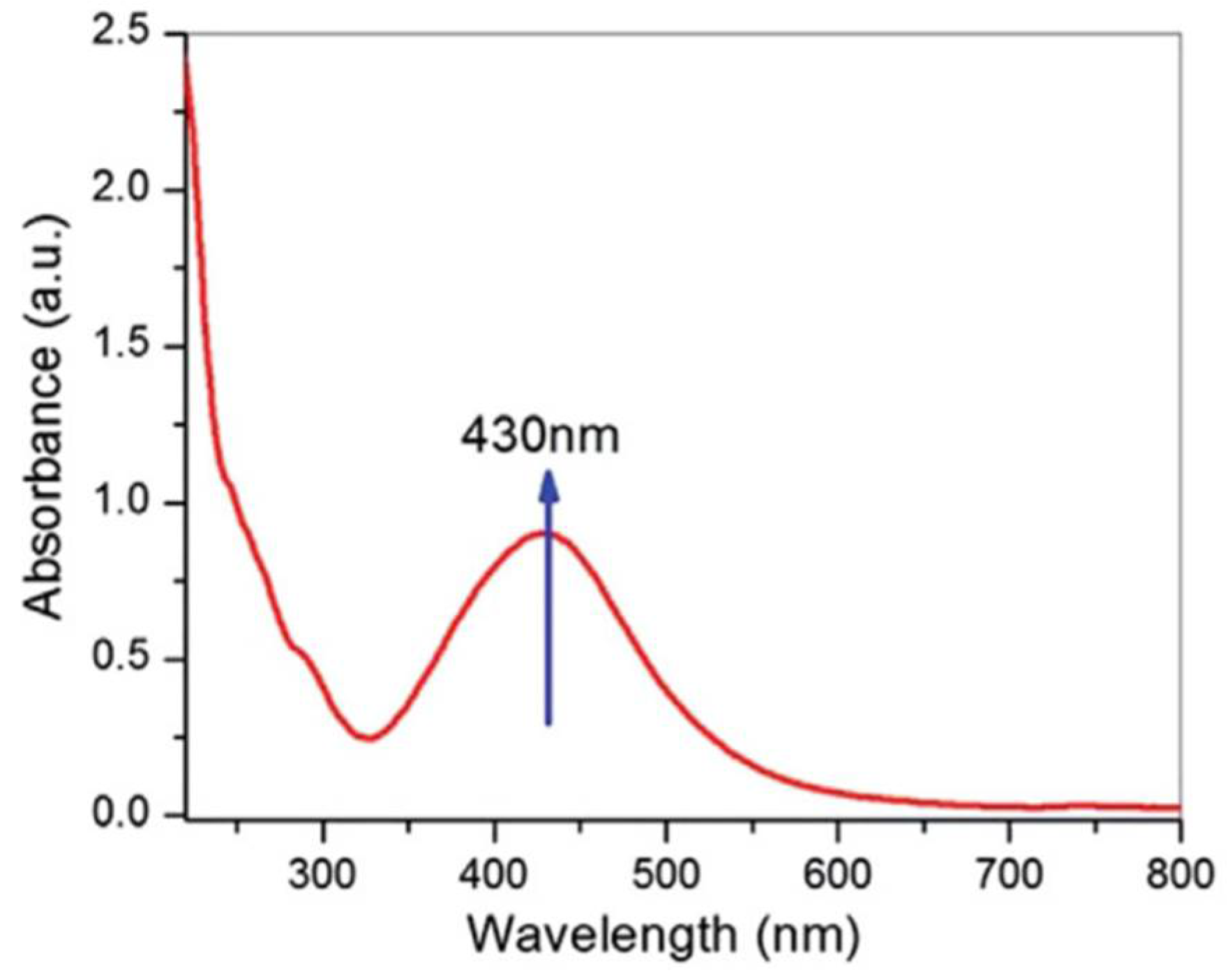
Due to their distinctive optical properties (Figure 1 and Figure 2), AgNPs have strong interactions with particular wavelengths of light [19]. The conduction band of silver nanoparticles is very close to the conductivity band, allowing electrons to move freely. These free electrons create a surface plasmon resonance (SPR) absorption band resulting from the collective oscillation of electrons of silver nano particles resonating with the light wave. The absorption and λmax value of metallic nanoparticles depend on the type of metal, the dimensions and the morphology of the particles, the dielectric medium, and the chemical environment [16]. The presence of metallic NPs can be confirmed by the SPR peaks in the visible region [7]. For spherical AgNPs, the LSPR absorption light band typically occurs around 400 nm and triangular silver nanoplates exhibit maximum absorption within the range of 500 nm to 1000 nm [20]. AgNPs also exhibit a unique resonance peak corresponding to their size. The UV–Vis absorption spectrum of silver nanoparticles synthesized using Cyprus rotundas is shown in Figure 4 where an SPR peak at 430 nm can be observed, confirming the existence of AgNPs. In unstable colloidal systems of AgNPs, the plasmon resonance peak shifts to longer wavelengths and widen due to an increase in diameter caused by aggregation [7]. However, the correlation between UV Vis spectra and size, shape, polydispersity and aggregation of AgNPs, albeit useful, is still qualitative, and thus lacks the rigorous quantitative understanding that is required to achieve detailed characterization of AgNPs.

2.2. Fourier Transform Infrared Spectroscopy (FTIR)

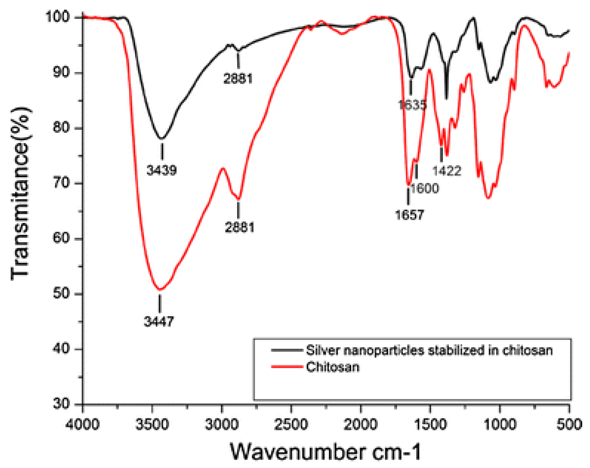
Fourier transform infrared spectroscopy (FTIR) is a technique based on the measurement of the absorption of light with wavelengths within the mid-infrared region (4000–400 cm−1) [21]. Upon irradiation of a material with infrared light, the radiation that is absorbed or transmitted is measured. The obtained spectrum serves as a unique fingerprint for samples, providing details about the sample’s nature, including the involved bonds, polarity, and oxidation state. FTIR is primarily employed for characterizing organic materials, such as determining the surface chemical composition or functionalization of nanoparticles and also for the identification of contaminants [10]. This method reveals the presence of amide (-CO-NH2), carbonyl (-CO), and hydroxyl (-OH) functional groups which play a role in the reduction, capping, and stability of Ag NPs. Figure 5 illustrates the FTIR spectrα of pure chitosan and of silver nanoparticles stabilized in chitosan. The absorption bands at 1657 cm−1 and 1600 cm−1, which represented the chitosan –CONH2 and –NH2 groups, vanished from the spectrum of AgNPs with chitosan, and a new band formed at 1635 cm−1, representing the bonding between silver and a nitrogen atom [22]. FTIR is fast, cost-effective and exhibits high reproducibility. However, it provides primarily qualitative information [23].

2.3. X-Ray Diffraction (XRD)

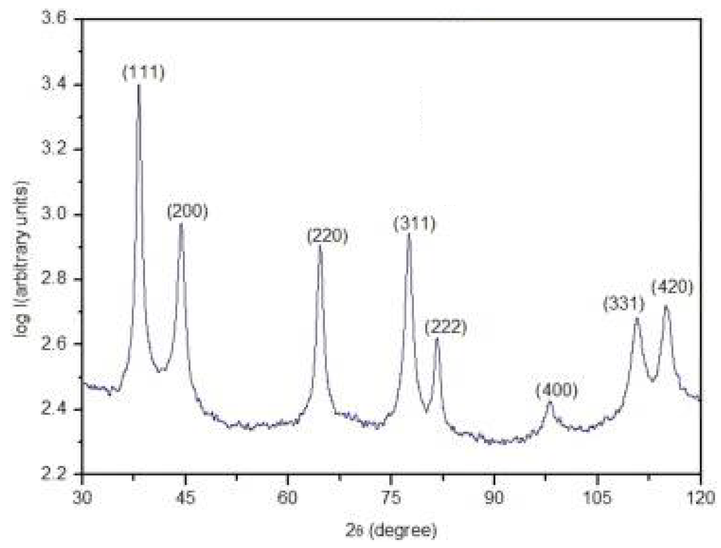
X-ray diffraction (XRD) is a widespread analytical technique that can be used to analyze the crystalline structure of AgNPs. In this method, a monochromatic X-ray beam is directed onto the surface of the crystalline NPs at a specific angle, resulting in the diffraction of the X-rays in specific directions. The fundamental principle behind XRD is Bragg’s law, which is based on the wide-angle elastic scattering of X-rays. If the Bragg equation holds for the wavelength of the incident X-ray under a specific angle: nλ = 2d sin(θ), n = 1, 2, 3. then the diffracted rays reinforce each other with an amplifying contribution and are detected during the analysis. The Bragg’s law estimates the relationship among the wavelength of the incoming electromagnetic radiation (λ), the diffraction angle (θ), and the spacing (d) between the crystalline planes [7,20]. Based on the angle and intensity of the diffracted beam, a diffraction pattern for the crystal is obtained. Most crystalline compounds have been measured, and their diffraction patterns are stored in databases. Identification of the material’s crystalline structure from the diffraction pattern can be made from these databases. The crystallinity of AgNPs is assessed using (XRD) an angle 2θ ranging from 20° to 80°, confirming their morphology. Typically, it reveals the formation of face-centered cubic (FCC) structures of metallic silver in which case the diffraction peaks are located at angles 2θ = 38.19°, 44.46°, 64.63°, and 77.34°, corresponding to planes (1 1 1), (2 0 0), (2 2 0), and (3 1 1), respectively (Figure 6) [7]. In this manner, the type and size of crystalline lattice of the AgNPs can be established. However, the application of this technique assumes the availability of relatively large amounts of AgNPs, which are not always available.

2.4. Scanning Electron Microscopy (SEM)

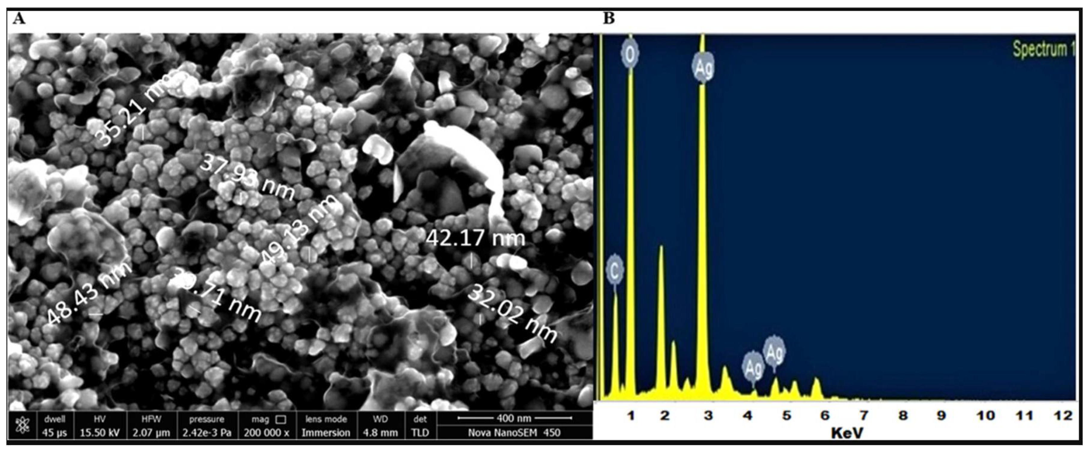
The evolution of nanotechnology has led to the need for the development of high-resolution microscopy techniques for the comprehensive study of nanomaterials using a high-energy electron beam that allows the investigation of objects on a very small scale. One such technique is Scanning Electron Microscopy (SEM). An electron beam scans the entire surface of a sample, and through detectors, the surface of the sample can be visualized by collecting signals generated as electrons interact with the atoms of the sample. This method is used for analyzing different particle sizes, size distributions, shapes of nanomaterials, and the nanoparticles’ surface structure. The investigation of particle morphology and the acquisition of histograms can be performed either by manually counting the particles from the images or by using JEOL SEM software v1.2.4.0. By combining SEM with Energy Dispersive X-ray Spectroscopy (EDX), both the morphology and the chemical composition can be analyzed, thus it is possible to determine the purity and degree of aggregation of nanoparticles, but it does not provide information about the internal structure (Figure 7) [7,16]. Despite the very informative nature of SEM in AgNPs research, its main drawbacks, are that it can not detect very small nanoparticles and that the obtained information relates only to a small area of the sample and not the properties of the bulk sample.

2.5. Tranmission Electron Microscopy (TEM)

Transmission Electron Microscopy (TEM) is a particularly significant and useful technique used for characterizing nanoparticles, quantitatively measuring particle sizes, size distributions, and for visualizing their morphology (Figure 8). TEM thus plays a critical role in advancing nanomaterials research by enabling accurate, direct visualization at atomic to nanometer scales [24]. In the TEM technique, a thin sample is bombarded by an electron beam with uniform current density, focused by condenser lenses. The sample undergoes appropriate processing to reduce its thickness to less than 100 nm, allowing the electron beam to pass through it. The signal of electrons passing through the sample is magnified 50–1,000,000 times by electromagnetic lenses, and it is projected onto a fluorescent screen where the image is formed. The observation of the image can be made directly on the microscope screen, and the image is digitized and stored through the integrated digital camera [7,16].
A major benefit of the technique is the ability to observe both electron microscope images (information in real space) and diffraction patterns (information in reciprocal space) for the same area by adjusting the electron lenses. By introducing a selected area aperture, a diffraction pattern can be obtained from a specific area as small as 100 nm in diameter [26]. However, the method also has some drawbacks, including the necessity for a high vacuum environment, the requirement for ultra-thin sample sections, and the time-intensive nature of sample preparation. Additionally, this method can only capture static information due to the physical and chemical fixation of the sample [7,16,27].
High-Resolution Transmission Electron Microscopy (HRTEM) is an imaging mode of TEM that relies on phase contrast, wherein the interference between unscattered (transmitted) and scattered electron waves produces enhanced image contrast. Phase-contrast imaging enhances the visualization of nanoscale structures by converting phase shifts in electron waves into amplitude variations, enabling the detection of atomic arrangements within crystalline materials. In contrast to typical TEM imaging, it demands a broader objective aperture to utilize both the direct beam and the Bragg diffraction beams for image formation. Among imaging techniques, HRTEM provides the most refined resolution, enabling the identification of atomic arrays within crystalline structures. Additionally, it offers significant information about the structure of nanoparticles, while traditional electron microscopy methods lack sufficient resolution to depict the crystalline structure of a particle. Therefore, HRTEM is more frequently used for characterizing the internal structure of NPs [21].
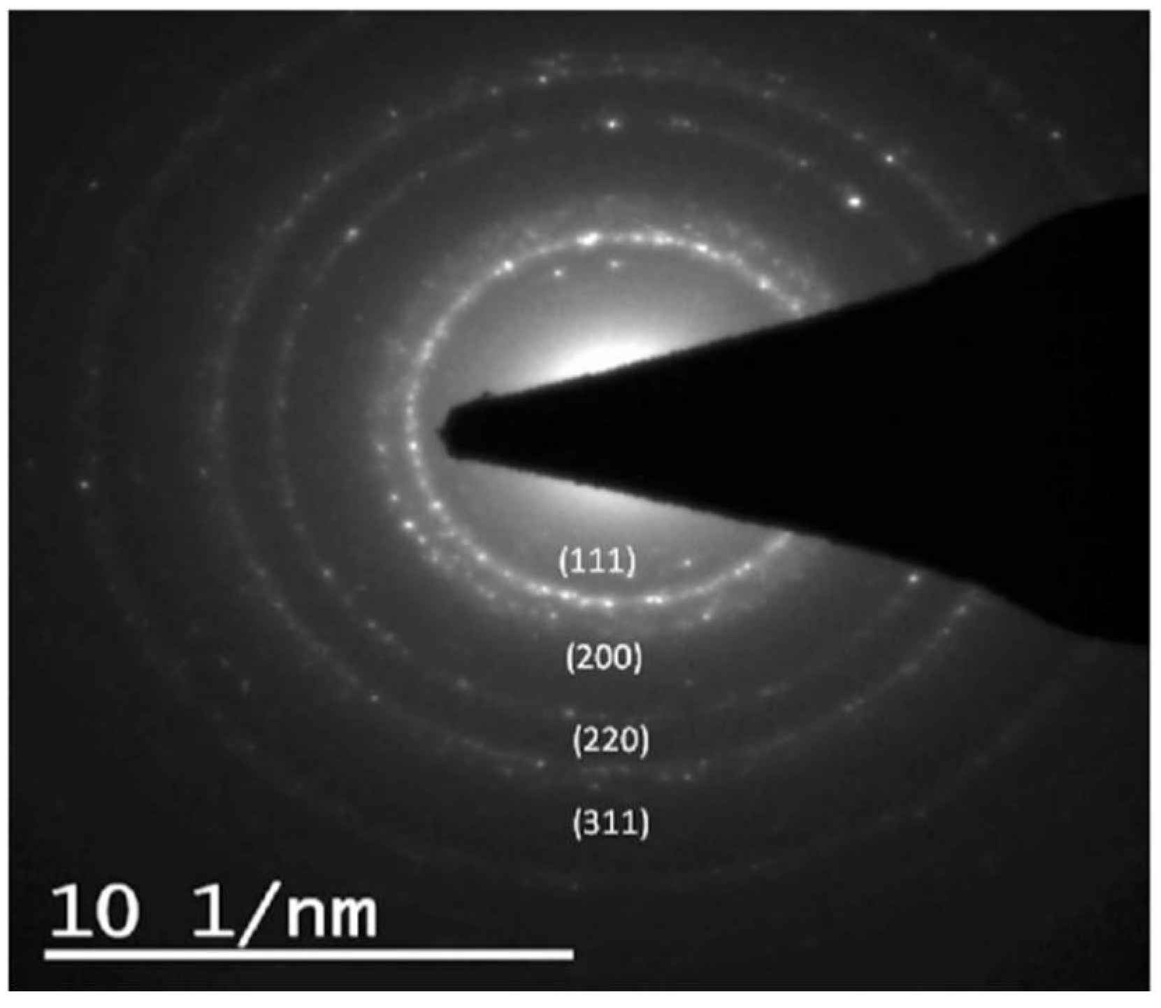
Electron diffraction through TEM is an effective technique for determining the internal architecture, encompassing both perfect crystals and defective structures. It presents advantages over other methods due to its exceptionally short wavelength, high atomic scattering, and capacity to analyze small volumes of matter (≈10 nm3). When an electron beam is directed onto a sample, the crystal lattice functions as a diffraction grating, scattering electrons in a predictable way, resulting in a diffraction pattern. The diffraction pattern provides information that allows the determination of atomic structure in a material [26]. Selected Area Electron Diffraction (SAED) is commonly employed to obtain a diffraction pattern from a specific region of interest, such as an individual grain within a polycrystalline sample (Figure 9). This is achieved by inserting a selected area aperture into the image plane of the objective lens, resulting in capturing a diffraction pattern from a specific sample area. The SAED technique can be used to determine if a sample is monocrystalline, polycrystalline, or amorphous, and to find the crystallographic structure, symmetry, and orientation. Studying individual nanoparticles with the SAED technique is challenging because many NPs contribute to the diffraction pattern due to the relatively large size of the selected area [21,26].

2.6. Dynamic Light Scattering (DLS)

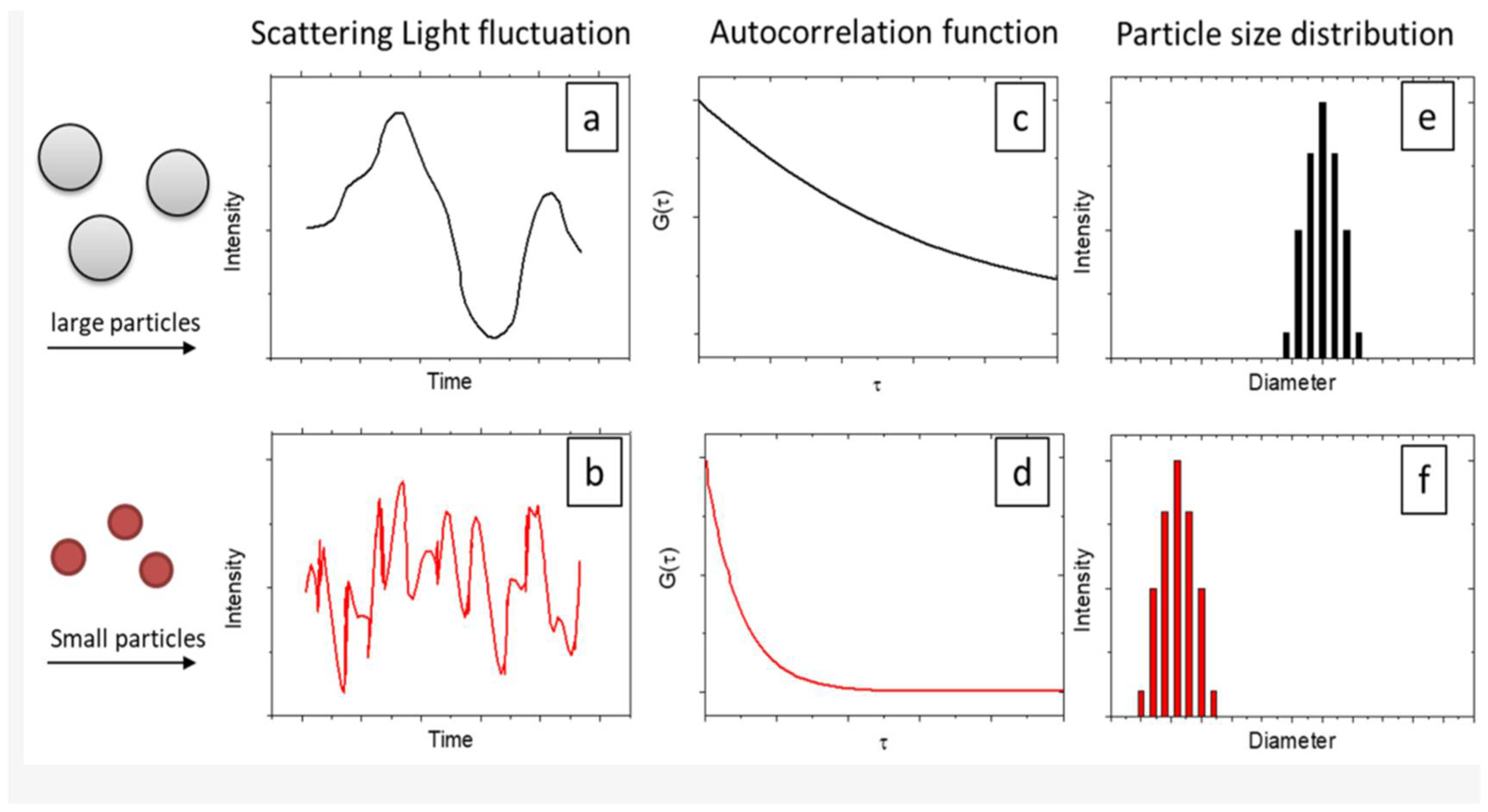
Dynamic Light Scattering (DLS) is a non-destructive analytical technique used to determine the size of small particles and size distributions in a suspension or colloidal suspension. This method relies on the interaction of light with particles in the range of 2–500 nm. DLS measures the scattered light from dispersed nanoparticles when a laser passes through a colloidal solution, primarily based on Rayleigh scattering (Figure 10). Subsequently, an analysis of the fluctuations in the intensity of scattered light over time is performed, which depends on the diffusion coefficient of the particles, to determine the hydrodynamic size of nanoparticles [16]. Particles dispersed in a liquid undergo random motion known as Brownian motion. Ordinarily, DLS measurements rely on the Doppler effect, which asserts that the frequency of light scattered by a particle shifts with particle velocity. Smaller particles move faster in the medium than larger particles, leading to a more rapid fluctuation in the intensity signal [29]. The impact of particle size on the intensity fluctuations of scattered light and the size distribution of particles is illustrated in the figure below.
The hydrodynamic size of the nanoparticles encompasses the actual diameter along with the diameter of the electrostatic potential surrounding nanoparticles. For this reason, the size obtained from the DLS method is usually larger than that of SEM and TEM analysis. DLS is based on the intensity that provides an average particle size of the sample, while TEM gives the actual geometric size of a particle. An advantage of DLS is the ability to detect numerous particles at once, in the sample and thus carry our measurements that may relate to bulk properties [7,16]. However, the analysis of DLS data and the quantitative information that it produces, depends largely on the model that is applied for the analysis of the DLS data, which can be quite arbitrary.

2.7. Zeta-Potential

Zeta potential is a measure of the electric potential at the slipping plane around a particle suspended in a liquid (Figure 11). This technique is widely employed to evaluate the stability of colloidal dispersions such as silver nanoparticles [30]. Various studies showed that silver nanoparticles with zeta potentials more negative than ca-30 mV or more positive than ca+30 mV are generally considered stable, due to electrostatic repulsion between charged particles, thus preventing aggregation, while values near zero indicate a tendency to aggregate [31].

2.8. Advanced Techniques

Advanced techniques are indispensable for thoroughly understanding the properties of nanoparticles, allowing researchers to precisely determine their physicochemical characteristics. These techniques cover a broad spectrum, including high-resolution microscopy, various spectroscopic methods, and surface analysis, each offering valuable insights into crucial aspects such as particle size, shape, composition, crystallinity, and surface chemistry.
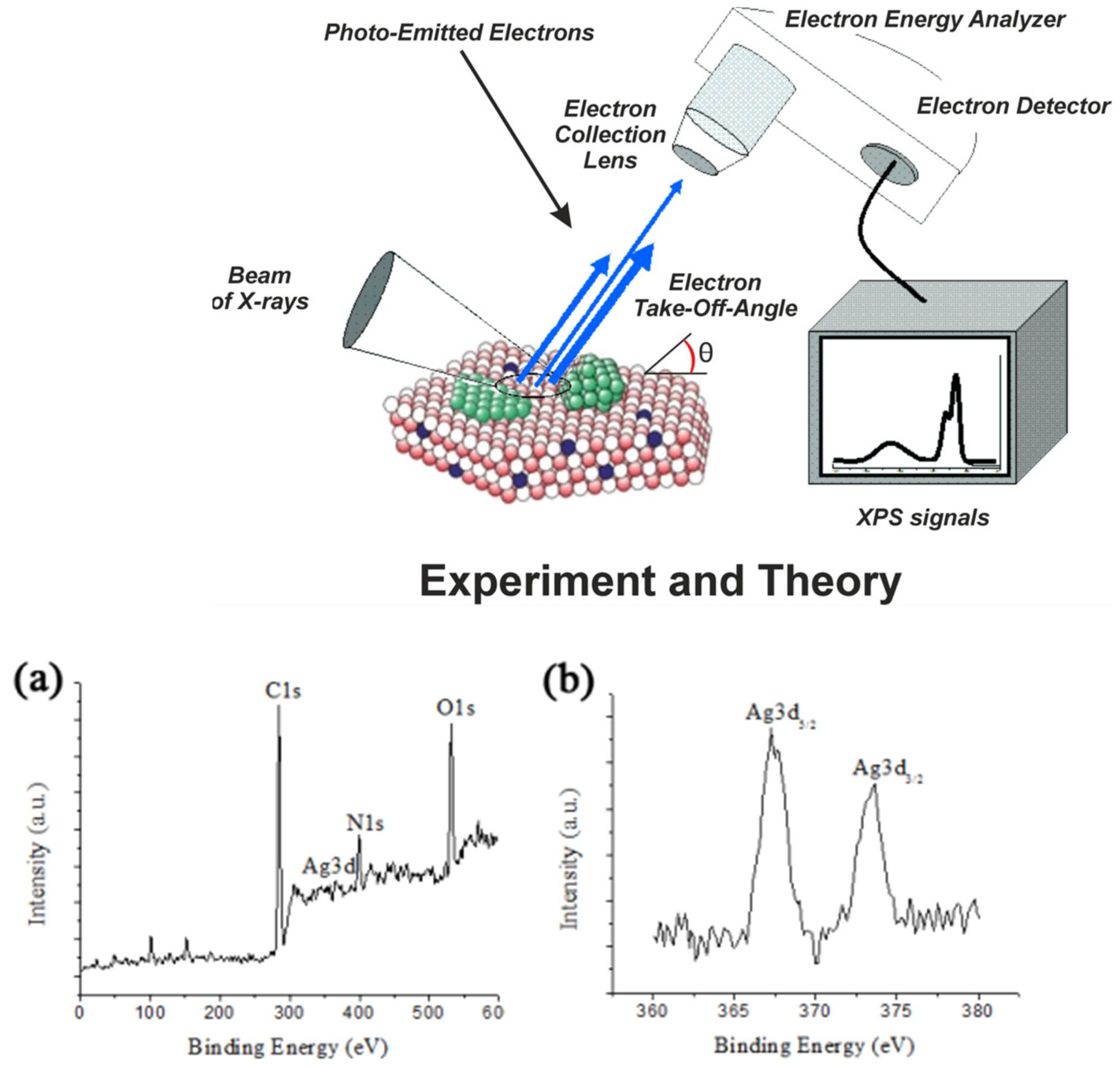
X-ray Photoelectron Spectroscopy (XPS) is a powerful surface-sensitive technique that provides detailed information about the elemental composition and chemical states of materials. The technique of XPS works by directing X-Rays in the surface of a material, causing atoms in the surface layer to emit electrons, due to the photoelectric effect (Figure 12). Each emitted electron has a specific kinetic energy that depends on the element it originated from and its chemical environment. By measuring these kinetic energies with a sensitive detector, XPS can determine the binding energies of electrons within the atoms. The measurement reveals which elements are present on the surface, because every element has unique electron binding energies. Moreover, slight shifts in these energies provide insight into the chemical states or oxidation states of these elements. XPS examines only the top few nanometers of the surface, making it a highly surface-sensitive technique.
Atomic Force Microscopy operates by scanning a sample surface with a sharp probe tip that is attached to a flexible cantilever. The cantilever, often made from silicon or silicon nitride, is extremely sensitive to forces when the tip approaches and interacts with the sample surface. The forces involved include attractive van der Waals forces when the tip is near but not touching, and repulsive forces if the tip comes too close. As the tip moves across the surface, the forces cause deflections or bending of the cantilever. This bending is detected by reflecting a laser beam off the top of the cantilever onto a position-sensitive photodetector. Any slight movement of the cantilever changes the direction of the reflected laser beam, allowing the photodetector to monitor nanoscale displacements.
This technique aids in visualizing the size, shape, and spatial distribution of silver nanoparticles, as well as the resulting surface roughness (Figure 13). Moreover, AFM can provide mechanical property data such as stiffness and adhesion forces, which are important for studying how nanoparticles interact with biological environments or polymers. Also, atomic Force Microscopy (AFM) is widely employed to study the interactions between silver nanoparticles (AgNPs) and cell membranes at the nanoscale. AFM imaging reveals detailed morphological changes in cell membranes exposed to AgNPs, such structural changes have been observed in bacterial cells like Escherichia coli, where AFM images show membrane disruption and leakage of cellular contents after AgNP treatment [34]. AFM thus provides critical insights into the antimicrobial and cytotoxic mechanisms of AgNPs by enabling real-time, high-resolution analysis of membrane-nanoparticle interactions [35].
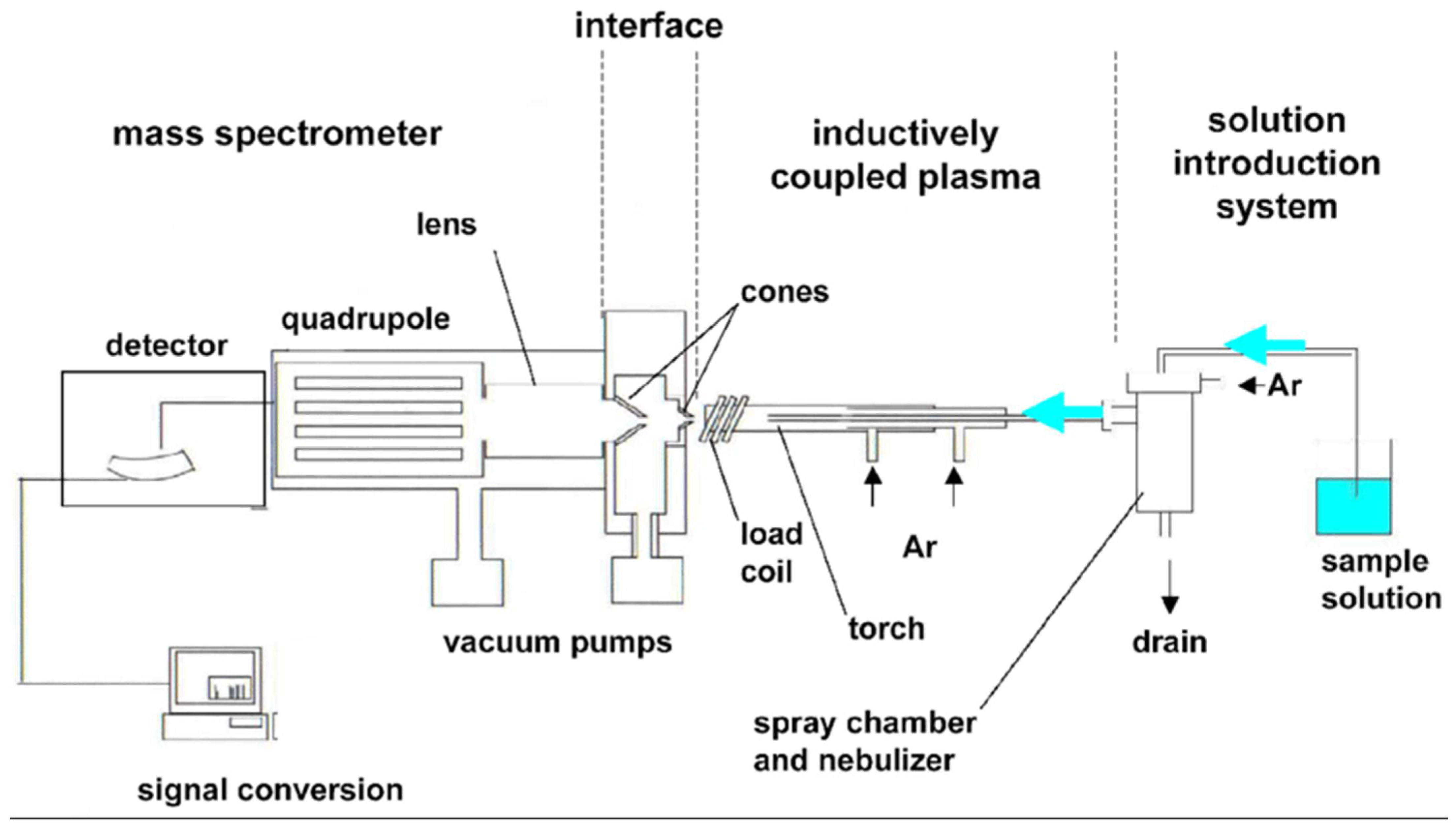
Inductively Coupled Plasma Mass Spectrometry (ICP-MS) is an advanced technique used to detect and quantify elements at extremely low concentrations. Its working principle involves multiple stages (Figure 14): First, a liquid sample is introduced as a fine aerosol into an inductively coupled plasma, typically generated using argon gas at very high temperatures (6000 to 10,000 K). This plasma acts as a highly energetic ion source, where the sample is atomized and ionize d, breaking down molecules into individual ions. These ions are then extracted from the plasma and guided through a series of lenses and electromagnetic fields known as ion optics, which focus the ion beam into the mass spectrometer. Inside the mass spectrometer, ions are separated based on their mass-to-charge ratio (m/z). The detector counts the ions according to this separation, enabling identification and quantification of elements and their isotopes.
This process allows ICP-MS to analyze multiple elements simultaneously with extremely high sensitivity, reaching concentrations as low as parts per trillion. It is widely utilized in environmental analysis, biomedical research, food safety, and materials science due to its precision and ability to detect trace and ultra-trace elements.
One of the primary advantages of ICP-MS is its capability to differentiate between dissolved silver ions and particulate silver without the need for extensive sample preparation or prior separation techniques, simplifying analysis and reducing potential errors. It offers rapid data acquisition with microsecond temporal resolution, enabling precise measurement of transient ion signals emitted from single nanoparticles as they are atomized and ionized in the plasma. This facilitates not only the detection of nanoparticles at very low environmental and biological concentrations but also real-time monitoring of their behavior, such as aggregation and dissolution dynamics [38].
However, despite these strengths, ICP-MS has notable limitations that warrant consideration. The method requires careful calibration and validation, including accurate determination of transport efficiency and correction for matrix effects, which can complicate quantitative analysis. Its reliance on ionized particle mass means ICP-MS does not provide direct information about nanoparticle morphology, surface chemistry, or aggregation state, thus necessitating complementary techniques for comprehensive characterization. Additionally, SP-ICP-MS sensitivity may be challenged by polydisperse samples or samples with extremely low particle concentrations, leading to potential underestimation or mischaracterization. The interpretation of data depends heavily on instrumental parameters such as dwell times and detector response, where suboptimal settings can lead to inaccurate particle counting or sizing [39,40].
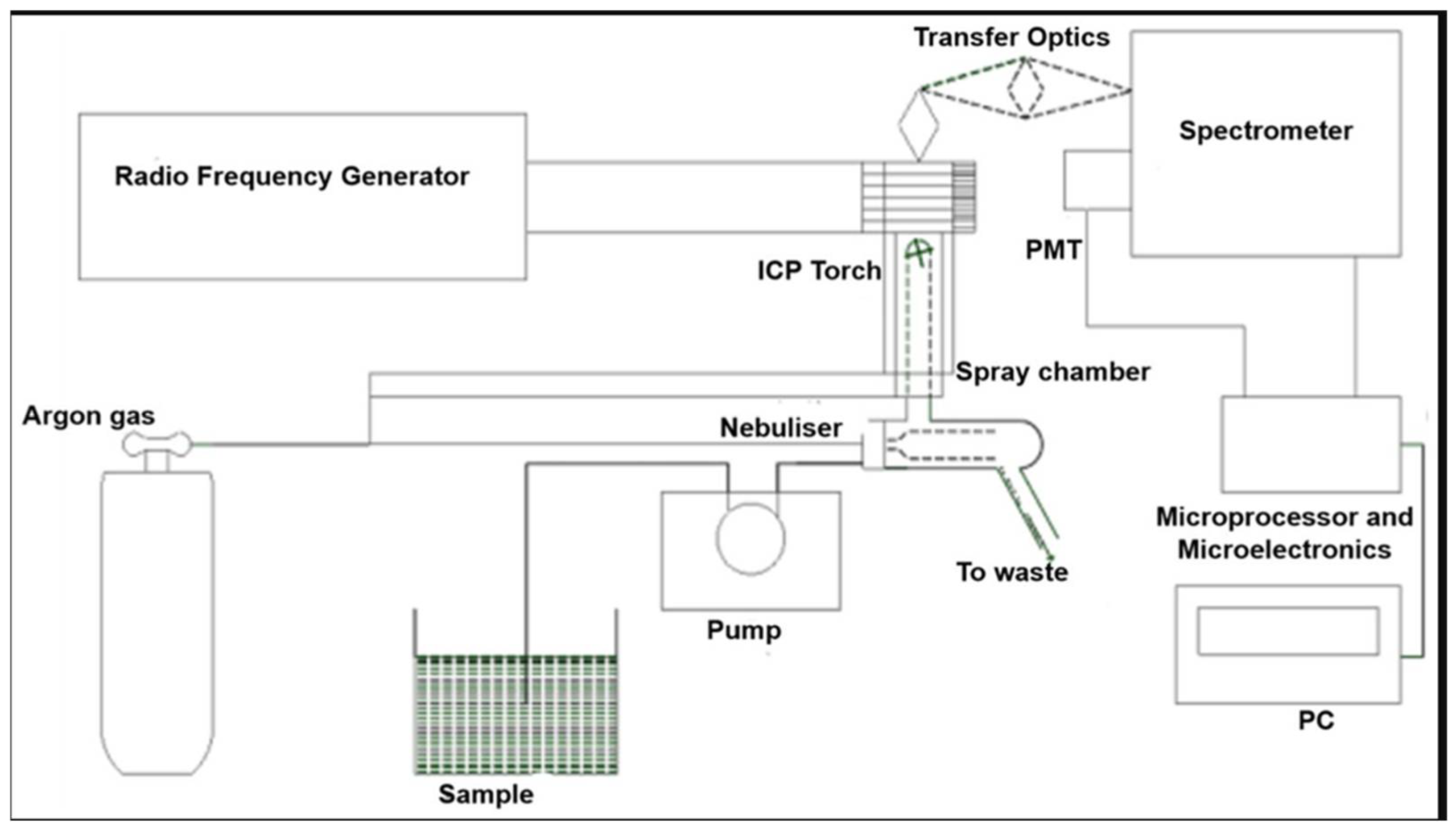
Inductively Coupled Plasma Optical Emission Spectroscopy (ICP-OES) is a technique widely used for the qualitative and quantitative determination of elemental composition in various sample types (Figure 15). Although ICP-OES and ICP-MS share a common principle in that both utilize inductively coupled plasma (ICP) to ionize sample elements, they differ fundamentally in their detection methods. ICP-OES measures the characteristic light emitted by excited atoms or ions as they return to their ground state, providing elemental identification and quantification based on optical emission spectra. In contrast, ICP-MS separates and quantifies ions based on their mass-to-charge ratio using a mass spectrometer, granting it superior sensitivity and the ability to detect trace elements with high precision. Thus, while both techniques rely on plasma ionization, their analytical approaches and detection capabilities are distinct.
Regarding its application to silver nanoparticles, ICP-OES can be used to measure the total silver content after appropriate sample preparation. However, ICP-OES does not differentiate between silver ions and silver nanoparticles directly because it detects elemental emissions regardless of their chemical or physical form. For detailed differentiation and quantitative analysis between silver ions and nanoparticles, ICP-OES is often complemented by other techniques such as single-particle ICP-MS or separation methods prior to measurement.
Advanced techniques for silver nanoparticles (AgNPs) characterization, while powerful, present several limitations. For example, in situ liquid-cell TEM requires expensive instrumentation and careful environmental control, which can limit experiment duration and throughput. Sample preparation for TEM can introduce artifacts, potentially skewing particle size and morphology data. Single-particle ICP-MS offers excellent sensitivity but requires extensive sample preparation and can struggle to distinguish different silver species in complex biological matrices. Furthermore, physicochemical properties of AgNPs can change dynamically in biological environments, complicating reproducibility and interpretation of results [42,43].
Density Functional Theory, or DFT, is a quantum mechanical tool that scientists widely use to explore interactions at the atomic level, especially on surfaces like those of silver nanoparticles. It helps us understand how electrons are arranged and behave, providing a detailed picture of how atoms bond together. For example, DFT studies have elucidated the formation of covalent bonds between silver surfaces and surrounding matrices, critical for understanding ion release and stability of AgNPs embedded in materials [44] and explored selective binding interactions on AgNP surfaces [45]. Modeling approaches like Density Functional Theory (DFT) for surface interactions and physiologically based pharmacokinetic (PBPK) models rely on simplifying assumptions and approximations that may not fully capture biological complexity. Therefore, integrated use of complementary experimental methods and theoretical modeling is necessary for comprehensive understanding and reliable safety assessment of AgNPs [46].
Physiologically Based Pharmacokinetic (PBPK) models use computer simulations to closely mimic how silver nanoparticles move through the body. These models incorporate detailed biological data, including how organs function, blood flow patterns, and nanoparticle behaviors within tissues. For instance, PBPK models can map out how nanoparticles of different sizes spread and accumulate in key organs such as the liver, spleen, kidneys, and lungs over time. This helps scientists predict where nanoparticles gather and how long they stay in each part of the body, which is crucial for understanding their safety. A landmark study by Bachler et al. (2013) [47] developed a PBPK model showing how both ionic silver and nanosilver distribute within rodents after exposure via skin, ingestion, or inhalation. They found that within typical consumer exposure levels, nanoparticle size and coating do not significantly alter their distribution, and that silver tends to be stored as insoluble salts rather than dissolving fully in tissues.
The physicochemical properties of silver nanoparticles are vital to their successful application in biomedical fields. Attributes such as particle size, morphology, surface charge, and chemical composition profoundly influence their interactions with biological systems, including cells and biomolecules, thereby affecting both efficacy and safety. Despite advances in analytical techniques, current methods remain insufficient to fully characterize the complex behaviors and transformations that silver nanoparticles undergo within biological environments. This limitation is mainly due to their dynamic interactions and the formation of biomolecular coronas, which obscure a complete understanding of their biological mechanisms. Therefore, further development of sophisticated characterization strategies is essential to comprehensively elucidate the role and impact of silver nanoparticles in biomedical contexts.

3. Biomedical Applications of Silver Nanoparticles

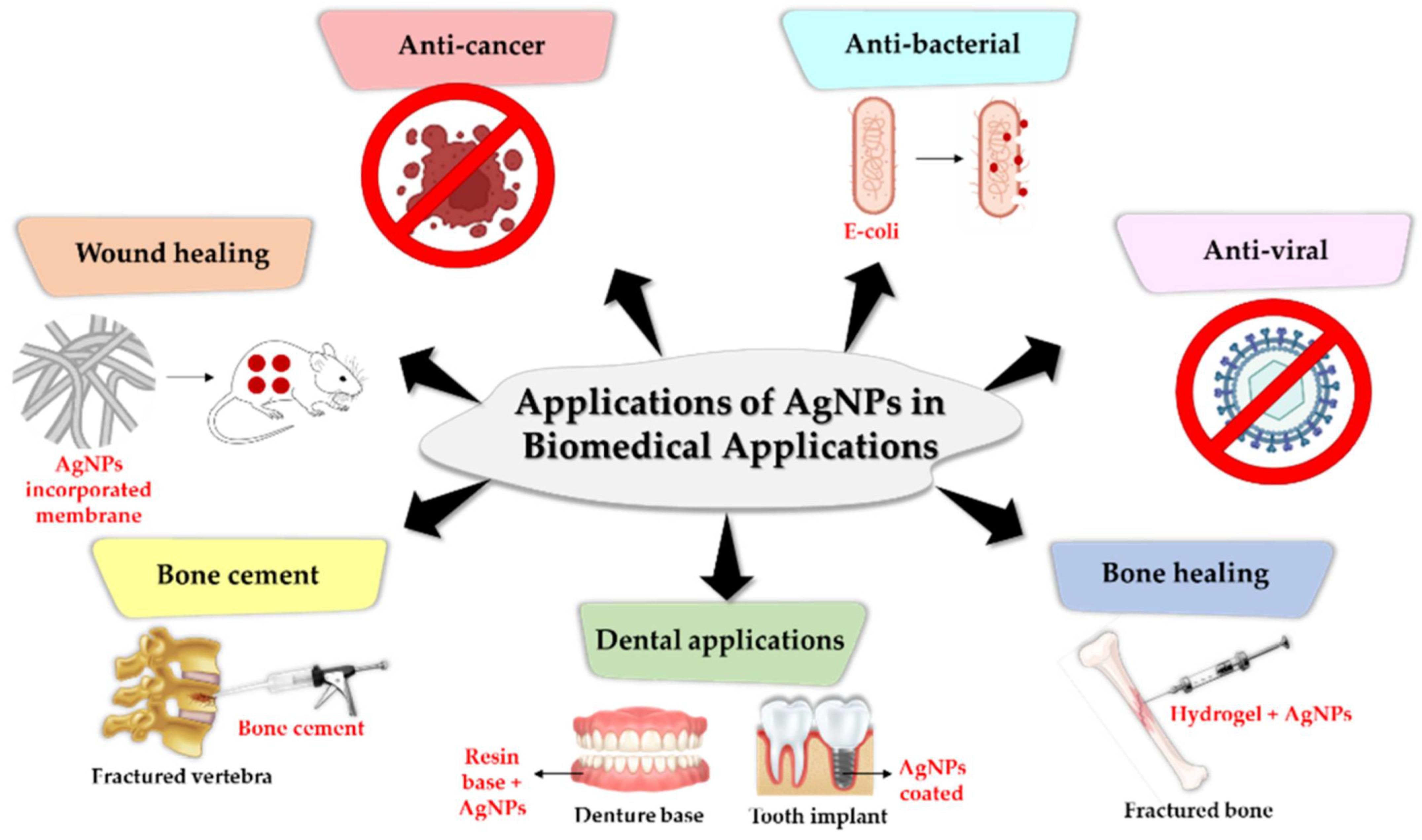
Due to their unique physicochemical properties, silver nanoparticles have captured significant interest from researchers across multiple scientific disciplines. Their applications span a wide array of industries, including textiles, water treatment, cosmetics, pharmaceuticals, medicine, and the food sector, specifically in food storage and household utensils [48,49,50]. Importantly, silver nanoparticles are a leading example of nanomaterials harnessed in biomedical engineering, where their distinctive characteristics enable diverse medical applications [51]. Additionally, silver nanoparticles exhibit a broad range of remarkable biological activities, such as antibacterial, antiviral, antifungal, anti-inflammatory, anti-angiogenic, antitumor, and antioxidative effects, making them invaluable in biomedical fields. They also play an essential role in imaging techniques like bio-sensing [6,16,52,53,54,55,56,57,58,59,60]. Below is a summary of some of the main biomedical applications of silver nanoparticles (Figure 16).

3.1. Antimicrobial Agents

In the current era, the increase in antibiotic resistance among bacteria, posing a global threat to public health, has led scientists to continually search for new antibacterial agents. Nanomaterials find increasingly diverse applications in medical, and pharmaceutical fields to harness their antimicrobial action derived from their large surface area. Various nanoparticles have been employed in antibacterial treatments, with silver nanoparticles (AgNPs) being among the most widely used due to their broad-spectrum antimicrobial effects and strong efficacy against a wide variety of bacteria, viruses, and fungi [62,63]. Notably, AgNPs also represent a promising therapeutic option against Mycobacterium tuberculosis, including multidrug-resistant strains, owing to their broad-spectrum antimycobacterial activity and ability to enhance traditional antibiotic efficacy, potentially reducing treatment duration [52,64].
Silver nanoparticles exhibit strong antimicrobial efficacy against both Gram-positive and Gram-negative bacteria [53,54,55,56], including multidrug-resistant strains, underscoring their significant potential in addressing the growing challenge of antimicrobial resistance [59]. Nevertheless, AgNPs seem to be more effective in killing Gram-negative bacteria compared to Gram-positive due to differences in the construction of their cell walls [65]. Gram-positive bacteria have a thick cell wall primarily composed of various layers of peptidoglycan (20–80 nm), while Gram-negative bacteria have a thin layer of peptidoglycan and an outer lipopolysaccharide membrane [66]. The rigid three-dimensional structure created by the thick peptidoglycan layer in Gram-positive bacteria makes it difficult for AgNPs to adhere and penetrate through the cell wall [65]. In contrast, in Gram-negative bacteria, the lipopolysaccharide layer consists of covalently linked lipids and negatively charged polysaccharides, creating a weak permeability barrier against positively charged AgNPs [67]. Findings indicate that AgNPs are able to cross both types of cell walls and penetrate inside the cell [65]. They are additionally able to kill multidrug-resistant (MDR) bacteria by disrupting their defense mechanisms [59,66]. Their action is based on multiple mechanisms through which they interact with cells, inhibit enzymatic activity, destabilize the cell membrane, and ultimately induce cell death. This is a significant advantage, as bacteria would need to target multiple mechanisms simultaneously to develop resistance against AgNPs [59,63].
Silver nanoparticles demonstrate potent antibacterial effects that are critically dependent on their colloidal stability, as stable nanoparticles maintain their small size and surface characteristics, thereby ensuring sustained antimicrobial activity [68] and preventing aggregation-induced loss of efficacy [69,70]. Research has shown that the size [71], shape, charge of nanoparticles, as well as their stability after synthesis, affect their final antibacterial action [72]. If silver nanoparticles have low stability after synthesis, they tend to aggregate and form larger particles with lower antibacterial activity [59]. A number of studies suggest that smaller nanoparticles exhibit enhanced stability and increased antimicrobial activity, particularly when below 10 nm [73]. This relationship between size and biological activity is attributed to the fact that smaller nanoparticles penetrate more effectively through microbial cell walls, leading to membrane damage and cellular disruption [74,75]. Furthermore, it has been discovered that smaller nanoparticles have a higher dissolution rate in diverse media, releasing silver ions which could contribute to the antibacterial activity of nanoparticles. The small hydrodynamic diameter of AgNPs (in some cases < 6 nm) allows their entry into the kidneys and their elimination from the body through the urinary system, significantly decreasing the risk of long-term toxicity [63,65]. Additionally, the stability of nanoparticles is influenced by charge and coating [76,77]. Different stabilizers can alter the surface charge, size, and silver ion release profile of the nanoparticles, which in turn affects their biological activity. Positively charged AgNPs, such as those stabilized with arginine, exhibited the strongest antibacterial effects, while negatively charged AgNPs stabilized with (-)-epigallocatechin gallate (EGCG) showed the most potent antifungal activity [78,79,80]. Considering their zeta potential, silver nanoparticles can be classified as stable when their surface charge is greater than +30 mV or less than −30 mV, which prevents their aggregation due to repulsive forces between nanoparticles. Moreover, it has been proven that nanoparticles with a positive charge exhibit greater antibacterial action, as electrostatic attraction between positively charged AgNPs and negatively charged bacterial cells contributes to their antibacterial effectiveness. The charge is determined by the synthesis process and the coating agent used. Studies have shown that coating nanoparticles with polymers or organic compounds eliminates their cytotoxicity toward mammalian cells, while preserving their antimicrobial effectiveness against tested bacteria [59]. Finally, the nanoparticle’s shape also affects its bioavailability and antimicrobial action. The highest toxicity has been observed in hexagonal nanoparticles, and this variation in toxicity is correlated to differences in the surface area among various nanoparticle shapes [69].

3.2. Antiviral Agents

Viral mediated diseases are prevalent globally and thus the development of antiviral agents is crucial [16]. Nanoparticles deliver an alternative therapeutic approach for the treatment and control of the proliferation of viral pathogens. AgNPs could be used as potent antiviral agents that limit virus functions in the biomedical field [42,43,81]. Understanding how silver nanoparticles exert their antiviral action is a key component of antiviral treatment [16]. AgNPs have demonstrated good antiviral activity against a variety of respiratory viruses, including coronaviruses, rhinovirus, influenza virus, adenoviruses, as well as viruses like hepatitis B (HBV), human immunodeficiency virus (HIV), and herpes simplex virus (HSV). AgNPs smaller than 10 nm display significant antiviral activity, likely attributed to their large reaction area and strong adhesion to the virus surface [82]. In a study, the antiviral activity of silver nanoparticles against feline calicivirus was investigated, with various dosages and sizes. It was found that AgNPs with diameter of 10 nm can effectively reduce the virus load, as the virus size falls within the range of 27 to 40 nm, allowing the smaller nanoparticles to interact with the virus more easily. Another study assessed the antiviral activity of AgNPs on SARS-CoV-2, and the results demonstrated that silver nanoparticles with a size of 10 nm were effective in inhibiting the extracellular SARS-CoV-2 [83]. While there have been some studies on the effect of AgNPs towards viruses, the details of these interactions remain limited, likely due to the complexity of virus structure [61]. The detailed mechanisms involved must be further investigated, to facilitate the development of AgNPs as efficient antimicrobial agents [82].

3.3. Wound Healing

Silver nanoparticles demonstrate remarkable antimicrobial, anticancer, and wound-healing properties, positioning them as invaluable tools in tackling pressing healthcare challenges such as antibiotic resistance, cancer treatment, and tissue repair. Their exceptional efficacy is largely attributed to their nanoscale size and high surface area, allowing them to interact extensively with biological systems. Notably, silver nanoparticles incorporated into biopolymers have found a significant role in wound management, where they not only prevent infections but also actively promote tissue regeneration. This integration enhances the effectiveness of wound dressings, leading to accelerated healing and improved patient outcomes. The ongoing research and development in this field underscore the transformative potential of silver nanoparticles in advancing modern medicine and improving clinical care [84]. The resulting biomedical products (AgNP-BMs) are considered to be non-cytotoxic, safe for patients, and play a significant role in the treatment of both acute and chronic wounds. AgNP-BMs are engineered to maintain a moist environment around the wound while preserving desirable mechanical properties [85]. Silver nanoparticles (AgNPs) have gained considerable recognition for their exceptional role in enhancing the wound healing process. Wound dressings containing AgNPs are designed to maintain permeability to oxygen, a vital factor that facilitates tissue repair, while simultaneously offering robust protection against microbial infections. The silver nanoparticles are uniformly distributed over cotton fibers without the need for additional binders, ensuring firm adhesion to the fabric and continuous antimicrobial efficacy. This coating effectively inhibits bacterial growth and biofilm formation. Importantly, these silver-coated gauzes demonstrate a significant reduction in bacterial proliferation in laboratory tests while preserving compatibility with essential skin cells such as fibroblasts and keratinocytes, which play a critical role in wound healing. This combination of properties makes AgNP-based dressings a promising option for improving clinical outcomes in wound care [53,86,87,88,89]. There are many types of silver-based dressings available today, crafted from polymers like chitosan, cellulose, collagen, polyvinyl alcohol, and polyethylene glycol. The choice of polymer and how it interacts with the silver nanoparticles greatly affects how well the dressing fights infection, its stability, and how controlled the release of silver ions is, which together enhance healing and tissue regeneration [90].
Moreover, when silver nanoparticles are woven into nanofibers, they significantly speed up wound closure. For example, a special nanofiber made with 1% licorice root extract and silver nanoparticles combined with PLGA showed the fastest healing compared to other tested materials. This is because licorice root extract has natural properties that fight microbes, reduce inflammation, and act as antioxidants, which work hand-in-hand with the silver nanoparticles to boost their healing power. Overall, silver nanoparticles offer a powerful, multifaceted approach to wound care, reducing infections and promoting faster, healthier tissue repair [22,86,91,92,93].

3.4. Dental Products

Silver nanoparticles (AgNPs) represent a significant innovation in dental products and therapeutics, attributable to their strong antimicrobial activity and broad applicability. They are used across various dental fields, including disinfection, infection control, restorative dentistry, endodontics, implantology, periodontics, and orthodontics [94,95,96,97,98,99].
The addition of AgNPs to dental products such as composite resins and acrylic-based materials has been well investigated [96]. Research indicates that nanosilver effectively inhibits a variety of medically relevant oral pathogens, including Streptococcus mutans, Candida albicans, Pseudomonas aeruginosa, Enterococcus faecalis, and Staphylococcus aureus. These antimicrobial properties suggest that AgNPs can play a crucial role in reducing secondary caries, preventing fungal infections, improving the outcomes of endodontic treatments, and minimizing failures of dental implants. Notably, antimicrobial tests reveal that AgNPs significantly inhibit the growth of Escherichia coli, although their effect on Staphylococcus aureus and Candida albicans is comparatively moderate. Importantly, the addition of AgNPs to dental alginates enhances their antibacterial effectiveness without compromising mechanical properties. This finding supports the potential use of silver nanoparticle-modified alginates as self-disinfecting dental impression materials, which may help reduce cross-contamination risks in clinical settings while preserving the necessary clinical performance [97]. It is essential to highlight that AgNPs have been proven to be biocompatible with mammalian cells, indicating they are safe for use in dental applications [98,99]. Further studies have revealed that adhesives modified with silver nanoparticles can sustain silver ion release for up to four months, providing prolonged antimicrobial efficacy while avoiding common adverse effects such as irritation or delayed hypersensitivity reactions typically observed with other antimicrobial agents [100]. Peri-implant infections, a major complication in implantology, may be effectively mitigated by coating titanium implant surfaces with AgNPs, as demonstrated by researchers in [101], who reported continuous ion release and pronounced antibacterial activity. Moreover, Zhou et al. (2017) [102] revealed that AgNPs not only prevent infection but may also enhance peri-implant bone density and osteogenesis, without eliciting adverse tissue responses [58,88,102].
Furthermore, various scientists have designed formulations incorporating silver nanoparticles, and assessed their minimum inhibitory concentration (MIC) and minimum bactericidal concentration (MBC), primarily against Streptococcus mutans. Findings indicated that they serve as effective antimicrobial agents in the oral environment, particularly when their dimensions are in the range of 80–100 nm, as dimensions smaller than 20 nm have demonstrated increased cytotoxicity [99]. However, more research is required to examine the release of Ag ions and the long-term properties of AgNPs in these products [98].

3.5. Medical Device Coatings (Catheters with Embedded Silver Nanoparticles)

The central venous catheter (CVC) is an essential medical device utilized primarily in patients with chronic health conditions, facilitating intravenous delivery of fluids, medications, hemodynamic monitoring, and nutritional support. Despite its critical role, CVCs are highly vulnerable to nosocomial infections, with a substantial risk of bacterial colonization. Various strains of Staphylococcus aureus, notably including 82% methicillin-resistant isolates, have been implicated in these infections. Numerous studies have highlighted the effectiveness of catheters embedded with silver nanoparticles (AgNPs) as biocompatible agents that mitigate infection-related complications through sustained silver ion release. Investigations have also demonstrated the antimicrobial efficacy of AgNP-coated catheters against coagulase-negative staphylococci (CoNS), which represent a significant group of pathogens responsible for device-associated infections. Furthermore, AgNP-coated CVCs have shown notable inhibition of biofilm formation by both Gram-positive and Gram-negative bacteria. In patients undergoing peritoneal dialysis, the deployment of silver ion-releasing catheters has been linked to a reduction in complications related to dialysis [88,103]. Recent advancements have led to the development and clinical implementation of a new generation of catheters incorporating silver for enhanced infection control [58].

3.6. Tissue Engineering and Regeneration

Silver nanoparticles have attracted a lot of interest in tissue engineering due to their strong antimicrobial effects, good biocompatibility, and ability to stimulate cellular activities that support tissue repair and regeneration. Incorporating AgNPs into biomaterials, scaffolds, and wound dressings has shown to enhance healing processes and help prevent infections, which often complicate tissue regeneration [104,105,106].
In the context of bone tissue engineering, AgNPs are integrated into composite scaffolds such as hydroxyapatite, collagen, alginate, and various biopolymers, offering both antimicrobial protection and support for osteoblast adhesion, growth, and differentiation. These nanocomposites create an optimal environment for bone regeneration, speeding up fracture healing and enhancing the mechanical strength of the grafts. The size, shape, and surface characteristics of AgNPs can be adjusted to maximize their interaction with bone cells and improve the delivery of growth factors [93,103,104,105,106,107,108]. For soft tissue repair, combining AgNPs with polymers like chitosan or silk fibroin enhances the antimicrobial properties of wound dressings, thereby reducing bacterial growth and biofilm formation in wounds and burns. These materials not only protect against infection but also promote cell survival, new blood vessel formation, and skin regeneration, leading to faster and more effective healing.
Vascular tissue engineering benefits from AgNPs, as their inclusion in vascular graft scaffolds helps prevent infections while supporting the function of vascular cells. Likewise, nerve and skin tissue repair are improved by AgNP-containing scaffolds that mitigate microbial infections and encourage cellular regeneration. Recent developments have focused on creating multifunctional AgNP-based medical products that allow controlled silver ion release, reducing cytotoxic effects while enhancing regenerative capabilities. Despite these advances, challenges persist in balancing antimicrobial effectiveness with long-term biocompatibility and addressing concerns over potential bacterial resistance [42,43,102].

3.7. Anticancer Agents

Cancer is related with the secondary complications, which are a major cause of mortality worldwide. This is due to the limited effectiveness and severe side effects of current chemotherapeutic drugs, as well as the often poor prognosis of affected patients. Conventional cancer treatments—including surgery, radiation, and chemotherapy—frequently result in suboptimal outcomes because of their low selectivity and specificity [61]. In contemporary research, nanotechnology-based strategies have introduced novel approaches in cancer therapy aimed at enhancing treatment efficacy and improving patient outcomes. Silver nanoparticles (AgNPs) have garnered considerable attention in recent years for their promising role in cancer diagnosis and therapy. Their unique physicochemical properties allow them to selectively accumulate in tumor tissues, thus enhancing the precision of drug delivery systems. This selective targeting helps to improve the efficiency of treatment while minimizing damage to healthy cells. AgNPs are being explored for use in tumor detection, diagnostics, and the development of controlled, externally triggered drug delivery platforms. Additionally, their therapeutic effects include inducing cancer cell death through mechanisms such as reactive oxygen species production, DNA damage, and apoptosis. These properties position AgNPs as multifunctional agents capable of combining diagnostic and therapeutic functions on a single platform, offering significant potential to improve cancer treatment outcomes [109,110]. The therapeutic efficacy of AgNPs depends on their ability to be internalized by cancer cells through mechanisms such as diffusion, phagocytosis, pinocytosis, and receptor-mediated endocytosis. Upon uptake, AgNPs release silver ions locally and induce oxidative stress, leading to cancer cell death either via apoptosis or by damaging critical cellular components through specific interactions with silver nanoparticles and ions [111]. Studies on lymphoma cell lines have confirmed the dose-dependent cytotoxicity of AgNPs in vitro and suggested their role in triggering apoptosis. Additionally, animal models demonstrated that treatment with AgNPs prolonged survival, while an increase in nanoparticle concentration correlated with greater inhibition of tumor cell growth [42,43]. Investigations involving hepatocellular carcinoma (HepG2) and breast cancer (MCF-7) cells treated with AgNPs synthesized using Ganoderma lucidum revealed that spherical nanoparticles of approximately 11 nm effectively reduced cancer cell proliferation. Similarly, AgNPs prepared with Acalypha indica Linn. showed a 40% inhibitory effect on human breast cancer cells (MDA-MB-231) [61].

3.8. Biosensing and Diagnostics

Inorganic biomaterials, including metallic nanoparticles, metal-organic frameworks (MOFs), and silica-based nanomaterials, offer unique physicochemical, mechanical, optical, and magnetic properties that make them highly effective for targeted drug delivery, bioimaging, and biosensing. These materials are particularly valuable due to their stability, biocompatibility, and tunable surface chemistry, enabling precise molecular recognition and controlled therapeutic release [112,113].
Silver nanoparticles have become key components in diagnostic and therapeutic monitoring tools thanks to their distinctive optical and electronic properties. Their large surface area, adjustable optical behavior, and excellent electrical conductivity make them well-suited for creating highly sensitive and selective biosensors [114].
One of the standout optical features of AgNPs is their strong Surface Plasmon Resonance (SPR), which reacts to tiny changes in the surrounding environment. This allows biosensors using AgNPs to detect subtle interactions between biological molecules and the nanoparticles without the need for labels, enabling real-time monitoring. These interactions can cause color changes that are easy to observe, making quick and straightforward diagnostics possible—even in point-of-care settings. AgNPs also boost the Raman scattering signals of nearby molecules, a process called Surface-Enhanced Raman Scattering (SERS), which further enhances the sensitivity and accuracy of biosensors [112,114]. Electrochemical biosensors take advantage of AgNPs’ high electrical conductivity and catalytic activity. When combined with transducer platforms, AgNPs help speed up electron transfer and increase the reactive surface area, resulting in quicker detection and lower detection limits. These sensors have successfully identified a wide range of clinical markers such as glucose, DNA, proteins, enzymes, and small molecules with high precision. Recent developments have introduced multiplex biosensors using AgNPs that can detect multiple targets at once, improving the efficiency and speed of diagnostics. The ease of attaching various biomolecules to the nanoparticles allows for selective targeting of specific analytes, enhancing the accuracy of these sensing systems.
Beyond clinical applications, AgNP-based biosensors also hold potential for use in environmental monitoring, food safety, and pathogen detection. They are attractive options for broad use due to cost-effective production and potential for miniaturization, which is particularly valuable in settings with limited resources. Although there are challenges related to the stability of AgNPs in biological environments and concerns about toxicity, ongoing research into surface modifications, core–shell designs, and biocompatible coatings aims to overcome these hurdles [115].

3.9. Toxicity of Silver Nanoparticles

The toxicity and bioactivity of silver nanoparticles (AgNPs) are fundamentally determined by their physicochemical properties, which govern their intricate interactions within biological systems. Critical parameters including particle size, morphology, surface chemistry, and colloidal stability significantly influence the absorption, biodistribution, cellular uptake, and accumulation of AgNPs, thereby modulating their therapeutic potential as well as possible adverse effects [42,43,116,117].
Particle size is widely recognized as a pivotal factor in biological activity. Smaller nanoparticles, particularly those ranging from 1 to 30 nm, possess a higher surface area-to-volume ratio resulting in enhanced chemical reactivity and an elevated rate of silver ion release. This ion release is a key mechanism underlying cytotoxic and genotoxic responses. Empirical observations indicate that AgNPs smaller than 20 nm penetrate cellular membranes more efficiently and accumulate in organs, exhibiting increased toxicity compared to larger particles which tend to aggregate and have reduced cellular internalization. However, such generalizations often overlook the complex interplay of surface modifications and tissue microenvironments that critically influence these effects [7,118].
In addition to size, the morphology of AgNPs—encompassing spherical, rod-like, cubic, triangular, and star-shaped forms—exerts a profound impact on their biological behavior. While spherical nanoparticles are generally associated with higher cellular uptake and uniform tissue dispersion, anisotropic shapes modulate oxidative stress induction and antimicrobial efficacy distinctively. Yet, the current understanding remains largely descriptive, lacking comprehensive mechanistic insights linking specific shapes to biological outcomes.
In order to develop successful biomedical applications of silver nanoparticles, it is crucial to limit the potential harmful effects of these nanoparticles on both human health and the environment [119]. The primary ways silver nanoparticles enter the human body are through ingestion, inhalation, and skin absorption. They may also enter directly into the bloodstream via intraperitoneal or intravenous injections [60]. This creates several potential pathways for exposure to silver nanoparticles.
Regarding oral uptake, the nanoparticles quickly reach the stomach, where the acidic conditions cause them to partially dissolve, subsequently they move through the intestines, are taken up by the intestinal lining, and are transported to different organs. Research shows that when silver nanoparticles enter the body through the respiratory system, they mostly accumulate in the lungs but, due to their small size, they can also be distributed to other tissues and organs throughout the body. The nanoparticles are also capable of penetrating both intact and damaged skin, reaching the epidermis and diffusing into deeper tissues. Therefore, nanosilver found in cosmetics, wound care products, and antimicrobial textiles is likely to diffuse through the skin in substantial amounts. Moreover, if introduced via intraperitoneal or intravenous injection, silver nanoparticles enter directly into the bloodstream and are then distributed to vital organs such as the heart, liver, kidneys, brain, and reproductive organs, potentially causing pathological effects [60,120], where because of their extremely small size, they have the potential to interfere with biological molecules, cells, and human organs at the nanoscale level [89]. Toxicity mechanisms primarily involve oxidative stress, mitochondrial damage, DNA disruption, and induction of apoptosis in cells. Understanding and minimizing these toxic effects through careful design and thorough in vitro and in vivo studies is essential for the responsible development of AgNP-based products [121].
In Figure 17 below, the cytotoxic effects of silver nanoparticles (AgNPs) on various tumor models and normal cell lines are illustrated on the right. On the left of Figure 17, their harmful impact on different anatomical regions of the body is depicted, demonstrating the wide-ranging biological effects of AgNP exposure.
Recent research has shed new light on how silver nanoparticles (AgNPs) affect cancer and normal cells, revealing that their biological activity strongly depends on their physical and chemical characteristics. Studies have shown that particle size, shape, surface coating, and exposure duration play crucial roles in determining cytotoxicity. For example, smaller AgNPs—typically below 20 nm—tend to generate higher levels of reactive oxygen species (ROS), which trigger DNA damage and apoptosis in tumor cells while sparing most normal cells. In a 2023 comparative analysis, neuroblastoma and breast cancer cells were found to be particularly sensitive to AgNP exposure, with effective inhibitory concentrations below 1 μg/mL, whereas normal fibroblasts remained largely unaffected at the same doses. Similarly, green-synthesized AgNPs around 12 nm in size significantly suppressed the viability of MCF-7 breast cancer cells with an IC50 of approximately 28 μg/mL, but normal fibroblast cells maintained viability up to nearly 300 μg/mL [124].
These findings highlight a delicate balance in the use of silver nanoparticles (AgNPs) for cancer treatment. Smaller and more uniform nanoparticles tend to have stronger anticancer effects because they can better induce oxidative stress and trigger programmed cell death in tumor cells, while generally sparing healthy tissues. This selective action relates closely to the release of free silver ions from the nanoparticles, which play a key role in their biological impact [89].
Furthemore, recent studies show that size also significantly influences toxicity: among particles sized 15, 50, and 100 nm, the smallest (15 nm) exhibit the highest toxicity against cancer cells. Larger particles, on the other hand, tend to clump together, which prevents them from entering cells effectively by endocytosis and limits their harmful potential. Therefore, designing AgNPs with the right size and uniformity is crucial, allowing them to maximize tumor-killing effects while minimizing harm to normal cells. This understanding is key to safely harnessing AgNPs as targeted anticancer therapies [60].
Additionally, research indicates that differences in the surface charge of silver nanoparticles can impact their uptake by cells, distribution to various tissues, and overall cytotoxicity. The toxicity is also associated with transformations that silver nanoparticles undergo in biological and environmental conditions, including their interactions with biological macromolecules, surface oxidation, and the subsequent release of silver ions [125].
Quantitative evidence on silver ion release and its distinction from particulate silver in silver nanoparticles (AgNPs) has been systematically studied using advanced analytical techniques. Controlled release experiments demonstrate that the rate of soluble silver ion (Ag+) release from nanoparticles strongly depends on particle size and surface area.
For instance, Liu et al. (2010) [126] reported that 4.8 nm AgNPs release silver ions at a first-order rate constant of 4.1 day−1, while larger 60 nm particles release ions at a rate of 0.74 day−1, and macroscopic silver foil at a much lower rate of approximately 1.1 × 10⁻⁵ day−1. Normalizing for surface area, the difference in release rate across these scales collapses to less than one order of magnitude, showing that surface area is the dominant factor controlling ion release. The study estimated a typical surface recession rate of about 2 nm/day for nanosilver under experimental conditions, which corresponds roughly to 0.7 µm/year for bulk silver surfaces.
Regarding the interaction between silver nanoparticles and biological systems, their behavior is strongly influenced by surface chemistry, including particle charge, functional groups, and surface coatings. Consequently, these surface characteristics dictate critical biological processes such as protein binding, cellular uptake, and toxicity.
These factors govern crucial processes such as colloidal stability, the formation of the protein corona, cellular uptake, and biodistribution patterns. Positively charged nanoparticles tend to interact more strongly with negatively charged cell membranes, leading to enhanced cellular internalization and increased cytotoxicity. Surface modifications such as polyethylene glycol (PEG) coatings are commonly applied to improve nanoparticle stability and biocompatibility, aiming to reduce toxicity. However, our understanding of the long-term effects of these modifications and their impact on immune responses remains incomplete. Moreover, silver nanoparticles undergo dynamic biotransformations in biological environments—such as oxidation and dissolution—that regulate the release of silver ions and complicate their toxicological profiles.
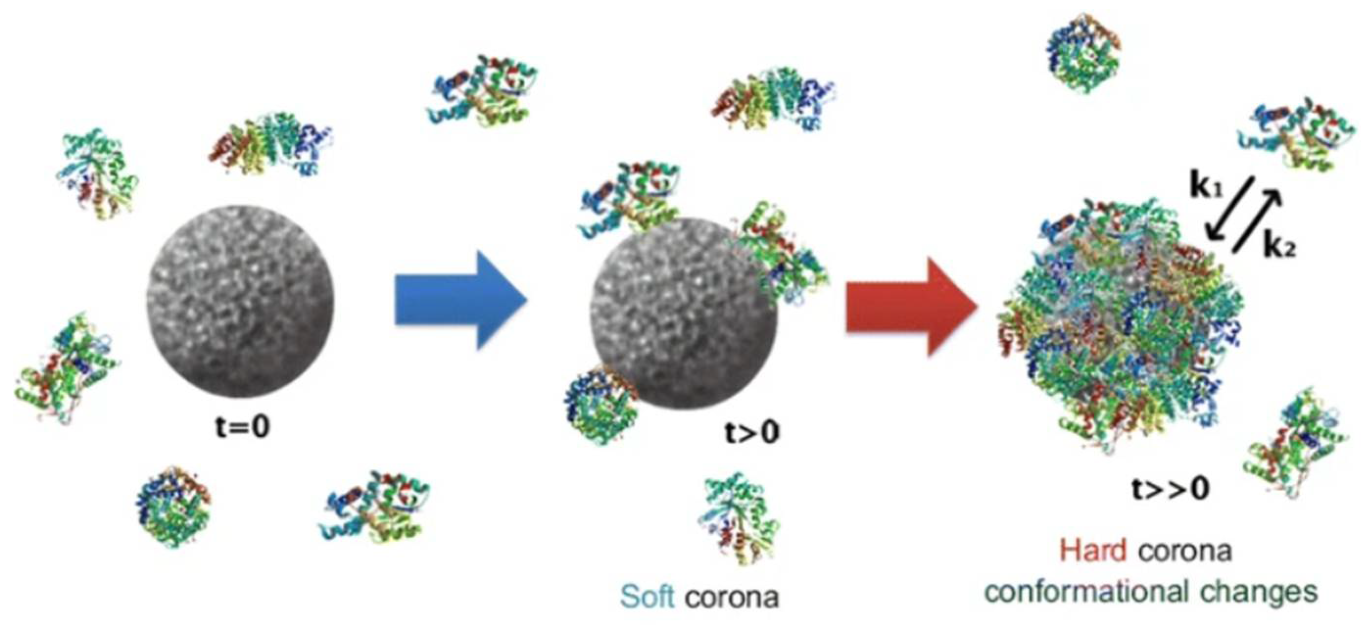
Concerning the protein corona, it is a dynamic and integral layer of biomolecules, predominantly proteins, that adsorb onto the surface of silver nanoparticles (AgNPs) when they enter biological fluids. This adsorption process rapidly transforms the nanoparticle’s synthetic surface into a new bio-identity, which governs how the nanoparticle interacts with cells, tissues, and organs. For silver nanoparticles specifically, the protein corona modulates not only biological identity but also chemical transformations on the nanoparticle surface. Recent studies have shown that the protein corona influences formation of silver sulfide (Ag2S) layers on AgNP surfaces, altering nanoparticle stability and toxicity. For example, the hard corona acts as a scaffold for sulfidation near the silver core, while the soft corona can reduce the formation of such sulfide by facilitating the removal of silver ions from the particle’s surface. Figure 18 shows the formation of protein corona around nanoparticles and includes a table comparing properties of soft and hard corona.
The relative abundance and composition of corona proteins are affected by environmental factors like serum concentration, which directly impacts nanoparticle aggregation, dissolution, and reactivity [128,129,130].
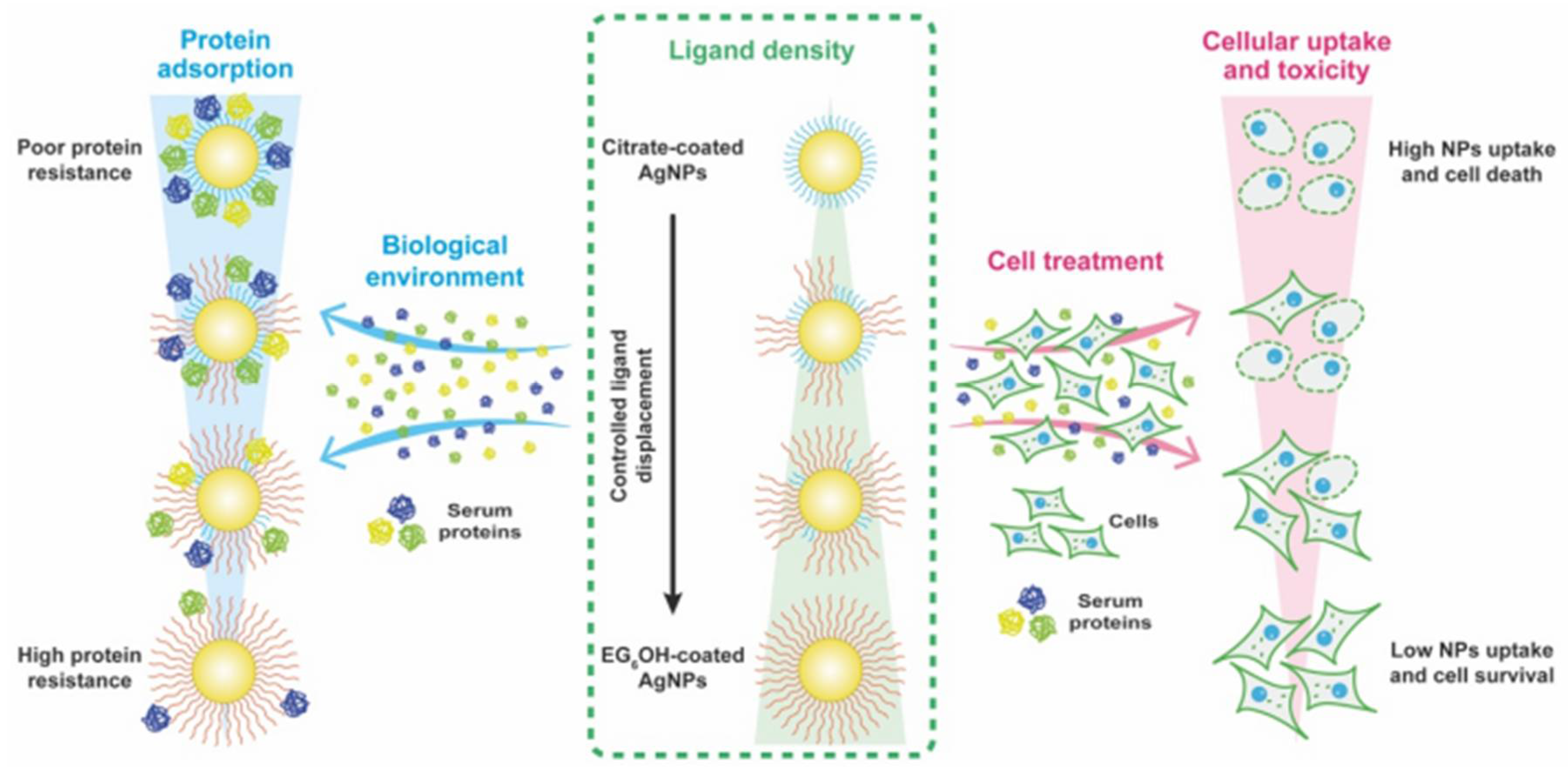
In Figure 19 illustrates the interplay between protein adsorption, ligand density on silver nanoparticles (AgNPs), and how these factors influence cellular uptake and toxicity. The figure highlights how variations in nanoparticle surface properties affect protein binding from the biological environment, which subsequently modulates nanoparticle interaction with cells, ultimately impacting toxicity levels.
On the left, it shows how protein adsorption varies with nanoparticle surface properties, highlighting that nanoparticles with poor protein resistance accumulate more serum proteins, while those with high protein resistance bind fewer proteins.
In the center panel, ligand density on the nanoparticle surface is depicted, comparing citrate-coated AgNPs (which have lower ligand density) to EG3OH-coated AgNPs (with higher ligand density). Increased ligand density controls the displacement of surface ligands by serum proteins, affecting nanoparticle stability.
On the right, the effects on cell treatment and cellular uptake are shown. Nanoparticles with low protein resistance and lower ligand density experience higher cellular uptake and toxicity, resulting in increased cell death. Conversely, nanoparticles with high protein resistance and higher ligand density have reduced cellular uptake and better cell survival.
This diagram emphasizes how surface chemistry modulates protein corona formation, which in turn influences biological responses such as cellular uptake and cytotoxicity.
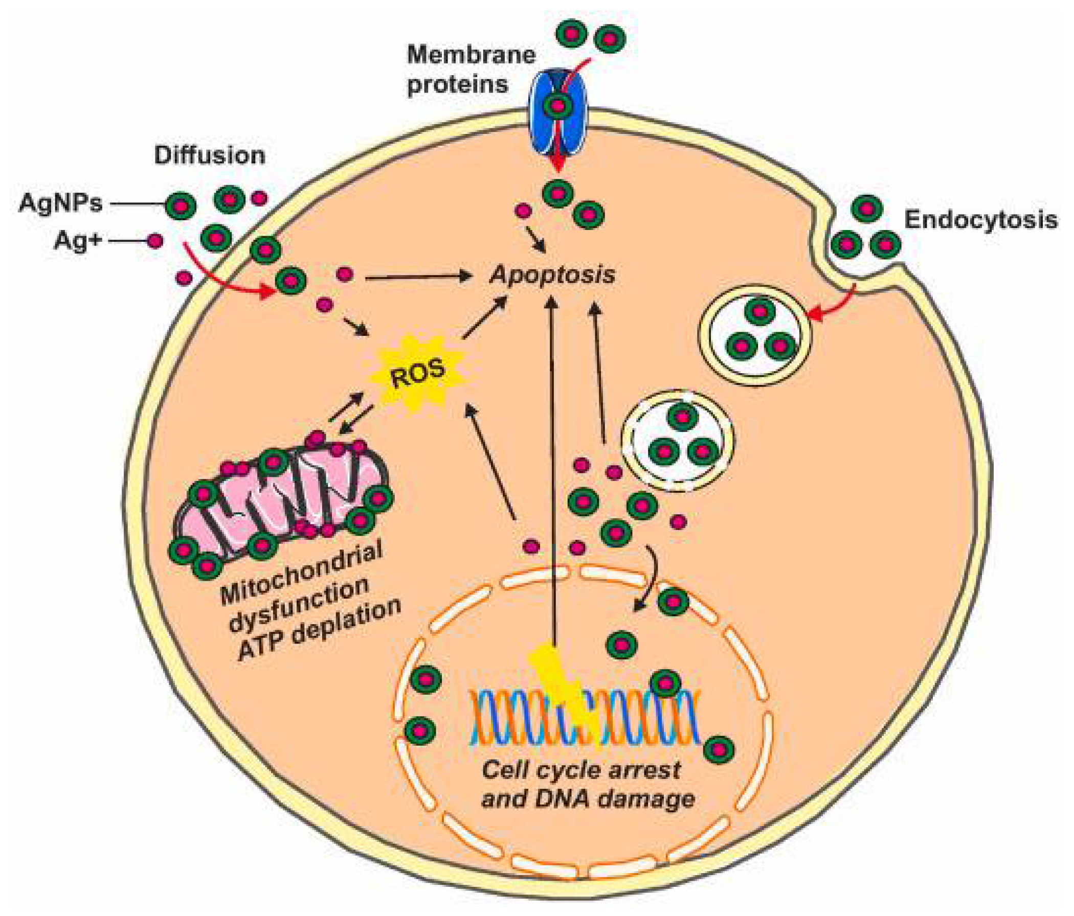
In Figure 20 illustrates the exposure to silver nanoparticles that can reduce cell viability through multiple pathways. Studies suggest one such pathway involves triggering apoptosis by activating genes related to the apoptotic process. Another pathway may involve the generation and buildup of reactive oxygen species (ROS) inside the cell, caused by disruptions to the mitochondrial electron transport chain, which can result in DNA damage. Silver nanoparticles also cause significant oxidative damage to cellular membranes and organelles such as the nucleus and lysosomes, which can trigger cell death through processes like apoptosis or necrosis.
Extensive research, including studies on both cells and animal models, is crucial to fully understand the toxicity of silver nanoparticles and how they affect bodily functions and tissue structures [42,43]. While many lab-based experiments have explored the harmful effects of AgNPs, the data so far are not consistent. Various cell studies have shown that nanosilver can be toxic to rat liver cells, nerve cells, mouse stem cells, and human lung cells [125]. For example, research on rat hepatocytes revealed that even minimal exposure to silver nanoparticles triggered considerable depletion of glutathione, increased oxidative stress, and impaired mitochondrial function [119].
Moreover, in vivo studies focusing on biocompatibility and biodistribution have demonstrated that AgNPs can cause structural and functional alterations in critical organs. Inhaled silver nanoparticles tend to accumulate in the alveoli of the lungs, potentially leading to lung damage, and can also cause notable changes in the nervous system along with liver and kidney tissues [125]. Hence, further in vivo investigations are necessary to properly evaluate the toxicity of silver nanoparticles before definitive conclusions can be made [119].

4. Conclusions

Silver nanoparticles (AgNPs) are promising candidates for biomedical applications due to their unique physicochemical properties and bioactivity. However, a major scientific challenge lies in the incomplete and often superficial characterization of these nanoparticles, which hinders a comprehensive understanding of their behavior and performance in biological settings. Conventional complementary techniques such as UV–Vis spectroscopy, FTIR, TEM, SEM, DLS, XRD, and zeta potential measurements provide important but limited information. While these methods can assess parameters like particle size, morphology, crystallinity, surface charge, and optical properties, they fall short of fully capturing the complexity of AgNPs. For instance, the critical surface chemistry aspects—including the nature of functional groups, their interaction with biomolecules, and dynamic biological transformations—are often underexplored or ambiguous. The in vivo behavior of silver nanoparticles (AgNPs) is complex and not yet fully understood, particularly regarding their aging processes such as oxidation, corrosion, and ion release. These mechanisms are fundamental to assessing both their toxicity and therapeutic efficacy. As nanoparticles age within biological systems, their chemical states and surface properties can change, influencing their interactions with cells and tissues.
Given the potential toxicity of AgNPs, careful evaluation and management of their release and exposure are essential. Proper regulation ensures that the benefits of nanotechnology can be harnessed safely, minimizing ecological impacts and protecting human health. Additional research is crucial to develop a comprehensive understanding of how AgNPs evolve over time in vivo, which will inform guidelines for their safe use in medical and industrial applications [133].
Advanced surface-sensitive and nanoscale characterization techniques offer more profound insights. X-ray Photoelectron Spectroscopy (XPS) enables precise identification of elemental composition and oxidation states at the nanoparticle surface, revealing the chemical environment and potential modification sites. Atomic Force Microscopy (AFM) provides nanoscale topographical and mechanical information, crucial for understanding surface roughness, agglomeration, and interactions with cells or proteins under physiologically relevant conditions. Auger Electron Spectroscopy (AES) and Low-Energy Ion Scattering (LEIS) supply complementary atomic-level surface compositional data, indispensable for studying thin coatings or surface modifications. These advanced methods are essential for deciphering the true ‘bio-identity’ of AgNPs but are limited by high cost, sample preparation complexity, and technical expertise demands, restricting widespread implementation.
This incomplete characterization landscape significantly limits the reproducibility of experiments and comparability across studies, complicating risk assessment and safety. Without comprehensive physicochemical knowledge, the biological effects of AgNPs remain ill-defined, undermining confidence in their biomedical application and regulatory evaluation. Therefore, establishing integrated characterization workflows that combine traditional bulk techniques with advanced surface-sensitive and in situ analyses is imperative. Such holistic approaches will better capture the multifaceted physicochemical landscape of silver nanoparticles, facilitating safer and more effective biomedical use while addressing outstanding questions about their long-term stability, bio-interactions, and toxicity.

Author Contributions

Conceptualization, A.N., Τ.C., N.M. & A.A.; methodology, A.N., T.C. and A.A.; writing—original draft preparation, A.N., T.C. and A.A.; writing—review and editing, A.N., T.C. and A.A.; visualization, A.N., T.C. and A.A.; supervision, A.A., N.M.; project administration, A.N. and A.A.; funding acquisition, N.M. and A.A. All authors have read and agreed to the published version of the manuscript.

Funding

This research received no external funding.

Data Availability Statement

No new data were created or analyzed in this study. Data sharing is not applicable to this article.

Conflicts of Interest

The authors declare no conflicts of interest.

References

  1. Silvestry-Rodriguez, N.; Sicairos-Ruelas, E.E.; Gerba, C.P.; Bright, K.R. Silver as a Disinfectant. Rev. Environ. Contam. Toxicol. 2007, 191, 23–45. [Google Scholar] [CrossRef]
  2. Sim, W.; Barnard, R.T.; Blaskovich, M.A.T.; Ziora, Z.M. Antimicrobial Silver in Medicinal and Consumer Applications: A Patent Review of the Past Decade (2007–2017). Antibiotics 2018, 7, 93. [Google Scholar] [CrossRef]
  3. Abbas, R.; Luo, J.; Qi, X.; Naz, A.; Khan, I.A.; Liu, H.; Yu, S.; Wei, J. Silver nanoparticles: Synthesis, structure, properties and applications. Nanomaterials 2024, 14, 1425. [Google Scholar] [CrossRef]
  4. Khan, I.; Saeed, K.; Khan, I. Nanoparticles: Properties, applications and toxicities. Arab. J. Chem. 2019, 12, 908–931. [Google Scholar] [CrossRef]
  5. Wang, L.; Kafshgari, M.H.; Meunier, M. Optical Properties and Applications of Plasmonic-Metal Nanoparticles. Adv. Funct. Mater. 2020, 30, 2005400. [Google Scholar] [CrossRef]
  6. Abbasi, E.; Milani, M.; Fekri Aval, S.; Kouhi, M.; Akbarzadeh, A.; Tayefi Nasrabadi, H.; Nikasa, P.; Joo, S.W.; Hanifehpour, Y.; Nejati-Koshki, K. Silver nanoparticles: Synthesis methods, bio-applications and properties. Crit. Rev. Microbiol. 2016, 42, 173–180. [Google Scholar] [CrossRef] [PubMed]
  7. Patil, R.B.; Chougale, A.D. Analytical Methods for the Identification and Characterization of Silver Nanoparticles: A Brief Review. Mater. Today Proc. 2021, 47, 5520–5532. [Google Scholar] [CrossRef]
  8. Ambreen, T.; Kim, M.-H. Influence of Particle Size on the Effective Thermal Conductivity of Nanofluids: A Critical Review. Appl. Energy 2020, 264, 114684. [Google Scholar] [CrossRef]
  9. Syafiuddin, A.; Salmiati Salim, M.R.; Beng Hong Kueh, A.; Hadibarata, T.; Nur, H. A Review of Silver Nanoparticles: Research Trends, Global Consumption, Synthesis, Properties, and Future Challenges. J. Chin. Chem. Soc. 2017, 64, 732–756. [Google Scholar] [CrossRef]
  10. Joudeh, N.; Linke, D. Nanoparticle Classification, Physicochemical Properties, Characterization, and Applications: A Comprehensive Review for Biologists. J. Nanobiotechnol. 2022, 20, 262. [Google Scholar] [CrossRef]
  11. Kandasamy, G.; Paramasivam, S.; Varudharajan, G.; Dhairiyasamy, R. Optimization of Silver Nanoparticle-Enhanced Nanofluids for Improved Thermal Management in Solar Thermal Collectors. Matéria 2024, 29, e20240363. [Google Scholar] [CrossRef]
  12. Lin, L.; Peng, X.; Voirin, E.; Donnio, B.; Rastei, M.V.; Vileno, B.; Gallani, J.L. Influence of the crystallinity of silver nanoparticles on their magnetic properties. Helv. Chim. Acta 2023, 106, e202200165. [Google Scholar]
  13. Koopmans, R.J.; Aggeli, A. Nanobiotechnology—Quo Vadis? Curr. Opin. Microbiol. 2010, 13, 327–334. [Google Scholar] [CrossRef] [PubMed]
  14. Stojkovska, J.; Zvicer, J.; Obradovic, B. Preclinical functional characterization methods of nanocomposite hydrogels containing silver nanoparticles for biomedical applications. Appl. Microbiol. Biotechnol. 2020, 104, 4643–4658. [Google Scholar] [CrossRef]
  15. Tassieri, M.; Waigh, T.A.; Trinick, J.; Aggeli, A.; Evans, R.M.L. Analysis of the Linear Viscoelasticity of Polyelectrolytes by Magnetic Microrheometry—Pulsed Creep Experiments and the One Particle Response. J. Rheol. 2010, 54, 117–131. [Google Scholar] [CrossRef]
  16. Zhang, X.F.; Liu, Z.G.; Shen, W.; Gurunathan, S. Silver Nanoparticles: Synthesis, Characterization, Properties, Applications, and Therapeutic Approaches. Int. J. Mol. Sci. 2016, 17, 1534. [Google Scholar] [CrossRef]
  17. Quevedo, A.C.; Guggenheim, Ε.B.; Sophie, M.; Adams, J.; Lofts, S.; Kwak, M.; Lee, T.G.; Johnston, C.; Wagner, S.; Holbrook, T.R.; et al. UV-Vis Spectroscopic Characterization of Nanomaterials in Aqueous Media. J. Vis. Exp. 2021, 176, e61764. [Google Scholar] [CrossRef] [PubMed]
  18. Grand, J.; Auguié, B.; Le Ru, E.C. Combined Extinction and Absorption UV–Visible Spectroscopy as a Method for Revealing Shape Imperfections of Metallic Nanoparticles. Anal. Chem. 2019, 91, 14639–14648. [Google Scholar] [CrossRef] [PubMed]
  19. Duman, H.; Bölükbaşı, K.; Yıldırım, M. Silver nanoparticles: A comprehensive review of synthesis methods and chemical and physical properties. Nanomaterials 2024, 14, 1527. [Google Scholar] [CrossRef]
  20. Pryshchepa, O.; Pomastowski, P.; Buszewski, B. Silver Nanoparticles: Synthesis, Investigation Techniques, and Properties. Adv. Colloid Interface Sci. 2020, 284, 102246. [Google Scholar] [CrossRef]
  21. Mourdikoudis, S.; Pallares, R.M.; Thanh, N.T.K. Characterization Techniques for Nanoparticles: Comparison and Complementarity upon Studying Nanoparticle Properties. Nanoscale 2018, 10, 12871–12934. [Google Scholar] [CrossRef] [PubMed]
  22. Venkatesham, M.; Ayodhya, D.; Madhusudhan, A.; Veera Babu, N.; Veerabhadram, G. A Novel Green One-Step Synthesis of Silver Nanoparticles Using Chitosan: Catalytic Activity and Antimicrobial Studies. Appl. Nanosci. 2012, 4, 113–119. [Google Scholar] [CrossRef]
  23. Dawadi, S.; Katuwal, S.; Gupta, A.; Lamichhane, U.; Thapa, R.; Jaisi, S.; Lamichhane, G.; Bhattarai, D.P.; Parajuli, N. Current Research on Silver Nanoparticles: Synthesis, Characterization, and Applications. J. Nanomater. 2021, 2021, e6687290. [Google Scholar] [CrossRef]
  24. Braidy, N.; Béchu, A.; de Souza Terra, J.C.; Patience, G.S. Experimental methods in chemical engineering: Transmission electron microscopy—TEM. Can. J. Chem. Eng. 2020, 98, 628–641. [Google Scholar] [CrossRef]
  25. Lara, H.H.; Romero-Urbina, D.G.; Pierce, C.; Lopez-Ribot, J.L.; Arellano-Jiménez, M.J.; Jose-Yacaman, M. Effect of silver nanoparticles on Candida albicans biofilms: An ultrastructural study. J. Nanobiotechnology 2015, 13, 91. [Google Scholar] [CrossRef] [PubMed]
  26. Bendersky, L.A.; Gayle, F.W. Electron Diffraction Using Transmission Electron Microscopy. J. Res. Natl. Inst. Stand. Technol. 2001, 106, 997. [Google Scholar] [CrossRef]
  27. Malatesta, M. Transmission Electron Microscopy as a Powerful Tool to Investigate the Interaction of Nanoparticles with Subcellular Structures. Int. J. Mol. Sci. 2021, 22, 12789. [Google Scholar] [CrossRef]
  28. Das, D.; Ghosh, R.; Mandal, P. Biogenic Synthesis of Silver Nanoparticles Using S1 Genotype of Morus Alba Leaf Extract: Characterization, Antimicrobial and Antioxidant Potential Assessment. SN Appl. Sci. 2019, 1, 498. [Google Scholar] [CrossRef]
  29. Jia, Z.; Li, J.; Gao, L.; Yang, D.; Kanaev, A. Dynamic light scattering: A powerful tool for in situ nanoparticle sizing. Colloids Interfaces 2023, 7, 15. [Google Scholar] [CrossRef]
  30. Sharma, S.; Shukla, P.; Misra, A.; Mishr, P.R.; Ohshima, H.; Makino, K. (Eds.) Colloid and Interface Science in Pharmaceutical Research and Development; Elsevier: Amsterdam, The Netherlands, 2014; pp. 149–172. [Google Scholar]
  31. Khan, S.S.; Mukherjee, A.; Chandrasekaran, N. Studies on interaction of colloidal silver nanoparticles (SNPs) with five different bacterial species. Colloids Surf. B Biointerfaces 2011, 87, 129–138. [Google Scholar] [CrossRef]
  32. Bagus, P.S.; Freund, H.-J. X-ray photoelectron spectroscopy as a useful tool to study surfaces and model systems for heterogeneous catalysts: A review and perspective. Surf. Sci. 2024, 745, 122471. [Google Scholar] [CrossRef]
  33. Huang, Z.; Jiang, H.; Liu, P.; Sun, J.; Guo, D.; Shan, J.; Gu, N. Continuous synthesis of size-tunable silver nanoparticles by a green electrolysis method and multi-electrode design for high yield. J. Mater. Chem. A 2015, 3, 1925–1929. [Google Scholar] [CrossRef]
  34. Prieto, E.I.; Kiat, A.A. The antimicrobial action of silver nanoparticles on Escherichia coli as revealed by atomic force microscopy. Philipp. Sci. Lett. 2017, 10, 123–129. [Google Scholar]
  35. Caniglia, G.; Valavanis, D.; Tezcan, G.; Magiera, J.; Barth, H.; Bansmann, J.; Kranz, C.; Unwin, P.R. Antimicrobial effects of silver nanoparticle-microspots on the mechanical properties of single bacteria. Analyst 2024, 149, 2637–2646. [Google Scholar] [CrossRef]
  36. Daphedar, A.; Taranath, T.C. Characterization and cytotoxic effect of biogenic silver nanoparticles on mitotic chromosomes of Drimia polyantha (Blatt. & McCann) Stearn. Toxicol. Rep. 2018, 5, 910–918. [Google Scholar] [CrossRef]
  37. Gilstrap, R.A., Jr. A Colloidal Nanoparticle Form of Indium Tin Oxide: System Development and Characterization. PhD Dissertation, Georgia Institute of Technology, Atlanta, GA, USA, 2009. [Google Scholar]
  38. Rievaj, M.; Culková, E.; Šandorová, D.; Durdiak, J.; Bellová, R.; Tomčík, P. A review of analytical techniques for the determination and separation of silver ions and its nanoparticles. Nanomaterials 2023, 13, 1262. [Google Scholar] [CrossRef]
  39. Laborda, F.; Abad-Álvaro, I.; Jiménez, M.S.; Bolea, E. Catching particles by atomic spectrometry: Benefits and limitations of single particle-inductively coupled plasma mass spectrometry. Spectrochim. Acta Part B At. Spectrosc. 2023, 199, 106570. [Google Scholar] [CrossRef]
  40. Li, Q.; Wang, Z.; Mo, J.; Zhang, G.; Chen, Y.; Huang, C. Imaging Gold Nanoparticles in Mouse Liver by Laser Ablation Inductively Coupled Plasma Mass Spectrometry. Sci. Rep. 2017, 7, 2965. [Google Scholar] [CrossRef]
  41. Nyika, J.; Onyari, E.; Dinka, M.O.; Mishra, S.B. A comparison of reproducibility of inductively coupled spectrometric techniques in soil metal analyses. Air Soil Water Res. 2019, 12, 1178622119869002. [Google Scholar]
  42. Almatroudi, A. Silver Nanoparticles: Synthesis, Characterisation and Biomedical Applications. Open Life Sci. 2020, 15, 819–839. [Google Scholar] [CrossRef] [PubMed]
  43. Almatroudi, A. Potential Toxic Effects and Clinical Implications of Silver Nanoparticles in Tissue Engineering: A Comprehensive Review. Toxicol. Rep. 2020, 7, 1039–1053. [Google Scholar]
  44. Balout, H.; Tarrat, N.; Puibasset, J.; Ispas, S.; Bonafos, C.; Benoit, M. Density functional theory study of the spontaneous formation of covalent bonds at the silver/silica interface in silver nanoparticles embedded in SiO2: Implications for Ag+ release. ACS Appl. Nano Mater. 2019, 2, 5179–5189. [Google Scholar] [CrossRef]
  45. Khan, J.; Naseem, I.; Bibi, S.; Ahmad, S.; Altaf, F.; Hafeez, Μ.; Almoneef, M.M.; Ahmad, K. Green Synthesis of Silver Nanoparticles (Ag-NPs) Using Debregeasia Salicifolia for Biological Applications. Materials 2022, 16, 129. [Google Scholar] [CrossRef]
  46. Zozulya, A.; Zyubin, A.; Samusev, I. A review for DFT in chemical mechanism of SERS studies. R. Soc. Open Sci. 2025, 12, 242000. [Google Scholar] [CrossRef]
  47. Bachler, G.; von Goetz, N.; Hungerbühler, K. A physiologically based pharmacokinetic model for ionic silver and silver nanoparticles. Int. J. Nanomed. 2013, 3365–3382. [Google Scholar] [CrossRef]
  48. Lasmi, F.; Hamitouche, H.; Laribi-Habchi, H.; Benguerba, Y.; Chafai, N. Silver Nanoparticles (AgNPs), Methods of Synthesis, Characterization, and Their Application: A Review. Plasmonics 2025, 1–34. [Google Scholar] [CrossRef]
  49. Malik, M.; Iqbal, M.A.; Iqbal, Y.; Malik, M.; Bakhsh, S.; Irfan, S.; Ahmad, R.; Pham, P.V. Biosynthesis of silver nanoparticles for biomedical applications: A mini review. Inorg. Chem. Commun. 2022, 145, 109980. [Google Scholar] [CrossRef]
  50. Kolahalam, L.A.; Viswanath, I.K.; Diwakar, B.S.; Govindh, B.; Reddy, V.; Murthy, Y.L.N. Review on nanomaterials: Synthesis and applications. Mater. Today Proc. 2019, 18, 2182–2190. [Google Scholar] [CrossRef]
  51. Bronzino, J.D.; Enderle, J.D.; Blanchard, S.M. Introduction to Biomedical Engineering; Elsevier Academic Press: Amsterdam, The Netherlands, 2005. [Google Scholar]
  52. Barua, N.; Buragohain, A.K. Therapeutic Potential of Silver Nanoparticles (AgNPs) as an Antimycobacterial Agent: A Comprehensive Review. Antibiotics 2024, 13, 1106. [Google Scholar] [CrossRef] [PubMed]
  53. Wahab, M.A.; Li, L.; Li, H.; Abdala, A. Silver nanoparticle-based nanocomposites for combating infectious pathogens: Recent advances and future prospects. Nanomaterials 2021, 11, 581. [Google Scholar] [CrossRef] [PubMed]
  54. More, P.R.; Pandit, S.; Filippis, A.D.; Franci, G.; Mijakovic, I.; Galdiero, M. Silver nanoparticles: Bactericidal and mechanistic approach against drug resistant pathogens. Microorganisms 2023, 11, 369. [Google Scholar] [CrossRef]
  55. Franci, G.; Falanga, A.; Galdiero, S.; Palomba, L.; Rai, M.; Morelli, G.; Galdiero, M. Silver nanoparticles as potential antibacterial agents. Molecules 2015, 20, 8856–8874. [Google Scholar] [CrossRef]
  56. Solomon, M.; Holban, A.M.; Bălăceanu-Gurău, B.; Dițu, L.M.; Alberts, A.; Grumezescu, A.M.; Manolescu, L.S.C.; Mihai, M.M. Silver Nanoparticles Functionalized with Polymeric Substances to Reduce the Growth of Planktonic and Biofilm Opportunistic Pathogens. Int. J. Mol. Sci. 2025, 26, 3930. [Google Scholar] [CrossRef]
  57. Campora, S.; Ghersi, G. Recent Developments and Applications of Smart Nanoparticles in Biomedicine. Nanotechnol. Rev. 2022, 11, 2595–2631. [Google Scholar] [CrossRef]
  58. Agrawal, S.; Bhatt, M.; Rai, S.K.; Bhatt, A.; Dangwal, P.; Agrawal, P.K. Silver nanoparticles and its potential applications: A review. J. Pharmacogn. Phytochem. 2018, 7, 930–937. [Google Scholar]
  59. Bruna, T.; Maldonado-Bravo, F.; Jara, P.; Caro, N. Silver nanoparticles and their antibacterial applications. Int. J. Mol. Sci. 2021, 22, 7202. [Google Scholar] [CrossRef] [PubMed]
  60. Zhang, J.; Wang, F.; Yalamarty, S.S.K.; Filipczak, N.; Jin, Y.; Li, X. Nano Silver-Induced Toxicity and Associated Mechanisms. Int. J. Nanomed. 2022, 17, 1851–1864. [Google Scholar] [CrossRef] [PubMed]
  61. Naganthran, A.; Verasoundarapandian, G.; Khalid, F.E.; Masarudin, M.J.; Zulkharnain, A.; Nawawi, N.M.; Karim, M.; Che, A.; Che, A.; Ahmad, S. Synthesis, Characterization and Biomedical Application of Silver Nanoparticles. Materials 2022, 15, 427. [Google Scholar] [CrossRef]
  62. Bamal, D.; Singh, A.; Chaudhary, G.; Kumar, M.; Singh, M.; Rani, N.; Mundlia, P.; Sehrawat, A.R. Silver Nanoparticles Biosynthesis, Characterization, Antimicrobial Activities, Applications, Cytotoxicity and Safety Issues: An Updated Review. Nanomaterials 2021, 11, 2086. [Google Scholar] [CrossRef] [PubMed]
  63. Crisan, C.M.; Mocan, T.; Manolea, M.; Lasca, L.I.; Tăbăran, F.A.; Mocan, L. Review on silver nanoparticles as a novel class of antibacterial solutions. Appl. Sci. 2021, 11, 1120. [Google Scholar] [CrossRef]
  64. Aguilar-Garay, R.; Lopez, M.; Sanchez, F.; Garcia, M. A comprehensive review of silver and gold nanoparticles as effective antibacterial agents. Pharmaceuticals 2024, 17, 1134. [Google Scholar] [CrossRef]
  65. Pulit-Prociak, J.; Banach, M. Silver Nanoparticles—A Material of the Future…? Open Chem. 2016, 14, 76–91. [Google Scholar] [CrossRef]
  66. Liao, C.; Li, Y.; Tjong, S.C. Bactericidal and Cytotoxic Properties of Silver Nanoparticles. Int. J. Mol. Sci. 2019, 20, 449. [Google Scholar] [CrossRef]
  67. Tamboli, D.P.; Lee, D.S. Mechanistic Antimicrobial Approach of Extracellularly Synthesized Silver Nanoparticles against Gram Positive and Gram Negative Bacteria. J. Hazard. Mater. 2013, 260, 878–884. [Google Scholar] [CrossRef]
  68. Mikhailova, E.O. Green Silver Nanoparticles: An Antibacterial Mechanism. Antibiotics 2024, 14, 5. [Google Scholar] [CrossRef]
  69. Vishwanath, R.; Negi, B. Conventional and Green Methods of Synthesis of Silver Nanoparticles and Their Antimicrobial Properties. Curr. Res. Green Sustain. Chem. 2021, 4, 100205. [Google Scholar] [CrossRef]
  70. Nguyen, N.P.U.; Dang, N.T.; Doan, L.; Nguyen, T.T.H. Synthesis of Silver Nanoparticles: From Conventional to ‘Modern’ Methods—A Review. Processes 2023, 11, 2617. [Google Scholar] [CrossRef]
  71. Chicea, D.; Ciocan, I.; Watz, C.; Socol, M. Comparative synthesis of silver nanoparticles: Evaluation of chemical reduction procedures, AFM and DLS size analysis. Materials 2023, 16, 5244. [Google Scholar] [CrossRef] [PubMed]
  72. Chakraborty, M.; Jain, S.; Rani, V. Nanotechnology: Emerging Tool for Diagnostics and Therapeutics. Appl. Biochem. Biotechnol. 2011, 165, 1178–1187. [Google Scholar] [CrossRef]
  73. Tang, S.; Zheng, J. Antibacterial Activity of Silver Nanoparticles: Structural Effects. Adv. Healthc. Mater. 2018, 7, 1701503. [Google Scholar] [CrossRef]
  74. Sampath, G.; Chen, Y.Y.; Rameshkumar, N.; Krishnan, M.; Nagarajan, K.; Shyu, D.J. Biologically synthesized silver nanoparticles and their diverse applications. Nanomaterials 2022, 12, 3126. [Google Scholar] [CrossRef]
  75. Menichetti, A.; Mavridi-Printezi, A.; Mordini, D.; Montalti, M. Effect of size, shape and surface functionalization on the antibacterial activity of silver nanoparticles. J. Funct. Biomater. 2023, 14, 244. [Google Scholar] [CrossRef]
  76. Javed, R.; Zia, M.; Naz, S.; Aisida, S.O.; Ain, N.U.; Ao, Q. Role of capping agents in the application of nanoparticles in biomedicine and environmental remediation: Recent trends and future prospects. J. Nanobiotechnology 2020, 18, 172. [Google Scholar] [CrossRef] [PubMed]
  77. Raj, S.; Trivedi, R.; Soni, V. Biogenic Synthesis of Silver Nanoparticles, Characterization and Their Applications—A Review. Surfaces 2021, 5, 67–90. [Google Scholar] [CrossRef]
  78. Qamer, S.; Romli, M.H.; Che-Hamzah, F.; Misni, N.; Joseph, N.M.; Al-Haj, N.A.; Amin-Nordin, S. Systematic review on biosynthesis of silver nanoparticles and antibacterial activities: Application and theoretical perspectives. Molecules 2021, 26, 5057. [Google Scholar] [CrossRef] [PubMed]
  79. Pedroso-Santana, S.; Fleitas-Salazar, N. The Use of Capping Agents in the Stabilization and Functionalization of Metallic Nanoparticles for Biomedical Applications. Part. Part. Syst. Charact. 2022, 40, 2200146. [Google Scholar] [CrossRef]
  80. Gibała, A.; Żeliszewska, P.; Gosiewski, T.; Krawczyk, A.; Duraczyńska, D.; Szaleniec, J.; Szaleniec, M.; Oćwieja, M. Antibacterial and antifungal properties of silver nanoparticles—Effect of a surface-stabilizing agent. Biomolecules 2021, 11, 1481. [Google Scholar] [CrossRef]
  81. Salleh, A.; Naomi, R.; Utami, N.D.; Mohammad, A.W.; Mahmoudi, E.; Mustafa, N.; Fauzi, M.B. The potential of silver nanoparticles for antiviral and antibacterial applications: A mechanism of action. Nanomaterials 2020, 10, 1566. [Google Scholar] [CrossRef]
  82. Xu, L.; Wang, Y.Y.; Huang, J.; Chen, C.Y.; Wang, Z.X.; Xie, H. Silver Nanoparticles: Synthesis, Medical Applications and Biosafety. Theranostics 2020, 10, 8996–9031. [Google Scholar] [CrossRef]
  83. Hussain, F.S.; Abro, N.Q.; Ahmed, N.; Memon, S.Q.; Memon, N. Nano-antivirals: A comprehensive review. Front. Nanotechnol. 2022, 4, 1064615. [Google Scholar] [CrossRef]
  84. Goel, M.; Sharma, A.; Sharma, B. Recent Advances in Biogenic Silver Nanoparticles for Their Biomedical Applications. Sustain. Chem. 2023, 4, 61–94. [Google Scholar] [CrossRef]
  85. Aldakheel, F.M.; Sayed, M.M.E.; Mohsen, D.; Fagir, M.H.; El Dein, D.K. Green synthesis of silver nanoparticles loaded hydrogel for wound healing; systematic review. Gels 2023, 9, 530. [Google Scholar] [CrossRef]
  86. Yang, Y.; Wang, P.; Zhang, G.; He, S.; Xu, B. Inorganic-nanomaterial-composited hydrogel dressings for wound healing. J. Compos. Sci. 2024, 8, 46. [Google Scholar] [CrossRef]
  87. Paladini, F.; Di Franco, C.; Panico, A.; Scamarcio, G.; Sannino, A.; Pollini, M. In vitro assessment of the antibacterial potential of silver nano-coatings on cotton gauzes for prevention of wound infections. Materials 2016, 9, 411. [Google Scholar] [CrossRef] [PubMed]
  88. Wang, J.; Li, J.; Guo, G.; Wang, Q.; Tang, J.; Zhao, Y.; Qin, H.; Wahafu, T.; Shen, H.; Liu, X.; et al. Silver-nanoparticles-modified biomaterial surface resistant to staphylococcus: New insight into the antimicrobial action of silver. Sci. Rep. 2016, 6, 32699. [Google Scholar] [CrossRef] [PubMed]
  89. Yin, I.X.; Zhang, J.; Zhao, I.S.; Mei, M.L.; Li, Q.; Chu, C.H. The Antibacterial Mechanism of Silver Nanoparticles and Its Application in Dentistry. Int. J. Nanomed. 2020, 15, 2555–2562. [Google Scholar] [CrossRef]
  90. Nicolae-Maranciuc, A.; Chicea, D.; Chicea, L.M. Ag Nanoparticles for Biomedical Applications—Synthesis and Characterization—A Review. Int. J. Mol. Sci. 2022, 23, 5778. [Google Scholar] [CrossRef]
  91. Singh, M.; Thakur, V.; Kumar, V.; Raj, M.; Gupta, S.; Devi, N.; Upadhyay, S.K.; Macho, M.; Banerjee, A.; Ewe, D.; et al. Silver nanoparticles and its mechanistic insight for chronic wound healing: Review on recent progress. Molecules 2022, 27, 5587. [Google Scholar] [CrossRef]
  92. Vijapur, L.S.; Shalavadi, M.; Desai, A.R.; Hiremath, J.N.; Gudigennavar, A.S.; Shidramshettar, S.L.; Hiremath, S.R.; Peram, M.R.; Kittur, B.S. Wound healing potential of green synthesized silver nanoparticles of Glycyrrhiza glabra linn root extract: A preclinical study. J. Trace Elem. Miner. 2025, 11, 100214. [Google Scholar] [CrossRef]
  93. Kumar, S.S.D.; Rajendran, N.K.; Houreld, N.N.; Abrahamse, H. Recent Advances on Silver Nanoparticle and Biopolymer-Based Biomaterials for Wound Healing Applications. Int. J. Biol. Macromol. 2018, 115, 165–175. [Google Scholar] [CrossRef] [PubMed]
  94. Guo, Y.; Hou, X.; Fan, S.; Jin, C. Research Status of Silver Nanoparticles for Dental Applications. Inorganics 2025, 13, 168. [Google Scholar] [CrossRef]
  95. Lee, J.J.; Niu, M.; Shakir, Z.; Hwang, G.; Chung, C.H.; Wolff, M.S.; Zheng, Z.; Li, C. Usage of Silver Nanoparticles in Orthodontic Bonding Reagents. J. Funct. Biomater. 2025, 16, 244. [Google Scholar] [CrossRef] [PubMed]
  96. Fernandez, C.C.; Sokolonski, A.R.; Fonseca, M.S.; Stanisic, D.; Araújo, D.B.; Azevedo, V.; Portela, R.D.; Tasic, L. Applications of silver nanoparticles in dentistry: Advances and technological innovation. Int. J. Mol. Sci. 2021, 22, 2485. [Google Scholar] [CrossRef]
  97. Rivera-Cortés, M.A.; Niño-Martínez, N.; Ruiz, F.; Félix-Sicairos, B.K.; Martínez-Castañón, G.A. Evaluation of Deformation and Antibacterial Properties of Dental Alginates Mixed with Silver Nanoparticles. Materials 2025, 18, 2069. [Google Scholar] [CrossRef]
  98. Corrêa, J.M.; Mori, M.; Sanches, H.L.; Cruz, A.D.D.; Poiate, E., Jr.; Poiate, I.A.V.P. Silver nanoparticles in dental biomaterials. Int. J. Biomater. 2015, 2015, 485275. [Google Scholar] [CrossRef]
  99. Butrón Téllez Girón, C.; Hernández Sierra, J.F.; DeAlba-Montero, I.; Urbano Peña, M.D.L.A.; Ruiz, F. Therapeutic use of silver nanoparticles in the prevention and arrest of dental caries. Bioinorg. Chem. Appl. 2020, 2020, 8882930. [Google Scholar] [CrossRef]
  100. Mazumder, M.M.U.; Sukul, A.; Saha, S.K.; Chowdhury, A.A.; Mamun, Y. A comprehensive in vitro biological investigation of metal complexes of tolfenamic acid. Alex. J. Med. 2018, 54, 23–26. [Google Scholar] [CrossRef]
  101. Lampé, I.; Beke, D.; Biri, S.; Csarnovics, I.; Csik, A.; Dombrádi, Z.; Hajdu, P.; Hegedűs, V.; Rácz, R.; Varga, I.; et al. Investigation of silver nanoparticles on titanium surface created by ion implantation technology. Int. J. Nanomed. 2019, 14, 4709–4721. [Google Scholar] [CrossRef]
  102. Zhou, Y.; Zhang, Y.; Li, X.; Zhou, Y.; Kuang, Y.; Sang, S.; Zhang, J.; Wang, Y. Silver nanoparticle coatings enhance bone density and formation without adverse tissue effects. Mater. Sci. Eng. C 2017, 75, 1089–1101. [Google Scholar]
  103. Kumar, A.; Vishakha, D.A.; Jeet, K.; Kumar, S. A review on silver nanoparticles focusing on applications in biomedical sector. Int. J. Pharmaceut. Sci. Drug Res. 2022, 8, 57–63. [Google Scholar]
  104. Saravanan, S.; Kumar, V.; Rajendran, A.; Thomas, S. A review on silver nanoparticles-based biomaterials for bone tissue engineering. Int. J. Chem. 2024, 16, 123–135. [Google Scholar]
  105. Aubert, T.; Buffière, J.; Montalent, P.; Bérenguer, F.; Boisse-Laporte, C.; Granier, C. Nanoparticles in tissue engineering: Applications, challenges, and prospects. J. Biomed. Nanotechnol. 2018, 14, 765–781. [Google Scholar]
  106. Kargozar, S.; Nazarnezhad, S.; Kermani, F.; Baino, F. Nanomaterials and scaffolds for tissue engineering and regenerative medicine. In Nanomaterials and Nanotechnology in Medicine; John Wiley & Sons: Hoboken, NJ, USA, 2022; pp. 279–302. [Google Scholar]
  107. Massironi, A.; Rizzo, F.; Gallo, A.; Giacomini, M.; Biffi, S.; Del Giudice, A. Development and characterization of highly stable silver nanoparticles as novel potential antimicrobial agents for wound healing hydrogels. Int. J. Mol. Sci. 2022, 23, 2161. [Google Scholar] [CrossRef]
  108. Przekora, A. A Concise Review on Tissue Engineered Artificial Skin Grafts for Chronic Wound Treatment: Can We Reconstruct Functional Skin Tissue In Vitro? Cells 2020, 9, 1622. [Google Scholar] [CrossRef]
  109. Chen, M. Application of Nanotechnology in Biomedicine: From Diagnosis to Therapy. J. Bioanal. Biomed. 2023, 15, 381. [Google Scholar]
  110. Takáč, P., Jr.; Michalková, R.; Čižmáriková, M.; Bedlovičová, Z.; Balážová, Ľ.; Laca Megyesi, Š.; Mačeková, Z.; Takáčová, G.; Moreno-Borrallo, A.; Ruiz-Hernandez, E.; et al. Do We Know Enough About the Safety Profile of Silver Nanoparticles in Oncology? A Focus on Novel Methods and Approaches. Int. J. Mol. Sci. 2025, 26, 5344. [Google Scholar] [CrossRef]
  111. Gherasim, O.; Puiu, R.A.; Bîrcă, A.C.; Burdușel, A.C.; Grumezescu, A.M. An updated review on silver nanoparticles in biomedicine. Nanomaterials 2020, 10, 2318. [Google Scholar] [CrossRef]
  112. Saikia, N. Functional Inorganic Biomaterials for Molecular Sensing and Biomedical Applications. Inorganics 2025, 13, 260. [Google Scholar] [CrossRef]
  113. Beck, F.; Loessl, M.; Baeumner, A.J. Signaling strategies of silver nanoparticles in optical and electrochemical biosensors: Considering their potential for the point-of-care. Microchim. Acta 2023, 190, 91. [Google Scholar] [CrossRef]
  114. Bouafia, A.; Gheribi, R.; Haddad, I.; Al-Douri, Y. The recent progress on silver nanoparticles: Synthesis and electronic applications. Nanomaterials 2021, 11, 2318. [Google Scholar] [CrossRef] [PubMed]
  115. Cao, W.; Tang, G.; Zhao, C.; Li, J.; Wang, Y. Silver nanoparticles: Comprehensive insights into bioanalytical and biomedical applications. ACS Omega 2025, 10, 1234–1245. [Google Scholar]
  116. Li, Y.; Zhang, L.; Chen, G.; Zhou, Q.; Wang, H. Constructing Fe-MOF-derived Z-scheme photocatalysts with enhanced charge transport: Nanointerface and carbon sheath synergistic effect. ACS Appl. Mater. Interfaces 2020, 12, 25494–25502. [Google Scholar] [CrossRef] [PubMed]
  117. Sarău, O.S.; Moacă, E.A.; Semenescu, A.D.; Dumitru, R.; Jijie, A.R.; Poenaru, M.; Dehelean, C.A.; Chevereşan, A. Physicochemical and toxicological screening of silver nanoparticle biosynthesis from Punica granatum peel extract. Inorganics 2024, 12, 160. [Google Scholar] [CrossRef]
  118. Laib, I.; Bououdina, M.; Bensaoula, A.; Benmansour, S. Tailoring innovative silver nanoparticles for modern medicine: The importance of size and shape control and functional modifications. Mater. Today Bio. 2025, 33, 102071. [Google Scholar]
  119. Prabhu, S.; Poulose, E.K. Silver Nanoparticles: Mechanism of Antimicrobial Action, Synthesis, Medical Applications, and Toxicity Effects. Int. Nano Lett. 2012, 2, 32. [Google Scholar] [CrossRef]
  120. Ferdous, Z.; Nemmar, A. Health Impact of Silver Nanoparticles: A Review of the Biodistribution and Toxicity Following Various Routes of Exposure. Int. J. Mol. Sci. 2020, 21, 2375. [Google Scholar] [CrossRef] [PubMed]
  121. Noga, M.; Milan, J.; Frydrych, A.; Jurowski, K. Toxicological aspects, safety assessment, and green toxicology of silver nanoparticles (AgNPs)—Critical review: State of the art. Int. J. Mol. Sci. 2023, 24, 5133. [Google Scholar] [CrossRef]
  122. Lee, H.A.; Kim, M.H.; Oh, J.H.; Park, E.H.; Lee, S.Y. Rhizome of Anemarrhena asphodeloides as mediators of the eco-friendly synthesis of silver and gold spherical, face-centred cubic nanocrystals and its anti-migratory and cytotoxic potential in normal and cancer cell lines. Artif. Cells Nanomed. Biotechnol. 2018, 46 (Suppl. S2), 285–294. [Google Scholar]
  123. Selvan, D.A.; Karuppusamy, I.; Veerappan, A.; Priya, C. Garlic, green tea and turmeric extracts-mediated green synthesis of silver nanoparticles: Phytochemical, antioxidant and in vitro cytotoxicity studies. J. Photochem. Photobiol. B Biol. 2018, 180, 243–252. [Google Scholar]
  124. Plotnikov, E.V.; Tretayakova, M.S.; Garibo-Ruíz, D.; Rodríguez-Hernández, A.G.; Pestryakov, A.N.; Toledano-Magaña, Y.; Bogdanchikova, N. A comparative study of cancer cells susceptibility to silver nanoparticles produced by electron beam. Pharmaceutics 2023, 15, 962. [Google Scholar] [CrossRef]
  125. Burdușel, A.C.; Gherasim, O.; Grumezescu, A.M.; Mogoantă, L.; Ficai, A.; Andronescu, E. Biomedical applications of silver nanoparticles: An up-to-date overview. Nanomaterials 2018, 8, 681. [Google Scholar] [CrossRef]
  126. Liu, J.; Sonshine, D.A.; Shervani, S.; Hurt, R.H. Controlled release of biologically active silver from nanosilver surfaces. ACS Nano 2010, 4, 6903–6913. [Google Scholar] [CrossRef]
  127. Shannahan, J.H.; Layne, J.; Gardella, J.; Ke, P.C. Formation of a protein corona on silver nanoparticles mediates cellular toxicity via scavenger receptors. Toxicol. Sci. 2015, 143, 136–146. [Google Scholar] [CrossRef] [PubMed]
  128. Breznica, P.; Rozafa, K.; Arlinda, D. A review of the current understanding of nanoparticles protein corona composition. Med. Pharm. Rep. 2020, 93, 342. [Google Scholar] [CrossRef]
  129. Miclăuş, T.; O’Connell, D.; Melinte, G.; Ivan, V.; Cristian, L.; Dinescu, G. Dynamic protein coronas revealed as a modulator of silver nanoparticle sulphidation in vitro. Nat. Commun. 2016, 7, 11770. [Google Scholar] [CrossRef] [PubMed]
  130. Eigenheer, R.; Hutchison, J.E.; Hess, K.L. Silver nanoparticle protein corona composition compared across engineered particle properties and environmentally relevant reaction conditions. Environ. Sci. Nano 2014, 1, 238–247. [Google Scholar] [CrossRef]
  131. Barbalinardo, M.; Bertacchini, J.; Bergamini, L.; Magarò, M.S.; Ortolani, L.; Sanson, A.; Palumbo, C.; Cavallini, M.; Gentili, D. Surface properties modulate protein corona formation and determine cellular uptake and cytotoxicity of silver nanoparticles. Nanoscale 2021, 13, 14119–14129. [Google Scholar] [CrossRef]
  132. Nikzamir, M.; Akbarzadeh, A.; Panahi, Y. An Overview on Nanoparticles Used in Biomedicine and Their Cytotoxicity. J. Drug Deliv. Sci. Technol. 2021, 61, 102316. [Google Scholar] [CrossRef]
  133. Panyala, N.R.; Peña-Méndez, E.M.; Havel, J. Silver or Silver Nanoparticles: A Hazardous Threat to the Environment and Human Health? J. Appl. Biomed. 2008, 6, 117–129. [Google Scholar] [CrossRef]
Figure 1. Graphic illustration of the resonance effect of surface plasmon SPR in nanoparticles [4].
Inorganics 13 00341 g001
Figure 2. Extinction spectra (absorption + scattering) of aqueous suspensions of Ag nanospheres with different diameters ranging between 10 and 100 nm (top) and Ag nanoplates with a diameter of 50–150 nm (bottom) [8].
Inorganics 13 00341 g002
Figure 3. Schematic illustration that conveys the way that effective biomedical applications of silver nanoparticles rely on the full, comprehensive physicochemical characterization of the nanoparticles, highlighting gaps in knowledge in this respect: The complete characterization of silver nanoparticles is depicted as a puzzle; currently the application of advanced complementary techniques allows us to add only some pieces in this puzzle; several parts of the puzzle are still missing, waiting for novel approaches in silver nanoparticle characterization to be introduced, in order to complete the full picture.
Inorganics 13 00341 g003
Figure 4. UV–Vis absorption spectrum of AgNPs synthesized from Cyprus rotundas [7].
Inorganics 13 00341 g004
Figure 5. FTIR spectra of AgNPs stabilized with chitosan and of pure chitosan [22].
Inorganics 13 00341 g005
Figure 6. XRD pattern of silver nanoparticles [7].
Inorganics 13 00341 g006
Figure 7. Electron microscopy study of silver nanoparticles: (A) SEM analysis, (B) EDX spectrum of silver nanoparticles [7].
Inorganics 13 00341 g007
Figure 8. (a) TEM micrograph of spherical silver nanoparticles (b) Log-normal size distribution histogram with average AgNPs size ≈ 1 nm [25].
Inorganics 13 00341 g008
Figure 9. SAED image of silver nanoparticles [28].
Inorganics 13 00341 g009
Figure 10. The impact of particle size on variations in scattered light intensity (a,b), the relevant autocorrelation function (c,d) and particle size distribution (e,f) [29].
Inorganics 13 00341 g010
Figure 11. Zeta potential of a negatively charged particle [30].
Inorganics 13 00341 g011
Figure 12. (top) XPS working principal and (bottom a,b) shows the XPS spectrum of the surface of silver nanoparticles capping with PVP [32,33].
Inorganics 13 00341 g012
Figure 13. Atomic force microscopy images of biogenic AgNPs using leaf extract of G. floribunda (a) 2D image (b) 3D image of AgNPs, and (c) Height of AgNPs [36].
Inorganics 13 00341 g013
Figure 14. Schematic diagram of main components of ICP-MS [37].
Inorganics 13 00341 g014
Figure 15. Schematic diagram of main components of ICP-OES [41].
Inorganics 13 00341 g015
Figure 16. Schematic overview of biomedical applications of AgNPs [61].
Inorganics 13 00341 g016
Figure 17. Silver nanoparticle exposure on human body and related cellular toxicity [122,123].
Inorganics 13 00341 g017
Figure 18. Schematic illustration of the formation of soft and hard corona-protein on silver nanoparticles, where k is the dissociation constant [127].
Inorganics 13 00341 g018
Figure 19. Schematic relationship between the protein adsorption, ligand density and cellular uptake and toxicity [131].
Inorganics 13 00341 g019
Figure 20. Proposed mechanisms of cytotoxicity of AgNPs [132].
Inorganics 13 00341 g020
Disclaimer/Publisher’s Note: The statements, opinions and data contained in all publications are solely those of the individual author(s) and contributor(s) and not of MDPI and/or the editor(s). MDPI and/or the editor(s) disclaim responsibility for any injury to people or property resulting from any ideas, methods, instructions or products referred to in the content.