1. Introduction
Nowadays there is a need to develop novel highly efficient luminescent compounds that can be used in a wide range of practical applications such as the active layer in organic light-emitting diodes (OLEDs) [
1,
2,
3,
4,
5,
6,
7,
8], biomarkers [
9], dyes for cell visualization [
10,
11,
12] and sensor materials [
13,
14,
15]. Coordination compounds of trivalent lanthanide ions with a partially filled 4f shell (from Ce to Yb) are among the prominent classes of materials that can be used in all these applications. They are characterized by narrow spectral lines of luminescence and a high theoretical quantum yield of emission [
16,
17]. The absorbance and luminescence of pure Ln
3+ ions originates from 4f–4f transitions, and since these transitions are forbidden by Laport rules [
18], the extinction coefficients are rather small, which causes weak light absorption even in resonant bands.
In contrast to pure Ln
3+ ions, the luminescence of complexes usually occurs as a result of energy transfer from a ligand to an ion, the so called antenna effect. The energy transfer efficiency is determined by the energies of the first excited singlet S
1 and triplet T
1 states of a ligand and by the energy gap between the triplet level of the ligand, and the resonant levels of the Ln
3+ ion are yet another crucial factor determining the efficiency [
19]. There is another crucial factor determining processes of vibrational and rotational relaxation in a complex [
20]. According to Latva’s rule, the energy gap must be within the range of 1800–2500 cm
−1 and 2500–4500 cm
−1 for Eu
3+ and Tb
3+, respectively, to sensitize the luminescence of Eu
3+ and Tb
3+ ions efficiently [
21]. However, this rule is not a mandatory condition and is violated in many cases [
22]. All the parameters described above can be tuned by modifying the chemical structure of ligands.
In order to create new highly efficient coordination compounds of Ln
3+, we need to understand how a minor change in the chemical structure of the ligand molecule would affect the energies of excited states in the ligand, and hence, the photophysical properties of the entire coordination compound. It is known that incorporation of atoms containing π electrons on the outer orbital causes an additional overlap of the π-orbitals and therefore increases the π-conjugation of the entire molecule [
23,
24]. In this case, the absorption spectrum shifts to longer wavelengths [
24], thus reducing the energy of the singlet and triplet states [
23].
Yuichi Kitagawa et al. studied Eu
3+ β diketonates bearing various aromatic moieties [
24]. It was found that the energy of the first triplet state decreased monotonically from the phenyl-substituted ligand (T
1 = 29470 cm
−1) to the anthracenyl-substituted one (T
1 = 14920 cm
−1), while for naphthalene-containing diketone, the T
1 energy was found to be 21250 cm
−1. The length of π conjugation is inversely proportional to the energy of the first excited triplet state of the ligand. S. Carlotto et al. [
23] studied the relationship between the π-conjugation and the excited states of coordination compounds with the central Gd
3+ ion. In this study, all the ligands were characterized by the presence of the same thienyl moiety, and polycyclic aromatic hydrocarbon substituents with various lengths of π-conjugation as the other moiety for the diketone fragment. An increase in the length of π-conjugation by a factor of three leads to a decrease in the first excited triplet state of the molecule by about 6000 cm
−1. However, in this paper, no studies on the dependence between the quantum yield and the length of π-conjugation were presented. Despite the fact that free ligands have been studied in detail and the values of the excited energy states have been established for them, it is impossible to predict the energies for ligands inside Ln
3+ coordination compounds. It is caused due to the fact that the strong electric field of the central ion is superimposed on the ligand located in the coordination sphere. In addition, currently it is poorly understood how π-conjugation in organic ligands affects the luminescence efficiency of Ln
3+ coordination compounds with these ligands.
The main goal of the present paper was to find out the relationship between the degree of π-conjugation in the aromatic moiety of symmetrically substituted diaroylmethanes and the photoluminescence efficiency of the central ion in the corresponding Eu
3+ complexes. The trivalent Eu
3+ ion was chosen as such an ion since it has a sufficiently low resonant level (17300 cm
−1) and therefore can be well sensitized by ligands with various triplet state energies. In addition, this ion has efficient radiative relaxation [
24] that leads to bright and easily observable luminescence. In total, six compounds of the [Eu(L)
3(H
2O)], [Eu(L)
3(phen)] and [Gd(L)
3(H
2O)] structural types were synthesized and studied. The studied compounds differ in the type of the main ligand (DBM or BiNaph) and the type of ancillary ligands (H
2O or phen). Since the naphthalene moiety has a longer π-conjugation length than the phenyl, the ligand containing napthyl moieties has lower excited state energies.
2. Results
2.1. Synthesis of the HL2 Ligand and the 2h, 2Gd and 2p complexes
Unlike 1,3-diphenyl-l,3-propanedione (dibenzoylmethane, HDBM,
HL1), which is readily available from the common commercial sources, 1,3-di(naphthalen-1-yl)propane-1,3-dione (HBiNaph,
HL2) is not so easily accessible. It was prepared previously by Claisen condensation of methyl 1-naphthoate (1) with 1-(naphthalen-1-yl)ethan-1-one (1-acetylnaphthalene, 2) in the presence of various bases, namely t-BuOK in benzene [
25], NaH in 1,4-dioxane [
26], NaNH
2 in Et
2O [
27] and NaH in THF [
28]. The latter method seems to be the most practical. Unfortunately, we could not reproduce it fully because the key procedure of the aforementioned method, i.e., the isolation of the solid Na salt of the diketone, failed. No measurable amount of the salt was obtained due to its high solubility in THF. Therefore, the isolation procedure was modified and an additional stage with precipitation of copper chelate 4 was added (
Scheme 1). The chelate then was decomposed by HCl / EtOAc in a biphase system as we reported previously [
29]. The resulting diketone was purified by recrystallization from hot heptane, and no chromatographic purification was necessary to obtain the pure diketone.
The known complexes
1h [Eu(L
1)
3(H
2O)],
1p [Eu(L
1)
3(phen)] and
1Gd [Gd(L
1)
3(H
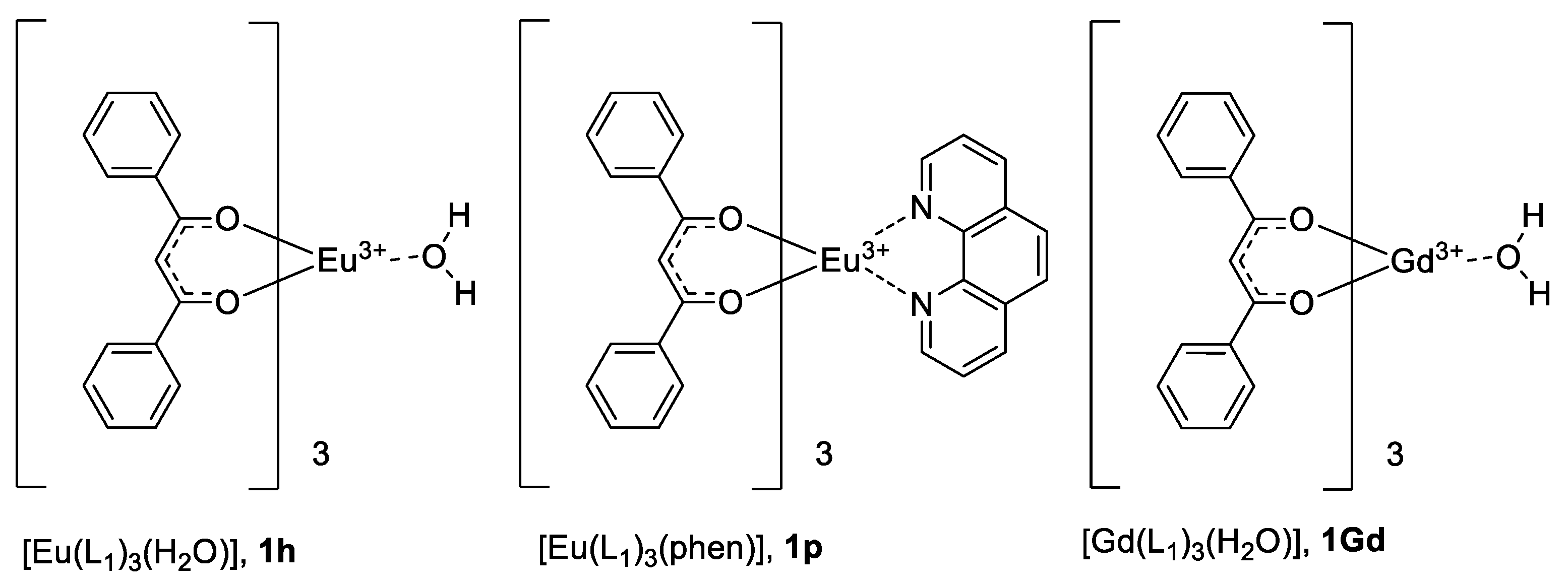
2O)] (
Figure 1) were prepared as described in the literature, namely, by the reaction of hydrated LnCl
3 with dibenzoylmethane (and additionally, with 1,10-phenanathroline in the case of complex
1p) and NaOH in EtOH solution.
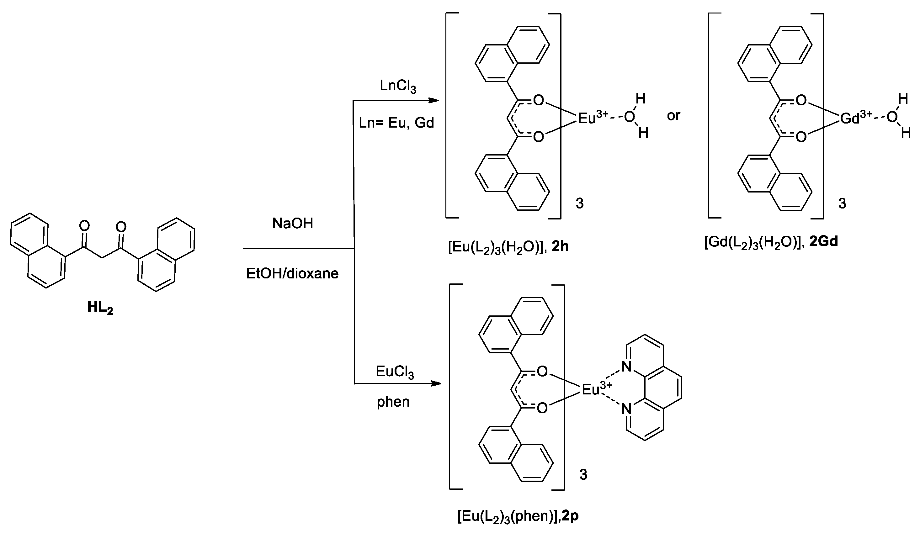
Due to the different solubility of both sodium salts of 1,3-di(naphthalen-1-yl)propane-1,3-dione and the corresponding hydrated complexes, known methods were not optimal for the preparation of the new compounds
2h,
2p and
2Gd. We developed a modified approach for such compounds, in which the EtOH-dioxane mixture was found to be the best solvent for the synthesis (
Scheme 2).
The chemical composition of all the three complexes was determined by elemental analysis. Only microcrystalline powders were obtained from solutions, but they became amorphous very quickly upon drying. Our multiple attempts to grow crystals suitable for X-ray analysis by various methods failed.
Although tris-complexes with a different number of coordinated water molecules are reported in the literature for dibenzoylmethane (e.g., [Ln(L
1)
3(H
2O)
2], [
30]), no rigorous data confirming this composition was provided. The mass-spectral data for hydrated complexes
2h and
2Gd are dubious due to the absence of molecular or other reliably interpreted ions. NMR data are also unreliable due to instability of complexes in solutions. Taking the elemental analysis data and literature data concerning the composition of similar complexes into account [
31,
32,
33], we identified hydrated complexes
2h and
2Gd as monohydrates [Eu(L
2)
3(H
2O)] and [Gd(L
2)
3(H
2O)], respectively. The composition of complex
2p was decisively identified as [Eu(L
2)
3(phen)].
2.2. X-ray Structure of the HL2 Ligand
The chemical composition of the
HL2 ligand was confirmed by single-crystal X-ray diffraction. According to the data, the asymmetric unit of the crystal structure contains one molecule of the ligand (
Figure 2). Due to the low quality of the crystal data, the coordinates of the H1 hydrogen atom could not be found from the electron-difference map and were calculated geometrically. The angle between the two naphthyl substituents is 128.8o. Analysis of crystal packing did not reveal strong intermolecular interactions other than the intramolecular hydrogen bond O1-H1…O2 (the O1…O2 distance is 2.466 Å) and weak π…π interactions.
2.3. FTIR Spectroscopy
For the 2h and 2Gd complexes, we observed a broad and very strong band at about 3700–3100 cm−1, which corresponds to the O-H vibrations. All spectra contain the very strong band at 1550 cm−1, corresponding to the C = O bond stretch. The bands for the C = C and C = N ring vibrations appear in the region 1650–1400 cm−1. Also, there are several intensive peaks at 1495 and 1510 cm−1 which we consider as C-C stretching vibrations in the naphthalene part of ligand. In the “fingerprint area”, there is a remarkable sharp band (C-H twisting bending vibration bands) at 780 cm−1.
2h IR spectrum: 3650–3120 (broad, vs); 3045 (vw); 1548 (vs); 1510 (s); 1494 (m); 1406 (m); 1381 (s); 1293 (w); 1250 (w); 1124 (vw); 1061 (vw); 873 (vw); 780 (m); 600 (w); 574 (w); 512 (w).
2p IR spectrum: 3650–3200 (broad, w); 3045 (w); 2854 (vw); 1548 (vs); 1510 (s); 1494 (m); 1406 (m); 1381 (s); 1337 (w); 1293 (w); 1250 (w); 1124 (vw); 1061 (vw); 872 (vw); 780 (m); 600 (w); 574 (w); 512 (w).
2Gd IR spectrum: 3700–3100 (broad, vs); 3047 (vw); 2972 (vw); 1623 (m); 1550 (s); 1511 (s); 1495 (m); 1405 (m); 1383 (s); 1295 (vw); 1249 (vw); 1120 (vw); 1061 (vw); 1045 (vw); 871 (vw); 779 (s); 600 (w); 574 (w); 514 (w).
2.4. Optical Absorption
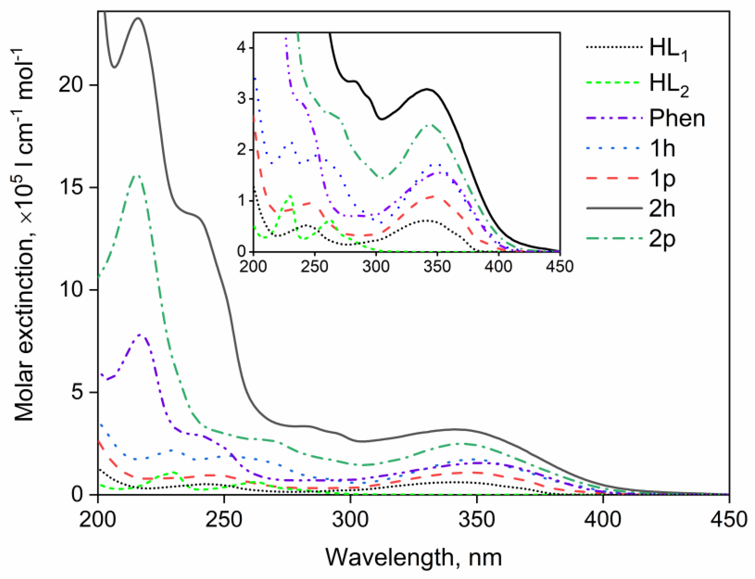
The optical absorption spectra for solutions of complexes
1h–
2p and free ligands
HL1,
HL2 and phen in MeCN are shown in
Figure 3.
All the complexes intensely absorb light in the spectral range from 200 to 450 nm. The absorption band with a maximum at 350 nm is associated with π-π* electronic transitions of the 1,3 diketone moiety in all the compounds [
34]. For complexes
1h and
1p, the maximum in the 250 nm band is associated with the absorption of the phenyl moiety [
35]. The bands with peaks at 225 nm and 250 nm for complex
2h are caused by the absorption of the naphthyl moiety, while for complex
2p, the feature in the 250 nm band is not observed. It is worthy of note that there are no characteristic absorption bands of the phen ligand in the spectra of complexes
1p and
2p. It can be seen from the spectra of the complexes that the additional ligand, phen, does not cause changes in the electronic structure of the
L1 and
L2 ligands.
The own absorption of the Eu3+ ion with characteristic values of molar extinction ϵ of the order of dozens mol−1cm−1l is not detected due to the strong ligand absorption (ϵ~105 mol−1cm−1l).
The energy of the first excited singlet state S
1 was estimated from the high-energy edge of the absorption band as a crossing point of fluorescence and absorption spectra [
36] for all the complexes and free ligands. The energy values obtained by this method are presented in
Table 1.
Substitution of more conjugated naphthalene fragments instead of phenyl rings in the diketone ligand expectedly lowers the energy of the S
1 state for the free ligands. However, the S
1 energy of the
HL1 ligand decreases by 1000 cm
−1 due to complexation, whereas the S
1 energy of
HL2 unexpectedly increases by 700 cm
−1 (see
Table 1 and
Figure S1). Binding of the Eu
3+ complexes studied with the auxiliary ligand phen does not result in significant changes in the S
1 energy.
2.5. Luminescence and Excitation Spectra
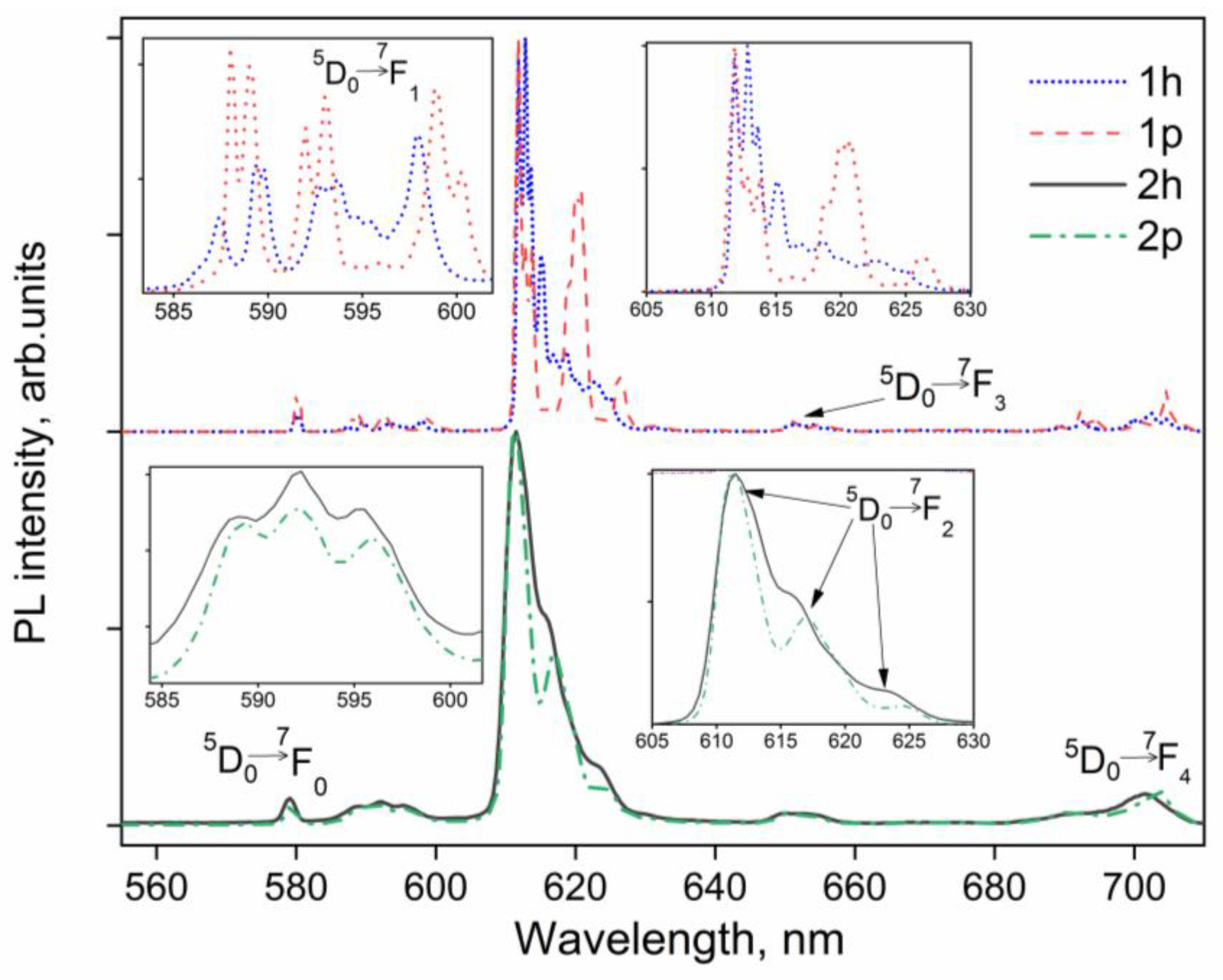
The luminescence spectra of the
1h–
2p complexes in crystalline phase under CW excitation at 360 nm at room temperature are shown in
Figure 4. All the spectra exhibit well-pronounced red emission corresponding to Eu
3+ ion luminescence. The observed narrow bands peaked at 580 nm, 590 nm, 615 nm, 650 nm and 710 nm originate from the
5D
0–
7F
J (J = 0–4) electronic
f-
f* transitions in the ion [
33].
Since
f-orbitals are shielded by the ligand electric field, the
7F
J state of the ion is degenerated (Stark effect). Therefore, the emission bands are split into several components whose maxima are listed in
Table 2. It is known that the symmetry of the nearest coordination environment of the central ion determines the crystal field energy matrix and associated B
kq coefficients of the electric field applied to the ion [
37,
38]. The symmetry of the Eu
3+ ion’s polyhedron is significantly higher in compound
1h than in
1p since the spectral band corresponding to the hypersensitive transition
5D
0→
7F
2 (611 nm) has an additional Stark component. In contrast, replacement of water molecules (compound
2h) with a phen ligand (compound
2p) does not change the splitting of the
5D
0→
7F
2 band, suggesting that the coordination polyhedrons of these compounds have the same spatial symmetry group. The
5D
0→
7F
0 transition exhibits a sole peak in all the spectra. It indicates the presence of a single center of symmetry of the Eu
3+ ion [
20].
The luminescence spectra of complexes 1h, 1p and 2h, 2p manifest no evident fluorescence or/and phosphorescence of ligands L1 and L2, respectively. It indicates that a relatively efficient energy transfer from the ligands to the central ion exists.
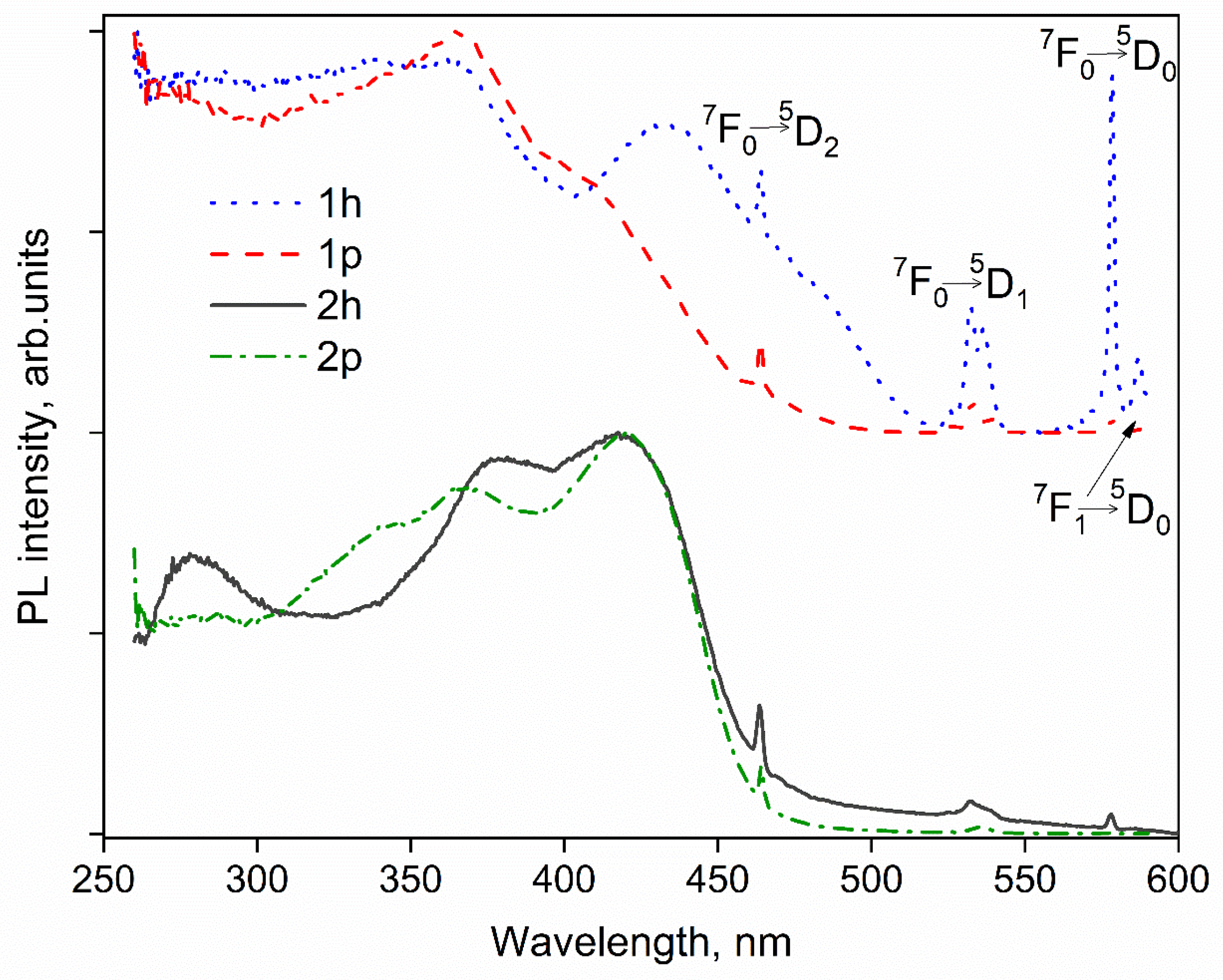
Upon an excitation in a wide spectral range from 260 to 450 nm, all the studied complexes exhibit bright red emissions visible to the naked eye. The optical excitation spectra of crystal complexes
1h–
2p recorded at room temperature are shown in
Figure 5.
The excitation spectra of complexes
2h and
2p qualitatively resemble each other. The local maxima in the bands at 370 and 375 nm for
2h and
2p, respectively, are associated with luminescence excitation through π-π* electronic transitions in the 1,3 diketone moiety. Moreover, a wide shoulder is observed in the range from 380 to 500 nm due to luminescence excitation through charge-transfer states in the spectra of both complexes. The excitation spectra of complexes
1h and
1p are identical in the range from 250 to 420 nm; however, a wide band associated with charge transfer states is observed for complex
1h in the range from 420 to 520 nm, while this band for complex
1p is practically not observed. Thus, it can be assumed that incorporation of a phen ligand into the
1p complex results in the blocking of states with charge transfer. Complex
1p is efficiently sensitized by a mixed-ligand combination of L
1 and phen. Narrow bands are also observed at wavelengths of 464 (
7F
0-
5D
2), 532 (
7F
0-
5D
1), 578 (
7F
0-
5D
0), and 587 nm (
7F
1-
5D
0) [
16] in the excitation spectra of complexes
1h–
2p.
2.6. PL Decays and Quantum Yields
The photoluminescence decays of crystalline complexes
1h–
2p in the visible spectral range at room temperature are shown in
Figure 6.
In the simplest case, the relaxation of excited states is explained by a two-level model (relaxation of exactly one excited state). It obeys the mono-exponential law:
where the lifetime τobs observed in the experiment is defined as:
where
krad and
knrad are the rate constants of radiative and nonradiative relaxation, respectively.
The luminescence decays for complexes
1h and
1p can be fitted well by a mono-exponential function, while the decays for
2h and
2p are bi-exponential. The calculated attenuation times of luminescence (τ
obs) are in the range from 3 to 414 μs. The maximum lifetime (414 μs) was obtained for complex
1p excited through the ligand environment (360 nm); the lifetimes for complexes
1h–
2p are presented in
Table 3.
The internal quantum yield can be estimated by the following formula
Since the rate constant of the magnetic-dipole transition
5D
0-
7F
1 in Eu
3+, k
MD = 14.65 s
−1, does not depend on the electric field induced by the ligand, the value of k
rad can be determined by the relationship:
where n is the refractive index and I
tot/I
MD is the ratio of the total integral luminescence intensity to the integral intensity of the magnetic dipole transition. The k
nrad constant was estimated using a simple dependence:
Comprising the calculated k
rad and the observed attenuation time τ
obs measured with resonant excitation of Eu
3+ ion at a wavelength of 532 nm. The calculated internal quantum yields Φ
Ln, the total quantum yields (Φ), measured by the absolute method, and the sensitization coefficients η are presented in
Table 4.
2.7. Energy of the First Excited Triplet Level
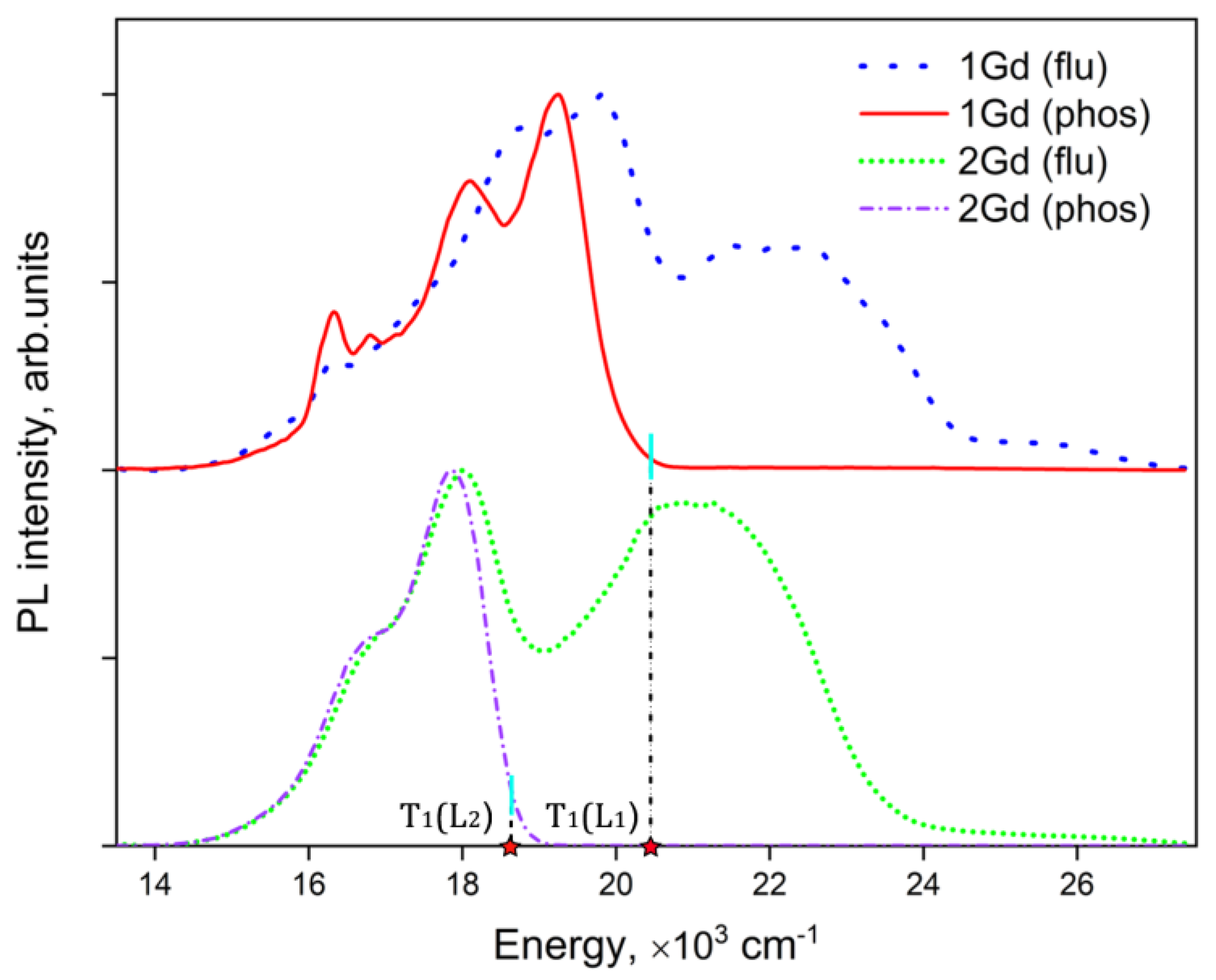
To determine the energy of the first excited triplet level (T1) of ligands L1 and L2, complexes of the Gd3+ ion (complexes 1Gd and 2Gd, respectively) with the same ligand environments as complexes 1h and 2h were synthesized. The triplet level positions of the majority of organic ligands, in particular those of the 1,3 diketone class, are below the resonant level of Gd3+. It prevents direct energy transfer from the ligand to the ion. Therefore, the T1 state relaxes radiatively to S0. However, this transition is prohibited by the selection rules (spin-selection), which results in a low emission intensity and relatively long lifetimes of the excited triplet state. Moreover, weak emission competes with non-radiative relaxation. Thus, complexes 1Gd and 2Gd should be cooled to 77 K in order to observe phosphorescence.
The energy level T
1 of ligands L
1 and L
2 was estimated as a 0-0 phonon transition [
39] using the method of tangent to the edge of the high-energy band in the phosphorescence spectra for complexes
1Gd and
2Gd recorded at 77 K (see
Figure 7). The energy of the first triplet level (T
1) obtained using this technique amounts to 20500 ± 100 and 18700 ± 100 cm
−1 for ligands
L1 and
L2, respectively.
2.8. Temperature-Dependent Luminescence Measurements
Aside from non-radiative relaxation processes on molecular oscillators, there is another pathway for energy relaxation of the lanthanide excited state, namely, the back energy transfer (BET) processes from a resonant energy state of the ion to an excited state of the ligand environment. In most cases, the triplet excited state T
1 may act as an energy acceptor in the BET process. Therefore, the BET rate has to be determined in order to understand how these processes affect the emission efficiency of complexes. Arrhenius analysis of the temperature sensitive luminescence decay kinetics makes it possible to calculate the back energy transfer process probability, the activation energy ΔE
A, and the frequency factor A. The Arrhenius equation is given by:
where
,
, Δ
EA,
R and
T are the emission lifetime at T
1, standard emission lifetime at room temperature, frequency factor, activation energy, gas constant and temperature, respectively.
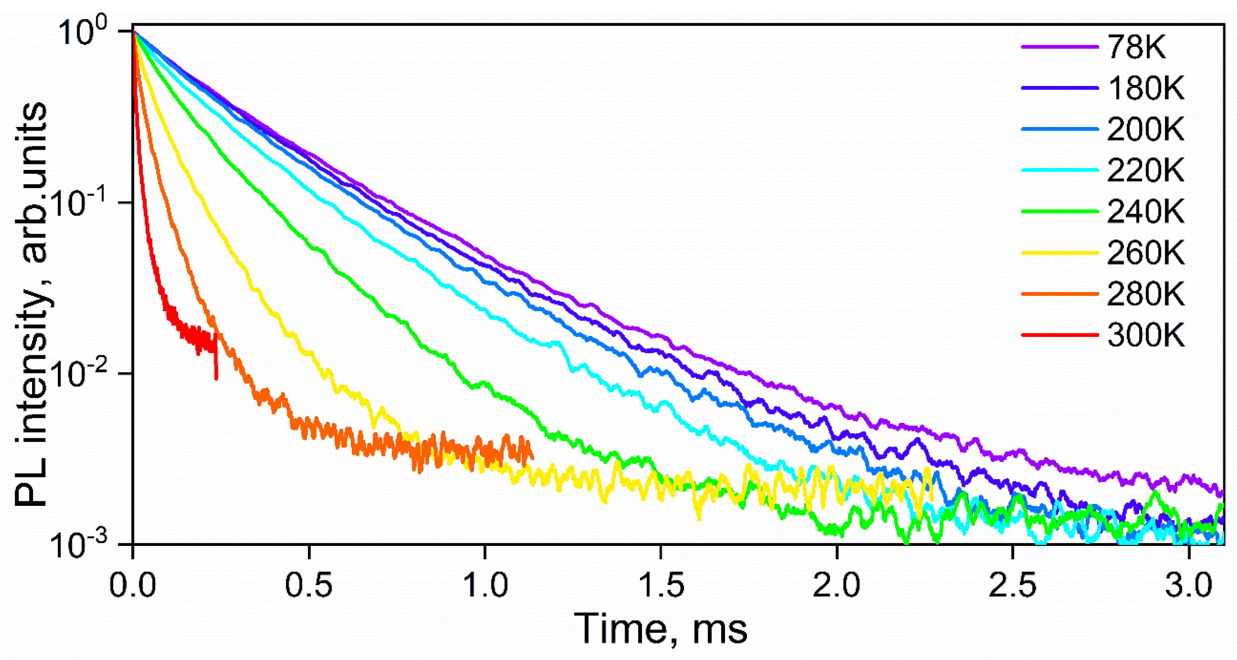
The recorded decays reveal bi-exponential behavior (see
Figure 8) with characteristic lifetimes of 22–208 μs and 74–425 μs (see
Table 3).
Both components were used for the Arrhenius analysis. The observed long- and short-time luminescence lifetimes increase monotonically upon sample cooling from 300 to 78 K. The estimated ln(k) has a linear dependence on T
−1 in the range of 260 to 78 K. Consequently, the fitting was carried out only in the selected temperature ranges for each sample. To exclude the contribution of non-radiative multiphonon relaxation, we studied the temperature dependence of emission lifetime at temperatures lower than 260 K. To exclude the contribution of vibrational relaxation on OH oscillations [
40], measurements were performed for the phen-containing compound
2p.
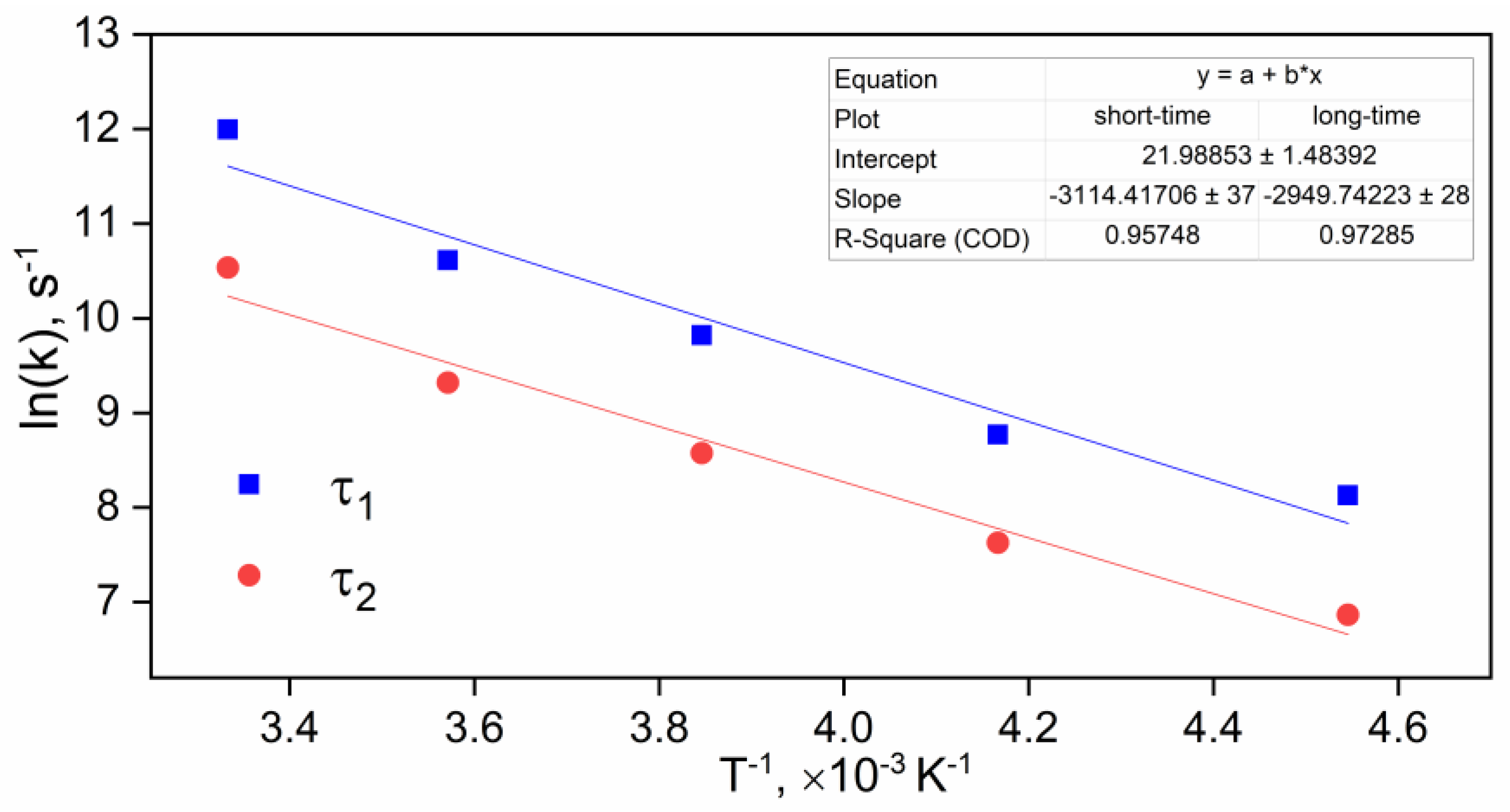
We estimated ΔE
A using the Arrhenius equation (see
Table S3). As shown in
Figure 8, the logarithm of the BET rates estimated for complex
2p linearly depends on inverse temperature since they are well fitted by linear functions (R
2 = 0.96–0.97). The linear dependence confirms that the reduction in the observed lifetime is precisely due to the back energy transfer from the
5D
0 state of the ion to the T
1 state of the ligand. As it is seen from
Figure 6, the ln(k) plots for both lifetime components are linear with similar slope values, suggesting that there is a sole energy transfer process to the ligand for two Eu
3+ symmetry sites. The activation energy of 2200 ± 26 cm
−1 is slightly higher than the energy gap ΔE = 1400 cm
−1 between the T
1 and
5D
0 states. Therefore, the BET process occurs through a small energy barrier of about 0.1 eV, which can be related to a ligand geometry reorganization. The subsequent analysis was conducted for the short-time component only. The considerable BET rate constant of 37160 ± 60 s
−1 and the high frequency factor of 3.5∙10
9 s
−1 suggest a large impact of the BET process on the nonradiative deactivation of the ion excited state for
2p. The observed k
nrad is higher than the BET rate by approximately 1600 cm
−1, whereas a nonradiative rate of 1200 cm
−1 was estimated for
1p. Therefore, the nonradiative relaxation of the
5D
0 state of Eu
3+_ion in the compounds with a bi-naphtyl diketone moiety occurs by 95% via the back energy transfer process, while the rest occur via multiphonon relaxation on molecular oscillators.
3. Experimental Part
3.1. Materials and Methods
All the reagents were purchased form Aldrich (St. Louis, MO, USA) and were used without further purification. THF was distilled over Na/benzophenone just prior to use. Complexes
1h—tris(1,3-diphenyl-l,3-propanediono)(aqua)europium(III) [Eu(L
1)
3(H
2O)],
1p—tris(1,3-diphenyl-l,3-propanediono)(1,10-phenenthroline)europium(III) [Eu(L
1)
3(phen)], and
1Gd—tris(1,3-diphenyl-l,3-propanediono)(aqua)gadolinium(III) [Gd(L
1)
3(H
2O)] were prepared by the procedures published previously [
31,
41,
42], respectively.
Elemental analysis was performed in the Laboratory of Microanalysis of the Nesmeyanov Institute of Organoelement Compounds (Moscow, Russian Federation). The 1H and 13C NMR spectra were recorded with a Bruker AV-300 instrument (Bruker AXS Handheld Inc., Kennewick, WA, USA) operated at 300 and 75.5 MHz, respectively, in CDCl3 solutions. FTIR spectra were recorded in KBr pellets on a Perkin Elmer Spectrum One instrument (Santa Barbara, CA, USA).
Data were collected using a Bruker D8 Quest diffractometer (Bruker AXS Handheld Inc., Kennewick, WA, USA) with a Photon III detector at a temperature of 110(2) K, MoK
α radiation (λ = 0.71073 Å), φ and ω scanning mode. Single crystals of compound
HL2 were obtained by slow evaporation of a saturated solution in EtOH at room temperature. The structure was solved by direct methods and refined by the least-squares method in the full-matrix anisotropic approximation vs. F
2 using the SHELXTL [
43,
44] and Olex2 software [
45] packages.
The
HL2 ligand crystallizes in the P2
1 chiral space group, but the absolute configuration could not be determined due to the absence of heavy atoms with high anomalous scattering. The hydrogen atoms were placed in the calculated positions and refined using the “riding” model. The crystallographic data, experimental parameters, and structural refinements are given in
Table S1. The atomic coordinates, bond lengths, bond angles, and thermal displacement parameters have been deposited in the Cambridge Structural Data Bank (CCDC No. 2214714).
The UV–Vis optical absorption spectra of the compounds studied in acetonitrile solutions (HPLC SuperGradient, Panreac, Barcelona, Spain) were recorded with a JASCO V-770 spectrophotometer (Jasco, Tokyo, Japan) operating within 200–2500 nm. The concentration of the solutions was 10−5 M. The measurements were performed using quartz cells with 1 cm path length. Photoluminescence spectra, luminescence excitation spectra and luminescence decays were obtained using a Horiba Fluorolog QM spectrofluorimeter (Horiba, Kioto, Japan) with a 75 W xenon arc lamp as the excitation source and a R13456 photomultiplier tube sensitive in the 200–980 nm emission range as the detector. Photoluminescence quantum yields were measured for solid samples by the absolute method using the same experimental setup equipped with a G8 (GMP, Renens, Switzerland) integration sphere. Luminescence decays were measured by the Multichannel Scaling (MCS) method using the same instrument. For this purpose, pulsed laser radiation of an Nd:YAG laser LQ529B (Solar LS, Minsk, Belarus) with a repetition rate of 20 Hz and a pulse duration of 8 ns was used. The excitation wavelength was selected using a system based on an optical parametric oscillator (OPO) LP604 and a second harmonic generator LG350 (Solar LS, Belarus), pumped by the second harmonic (532 nm) of LQ529B. The setup provides continuous tuning of the laser wavelength in the range of 340–2500 nm. The laser radiation was directed into the sample compartment of the Fluorolog spectrofluorimeter by a dedicated in-home designed adaptor. The laser pulses and timing electronics of the Fluorolog QM were synchronized using a Si amplified photodiode (Thorlabs, DET10A2) and a Horiba Tb-01 module.
The temperature-dependent luminescence measurements were recorded with a special in-house made setup. Complexes sandwiched between two quartz slides were placed into a vacuum optical cryostat LN-121-SPECTR (Cryotrade, Moscow, Russian Federation) cooled with liquid nitrogen. The residual pressure was maintained below 10−5 Torr by a Leybold Turbovac 90i vacuum pump (Leybold Vacuum GmbH, Köln, Germany). The temperature of the sample was measured by a resistive wire sensor connected to a LakeShore 325 temperature controller (Lake Shore Cryotronics, Westerville OH, USA) placed on a copper sample holder inside the cryostat in close proximity to the sample. The temperature in the range of 78–300 K was maintained by a special heater that was mounted on the sample holder and controlled by the same temperature controller. The cryostat was placed into the sample compartment of a Horiba Fluorolog QM spectrofluorimeter.
3.2. Synthesis of the HL2 Ligand and the 2p, 2h and 2Gd Complexes
3.2.1. 1,3-Di(naphthalen-1-yl)propane-1,3-dione (HL2, 3)
To a magnetically stirred suspension of 2.5 g NaH (60% dispersion in paraffin oil, 62.5 mmol) in 50 mL of dry THF under Ar atmosphere, 4.0 g of methyl 1-naphthoate (21.5 mmol) was added and the resulting mixture was stirred at 25 °C for 30 min. Then, a solution of 1-(naphthalen-1-yl)ethan-1-one (2.9 g, 17 mmol) in 8 mL of THF was added slowly and the resulting dark mixture was left overnight with stirring at room temperature. The excess solvent was evaporated under reduced pressure and the resulting tarry material was decomposed by shaking with a mixture of crushed ice (50 g) and conc. HCl (5.8 mL). The dark brown dense liquid was separated from the aqueous layer and mixed with a hot solution of Cu(OAc)2 (4.3 g, 24 mmol) in 50 mL of water. After cooling to room temperature, the dark oily precipitate of copper chelate 4 was filtered off, recrystallized from hot 2-propanol, and air-dried. Diketone 3 was recovered as follows: the solid chelate was added to a biphase mixture of EtOAc (50 mL) and 5 % aqueous HCl (20 mL), and placed in a separating funnel. The funnel was shaken until all the solids decomposed, and the organic layer was separated. Extraction was repeated 3 times with 50 mL portions of EtOAc. The combined extracts were dried over Na2SO4 and evaporated to dryness. The brown residue was heated with heptane (50 mL) to boiling, decanted hot from the resin, and the clear solution was cooled to room temperature. The precipitate was filtered off, washed with hexane, and air dried. The yield of the slightly yellow compound was 1.9 g (38% based on ketone). M.p. 115–116 °C (heptane-CHCl3). 1H NMR (ppm): δ 16.87 (broad s, 1H, -OH), 8.65 (m, 2H), 8.00 (m, 2H), 7.93 (m, 2H), 7.63 (m, 2H), 7.57 (m, 6H), 6.61 (s, 1H, CH = C(OH)). 13C NMR (ppm): δ 189.15, 134.32, 133.79, 131.89, 130.12, 128.51, 127.32, 127.28, 126.37, 126.56, 124.74, 102.99.
3.2.2. Tris(1,3-di(naphthalen-1-yl)-1,3-propanediono)(aqua)europium(III) [Eu(L2)3(H2O)], 2h
The HL2 ligand (0.195 g, 0.6 mmol) was dissolved in a mixture of EtOH (10 mL) and dioxane (5 mL), and 0.6 mL (0.6 mmol) of 1M NaOH solution in EtOH was added at 45 °C with continuous magnetic stirring. The mixture was kept at that temperature for 10 min, and EuCl3∙6H2O (0.073 g, 0.2 mmol) dissolved in 1 mL of hot EtOH was added slowly. The resulting cloudy solution was stirred at 45 °C for 2 h, filtered through a 0.22 μm PTFE syringe filter and left at room temperature in an open vial until only 1/3 of the original volume remained. No crystallization occurred. Deionized water (5 mL) was added to the clear solution with vigorous stirring and the mixture was stirred until spontaneous crystallization occurred. The resulting precipitate was filtered off, washed successively with 50% aqueous EtOH (5mL), deionized water (5mL) and hexane (5mL), and dried in air. The complex was additionally recrystallized from 50% aqueous EtOH and air-dried to afford 2h as a slightly yellow powder, yield 0.155g (68%). Anal. calcd. for C69H47EuO7 (1140.07): C, 72.69; H, 4.16; Eu, 13.33. Found: C, 72.74; H, 4.18; Eu, 13.39%.
3.2.3. Tris(1,3-di(naphthalen-1-yl)-1,3-propanediono)(aqua)gadolinium(III) [Gd(L2)3(H2O)], 2Gd
This complex was prepared similarly to 2h, starting from 0.075 g (0.2 mmol) of GdCl3∙6H2O. Light yellow powder, yield 0.162 g (71%). Anal. calcd. for C69H47GdO7 (1145.36): C, 72.36; H, 4.14; Gd, 13.73. Found: C, 72.41; H, 4.19; Gd, 13.86%.
3.2.4. Tris(1,3-di(naphthalen-1-yl)-1,3-propanediono)(1,10-phenenthroline)europium(III) [Eu(L2)3(phen)], 2p
This complex was prepared similarly to 2h, but 0.036 g (0.2 mmol) of 1,10-phenanthroline was added to an alkaline solution of 1,3-di(naphthalen-1-yl)propane-1,3-dione. After addition of a EuCl3∙6H2O solution, a precipitate of complex 2p gradually formed. The suspension was stirred at 45 °C for 2 h, cooled to room temperature and filtered. The precipitate was washed successively with 50% aqueous EtOH (7 mL), deionized water (10 mL) and hexane (5 mL), and dried in air. The yield of the light yellow powder was 0.203 g (78%). Anal. calcd. for C81H53EuN2O6 (1302.26): C, 74.71; H, 4.10; N, 2.15, Eu, 11.67. Found: C, 74.77; H, 4.16; N, 2.21, Eu, 11.73%.
4. Conclusions
In this work, we report the synthesis and photophysical investigation of a series of novel Eu3+ coordination compounds with a ligand environment from the β-diketone class. Highly π-conjugated compounds containing a bi-naphtyl diketone moiety were compared with DBM compounds. The increase in π-conjugation in the aromatic substituent of the β-diketone ligand lowers the energies of the first exited singlet and triplet states. However, the first excited singlet energies unusually change upon complexation of a ligand with the ion. An increase in π-conjugation results in a decrease in the S1 energy of free ligands from 25500 to 24300 cm−1. The reverse dependence is observed for the coordination compounds studied. The value of the first excited singlet energy state is 24550 cm−1 for 1h and 25000 cm−1 for 2h. Thus, π-conjugation of the ligand’s aromatic moiety does not allow targeted tuning of the S1 energy of a ligand in a coordination compound to be performed.
We found that using a more strongly π-conjugated ligand provides a significant decrease in the luminescence quantum yield, up to 15-fold for the phen-containing compound. This is explained by a reduction in the triplet state energy from 2050 cm−1 to 18500 cm−1. Consequently, the highly efficient back energy transfer process with a rate of 37160 s−1 favors the quenching of Eu3+ ion luminescence.

