A Scoping Review of the Efficacy of Diode Lasers Used for Minimally Invasive Exposure of Impacted Teeth or Teeth with Delayed Eruption
Abstract
:1. Introduction
2. Methodology
2.1. PICOS
2.2. Eligibility Criteria and Information Sources
3. Results
4. Case Reports
4.1. Case Report 1
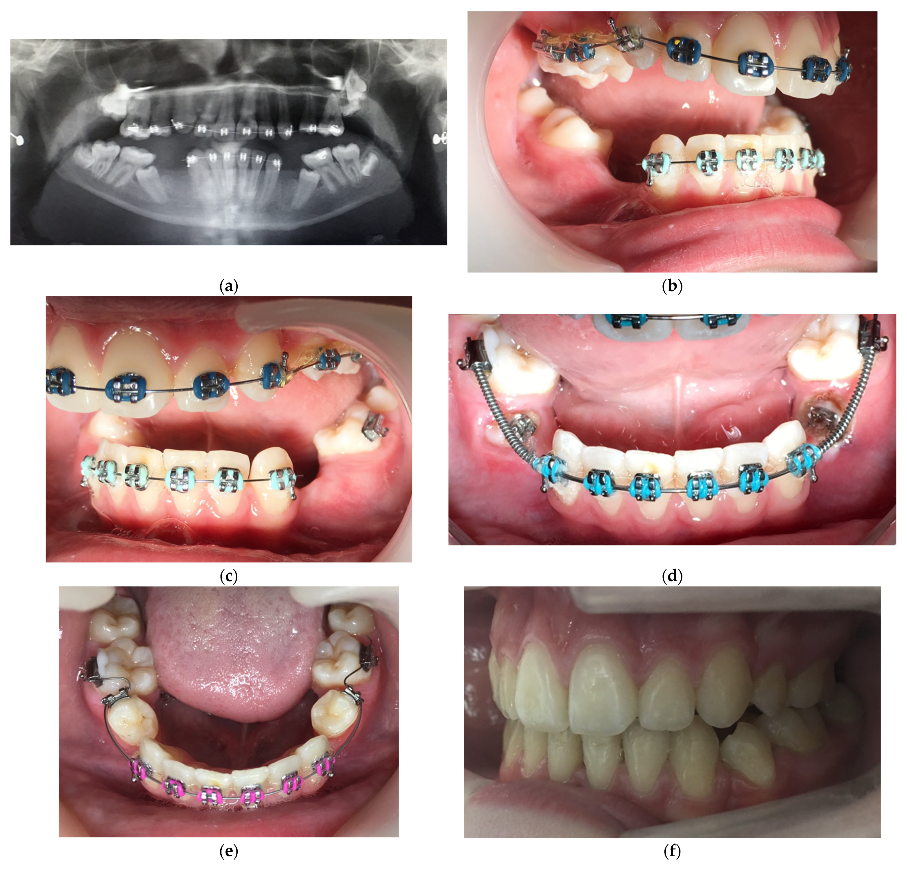
Impacted Mandibular Second Premolars
4.2. Case Report 2
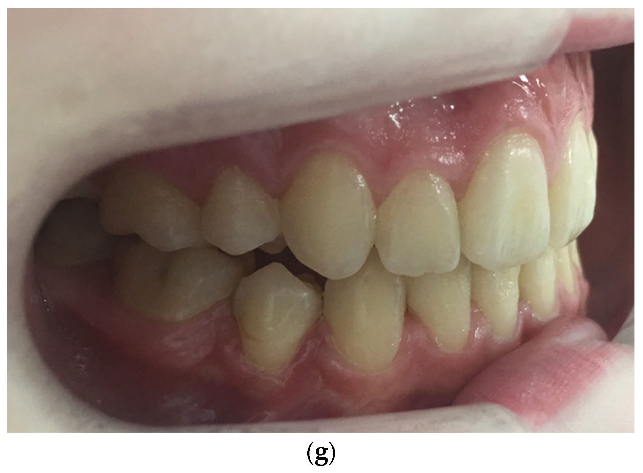
Impacted Mandibular Left Canine
5. Discussion
6. Conclusions
Funding
Institutional Review Board Statement
Informed Consent Statement
Conflicts of Interest
Abbreviations
| RCT | Randomized clinical trial |
| LL3 | Mandibular left canine |
References
- Hartman, B.; Adlesic, E.C. Evaluation and management of impacted teeth in the adolescent patient. Dent. Clin. N. Am. 2021, 65, 805–814. [Google Scholar] [CrossRef] [PubMed]
- Uslu, O.; Akcam, M.O.; Evirgen, S.; Cebeci, I. Prevalence of dental anomalies in various malocclusions. Am. J. Orthod. Dentofac. Orthop. 2009, 135, 328–335. [Google Scholar] [CrossRef] [PubMed]
- Fardi, A.; Kondylidou-Sidira, A.; Bachour, Z.; Parisis, N.; Tsirlis, A. Incidence of impacted and supernumerary teeth-a radiographic study in a north Greek population. Med. Oral Patol. Oral Cir. Bucal. 2011, 16, e56–e61. [Google Scholar] [CrossRef] [PubMed] [Green Version]
- Grover, P.S.; Lorton, L. The incidence of unerupted permanent teeth and related clinical cases. Oral Surg. Oral Med. Oral Pathol. 1985, 59, 420–425. [Google Scholar] [CrossRef]
- Dachi, S.F.; Howell, F.V. A survey of 3874 routine full-month radiographs. II A study of impacted teeth. Oral Surg. Oral Med. Oral Pathol. 1961, 14, 1165–1169. [Google Scholar] [CrossRef]
- Thilander, B.; Pena, L.; Infante, C.; Parada, S.S.; de Mayorga, C. Prevalence of malocclusion and orthodontic treatment need in children and adolescents in Bogota, Colombia. An epidemiological study related to different stages of dental development. Eur. J. Orthod. 2001, 23, 153–167. [Google Scholar] [CrossRef]
- Aitasalo, K.; Lehtinen, R.; Oksala, E. An orthopantomographic study of prevalence of impacted teeth. Int. J. Oral Surg. 1972, 1, 117–120. [Google Scholar] [CrossRef]
- Hou, R.; Kong, L.; Ao, J.; Liu, G.; Zhou, H.; Qin, R.; Hu, K. Investigation of impacted permanent teeth except the third molar in Chinese patients through an X-ray study. J. Oral Maxillofac. Surg. 2010, 68, 762–767. [Google Scholar] [CrossRef]
- Jain, S.; Raza, M.; Sharma, P.; Kumar, P. Unraveling impacted maxillary incisors: The why, when, and how. Int. J. Clin. Pediatr. Dent. 2021, 14, 149–157. [Google Scholar]
- Barone, S.; Antonelli, A.; Averta, F.; Diodati, F.; Muraca, D.; Bennardo, F.; Giudice, A. Does mandibular gonial angle influence the eruption pattern of the lower third molar? A three-dimensional study. J. Clin. Med. 2021, 10, 4057. [Google Scholar] [CrossRef]
- Mah, M.; Takada, K. Orthodontic management of the impacted mandibular second molar tooth. Orthod. Fr. 2016, 87, 301–308. [Google Scholar] [CrossRef] [PubMed]
- Dalessandri, D.; Parrini, S.; Rubiano, R.; Gallone, D.; Migliorati, M. Impacted and transmigrant mandibular canines incidence, aetiology, and treatment: A systematic review. Eur. J. Orthod. 2017, 39, 161–169. [Google Scholar] [CrossRef] [PubMed] [Green Version]
- Felsenfeld, A.L.; Aghaloo, T. Surgical exposure of impacted teeth. Oral Maxillofac. Surg. Clin. N. Am. 2002, 14, 187–199. [Google Scholar] [CrossRef]
- de Latour, F.; Burke, V. Management of impacted teeth. In Office-Based Maxillofacial Surgical Procedures; Ferneini, E., Goupil, M., Eds.; Springer: Cham, Switzerland, 2019. [Google Scholar]
- Borzabadi-Farahani, A. The adjunctive soft-tissue diode laser in orthodontics. Compend. Contin. Educ. Dent. 2017, 38, e18–e31. [Google Scholar] [PubMed]
- Migliario, M.; Rizzi, M.; Lucchina, A.G.; Renò, F. Diode laser clinical efficacy and mini-invasivity in surgical exposure of impacted teeth. J. Craniofac. Surg. 2016, 27, e779–e784. [Google Scholar] [CrossRef]
- Aoki, A.; Mizutani, K.; Schwarz, F.; Sculean, A.; Yukna, R.A.; Takasaki, A.A.; Romanos, G.E.; Taniguchi, Y.; Sasaki, K.M.; Zeredo, J.L.; et al. Periodontal and peri-implant wound healing following laser therapy. Periodontol. 2000 2015, 68, 217–269. [Google Scholar] [CrossRef]
- Borzabadi-Farahani, A.; Cronshaw, M. Lasers in Orthodontics. In Lasers in Dentistry—Current Concepts. Textbooks in Contemporary Dentistry; Coluzzi, D., Parker, S., Eds.; Springer: Cham, Switzerland, 2017. [Google Scholar] [CrossRef]
- Munn, Z.; Peters, M.D.J.; Stern, C.; Tufanaru, C.; McArthur, A.; Aromataris, E. Systematic review or scoping review? Guidance for authors when choosing between a systematic or scoping review approach. BMC Med. Res. Methodol. 2018, 18, 143. [Google Scholar] [CrossRef]
- Tricco, A.C.; Lillie, E.; Zarin, W.; O’Brien, K.K.; Colquhoun, H.; Levac, D.; Moher, D.; Peters, M.D.J.; Horsley, T.; Weeks, L.; et al. PRISMA Extension for Scoping Reviews (PRISMAScR): Checklist and Explanation. Ann. Intern. Med. 2018, 169, 467–473. [Google Scholar] [CrossRef] [Green Version]
- Ize-Iyamu, I.N.; Saheeb, B.D.; Edetanlen, B.E. Comparing the 810 nm diode laser with conventional surgery in orthodontic soft tissue procedures. Ghana Med. J. 2013, 47, 107–111. [Google Scholar]
- Seifi, M.; Vahid-Dastjerdi, E.; Ameli, N.; Badiee, M.R.; Younessian, F.; Amdjadi, P. The 808 nm laser-assisted surgery as an adjunct to orthodontic treatment of delayed tooth eruption. J. Lasers Med. Sci. 2013, 4, 70–74. [Google Scholar]
- Yossif, R.S.; El-Destawy, M.T.; El-Patal, M.A.; Elbaiomy, S.Y. Clinical outcome of diode laser usage versus conventional surgical technique in management of delayed erupted tooth. Al-Azhar J. Dent. Sci. 2017, 20, 201–208. [Google Scholar]
- Sant’Anna, E.F.; Araújo, M.T.S.; Nojima, L.I.; Cunha, A.C.D.; Silveira, B.L.D.; Marquezan, M. High-intensity laser application in orthodontics. Dent. Press J. Orthod. 2017, 22, 99–109. [Google Scholar] [CrossRef] [PubMed] [Green Version]
- Impellizzeri, A.; Horodynski, M.; Serritella, E.; Palaia, G.; De Stefano, A.; Polimeni, A.; Galluccio, G. Uncovering and autonomous eruption of palatally impacted canines-a case report. Dent. J. 2021, 9, 66. [Google Scholar] [CrossRef] [PubMed]
- Amaroli, A.; Colombo, E.; Zekiy, A.; Aicardi, S.; Benedicenti, S.; De Angelis, N. Interaction between laser light and osteoblasts: Photobiomodulation as a trend in the management of socket bone preservation—A review. Biology 2020, 9, 409. [Google Scholar] [CrossRef]
- Dumić, A.K.; Pajk, F.; Olivi, G. The effect of post-extraction socket preservation laser treatment on bone density 4 months after extraction: Randomizedcontrolled trial. Clin. Implant. Dent. Relat. Res. 2021, 23, 309–316. [Google Scholar] [CrossRef]
- Naoumova, J.; Rahbar, E.; Hansen, K. Glass-ionomer open exposure (GOPEX) versus closed exposure of palatally impacted canines: A retrospective study of treatment outcome and orthodontists’ preferences. Eur. J. Orthod. 2018, 40, 617–625. [Google Scholar] [CrossRef]
- Gharaibeh, T.M.; Al-Nimri, K.S. Postoperative pain after surgical exposure of palatally impacted canines: Closed-eruption versus open eruption, a prospective randomized study. Oral Surg. Oral Med. Oral Pathol. Oral Radiol. Endod. 2008, 106, 339–342. [Google Scholar] [CrossRef]
- Parkin, N.A.; Deery, C.; Smith, A.M.; Tinsley, D.; Sandler, J.; Benson, P.E. No difference in surgical outcomes between open and closed exposure of palatally displaced maxillary canines. J. Oral Maxillofac. Surg. 2012, 70, 2026–2034. [Google Scholar] [CrossRef] [Green Version]
- Smailiene, D.; Kavaliauskiene, A.; Pacauskiene, I.; Zasciurinskiene, E.; Bjerklin, K. Palatally impacted maxillary canines: Choice of surgical-orthodontic treatment method does not influence post-treatment periodontal status. A controlled prospective study. Eur. J. Orthod. 2013, 35, 803–810. [Google Scholar] [CrossRef]
- Fornaini, C.; Rocca, J.P.; Bertrand, M.F.; Merigo, E.; Nammour, S.; Vescovi, P. Nd:YAG and diode laser in the surgical management of soft tissues related to orthodontic treatment. Photomed. Laser Surg. 2007, 25, 381–392. [Google Scholar] [CrossRef] [Green Version]
- Lione, R.; Pavoni, C.; Noviello, A.; Clementini, M.; Danesi, C.; Cozza, P. Conventional versus laser gingivectomy in the management of gingival enlargement during orthodontic treatment: A randomized controlled trial. Eur. J. Orthod. 2020, 42, 78–85. [Google Scholar] [CrossRef] [PubMed]
- Narayanan, M.; Laju, S.; Erali, S.M.; Erali, S.M.; Fathima, A.Z.; Gopinath, P.V. Gummy smile correction with diode laser: Two case reports. J. Int. Oral Health 2015, 7 (Suppl. S2), 89–91. [Google Scholar] [PubMed]
- To, T.N.; Rabie, A.B.; Wong, R.W.; McGrath, C.P. The adjunct effectiveness of diode laser gingivectomy in maintaining periodontal health during orthodontic treatment. Angle Orthod. 2013, 83, 43–47. [Google Scholar] [CrossRef] [PubMed] [Green Version]
- Bhat, P.; Thakur, S.L.; Kulkarni, S.S. Evaluation of soft tissue marginal stability achieved after excision with a conventional technique in comparison with laser excision: A pilot study. Indian J. Dent. Res. 2015, 26, 186–188. [Google Scholar]
- Amin, N.; Watt, E.; Noar, J. The punch technique for the soft tissue exposure of superficial, buccally impacted teeth. J. Orthod. 2020, 47, 78–81. [Google Scholar] [CrossRef]


| Authors | Study Design/Groups/Funding Source | Exposed Tooth/Region | Diode Laser Characteristics | Main Findings/Adverse Events |
|---|---|---|---|---|
| Migliario et al. [16] | Prospective study. Funding source not clear. 16 orthodontic patients (overall, 20 impacted teeth, 4 patients had 2 impacted teeth). 9 males and 7 females. Age range = 10 years and 7 months to 24 years and 4 months. Control group (N = 10) received exposure of impacted teeth by scalpel. Experimental group (N = 10) received laser exposure to uncover the impacted teeth, including 60 s of laser biostimulation of tissues covering impacted tooth crown to reduce pain. 15% Lidocaine spray was used as topical anaesthsia. Pain assessed using a numerical rating scale (NRS, 1–10). 14-day follow-up. | Impacted teeth Mainly maxillary canines, maxillary lateral incisors, and mandibular 2nd molars | 980 nm diode laser Pulsed mode (20 s on/10 s off) Power = 1.5 W Fibre diameter tip = 320 μm | Of the 10 patients in the laser-treated group, only 3 needed infiltrative anaesthesia, and of those only 2 needed to take analgesics post-surgically (slight pain (NRS = 2)). None had bleeding or needed suturing. Brackets or attachments were bonded to the impacted teeth for all 10 impacted teeth. The laser surgical procedure was completed in 8–23 min. No adverse event was reported for laser use. All patients in the conventional group needed infiltrative anaesthesia and almost all (9/10) had pain for up to 5 days (average NRS = 4) and were treated with post-surgical analgesics. All had bleeding and 6 needed suturing. Only in 6 patients were brackets or the attachment bonded to the impacted teeth. The whole surgical procedure took 21–43 min. |
| Ize-Iyamu et al. [21] | Prospective study. Funding source not clear. 23 orthodontic patients (17 females and 6 males, age range = 10–30 years). A mixed sample of patients who had either conventional surgery or laser surgery for gingivectomy, aesthetic recontouring, maxillary frenectomy, operculectomy, or tooth impaction exposure surgery (all had conventional bone removal, i.e., palatally impacted canines, to uncover the tooth initially and to bond it with a bracket followed by flap closure). Control group (N = 11) received conventional surgical intervention (including 5 cases of tooth exposure). Experimental group (N = 12) received laser surgery (including 6 cases of tooth exposure). WHO bleeding scale (0–4) and Visual Analogue Scale (VAS, 0–10) were used and recorded. The length of follow-up not clear. | Sample included some impacted teeth but their locations were not clear | 810 nm diode laser The laser brand, mode of laser delivery, and the tip diameter were not specified | None of the laser procedures required suturing, while 8 (72.7%) of the conventional surgical procedures required suturing. Only 2 (16.7%) of the laser surgical procedures required infiltration anaesthesia compared to 10 (90.9%) with conventional surgery (p < 0.001). Post-operative pain was significantly reduced in all cases treated with the diode laser (p < 0.001). There was a significant reduction (p < 0.05) in post-operative bleeding in all cases treated with the diode laser. About 83% (10/12) of the laser surgery cases took ≤ 20 min to finish vs. 27% (3/11) in conventional surgery group. No adverse event reported for laser use. |
| Seifi et al. [22] | Prospective study. Funding source not clear. 16 orthodontic patients with delayed tooth eruption and no sign of impaction. Female/male data only available for the laser group (6 females and 2 males, mean age = 14 ± 0.9 years). Control group (N = 8) did not receive any surgical (conventional or laser) intervention. Experimental group (N = 8) received laser exposure to uncover the unerupted 2nd premolars after the utilization of topical and local anaesthetic. | 2nd premolars with delayed eruption | 808 nm diode laser Continuous wave mode Power = 1.6 watt Fibre diameter tip = 0.3 mm | Laser intervention accelerated the tooth eruption significantly (11 ± 1.1 vs. 25 ± 1.8 weeks to be able to access the facial axis of the clinical crown). No significant bleeding during or immediately after the surgery. No adverse event reported for laser use. |
| Yossif et al. [23] | Randomized clinical trial study. Funding source not clear. Study sample size is not clear (two figures of 30 and 20 were cited in the paper). 18 females and 12 males. Mean age = 11.2 (2.2) years. Control group (N = 15) received exposure of delayed erupted tooth by conventional method (scalpel). Experimental group (N = 15) received laser exposure to uncover the unerupted tooth/teeth. 7-day follow-up. | Teeth with delayed eruption but their locations were not clear | 935 nm diode laser Continuous wave mode Power = 1.6 watt Fibre diameter tip = 0.4 mm | The pain VAS score on days 1 and 7 were significantly lower in the laser group compared to the surgical group. The laser group showed less bleeding (the WHO bleeding criteria) than the conventional surgical group. Patients in the surgical group took more analgesics on the 1st day than patients in the laser group. No adverse event reported for laser use. |
Publisher’s Note: MDPI stays neutral with regard to jurisdictional claims in published maps and institutional affiliations. |
© 2022 by the author. Licensee MDPI, Basel, Switzerland. This article is an open access article distributed under the terms and conditions of the Creative Commons Attribution (CC BY) license (https://creativecommons.org/licenses/by/4.0/).
Share and Cite
Borzabadi-Farahani, A. A Scoping Review of the Efficacy of Diode Lasers Used for Minimally Invasive Exposure of Impacted Teeth or Teeth with Delayed Eruption. Photonics 2022, 9, 265. https://doi.org/10.3390/photonics9040265
Borzabadi-Farahani A. A Scoping Review of the Efficacy of Diode Lasers Used for Minimally Invasive Exposure of Impacted Teeth or Teeth with Delayed Eruption. Photonics. 2022; 9(4):265. https://doi.org/10.3390/photonics9040265
Chicago/Turabian StyleBorzabadi-Farahani, Ali. 2022. "A Scoping Review of the Efficacy of Diode Lasers Used for Minimally Invasive Exposure of Impacted Teeth or Teeth with Delayed Eruption" Photonics 9, no. 4: 265. https://doi.org/10.3390/photonics9040265
APA StyleBorzabadi-Farahani, A. (2022). A Scoping Review of the Efficacy of Diode Lasers Used for Minimally Invasive Exposure of Impacted Teeth or Teeth with Delayed Eruption. Photonics, 9(4), 265. https://doi.org/10.3390/photonics9040265


