Article

Cascaded Cavitation Bubble Excited by a Train of Microsecond Laser Pulses

by
Nadezhda A. Kudasheva
1,
Nikita P. Kryuchkov
2,
Arsen K. Zotov
1,
Polina V. Aleksandrova
1,
Oleg I. Pokhodyaev
2,
Kseniya A. Feklisova
1,
Yurii A. Suchkov
1,
Anatoly L. Bondarenko
1,
Ivan V. Simkin
2,
Vladislav A. Samsonov
2,
Sergey G. Ivakhnenko
3,
Irina N. Dolganova
1,4,*,
Stanislav O. Yurchenko
2,
Sergey V. Garnov
1,
Kirill I. Zaytsev
1,
David G. Kochiev
1 and
Egor V. Yakovlev
2,*
1
Prokhorov General Physics Institute of the Russian Academy of Sciences, Moscow 119991, Russia
2
Centre for Soft Matter and Physics of Fluids, Cluster for Engineering for Life Sciences (CELS), Bauman Moscow State Technical University, Moscow 105005, Russia
3
SEC “Ion Plasma Technologies”, Bauman Moscow State Technical University, Moscow 105005, Russia
4
Osipyan Institute of Solid State Physics of the Russian Academy of Sciences, Chernogolovka 142432, Russia
*
Authors to whom correspondence should be addressed.
Photonics 2025, 12(9), 927; https://doi.org/10.3390/photonics12090927
Submission received: 6 August 2025 / Revised: 10 September 2025 / Accepted: 15 September 2025 / Published: 18 September 2025
(This article belongs to the Section Lasers, Light Sources and Sensors)

Abstract

Although laser cavitation was discovered half a century ago, novel geometries and regimes to excite this effect have been vigorously explored during the past few decades. This research is driven by a variety of applications of laser cavitation in demanding branches of science and technology, such as microfabrication, synthesis of nanoparticles, manipulation of cells, surgery, and lithotripsy. In this work, we combine experimental studies using high-repetition-rate imaging and numerical simulations to uncover a novel regime of the laser cavitation observed upon excitation of a liquid by a train of laser pulses with the pulse energy of 140 mJ and duration of 1.2 μs delivered through a quartz optical fiber. Once the lifetime of the initial cavitation bubble (excited by the first laser pulse) is larger than the period between pulses, which is 34.3 μs, the secondary pulses in the train pass the gas in a bubble and evaporate additional liquid. This results in the formation of a cascaded cavitation bubble of larger volume and elongated shape of 4.6 mm length compared to 3.8 mm in case of excitation by a single laser pulse. In addition, the results of acoustic measurements confirm the presence of shock waves in the applied liquid. Finally, potential applications of the uncovered laser cavitation regime are discussed.

1. Introduction

Since the discovery of laser cavitation, considerable research and engineering efforts have been focused on investigating this effect [1,2,3], due to the destructive action of the cavitation bubble collapse and the emitted acoustic transients (shock waves) on solid surfaces [4,5,6], multilayer systems [7,8], and complex-shaped objects [9,10,11]. This effect paves the way for novel laser applications in fundamental and practical fields, such as laser-induced breakdown spectroscopy [12], microfabrication [13,14,15], nanoparticle synthesis [16], control of crystallization from solutions [17], drug delivery [18], microfluidics [19,20], management of membrane permeability [21,22,23], and lysis of thrombi, blood cells, and vesicles [24,25,26]. Special attention is paid to the medical applications of laser cavitation [27], with an emphasis on ocular surgery [28], solid tumor surgery [29], and lythotripsy [30,31].
To meet the demands of these applications, different geometries and regimes of laser cavitation have been studied, including illumination by pulsed and continuous waves [32,33,34]. The shape and dynamics of the cavitation bubble, along with the shock wave parameters, were managed by changing the pressure [35] or introducing an additional shock wave [36,37] into a liquid. By doping a liquid with plasmonic nanoparticles [38,39], or by combining laser and acoustic cavitation [40], the laser cavitation threshold was considerably reduced to comply with the laser dosimetry guidelines. Aimed at exciting prospective multi-bubble systems, [41,42] holographic laser fields were applied [43]. Meanwhile, a pair of laser cavitation bubbles with collinear or non-collinear excitation, variable phases, shapes, and dimensions demonstrated a potential to tune the shock wave parameters in wide limits [44,45,46,47]. The duration of a laser pulse determines the shape of the cavitation bubble. While short pulses with a duration of less than 1 μs induce relatively regular circular bubbles in a liquid [48], long pulses with duration > 100 μs commonly cause elongated and irregular bubbles, whose length depends on the temperature of the liquid [49]. In Refs. [50,51], the dynamics of elongated bubble were studied numerically. It was shown that the dynamics of vapor bubbles induced by the exposure of long-pulsed laser are driven by the inertia of the bubble nuclei as well as by the continuation of vaporization. Such cavitation bubbles are attractive for medical applications, including ocular laser surgery and lithotripsy. Additionally, recent studies have provided evidence of their benefit for clearing of fallopian tube obstructions [52]. However, nanosecond and sub-nanosecond laser pulses can also induce nonspherical cavitation bubbles when an additional holographic element is applied [53], or as an effect of weak focusing [54]. Studies focused on the interactions of cavitation bubbles with liquid–liquid or liquid–solid interfaces [7,55,56,57] as well as these bubbles’ propagation in finite medium [58] are aimed at understanding the process in conditions close to practice. For example, the study of laser-induced bubbles within microchannels [59] can be applied for the development of bubble-powered micromachines.
To further extend the capabilities of this effect, in this paper, we uncover a novel regime of laser cavitation, observed upon exposure of a liquid to a train of near-infrared microsecond laser pulses delivered through a quartz optical fiber. When the lifetime of the initial cavitation bubble (excited by the first laser pulse; Figure 1a) is larger than the delay between pulses, the secondary pulses within a train pass through the gas phase in a bubble, radiate the gas–liquid interface, and evaporate additional liquid (Figure 1b). This results in a cascaded cavitation bubble with a complex elongated shape. In this work, we experimentally demonstrate the formation of such cascaded laser cavitation, apply numerical analysis and acoustic measurements to interpret this effect, and discuss its applications.

2. Materials and Methods

2.1. Experimental Setup

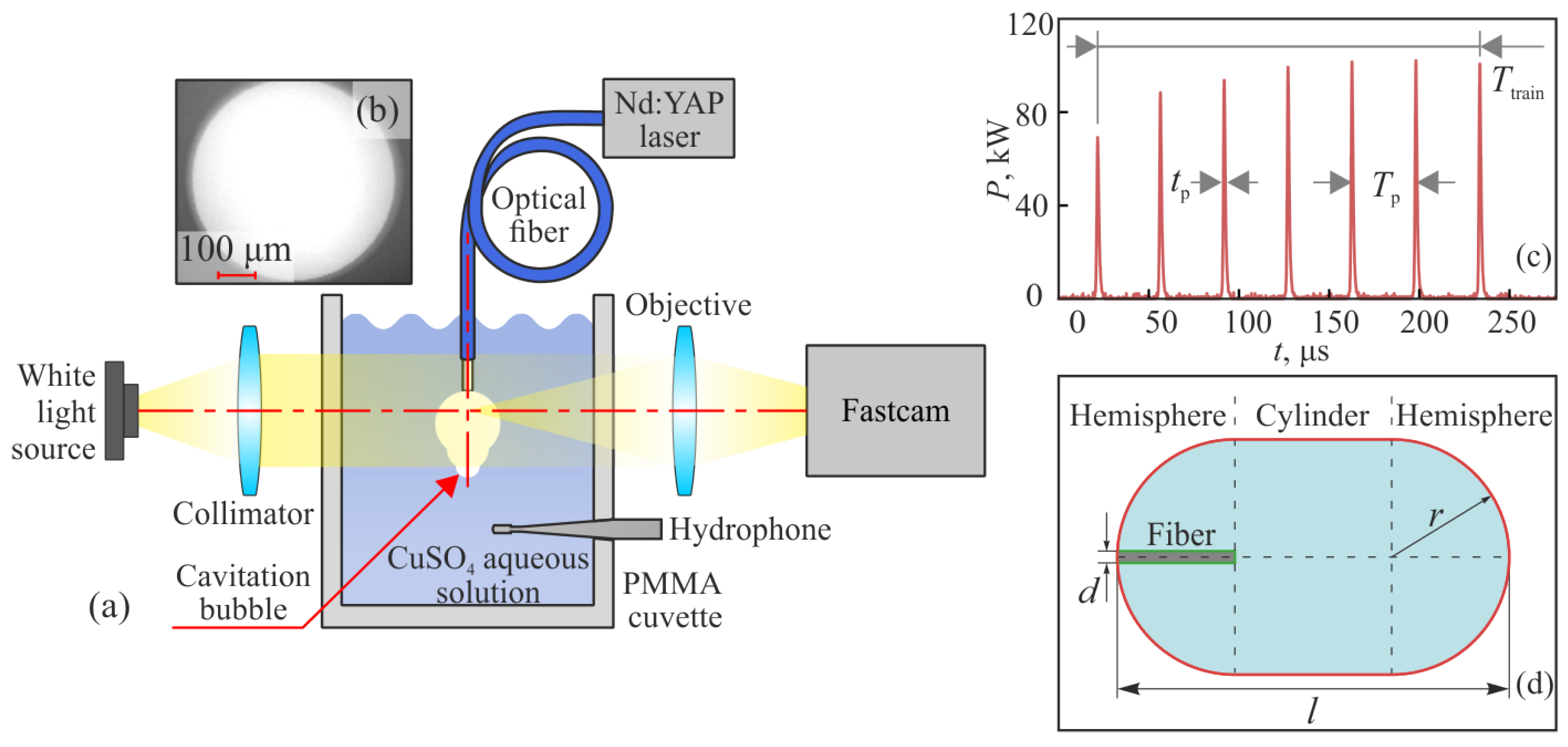
To experimentally uncover a cavitation regime induced by a train of microsecond laser pulses, we assembled the setup shown in Figure 2a. The in-house Nd:YAlO3-crystal pulsed laser (GPI RAS, Moscow, Russia) with the output wavelength of λ = 1079.6 nm is a key element of this setup. A favorable combination of a passive Q-switching mode (with the Cr4+:YAG crystal used as a saturable absorber) and a fiber-optic delay (integrated in a resonator) makes it possible to generate a train of laser pulses. The duration of each pulse in the train is τ p = 1.2 μs at the full-width of half-maximum (FWHM). The number of laser pulses in the train can vary from 1 to 7, which is a function of the duration of the laser pump pulse. In Figure 2c, we show the train formed by 7 pulses with a total duration of T train = 213 μs and a period (between pulses) of T p = 34.3 μs, which is considered in our experiments. Each pulse in the train has a peak power of P peak 103.3 kW, resulting in pulse energy as high as 140 mJ and total train energy of 980 mJ. The laser radiation is coupled into an all-silica fiber with a core diameter of 550 μm, an outer diameter of 600 μm, a numerical aperture of 0.22   NA , and a flat polished distal end (Figure 2b). This allows us to deliver the laser radiation to an exposed object. The fiber tip and the cavitation bubble are illuminated by the white light source and visualized by the high-speed digital camera, so the optical axis of the laser beam and that of the imaging system are orthogonal (Figure 2a). The camera (Fastcam SA6, Photron, Tokyo, Japan) provides a repetition rate up to f cam = 75 kHz and an exposure time down to τ cam = 13 μs. It is equipped with the lens (AF–S VR Micro-ikkor, Nikon, Tokyo, Japan), with a focal lengths of f = 105 mm, a focal ratio of f / 2.8 , and a diffraction-limited image quality. The optical fiber is introduced into the transparent cuvette made of polymethyl methacrylate (PMMA) and filled with a liquid medium, in which the cavitation is excited (Figure 2a). The cuvette has the dimensions 10.5 × 6 × 15 cm that eliminate the interference of its walls in the cavitation process. To find out the features of the cascaded cavitation, we use a train of 7 laser pulses for exposure. Then we compare the results with those of the ordinary cavitation performed by a single laser pulse. Finally, the results are compared with each other.
In addition to the time-resolved imaging, an acoustic signal produced by the cavitation process was measured by a polyvinylidenfluorid (PVDF) hydrophone (BARI-NN, Nizhny Novgorod, Russia). It features a high sensitivity 0.48 μV/Pa and wide bandwidth 100 kHz–100 MHz. The hydrophone was mounted inside the cuvette so that the distance between its receiving platform and the distal end of the fiber was set to 15 mm. It was oriented perpendicular to the optical axis (Figure 2a), as described in several previous works [15,58,60,61,62], which helped to reduce the risk of direct laser exposure to the sensor, preventing damage to the latter; the occurrence of parasitic noise, such as that caused by medium heating; and the corresponding measurement errors. At the same time, the symmetric collapse of the cavitation bubble ensured spherical propagation of the generated acoustic wave. Note that imaging and acoustic measurements were performed independently.

2.2. Sample Preparation

As a liquid medium, we used an aqueous solution of copper sulfate pentahydrate (CuSO4·5H2O) with a molar concentration of 0.7 M. It was prepared using the analytical laboratory balance (AND HR-250AZG, A&D Company Limited, Tokyo, Japan), with a maximal weight limit of 210 mg and precision as high as 0.1 mg. As reported in Ref. [63], the considered CuSO4 aqueous solution has the absorption coefficient μ a 10.705 cm−1 at λ 1000 nm. It mimicked (to some extent) the absorption properties of the liver tissue ( μ a 4 cm−1 at λ = 1000 nm) [64]; the brain (≃5 cm−1 at 1000 nm) [65]; and the stomach (≃2.65 cm−1 at 980 nm) [66].
The sample medium was kept at room temperature. No additional temperature maintenance was applied.

2.3. Numerical Simulations

To qualitatively reproduce and interpret the experimental data, we resort to numerical hydrodynamic modeling within the OpenFOAM package, version 2406 [67] (https://www.openfoam.com/ accessed on 2 January 2025). Namely, we use the compressible InterFoam solver to simulate the behavior dynamics of a mixture of the two immiscible compressible fluids, governed by the following equations:
ρ t + ρ u = 0 ,
ρ u t + ρ u u = p + τ + F σ ,
( ρ T ) t + ( ρ u T ) κ T = = p t + ( τ u ) ( ρ K ) t ( ρ u K ) α l C v l + α g C v g
α k ρ k t + α k ρ k u = 0 ,
τ = μ u + u T 2 3 u I .
Equation (1) is the general continuity equation for a mixture with density of ρ = α l ρ l + α g ρ g , moving at velocity u . The parameters α l and α g represent the volume fractions of liquid and gas, respectively, and satisfy the condition α l + α g = 1 , while ρ l and ρ g denote the local densities of the liquid and gas, respectively, and finally, ∇ is the nabla operator. The indices l and g indicate that the quantity refers to the liquid or gas, respectively. Equations (2) and (3) describe the momentum and energy balance, where μ = α l μ l + α g μ g and κ = α l κ l + α g κ g are the effective viscosity and thermal conductivity of the mixture, respectively; p is pressure, T is temperature, K = 0.5 | u | 2 is kinetic energy per unit mass, τ is the stress tensor for Newtonian fluid, C v l and C v g represent the isochoric heat capacities of the respective phases, F σ is the surface tension force calculated using the Continuum Surface Force (CSF) method, and I is the identity tensor. Similarly to studies [68], we considered a simplified case, where thermal conductivity could be neglected, and hence, κ l , g was set to zero. Finally, Equations (4) and (5) correspond to the continuity equations for each phase. Similarly to Ref. [69], we used the ideal gas equation of state to describe the vapor within the cavitation:
ρ = p R T ,
where R is the universal gas constant. To describe the liquid, we used the equation of state for a perfect fluid:
ρ = ρ 0 + p R f T ,
where R f is the fluid constant and ρ 0 is the initial density.
As the modeling domain, we considered a 3D capsule-shaped region formed by two hemispheres and a cylinder with a radius r = 6 mm and a total length l = 16 mm (Figure 2d). The cylindrical region with a diameter of 600 μ m and a height of 6 mm was removed from the simulation domain to represent the fiber through which the laser radiation enters the system. Using the Salome package and the NETGEN 3D utility, we generated a mesh within the simulation domain. Since the goal of the simulation was to qualitatively reproduce the experimentally observed processes, we used a relatively coarse mesh consisting of 7.4 × 10 6 tetrahedrons. We applied wave transmission boundary conditions (waveTransmissive in OpenFoam realization) to the capsule surfaces and set zero mass flux across the boundary corresponding to the fiber, while keeping the temperature fixed on its surface.
Initially, the entire system was filled with liquid ( α = 1 ) at a temperature of 300 K and a pressure of p = 10 5 Pa , with no flow present u = 0 . Subsequently, directly adjacent to the fiber, along the axis of symmetry of the system, within a 300-μm-diameter spherical region centered at the optical axis, the liquid was abruptly replaced by gas ( α = 0 ) with a pressure of p = 10 7 Pa , which was intended to simulate water evaporation due to laser absorption during the pulse. When simulating the cascade of cavitation bubbles, similar gas regions were placed at the liquid–gas interface along the axis of symmetry at times 35 μs and 70 μs from the start of the simulation. The initial pressure in each subsequent region was taken to be half of that in the previous one. Note that in this study, we only aimed to qualitatively reproduce the phenomenon observed in the experiment. Therefore, we did not fully replicate the actual pulse laser aeration process. Instead, we employed an empirical approach based on abruptly replacing a small region of the liquid phase with a gas phase at high pressure at the intended radiation absorption site. Furthermore, the initial sizes of the gas cavities, as well as the pressure values, were selected empirically to ensure a good visual agreement with the experiment. At the same time, the task of conducting a detailed comparison of the simulation results and the experiment, and thus determining the most accurate parameters of the gas cavities, is beyond the scope of the present study.
During the simulation, we used a dynamically varying time step that ranged from 8.0 × 10 11 to 1.0 × 10 7 s throughout the simulation. Within the scope of our simulation, we used the following approximate values for the thermodynamic parameters of water and vapor: ρ 0 = 1027 kg / m 3 , R f = 7400 J / kg / K , C p , l = 4200 J / kg / K , C p , g = 2000 J / kg / K , μ l = 8.9 × 10 4 N s / m 2 , μ g = 1.24 × 10 5 N s / m 2 . Finally, the results of numerical simulations provide us with cavitation bubble images in the form of spatial distributions of liquid volume fraction α l r at the different time steps; here, r is a radius vector.
Despite the fact that such a numerical analysis is not self-consistent and does not directly account for the laser beam, below, we confirm that it yields adequate predictions for the cavitation bubble geometry and dynamics [68].

2.4. Estimation of the Effective Radius of Cavitation Bubble

To quantify the dimensions of cavitation bubbles and the differences between theory and experiment, an in-house computer vision algorithm based on the OpenCV2 open source Python library (https://opencv.org/ accessed on 2 January 2025) was developed. It allows for determining and tracking (frame-by-frame) the cavitation bubble parameters in both numerical and experimental data. To achieve this, all images were converted to grayscale and underwent a thresholding procedure and a morphological transform (which was aimed at smoothing and reducing noise). Then, contour fitting was applied along the bubble boundary. Next, for each image, the minimal enclosing circle that fits the entire bubble contour in case of a single pulse and the first bubble in the case of a pulse train was determined. Finally, the radius of this circle was considered as the effective radius of a cavitation bubble R eff . As a result, R eff was estimated at each time step (even if the bubble shape is non-spherical).
Determining R eff is a viable approach because cavitation bubbles generally retain a spherical shape and are often too large to be fully captured within the frame. The method described above addressed this issue by using the minimal enclosing circle. It inherently approximated the shape of bubbles that extend beyond the frame. Thus, R eff was the radius of the circumscribed circle that completely enclosed the bubble (the first bubble in the case of a pulse train). This approach is easily implemented using sequential post-processing of the cavitation bubble evolution process and describes the transverse dimensions of the bubble well.

3. Results

3.1. Imaging of the Cavitation Process

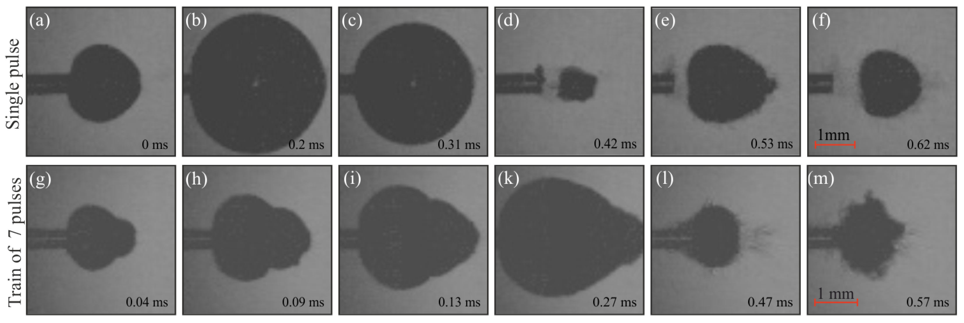
The selected experimental data for laser cavitation in the CuSO4 aqueous solution exposed to a single 1.2-μs-long laser pulse and a train of 7 pulses are presented in Figure 3. Each panel shows the axial cross-section of the gray-scale frames. The two rows of panels (a)–(f) and (g)–(m) give a general overview of the cavitation processes observed in the case of a single-pulsed and train-pulsed laser exposure, respectively. The specified time step is counted from the moment of bubble excitation by the laser pulse in the experiment. Unlike the symmetrical shape of the cavitation bubble, which appears under a single-pulsed exposure, the cascaded cavitation is characterized by an elongated shape. The experimental observation of this process revealed that the maximum depth of the cascaded bubble was 4.6 mm, while for a single bubble it was 3.8 mm. At the same time, the bubble widths (in the vertical direction in Figure 3) in both cases are almost equal ∼3.8 mm. We also can see the sequence of bubble expansion, its collapse, and the following rebound.

3.2. Comparison of Numerical and Experimental Data

In this section, we qualitatively compare the calculated spatial distribution of the liquid volume fractions α l r with the experimental data in Figure 4 in case of exposure to a single 1.2-μs-long laser pulse (panels (a)–(f)), and a train of 7 pulses (panels (g)–(m)). Each panel shows the axial cross section of α l r (top part, blue-to-white) and the video frames I r (bottom part, gray-scale). The frames of numerical data are selected according to the corresponding stage of the bubble collapse.
Qualitative agreement is observed between the numerical results and experiments in terms of the shapes and dimensions of the two cavitation bubbles. In fact, the applied theoretical model has also revealed a predictably symmetric spherical cavitation bubble excited by a single pulse, as well as an elongated cascaded cavitation bubble of a much larger volume, upon exposure of the liquid to a train of pulses. Meanwhile, some differences between the theory and experiment can be noticed for the stages of cavitation bubble collapse (see panels (c) and (m) in Figure 4) and appearance of the rebound bubble (panels (d)–(f)).
In Figure 5a,b, the effective radius of cavitation bubble R eff is estimated as a function of time based on the experimental and numerical data, respectively, for both the ordinary cavitation bubble excited by a single laser pulse and the cascaded one produced by a train of pulses. A detailed explanation of R eff extraction is described in Section 2.4. In panel (c), we compare the obtained results in the form of data contours. Panels (d)–(g) illustrate the estimation of R eff from the experiments and simulations.
On the one hand, both the experiment and simulations reveal the oscillatory character of the two cavitation bubbles. Next, they both demonstrate an increase in the lifetime of the cascaded bubble compared to the ordinary one. On the other hand, the experimental data demonstrate the moderate increase in R eff for the first cascaded cavitation bubble over the first single bubble (Figure 5a). The simulated data do not reveal the same obvious excess (Figure 5b). However, from Figure 5c, it is possible to observe the small difference for the simulated R eff . At the same time, the first peak of the cascaded cavitation bubble is almost the same for the simulations and experiment, at R eff 2 mm.
From Figure 5c, some discrepancies between the experiment and theory are evident, such as the different periods and damping constants of oscillatory processes. This might be attributed to the somewhat non-consistent character of the applied purely hydrodynamic numerical scheme (see Section 2.3), which does not account for the real shape, divergence, scattering, or attenuation of the laser beam. Another option is that we did not take into account the phase transition and used the oversimplified equation of states. Finally, this study did not aim to determine the model parameters (such as the initial radius of secondary cavitation bubbles and their energy) that would ensure the closest agreement with the experimental results, which also contributes to the observed discrepancies. Solving this problem requires an extensive series of additional simulations, which is far beyond the scope of the present paper. However, the key features of our experiment are reproduced by the performed simulations. Thus, they justify that the observed cascaded cavitation occurs owing to the evaporation of additional liquid by secondary pulses in a train passing the gas phase and interacting with the gas–liquid interface. Evidently, such a laser cavitation regime can manifest itself only if the period between pulses in a train is smaller than the lifetime of the initial cavitation bubble excited by the first pulse within a train, as predicted in Figure 1.

3.3. Acoustic Measurements

For a more comprehensive study of the observed process, we have measured the acoustic data in the liquid, which was exposed to the single laser pulse and to the train of pulses. Figure 6 shows the time-dependent acoustic signal for the cascaded and ordinary cavitation processes. In both cases, several distinct parts of the acoustic response are clearly observed. The first part reflects the initial stage of cavitation. In contrast to the signal caused by the single-pulsed bubble formation, which corresponds to pressure P single , 0 = 0.018 MPa, the laser pulse train produces a sequence of almost equal acoustic pulses with an average peak pressure P train , 0 = 0.020 MPa. The following parts reflect the acoustic emissions during the bubble collapse and further oscillations [58], so that we can clearly distinguish several peaks, which refer to first, second, and even third (in the case of the cascaded bubble) collapses. The corresponding pressures were measured: for the single bubble P single , 1 = 0.273 MPa and P single , 2 = 0.074 MPa; for the cascaded bubble P train , 1 = 0.277 MPa, P train , 2 = 0.065 MPa, P train , 3 = 0.014 MPa. The observed strong tensile pressures may stem from measurement artifacts associated with reflections of bubble collapse waves at the hydrophone interface [70], as well as from the use of extended cabling in conjunction with the oscilloscope’s 1 M Ω input resistance [60,71].
The acoustic data approves the presence of shock waves in the liquid, as well as the increased periods of cavitation bubble oscillation in the case of a pulse train compared to the single pulse illumination. The observed acoustic signals qualitatively correlate with the video frames. The pulse sequence provides an evaporation boost in the cavitation bubble, which leads to an increase in the size and lifetime of bubbles, as well as the more subsequent rebounds. Such artifacts are evident from Figure 6, while the video data confirm the prolonged lifetime.
Meanwhile, some differences can be observed between the results of the imaging and acoustic measurements. In particular, the exact moments of bubble collapses had a moderate time shift. This occurred due to a possible technical mismatch between the system alignment, sample preparation, and external environment conditions during two separate experiments. However, numerical simulations, imaging, and acoustic measurements qualitatively demonstrate the distinctive features of the studied cascaded cavitation.

4. Discussion

This work demonstrates the first qualitative observation of the cascaded cavitation caused by a sequence of microsecond laser pulses. Compared with the ordinary cavitation caused by a single pulse, the cascaded regime is characterized by distinct features, such as increases in the lifetime and size of the bubble. Moreover, the bubble has a complex shape due to the short period between pulses in the applied train. Previously, a similar shape was observed during the double-bubble interaction [14,46], when two laser sources were applied separately, or when a complex optical arrangement was used. Obviously, the studied pulse-train regime provides more practical solutions for making such bubbles, offering more options and possibilities.
The larger dimensions of a cascaded cavitation bubble as compared to an ordinary one should lead to more pronounced effects of liquid, soft and solid objects’ exposure to laser radiation and the emitted acoustic transients, which may be interesting for a number of applications, including surgical resection and non-thermal ablation. The increased response in an object arises from the accumulation of energy and more inertial dynamics associated with the growth of larger bubbles, which enhances hydrodynamic stresses and localized fluid–structure interactions during collapse.
Although in this work we have studied the cascaded cavitation in a simple homogeneous liquid medium, this regime has certain prospects in heterogeneous media, such as colloidal suspensions and emulsions or biological tissues [9,11,27]. When considering tissues, among possible applications in therapeutic and diagnostic fields, we can highlight non- and minimally invasive diagnosis and targeted drug delivery, in which cascaded cavitation using a microsecond laser can be useful. At the same time, the realization of cavitation in microfluidic cells [19,20] is also a prospective research topic. It enables precise control of microscale fluid dynamics, which has high potential for creating and improving lab-on-a-chip platforms, droplet generation, and microscale mixing.
Successful initial observation of the reported cavitation effect, which has a general character, can be reproduced using other experimental conditions. Together with the possible future applications, it initiates possible directions for further research. Firstly, significant attention should be paid to the experimental arrangement and equipment. It is important to study the impact of laser wavelength, pulse energy, duration, and period, as well as fiber parameters, such as the diameter, position and geometry of the distal end, on the shape, size, and dynamics of the cavitation bubble. Since, in this work, the energy of each laser pulse was almost equal, the cascaded cavitation was caused by a significantly higher energy input than the ordinary one. This may have caused some additional differences in the bubble dynamics. For a more detailed description of this process, the equal energy of the exciting radiation should be considered in a future study.
Next, the process should be studied in various media, including heterogeneous and microfluidics environments, and living soft and liquid tissues. The influence of sample parameters such as chemical composition, temperature, and pressure on the possible changes in the cavitation bubble lifetime should also be investigated. This will allow for effective adjustment of the laser parameters aimed at a desirable result. For example, an increase in the pressure of a liquid reduces the lifetime of a cavitation bubble [35], putting forward the demands for a smaller duration and period of laser pulses. Finally, a more thorough theoretical description should be developed to provide a deeper understanding of the underlying physics and provide an ability to simulate and predict the behavior of the cascaded bubbles in complex media.
Experiments with acoustic measurements have shown that the process of cavitation is accompanied by the generation and propagation of sound waves in the medium, which has a significant effect on the bubbles. This phenomenon will be taken into account in future modeling. Although the performed numerical simulations confirmed the results of high-repetition-rate imaging, the applied purely hydrodynamic numerical scheme can be considered only as a first approximation in the interpretation of the cascaded cavitation process. For predictive modeling, the parameters of the environment and radiation that generate the cavitation bubble will be determined more accurately. Moreover, it is important to account for the change in the scattering and absorption during the irradiation by the pulse train. Such alteration may lead to each pulse in a train having a different contribution to the bubble energy. All of these further improvements are in order.

5. Conclusions

The novel regime of laser cavitation was uncovered, and it is referred to as cascaded laser cavitation. It takes place upon exposure of a liquid to a train of microsecond laser pulses, the period of which is smaller than the lifetime of the initial cavitation bubble excited by a first pulse within a train. It was observed experimentally using an in-house near-infrared laser and high-repetition-rate imaging, as well as modeled theoretically using numerical methods of hydrodynamics. The results demonstrated the appearance of a larger cavitation bubble with a length of 4.6 mm compared to a 3.8 mm length ordinary bubble in the same conditions. It also has a prolonged lifetime. The results of this work demonstrate the straightforward ability to switch between the cavitation bubble shape just by choosing the number of microsecond laser pulses. Possible applications of this regime and the scope of further work in this research field were also discussed. This study paves the way for further investigations of the complex dynamics of laser-induced cavitation and its practical applications.

Author Contributions

Supervision, N.A.K., S.O.Y. and S.V.G.; conceptualization, K.I.Z.; methodology, D.G.K. and E.V.Y.; numerical simulations, N.P.K.; measurements, A.K.Z., E.V.Y., P.V.A. and I.V.S.; signal processing, O.I.P., K.A.F., V.A.S. and S.G.I.; experimental setup, Y.A.S. and A.L.B.; writing—original draft preparation, A.K.Z., E.V.Y. and N.P.K.; writing—review and editing, I.N.D. and K.I.Z. All authors have read and agreed to the published version of the manuscript.

Funding

Russian Science Foundation, project # 25–79–30006.

Data Availability Statement

Data underlying the results of this paper are not publicly available at this time, but may be obtained from the corresponding authors IND (dolganova@issp.ac.ru) and EVY (yakov.egor@bmstu.ru) upon reasonable request.

Conflicts of Interest

The authors declare no conflicts of interest.

References

  1. Lauterborn, W.; Bolle, H. Experimental investigations of cavitation-bubble collapse in the neighbourhood of a solid boundary. J. Fluid Mech. 1975, 72, 391–399. [Google Scholar] [CrossRef]
  2. Lauterborn, W. Optische Kavitation. Phys. Bl. 1976, 32, 553–563. [Google Scholar] [CrossRef]
  3. Liang, X.X.; Linz, N.; Freidank, S.; Paltauf, G.; Vogel, A. Comprehensive analysis of spherical bubble oscillations and shock wave emission in laser-induced cavitation. J. Fluid Mech. 2022, 940, A5. [Google Scholar] [CrossRef]
  4. Tomita, Y.; Shima, A. Mechanisms of impulsive pressure generation and damage pit formation by bubble collapse. J. Fluid Mech. 1986, 169, 535–564. [Google Scholar] [CrossRef]
  5. Vogel, A.; Lauterborn, W.; Timm, R. Optical and acoustic investigations of the dynamics of laser-produced cavitation bubbles near a solid boundary. J. Fluid Mech. 1989, 206, 299–338. [Google Scholar] [CrossRef]
  6. Lechner, C.; Lauterborn, W.; Koch, M.; Mettin, R. Fast, thin jets from bubbles expanding and collapsing in extreme vicinity to a solid boundary: A numerical study. Phys. Rev. Fluids 2019, 4, 021601. [Google Scholar] [CrossRef]
  7. Rosselló, J.M.; Reese, H.; Ohl, C.D. Dynamics of pulsed laser-induced cavities on a liquid–gas interface: From a conical splash to a ‘bullet’ jet. J. Fluid Mech. 2022, 939, A35. [Google Scholar] [CrossRef]
  8. Ohl, S.W.; Reese, H.; Ohl, C.D. Cavitation bubble collapse near a rigid wall with an oil layer. Int. J. Multiph. Flow 2024, 174, 104761. [Google Scholar] [CrossRef]
  9. Lin, C.; Kelly, M. Cavitation and acoustic emission around laser-heated microparticles. Appl. Phys. Lett. 1998, 72, 2800–2802. [Google Scholar] [CrossRef]
  10. Long, J.; Eliceiri, M.; Vangelatos, Z.; Rho, Y.; Wang, L.; Su, Z.; Xie, X.; Zhang, Y.; Grigoropoulos, C. Early dynamics of cavitation bubbles generated during ns laser ablation of submerged targets. Opt. Express 2020, 28, 14300–14309. [Google Scholar] [CrossRef] [PubMed]
  11. Sieber, A.; Preso, D.; Farhat, M. Cavitation bubble dynamics and microjet atomization near tissue-mimicking materials. Phys. Fluids 2023, 35, 027101. [Google Scholar] [CrossRef]
  12. Cristoforetti, G.; Tiberi, M.; Simonelli, A.; Marsili, P.; Giammanco, F. Toward the optimization of double-pulse LIBS underwater: Effects of experimental parameters on the reproducibility and dynamics of laser-induced cavitation bubble. Appl. Opt. 2012, 51, B30–B41. [Google Scholar] [CrossRef] [PubMed]
  13. Chen, X.; Xu, R.Q.; Chen, J.P.; Shen, Z.H.; Jian, L.; Ni, X.W. Shock-wave propagation and cavitation bubble oscillation by Nd:YAG laser ablation of a metal in water. Appl. Opt. 2004, 43, 3251–3257. [Google Scholar] [CrossRef] [PubMed]
  14. Robles, V.; Gutierrez-Herrera, E.; Devia-Cruz, L.; Banks, D.; Camacho-Lopez, S.; Aguilar, G. Soft material perforation via double-bubble laser-induced cavitation microjets. Phys. Fluids 2020, 32, 042005. [Google Scholar] [CrossRef]
  15. Soyama, H.; Iga, Y. Laser Cavitation Peening: A Review. Appl. Sci. 2023, 13, 6702. [Google Scholar] [CrossRef]
  16. Streubel, R.; Barcikowski, S.; Gokce, B. Continuous multigram nanoparticle synthesis by high-power, high-repetition-rate ultrafast laser ablation in liquids. Opt. Lett. 2016, 41, 1486–1489. [Google Scholar] [CrossRef]
  17. Nagalingam, N.; Raghunathan, A.; Korede, V.; Poelma, C.; Smith, C.; Hartkamp, R.; Padding, J.; Eral, H. Laser-Induced Cavitation for Controlling Crystallization from Solution. Phys. Rev. Lett. 2023, 131, 124001. [Google Scholar] [CrossRef]
  18. Schoppink, J.; Fernandez Rivas, D. Jet injectors: Perspectives for small volume delivery with lasers. Adv. Drug Deliv. Rev. 2022, 182, 114109. [Google Scholar] [CrossRef]
  19. Zwaan, E.; Le Gac, S.; Tsuji, K.; Ohl, C.D. Controlled Cavitation in Microfluidic Systems. Phys. Rev. Lett. 2007, 98, 254501. [Google Scholar] [CrossRef]
  20. Yuan, F.; Sankin, G.; Zhong, P. Dynamics of tandem bubble interaction in a microfluidic channel. J. Acoust. Soc. Am. 2011, 130, 3339–3346. [Google Scholar] [CrossRef]
  21. Prentice, P.; Cuschieri, A.; Dholakia, K.; Prausnitz, M.; Campbell, P. Membrane disruption by optically controlled microbubble cavitation. Nat. Phys. 2005, 1, 107–110. [Google Scholar] [CrossRef]
  22. Dijkink, R.; Le Gac, S.; Nijhuis, E.; van den Berg, A.; Vermes, I.; Poot, A.; Ohl, C.D. Controlled cavitation–cell interaction: Trans-membrane transport and viability studies. Phys. Med. Biol. 2007, 53, 375–390. [Google Scholar] [CrossRef]
  23. Sankin, G.N.; Yuan, F.; Zhong, P. Pulsating Tandem Microbubble for Localized and Directional Single-Cell Membrane Poration. Phys. Rev. Lett. 2010, 105, 078101. [Google Scholar] [CrossRef]
  24. Rooney, J. Hemolysis Near an Ultrasonically Pulsating Gas Bubble. Science 1970, 169, 869–871. [Google Scholar] [CrossRef] [PubMed]
  25. Everbach, E.; Francis, C. Cavitational mechanisms in ultrasound-accelerated thrombolysis at 1 MHz. Ultrasound Med. Biol. 2000, 26, 1153–1160. [Google Scholar] [CrossRef] [PubMed]
  26. Marmottant, P.; Hilgenfeldt, S. Controlled vesicle deformation and lysis by single oscillating bubbles. Nature 2003, 423, 153–156. [Google Scholar] [CrossRef]
  27. Vogel, A.; Venugopalan, V. Mechanisms of Pulsed Laser Ablation of Biological Tissues. Chem. Rev. 2003, 103, 577–644. [Google Scholar] [CrossRef]
  28. Vogel, A.; Hentschel, W.; Holzfuss, J.; Lauterborn, W. Cavitation Bubble Dynamics and Acoustic Transient Generation in Ocular Surgery with Pulsed Neodymium:YAG Lasers. Ophthalmology 1986, 93, 1259–1269. [Google Scholar] [CrossRef] [PubMed]
  29. Kennedy, J. High-intensity focused ultrasound in the treatment of solid tumours. Nat. Rev. Cancer 2005, 5, 321–327. [Google Scholar] [CrossRef]
  30. Ho, D.; Scialabba, D.; Terry, R.; Ma, X.; Chen, J.; Sankin, G.; Xiang, G.; Qi, R.; Preminger, G.; Lipkin, M.; et al. The Role of Cavitation in Energy Delivery and Stone Damage During Laser Lithotripsy. J. Endourol. 2021, 35, 860–870. [Google Scholar] [CrossRef]
  31. Chen, J.; Ho, D.; Xiang, G.; Sankin, G.; Preminger, G.; Lipkin, M.; Zhong, P. Cavitation Plays a Vital Role in Stone Dusting During Short Pulse Holmium:YAG Laser Lithotripsy. J. Endourol. 2022, 36, 674–683. [Google Scholar] [CrossRef]
  32. Rastopov, S.; Sukhodol’sky, A. Cluster nucleation in the process of CW laser induced thermocavitation. Phys. Lett. A 1990, 149, 229–232. [Google Scholar] [CrossRef]
  33. Ramirez-San-Juan, J.; Rodriguez-Aboytes, E.; Martinez-Canton, A.; Baldovino-Pantaleon, O.; Robledo-Martinez, A.; Korneev, N.; Ramos-Garcia, R. Time-resolved analysis of cavitation induced by CW lasers in absorbing liquids. Opt. Express 2010, 18, 8735–8742. [Google Scholar] [CrossRef] [PubMed]
  34. Padilla-Martinez, J.; Berrospe-Rodriguez, C.; Aguilar, G.; Ramirez-San-Juan, J.; Ramos-Garcia, R. Optic cavitation with CW lasers: A review. Phys. Fluids 2014, 26, 122007. [Google Scholar] [CrossRef]
  35. Pecha, R.; Gompf, B. Microimplosions: Cavitation Collapse and Shock Wave Emission on a Nanosecond Time Scale. Phys. Rev. Lett. 2000, 84, 1328–1330. [Google Scholar] [CrossRef] [PubMed]
  36. Sankin, G.; Simmons, W.; Zhu, S.; Zhong, P. Shock Wave Interaction with Laser-Generated Single Bubbles. Phys. Rev. Lett. 2005, 95, 034501. [Google Scholar] [CrossRef] [PubMed]
  37. Sankin, G.; Zhong, P. Interaction between shock wave and single inertial bubbles near an elastic boundary. Phys. Rev. E 2006, 74, 046304. [Google Scholar] [CrossRef]
  38. Ju, H.; Roy, R.; Murray, T. Gold nanoparticle targeted photoacoustic cavitation for potential deep tissue imaging and therapy. Biomed. Opt. Express 2013, 4, 66–76. [Google Scholar] [CrossRef]
  39. Wei, C.w.; Xia, J.; Lombardo, M.; Perez, C.; Arnal, B.; Larson-Smith, K.; Pelivanov, I.; Matula, T.; Pozzo, L.; O’Donnell, M. Laser-induced cavitation in nanoemulsion with gold nanospheres for blood clot disruption: In vitro results. Opt. Lett. 2014, 39, 2599–2602. [Google Scholar] [CrossRef]
  40. Kim, H.; Kang, J.; Chang, J. Thermal therapeutic method for selective treatment of deep-lying tissue by combining laser and high-intensity focused ultrasound energy. Opt. Lett. 2014, 39, 2806–2809. [Google Scholar] [CrossRef]
  41. Tiwari, A.; Pantano, C.; Freund, J. Growth-and-collapse dynamics of small bubble clusters near a wall. J. Fluid Mech. 2015, 775, 1–23. [Google Scholar] [CrossRef]
  42. Bempedelis, N.; Ventikos, Y. Energy focusing in shock-collapsed bubble arrays. J. Fluid Mech. 2020, 900, A44. [Google Scholar] [CrossRef]
  43. Quinto-Su, P.A.; Venugopalan, V.; Ohl, C.D. Generation of laser-induced cavitation bubbles with a digital hologram. Opt. Express 2008, 16, 18964–18969. [Google Scholar] [CrossRef] [PubMed]
  44. Han, B.; Köhler, K.; Jungnickel, K.; Mettin, R.; Lauterborn, W.; Vogel, A. Dynamics of laser-induced bubble pairs. J. Fluid Mech. 2015, 771, 706–742. [Google Scholar] [CrossRef]
  45. Tomita, Y.; Sato, K. Pulsed jets driven by two interacting cavitation bubbles produced at different times. J. Fluid Mech. 2017, 819, 465–493. [Google Scholar] [CrossRef]
  46. Luo, J.; Niu, Z. Jet and Shock Wave from Collapse of Two Cavitation Bubbles. Sci. Rep. 2019, 9, 1352. [Google Scholar] [CrossRef]
  47. Fan, Y.; Bußmann, A.; Reuter, F.; Bao, H.; Adami, S.; Gordillo, J.; Adams, N.; Ohl, C.D. Amplification of Supersonic Microjets by Resonant Inertial Cavitation-Bubble Pair. Phys. Rev. Lett. 2024, 132, 104004. [Google Scholar] [CrossRef] [PubMed]
  48. Verma, S.; Arba Mosquera, S. A Refined Model for Ablation Through Cavitation Bubbles with Ultrashort Pulse Lasers. Photonics 2024, 11, 1047. [Google Scholar] [CrossRef]
  49. Wang, D.; Bi, Y. Investigation of the influence of different liquid temperatures on the dynamics of long-pulse laser-induced cavitation bubbles. AIP Adv. 2024, 14, 025013. [Google Scholar] [CrossRef]
  50. Zhao, X.; Ma, W.; Wang, K. Simulating laser-fluid coupling and laser-induced cavitation using embedded boundary and level set methods. J. Comput. Phys. 2023, 472, 111656. [Google Scholar] [CrossRef]
  51. Zhao, X.; Ma, W.; Chen, J.; Xiang, G.; Zhong, P.; Wang, K. Vapour bubbles produced by long-pulsed laser: A race between advection and phase transition. J. Fluid Mech. 2024, 999, A103. [Google Scholar] [CrossRef]
  52. Wang, D.; Yang, F.; Gao, W.; Bi, Y.; Zhu, X.; Lin, Z. Exploratory study on the application of laser-induced cavitation bubble in the treatment of fallopian tube obstruction. PLoS ONE 2025, 20, e0317022. [Google Scholar] [CrossRef]
  53. Lim, K.Y.; Quinto-Su, P.A.; Klaseboer, E.; Khoo, B.C.; Venugopalan, V.; Ohl, C.D. Nonspherical laser-induced cavitation bubbles. Phys. Rev. E 2010, 81, 016308. [Google Scholar] [CrossRef]
  54. Vogel, A.; Noack, J.; Nahen, K.; Theisen, D.; Busch, S.; Parlitz, U.; Hammer, D.; Noojin, G.; Rockwell, B.; Birngruber, R. Energy balance of optical breakdown in water at nanosecond to femtosecond time scales. Appl. Phys. B 1999, 68, 271–280. [Google Scholar] [CrossRef]
  55. Li, M.; Huang, Z.; Yang, X. Collapsing dynamics and microjet formation of laser-induced cavitation bubble near corrugated solid wall. Appl. Phys. A 2025, 131, 242. [Google Scholar] [CrossRef]
  56. Park, S.H.; Phan, T.H.; Park, W.G. Numerical investigation of laser-induced cavitation bubble dynamics near a rigid surface based on three-dimensional fully compressible model. Int. J. Heat Mass Transf. 2022, 191, 122853. [Google Scholar] [CrossRef]
  57. Zhou, R.; Li, K.; Cao, Y.; Shi, W.; Yang, Y.; Tan, L.; Hu, R.; Jin, Y. Experimental Study of Laser-Induced Cavitation Bubbles near Wall: Plasma Shielding Observation. Water 2024, 16, 1324. [Google Scholar] [CrossRef]
  58. Fu, L.; Wang, J.; Wang, S.; Zhang, Z.; Vogel, A.; Liang, X.; Yao, C. Secondary cavitation bubble dynamics during laser-induced bubble formation in a small container. Opt. Express 2024, 32, 9747–9766. [Google Scholar] [CrossRef]
  59. Nagalingam, N.; Korede, V.; Irimia, D.; Westerweel, J.; Padding, J.T.; Hartkamp, R.; Eral, H.B. Unified framework for laser-induced transient bubble dynamics within microchannels. Sci. Rep. 2024, 14, 18763. [Google Scholar] [CrossRef] [PubMed]
  60. Fu, L.; Liang, X.X.; Wang, S.; Wang, S.; Wang, P.; Zhang, Z.; Wang, J.; Vogel, A.; Yao, C. Laser induced spherical bubble dynamics in partially confined geometry with acoustic feedback from container walls. Ultrason. Sonochem. 2023, 101, 106664. [Google Scholar] [CrossRef] [PubMed]
  61. Soyama, H. Development of Laser Cavitation Peening Using a Normal-Oscillation Nd:YAG Laser. Coatings 2023, 13, 1395. [Google Scholar] [CrossRef]
  62. Soyama, H.; Kuji, C. Improving effects of cavitation peening, using a pulsed laser or a cavitating jet, and shot peening on the fatigue properties of additively manufactured titanium alloy Ti6Al4V. Surf. Coat. Technol. 2022, 451, 129047. [Google Scholar] [CrossRef]
  63. Fonseca, M.B.; An, L.; Cox, B.T. Sulfates as chromophores for multiwavelength photoacoustic imaging phantoms. J. Biomed. Opt. 2017, 22, 125007. [Google Scholar] [CrossRef]
  64. Carneiro, I.; Carvalho, S.; Henrique, R.; Oliveira, L.; Tuchin, V. Measuring optical properties of human liver between 400 and 1000 nm. Quantum Electron. 2019, 49, 13. [Google Scholar] [CrossRef]
  65. Gonçalves, T.M.; Martins, I.S.; Silva, H.F.; Tuchin, V.V.; Oliveira, L.M. Spectral Optical Properties of Rabbit Brain Cortex between 200 and 1000 nm. Photochem 2021, 1, 190–208. [Google Scholar] [CrossRef]
  66. Wei, H.; Xing, D.; He, B.; Gu, H.; Wu, G.; Chen, X. Using an oblique incident laser beam to measure the optical properties of stomach mucosa/submucosa tissue. BMC Gastroenterol. 2009, 9, 64. [Google Scholar] [CrossRef]
  67. OpenCFD Ltd. OpenFOAM User Guide; OpenCFD Ltd.: Reading, UK, 2024. [Google Scholar]
  68. Koch, M.; Lechner, C.; Reuter, F.; Köhler, K.; Mettin, R.; Lauterborn, W. Numerical modeling of laser generated cavitation bubbles with the finite volume and volume of fluid method, using OpenFOAM. Comput. Fluids 2016, 126, 71–90. [Google Scholar] [CrossRef]
  69. Bußmann, A.; Riahi, F.; Gökce, B.; Adami, S.; Barcikowski, S.; Adams, N.A. Investigation of cavitation bubble dynamics near a solid wall by high-resolution numerical simulation. Phys. Fluids 2023, 35, 016115. [Google Scholar] [CrossRef]
  70. Gonzalez-Avila, S.R.; Denner, F.; Ohl, C.D. The acoustic pressure generated by the cavitation bubble expansion and collapse near a rigid wall. Phys. Fluids 2021, 33, 032118. [Google Scholar] [CrossRef]
  71. Olmes, A.; Lohmann, S.; Lubatschowski, H.; Ertmer, W. An improved method of measuring laser induced pressure transients. Appl. Phys. B 1997, 64, 677–682. [Google Scholar] [CrossRef]
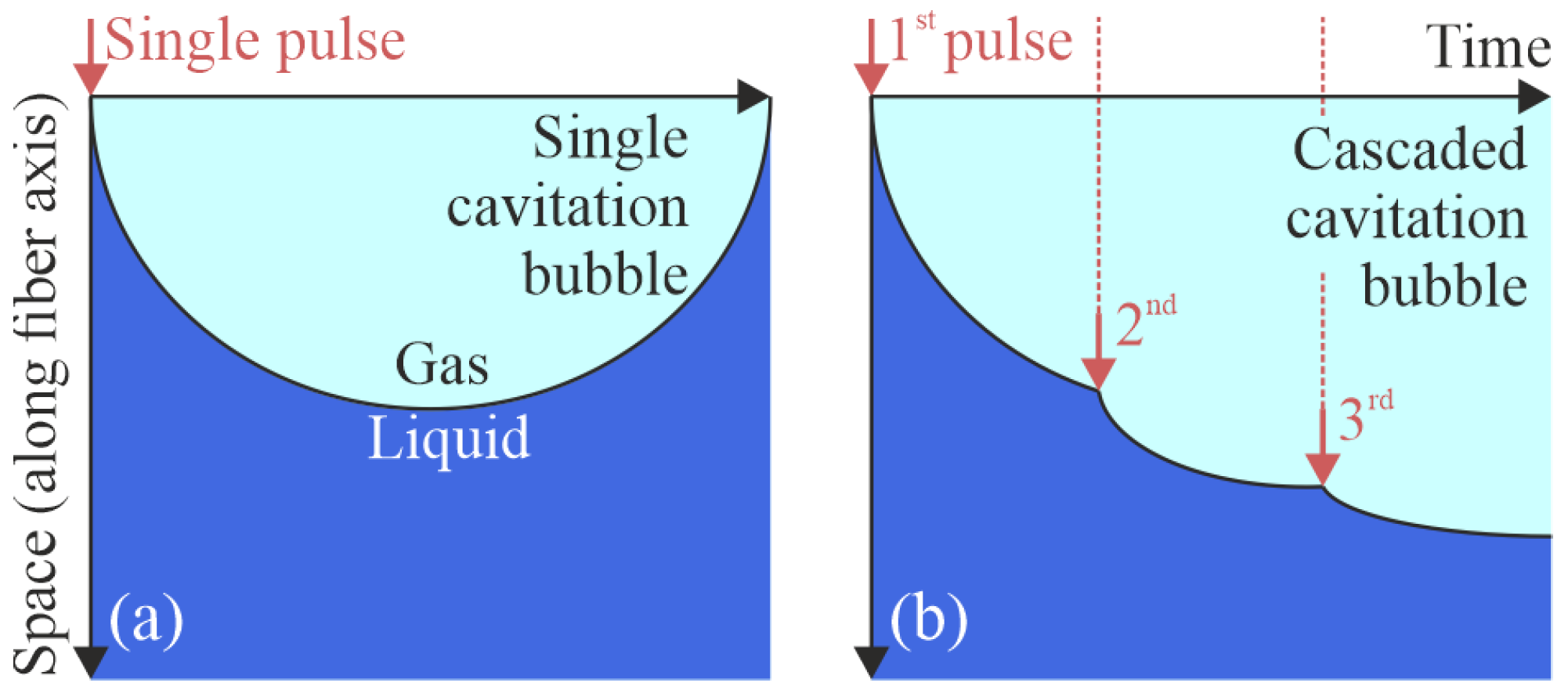
Figure 1. Time–space diagrams for the cavitation bubble formation upon liquid excitation by (a) a single laser pulse and (b) a train of such pulses.
Photonics 12 00927 g001
Figure 2. Experimental setup for studying laser cavitation under exposure of a liquid to a single microsecond laser pulse and a train of such pulses. (a) Schematic of the experimental setup. (b) Microscopy of the distal end of the optical fiber. (c) The temporal profile of the train of laser pulses. (d) Schematic of the computational domain.
Photonics 12 00927 g002
Figure 3. Frames of high-speed imaging of the laser cavitation in CuSO4 aqueous solution. (af) Results of exposure by a single 1.2-μs-long laser pulse; (gm) data for a train of 7 pulses.
Photonics 12 00927 g003
Figure 4. Comparison of the theoretical and imaging data of laser cavitation in CuSO4 aqueous solution, exposed to a single laser pulse (af) and to a train of 7 pulses (gm). Each panel is divided into two parts, where top part shows the calculated α l r -distribution (in blue to white) and bottom part shows the video frames for the same moment.
Photonics 12 00927 g004
Figure 5. Dynamics of the laser cavitation bubble excited by a single laser pulse (in blue) or a train of pulses (in red). (a) Time-dependent effective radius R eff of the two laser cavitation bubbles calculated based on the experimental high-speed video images. (b) Equal data from the numerical analysis. (c) Comparison of the data contours for the experimental and numerical results. (dg) Estimation of R eff -parameter based on experimental (d,e) and numerical (f,g) data, respectively.
Photonics 12 00927 g005
Figure 6. Time dependence of the acoustic signal in the liquid sample illuminated by the single laser pulse (a) and by the train of pulses (b). Black arrows indicate the acoustic peaks corresponding to the bubble collapses.
Photonics 12 00927 g006
Disclaimer/Publisher’s Note: The statements, opinions and data contained in all publications are solely those of the individual author(s) and contributor(s) and not of MDPI and/or the editor(s). MDPI and/or the editor(s) disclaim responsibility for any injury to people or property resulting from any ideas, methods, instructions or products referred to in the content.