Article

Label-Free Detection of Cellular Senescence in Fibroblasts via Third Harmonic Generation

1
Institute of Electronic Structure and Laser (I.E.S.L.), Foundation for Research and Technology-Hellas (FO.R.T.H.), 71110 Heraklion, Greece
2
Department of Biology, University of Crete, 70013 Heraklion, Greece
3
Department of Materials Science and Technology, University of Crete, 70013 Heraklion, Greece
4
Department of Physics, University of Crete, 70013 Heraklion, Greece
*
Authors to whom correspondence should be addressed.
These authors contributed equally to this work.
Photonics 2025, 12(9), 919; https://doi.org/10.3390/photonics12090919
Submission received: 8 August 2025 / Revised: 9 September 2025 / Accepted: 11 September 2025 / Published: 15 September 2025
(This article belongs to the Special Issue New Perspectives in Biomedical Optics and Optical Imaging)

Abstract

Cellular senescence, a state of irreversible growth arrest in response to stress, plays a dual role in physiology and pathology. While essential for processes such as embryogenesis, wound healing, and tumor suppression, senescence also contributes to aging and age-related diseases, including cancer and neurodegeneration. The accumulation of senescent cells is linked to aging and numerous age-associated pathologies, making the detection of these cells crucial for understanding and potentially mitigating age-related diseases. Lipid metabolism is a key feature of senescent cells, which undergo significant alterations in lipid composition that influence membrane remodeling and cellular function. Here, we propose the use of third harmonic generation (THG) microscopy, a label-free imaging modality, to assess lipid profiles in senescent and nonsenescent cells. Our study demonstrated that THG can discriminate between senescent and nonsenescent fibroblasts based on their lipid content, suggesting a noninvasive approach for the detection and characterization of cellular senescence. In addition, these findings reveal that lipid content is increased in senescent cells. This methodology has potential applications in the diagnosis and study of age-related pathologies where lipid dysregulation is a hallmark feature.

1. Introduction

Cellular senescence was first described in 1961 by Hayflick and Moorhead and refers to permanent cell growth arrest, despite remaining metabolically active, in response to damage or stress [1]. As a physiological mechanism, it contributes to embryogenesis, wound healing, and tumor suppression, although it can result in antagonistic effects on aging, degenerative disorders, and cancer [2,3,4]. This dual role of senescence has driven significant research in understanding both normal tissue homeostasis and disease progression [5].
Senescence can be triggered by various endogenous and exogenous stressors, including telomere shortening [6], oxidative stress [7], oncogene activation [8], and mitochondrial dysfunction [9]. When cells fail to repair this damage, they “switch” to the senescent state as a defense mechanism [10] to avoid further genomic instability [11,12,13]. Distinct from quiescent cells and terminally differentiated cells [14], senescent cells undergo distinct phenotypic alterations, including chromatin remodeling, tumor suppressor activation [15,16], and a unique senescence-associated secretory phenotype (SASP) [17]. They become flattened and enlarged with an increased cytoplasm-to-nucleus ratio and elevated lysosomal activity, resulting in increased activity of senescence-associated β-galactosidase (SA-β-gal) [18]. The two key signaling pathways upregulated during senescence are p53/p21 and p16, which are proteins that inhibit the cyclin-dependent kinases (CDKs) involved in the cell cycle [19].
Experimentally, senescence can be induced in vitro by continuous cell passaging [1,20]; oxidative stress via reactive oxygen species (ROS), such as hydrogen peroxide (H2O2) [21]; ultraviolet (UV) or infrared (IR) irradiation [22,23,24]; or chemotherapeutic agents [25]. However, identifying senescent cells remains challenging. Not all senescent cells display all biomarkers of senescence, and some overlap with those of apoptotic or quiescent cells [2]. Thus, advanced noninvasive, label-free microscopy techniques hold promise for detecting senescent cells and serve as important tools in the diagnosis of age-associated pathologies, including cancer, neurodegeneration, and cardiovascular diseases.
Senescent cells exhibit global lipid remodeling [26], altered lipid-regulating gene expression [27,28], and accumulation of lipid bodies (LBs) [29,30,31]. These changes are linked to cell enlargement, SASP secretion via lipid-rich extracellular vesicles [29,30,31], and dysregulated lipid metabolism with aging [32,33]. However, it remains unclear whether these lipid changes are directly caused by cellular senescence. Current detection methods rely on staining and fixation-based assays, which are invasive, alter cell physiology, and limit long-term observation [34,35]. Accurate identification of senescent cells is critical for studying aging and related diseases, yet current methods are invasive and unreliable. Therefore, microscopy techniques that detect LBs in unstained samples can be necessary for studying their physiological activity.
Previously, chromatography coupled mass spectrometry has been employed to detect and quantify the intracellular lipid composition [36]. Moreover, lipid quantification with MRI in tissues has also been carried out [37,38]. Owing to the development of Raman imaging techniques, coherent Raman scattering microscopy (CRS), which has relatively high imaging speeds and rates, enables the noninvasive and label-free detection of lipid molecules [39]. CRS has two imaging modalities: coherent anti-Stokes Raman scattering (CARS) and stimulated Raman scattering (SRS) microscopy. CARS microscopy was first used for lipid imaging in 1999 [40], followed by lipid quantification in 2002 [41]. SRS was used for lipid imaging in 2008 [42].
Few methodologies can provide direct visualization of unstained LBs in situ. CARS microscopy [43] and third harmonic generation (THG) microscopy [44] have been proposed as promising imaging techniques. CARS is a four-wave mixing process that requires two spatiotemporally aligned laser beams, thus expanding the cost of the equipment needed. In the case of THG, a simpler experimental configuration is needed, and a single source is necessary for the experiments.
THG microscopy is a noninvasive label-free imaging modality that is capable of quantifying and correlating lipid profiles of various cells with metabolic syndrome [45,46] and cancer pathologies [47,48]. THG microscopy is a third-order nonlinear optical process that occurs upon near-infrared (NIR) irradiation of a medium with intrinsic inhomogeneities due to refractive index variations [49]. Three excitation photons [50] are combined to be converted into a single photon emitted at triple the incident photon energy [51]. The THG arises at the interface between two optically different materials [51]. Due to the Gouy phase shift of the fundamental beam, the nonlinear signals generated at symmetric planes, with respect to the focus, interfere destructively, resulting in zero output from a homogeneous medium.
THG microscopy allows the imaging of interfaces, for example, between aqueous interstitial fluids and lipid-rich domains, such as cellular membranes and lipid droplets [52]. LBs are the main intracellular sources of high THG signals [44]. THG can perform 3D imaging at high spatial resolution, proving its capacity to study LBs in complex environments in cells [47,53,54], tissues [48], and living organisms [46,55]. The advantage of THG in bioimaging is that the dominant signal is generated within intact live cells and tissues without the need for any staining [50].
Our previous studies revealed that it was feasible to detect the state of unstained cells (activated versus resting) based on the quantification of their LBs [47,53,56]. Moreover, noninvasive and label-free SHG and THG imaging techniques were used to monitor and quantify ectopic lipid deposition and delineate the precise connection between ectopic fat accumulation and aging in C. elegans samples. Our studies revealed that fat content gradually increases with age in the nonadipose tissues, such as body wall muscles, pharyngeal muscles, and neurons, of C. elegans [45,55].
The most recent study of THG on Drosophila haemocytes [57] presented the potential of this technique to record the dynamic change in the lipid accrual of haemocytes upon encountering tumor cells, which could be a useful tool to assess the phagocytic capacity or activation state of tumor-associated haemocytes. In this work, we suggest a noninvasive approach in which label-free THG imaging is used as a diagnostic tool to detect and discriminate senescent cells from nonsenescent cells based on the quantification of their lipid content. As previously reported [26,27], increased lipid content is a known feature of senescent cells. Our contribution lies in presenting a label-free, noninvasive THG imaging method to detect this phenotype in situ. By demonstrating significant differences in lipid accumulation between senescent and nonsenescent fibroblasts, we provide an innovative technical approach to detect cellular senescence. This method underscores the relevance of lipid accumulation as a detectable phenotype of senescence, supports further investigation into the metabolic remodeling that accompanies this state, and opens new avenues for detecting and studying age-related diseases where senescence plays a crucial role.

2. Materials and Methods

2.1. Cell Culture and Induction of Senescence

For all cell cultures in this study, murine NIH 3T3 fibroblasts (LGC Standards GmbH, Wesel, Germany) were used. NIH 3T3 cells were grown in cell culture flasks in Dulbecco’s modified Eagle’s medium (DMEM)–high glucose (HG) (4500 mg/L glucose) (Thermo Fischer Scientific, Gibco™, Waltham, MA, USA) supplemented with 10% fetal bovine serum (FBS) (GibcoTM, USA) and 1% penicillin/streptomycin solution (PS) (Thermo Fischer Scientific, GibcoTM, Waltham, MA, USA) at 37 °C in a 5% CO2 incubator, with medium renewal every 2–3 days. For the study of senescence (Figure 1), 50,000 cells/mL in culture medium were seeded on glass coverslips (Corning Inc., New York, NY, USA) in 24-well plates (Corning Inc., New York, NY, USA) and left to grow overnight. Then, the standard medium was removed, and fresh DMEM–HG supplemented with various concentrations of H2O2 (0 μM, 200 μM, 400 μM, or 800 μM; 30% w/w in H2O, Sigma‒Aldrich, Saint Louis, MO, USA) was added to the cells, which were incubated for 1 h at 37 °C. Finally, the media was replaced with fresh standard DMEM–HG, and the cultures were left for 1, 3, 7, 10, and 14 days. The medium was renewed every 2–3 days. For comparison, untreated samples (without the addition of H2O2) were also investigated. All experiments presented in Figure 1, Figure 2, Figure 3 and Figure 4, were performed with a minimum of three independent biological replicates.

2.2. Immunocytochemical Assay

For this assay, the medium was removed from the samples, and the samples were washed twice with 1X PBS (Sigma-Aldrich, Saint Louis, MO, USA) (pH = 7.4) for 5 min and then fixed with 4% paraformaldehyde (PFA) for 15 min at RT. After the removal of the PFA solution, the samples were washed again twice with 1X PBS and treated with 0.1% Triton X-100 (Sigma-Aldrich, Saint Louis, MO, USA) in PBS for 5 min to permeabilize the cell membranes. The samples were subsequently washed twice with 1X PBS for 5 min, and the nonspecific binding sites were blocked with 2% bovine serum albumin (BSA) (Sigma‒Aldrich, Saint Louis, MO, USA) in PBS for 30 min. Subsequently, the cells were incubated with the primary antibody [mouse Ki-67 (1:800 in 1X PBS-BSA 1%) (Cell Signaling Technology, Danvers, MA, USA)] overnight at 4 °C. The next day, the samples were washed twice with 1X PBS and incubated with the secondary antibody [CF488 goat anti-mouse (1:500 in 1X PBS-BSA 1%) (Biotium Inc., Fremont, CA, USA) or CF568 goat anti-mouse (1:500 in 1% PBS-BSA 1%) (Biotium Inc., Fremont, CA, USA)] for 2 h, after which nuclear staining was carried out by (i) 4,6-diamidino-2-phenylindole (DAPI) (Thermo Fischer Scientific, InvitrogenTM, Waltham, MA, USA) or (ii) Hoechst 33342, trihydrochloride, and trihydrate (Thermo Fischer Scientific, Invitrogen TM, Waltham, MA, USA) (1:2000 in PBS) for 15 min at RT. For Figure 1, the samples were transferred to microscope slides for observation via a Leica SP8 laser inverted scanning confocal microscope (Leica Microsystems, Wetzlar, Germany). A 40× objective and a z-stack of the confocal microscope were used.

2.3. SA-β-Galactosidase Staining

The Senescence β-Galactosidase Staining Kit was purchased from Cell Signaling Technology (Danvers, MA, USA). For this method, the medium was removed from the samples (treated and untreated fibroblasts, after 10 days of culture), and the samples were washed with 1X PBS and then fixed with 1X fixative solution provided from the kit for 10–15 min in the dark at RT. After incubation, the fixative was removed, and the cells were washed twice with 1X PBS. The β-galactosidase staining solution was prepared according to the kit instructions by mixing the 1X staining solution with X-gal reagents (Solutions A and B). The stain was added to the cells, which were incubated at 37 °C in a CO2-free environment overnight (for 12–16 h). Finally, the fibroblasts were observed under a Leica DFC310 FX Digital Color Camera Optical Microscope (Leica Microsystems, Wetzlar, Germany) with transmitted light and the ×10 objective for acquisition. In Figure 2, senescent cells display blue coloration in the cytoplasm due to β-galactosidase activity.

2.4. Confocal Image Analysis

In Figure 3, image processing was performed via ImageJ software (1.54p National Institute of Health, Bethesda, MD, USA) [58] and CellProfiler 4.2.8 software (Broad Institute, Cambridge, MA, USA). CellProfiler was used to analyze the following nuclear morphological parameters: the nuclear area and perimeter and the nuclear aspect ratio. Different image processing pipelines were generated to load 1-channel immunofluorescence images for each analysis. This was followed by automated detection of cell nuclei, the nuclear area and perimeter, and the nuclear aspect ratio. For quantitative measurements between images, the same procedures and parameters were used in the software [59,60,61]. All confocal images were analyzed using maximum intensity projections generated from z-stacks consisting of 10–12 optical planes.

2.5. Nonlinear Microscope

The study of senescence involves the simultaneous detection of THG and TPEF signals from cells. THG arises from the lipid domains of the fibroblasts, whereas the TPEF signals signify the expression of the Ki-67 mouse antibody (2nd antibody: CF568 goat anti-mouse) in the nucleus. The experimental apparatus employed in this work allows the simultaneous collection of two nonlinear signals and has been previously described in detail [57].
Briefly, the experimental setup consisted of an Axon pulse femtosecond laser (Coherent, 1064 nm, 80 MHz, 2 W, 150 fs) and a modified Nikon upright microscope (Nikon Eclipse ME600D, Tokyo, Japan). The energy per pulse at the sample plane was 0.6 nJ. A high numerical aperture objective lens (Carl Zeiss, N-Achroplan 32x, NA 0.85, water immersion, WD: 1.1 mm) could tightly focus the laser beam on the sample. A pair of galvanometric mirrors (Cambridge Tech. 6210H) performed the scanning of the samples, which were fitted onto a motorized xyz translation stage (Standa 8MT167-100, Vilinius, Lithuania), with 1 μm resolution. The scanning process and the data acquisition were controlled by LabVIEW. A photomultiplier tube (PMT Hamamatsu H9305-04, Hamamatsu city, Japan) collected the THG signals in the forward direction. A colored glass filter (U 340 nm Hoya, UOG Optics, Cambridge, UK) cut off the reflected laser light and allowed the transmission of solely the ΤHG signals arising from the samples. TPEF signals were collected by a second photomultiplier tube (Hamamatsu H9305-04, Hamamatsu city, Japan) in the backward direction in combination with a bandpass filter (CHROMA, 605/70 M-2p, Bellows Falls, VT, USA) for CF568 detection.
The scanning duration of a single-plane (2D), 500 × 500-pixel nonlinear image was one (1) second. A total of 20 scans were performed for each sample plane of 3D optical sections at 2 μm intervals (z stack). They were either presented as a montage or projected (maximum intensity projection) onto a single plane. All nonlinear images were z-projections of 7–10 optical planes divided by 2 μm, depending on the sample thickness/size of the fibroblasts. ImageJ software (1.54p National Institute of Health, Bethesda, MD, USA) supported the data viewing and processing https://imagej.net/ij/ (accessed on 31 October 25).

2.6. Quantification of the THG Signals Specific to the Lipid Content of the Cells

The separation of senescent and nonsenescent cells may be directly correlated with their lipid metabolism and lipid content. Variations in lipid metabolism are often indicated by LB morphology alterations. Although such variations might be qualitatively displayed by a selection of microscopy techniques [62,63,64,65], the quantification of LB amount and size could be crucial to characterize the identified cell phenotype.
The quantification of the lipids of fibroblasts includes Fiji thresholding in combination with in-house-created MATLAB algorithms to process all 3D slices of the cells (Supplementary Figure S1). THG signals outside of the cells were eliminated from the quantification analysis. As the shape and size of the fibroblasts may vary, it was necessary to normalize the signals by dividing the total lipid area of each cell (lipid sum of all slices) by the total projected cell area (Supplementary Figure S1).
All data were recorded under steady laser power on the sample and the same amplification of the PMT units. The major signals of THG arose from the lipid droplets within the fibroblasts, simplifying the analysis. The cells were isolated from other surrounding structures (Supplementary Figure S1b). The THG signals were processed evenly by applying a constant threshold, resulting in a binary set of images (Supplementary Figure S1c). The total lipid area resulted from the summation of all slices of the remaining pixels of the THG channel. The z-projected intensity THG channel was adjusted to reveal cell contours that could be easily measured. The final lipid index (a.u.) was a ratio of the total lipid area/cell area and was not only an indicator of the lipid content of every cell but also a way of discriminating among different types of cells.
Finally, all quantitative analyses were based on three independent biological experiments. The minimum number of cells quantified from each independent experiment was as follows: untreated (Nset1 = 15, Nset2 = 17, Nset3 = 16) and treated (Nset1 = 16, Nset2 = 18, Nset3 = 24). Figure 4e presents one dot per independent experiment, representing the average lipid index per condition.

2.7. Statistical Analysis

Statistical analyses were performed with GraphPad PRISM 8.0 software. Variables were compared between two independent groups with an unpaired t-test. For multigroup comparisons, an one-way analysis of variance (ANOVA) with a post hoc Tukey’s test was employed. The results are presented as the means ± standard deviations (SDs). p > 0.05 was considered a nonsignificant (ns) difference, whereas * p < 0.05, ** p < 0.01, *** p < 0.001 and **** p < 0.0001 were considered significantly different.

3. Results

3.1. Establishing a Senescent Phenotype of Fibroblasts
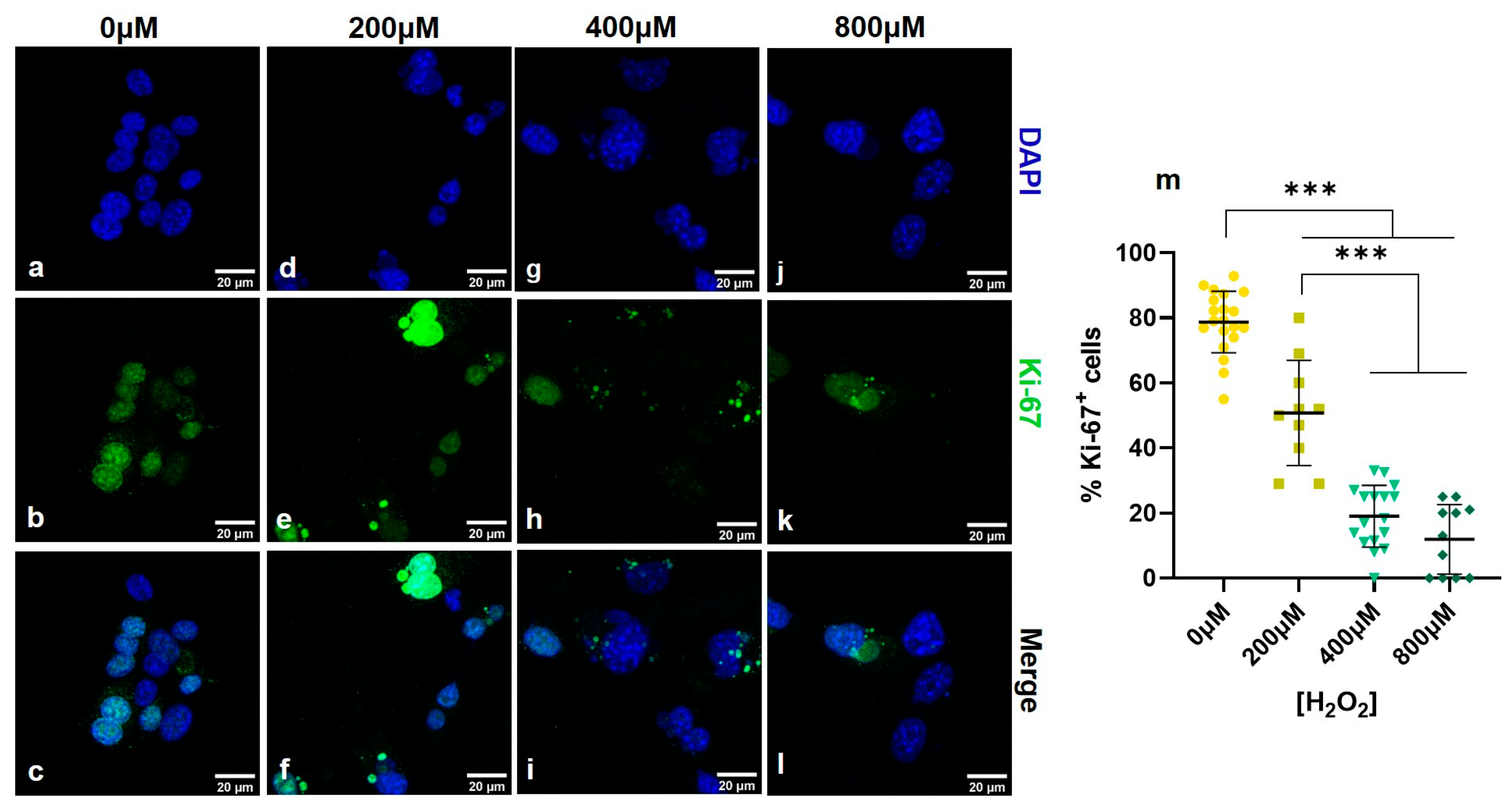
In this study, different concentrations of H2O2 were used to develop a reliable model of fibroblast senescence. Following an overnight proliferation period, the cells were treated with H2O2 for one hour, as described in the Materials and Methods Section, to induce senescence while avoiding detrimental cytotoxicity. Analysis of H2O2 concentrations revealed a dose-dependent reduction in cell proliferative capacity. Specifically, the expression of Ki-67, a nuclear marker of cell proliferation [59], was prevalent in most untreated cells (Figure 1a–c) but significantly decreased in cells treated with 800 μM H2O2, where only a small subset of cells remained Ki-67-positive (Figure 1j–l). Quantification of Ki-67 staining revealed a decrease in the percentage of positive cells from 80% in the untreated samples to less than 20% in cultures treated with relatively high concentrations of H2O2 after 10 days in culture (Figure 1m). Although H2O2 is generally regarded as a cytotoxic agent, 800 μM H2O2 is optimal for inducing senescence without causing significant cell death or detachment.
Figure 1. Confocal microscope images of the stained nuclei (40× magnification, scale bar: 20 μm) (al) of NIH3T3 fibroblasts cultured on glass coverslips for 10 days after treatment with different concentrations of H2O2 [0 μM (ac), 200 μM (df), 400 μM (gi) and 800 μM (jl)]. The nuclei of the cells are visualized with blue (DAPI), while the nuclei of proliferating cells are shown in green (anti-Ki-67 antibody). The graph (m) presents the percentage of Ki-67-positive cells, indicating their proliferative capacity, at various concentrations of H2O2. The graph shows a significant decrease in the population of Ki-67-positive cells with increasing treatment concentrations. The data were subjected to one-way ANOVA with a post hoc Tukey’s HSD test for multiple comparisons between the groups (*** p < 0.001). The error bars denote the standard deviation of the mean. The experiments were performed with a minimum of three independent biological replicates. The confocal images were analyzed using maximum intensity projections generated from z-stacks consisting of 10–12 optical planes.
Photonics 12 00919 g001
This was further indicated by the examination of β-galactosidase activity, a hallmark of cellular senescence. During this pH-sensitive staining procedure, the chromogenic substrate 5-bromo-4-chloro-3-indolyl β-D-galactopyranoside (X-gal) was used to detect cytochemically endogenous β-galactosidase activity in mammalian cells [18,60,61]. The optical microscopy images revealed that, after 10 days of culture, the untreated cells exhibited no morphological alterations (Figure 2a). In contrast, cells treated with 200 μM and 400 μM H2O2 presented initial morphological changes and an increase in β-galactosidase-stained cells (blue color). Similarly to Ki-67 expression, the most profound changes in cell size, as well as β-galactosidase activity, were observed in fibroblasts treated with 800 μΜ H2O2 (Figure 2d). This senescence-associated enzyme activity, which is absent in pre-senescent, quiescent, or terminally differentiated cells [18], strongly indicates that treatment with 800 μM H2O2 for one hour is an effective method for inducing senescence in fibroblasts.
Figure 2. Optical microscope images (10× magnification, scale bar: 200 μm) of β-galactosidase staining after 10 days of (a) untreated fibroblast cultures treated with (b) 200 μM, (c) 400 μM, and (d) 800 μM H2O2. The majority of senescent fibroblasts were observed after 800 μM treatment because of their distinct enlarged morphology, as well as blue staining of β-galactosidase. The experiments were performed with a minimum of three independent biological replicates.
Photonics 12 00919 g002
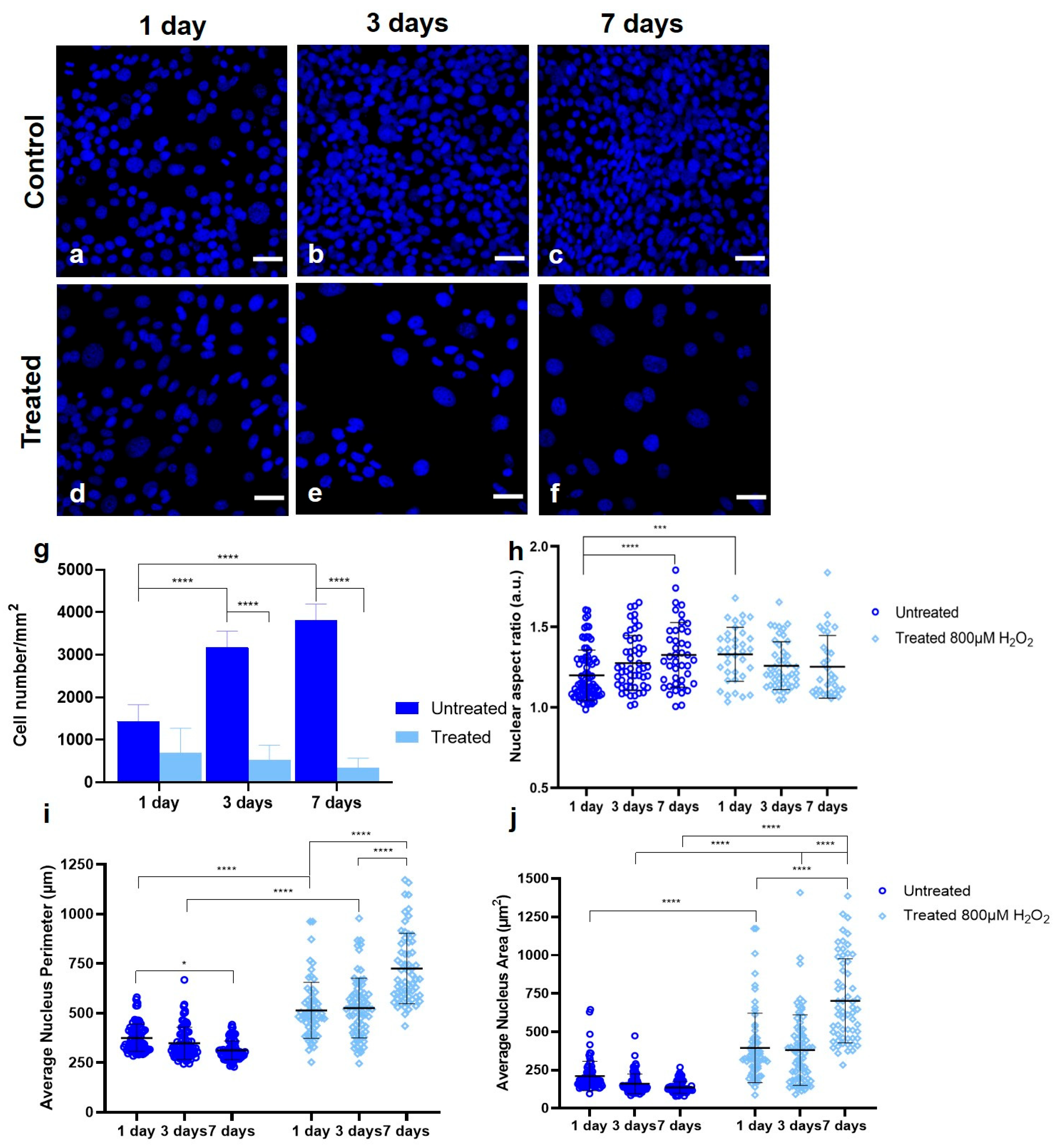
The increase in the number of senescence-associated β-galactosidase-positive cells (Figure 2b–d) can be correlated with a reduction in cell density (Figure 3d–g) and a notable increase in both cell (Figure 3g) and nuclear size (Figure 3i,j). To further characterize the established senescent fibroblast model induced by 800 μM H2O2, nuclear changes were examined. Untreated cells exhibited extended proliferative capacity, as also reported by Ki-67 expression (Figure 1), with a progressive increase in cell number from day 1 to day 7 of culture (Figure 3g). Notably, after 1 day of culture, fewer senescent cells than untreated cells were observed, indicating the onset of senescence, as well as the relative cytotoxicity of H2O2. In addition, the number of senescent (treated) fibroblasts decreased over time, although the difference was not statistically significant. These findings further support the effectiveness of this senescence induction method, which limits cell proliferation without causing extensive cell death.
Quantification of the nuclear aspect ratio (long to short nuclear axis) revealed that the nuclear shape remained consistent across conditions, with values close to 1, indicating a generally circular nucleus (Figure 3h). However, the nuclear perimeter and area were significantly greater in senescent cells than in control samples after 7 days of culture (Figure 3i,j), highlighting the pronounced hypertrophic characteristic of senescent fibroblasts. According to Wallis et al. [62], the original observations of cell and nuclear morphology during senescence could facilitate the use of “first-pass” tools to detect senescent and proliferating cells, since it is currently established that cellular and nuclear enlargement may trigger mechanosensitive calcium channels, causing alterations in lipid metabolism and chromatin reorganization, among other processes [63].
Figure 3. Confocal microscope images of the stained nuclei (40× magnification, scale bar: 50 μm) (af) of NIH3T3 fibroblasts cultured on glass coverslips for 1, 3, and 7 days after treatment with H2O2 for 1 hr [0 μM (ac) and 800 μM (df)]. The nuclei of the cells are visualized with a blue color (Hoechst 33342). Quantification graphs of (g) the number of fibroblasts/mm2, (h) the nuclear aspect ratio (long nuclear axis to short nuclear axis), (i) the average nucleus perimeter, and (j) the average nucleus area for each timepoint (1, 3, and 7 days). The untreated fibroblasts are presented in dark blue (circles), and those treated with light blue (rhombuses) are presented. The data were subjected to one-way ANOVA with a post hoc Tukey’s HSD test for multiple comparisons between the groups (* p < 0.05, *** p < 0.001 and **** p < 0.0001). The error bars denote the standard deviation of the mean. The experiments were performed with a minimum of three independent biological replicates. The confocal images were analyzed using maximum intensity projections generated from z-stacks consisting of 10–12 optical planes.
Photonics 12 00919 g003

3.2. Simultaneous THG Imaging of Unstained Lipids and TPEF of Ki-67-Positive Nuclei

Three-dimensional scanning was performed on untreated (Figure 4a,b) and treated (Figure 4c,d) fibroblasts that were cultured on glass coverslips for 7 days. THG signals were detected from the lipid domains (cyan), and TPEF signals arose from the Ki-67-positive nuclei (red hot), reflecting their proliferative tendency.
Compared with untreated fibroblasts, treated fibroblasts (Figure 4c,d) presented a substantial increase in cell size, with most appearing two or three times larger. The nuclei were significantly enlarged, and the lipid droplets were both larger and more abundant (Figure 4e–g). Furthermore, the fluorescence intensity of Ki-67 was greatly reduced, indicating a decrease in the proliferative capacity of the treated fibroblasts. The findings obtained via nonlinear microscopy were in good agreement with those reported via confocal microscopy (Figure 3), indicating an increase in cell and nuclear size as well as a decrease in Ki-67 intensity in the treated cells.
Figure 4. Nonlinear imaging of (i) untreated fibroblasts (a,b) and (ii) those treated with 800 μM H2O2 (c,d) after 7 days of culture. THG (cyan) reveals the label-free lipid domains, whereas TPEF (red hot) arises from the stained Ki-67 (2nd antibody: CF568 goat anti-mouse)-positive cells in the nucleus. Quantification graphs of (e) the lipid index (a.u.), (f) the mean lipid droplet size (μm2) and (g) comparison of the total lipid area (μm2) of untreated (yellow circles) and treated fibroblasts with 800 μΜ H2O2 (blue diamonds) after 7 days of culture; (eg) data points per replicate (Set 1, Set 2, Set 3) of each condition; untreated—yellow circles (Nset1 = 15, Nset2 = 17, Nset3 = 16) and treated—blue diamonds (Nset1 = 16, Nset2 = 18, Nset3 = 24) are presented. The data were subjected to one-way ANOVA with a post hoc Tukey’s HSD test for multiple comparisons between the groups. The significance between the groups of data is denoted from the bars on top (* p < 0.05, ** p < 0.01, *** p < 0.001). The error bars denote the standard deviation of the mean. A total of 48 untreated fibroblasts and 58 fibroblasts treated with 800 μΜ H2O2 from three independent experiments were analyzed.
Photonics 12 00919 g004
A total of 48 untreated and 58 treated cells were imaged and quantified, revealing a significant difference in total lipid content per cell area (Figure 4e) and total lipid area per cell (Figure 4g). In addition, the senescent cells presented a significant increase in the average droplet size per cell (Figure 4f). This is shown in Figure 4a–d qualitatively but also quantitatively through the analysis revealed in Figure 4e–g. For the quantification of the THG signals, three independent biological experiments were performed for each population. The minimum number of cells quantified from each independent experiment was as follows: untreated (Nset1 = 15, Nset2 = 17, Nset3 = 16) and treated (Nset1 = 16, Nset2 = 18, Nset3 = 24). The graphs in Figure 4e–g present the data values for e, the lipid content per cell area (a.u), f, the mean size of the lipid droplets per cell (μm2), and g, the total lipid area per cell for every independent experiment (μm2). As expected, the total lipid area per cell increased significantly upon senescence, and this was noted not only in the significant increase in the lipid droplet size, but also in the significant increase in the lipid index. The total lipid content was normalized to the size of the cell. The process of the quantification is described in the Materials and Methods Section and in Supplementary Figure S1.
These findings suggest that lipid metabolism is correlated with senescence and, specifically, that both the cell size, the nuclear size, and lipid content of fibroblasts increase as cells reach senescence. THG could be a promising technique for revealing senescence through lipid content quantification.
To further support the label-free imaging capability of THG microscopy, we performed proof-of-concept experiments on nonfixed NIH 3T3 cells. Cells were cultured for 7 days and then imaged in PBS to preserve viability. Supplementary Figure S2 shows increased lipid signal in H2O2-treated cells (Figure S2c,d) compared to untreated controls (Figure S2a,b), confirming that THG can detect lipid accumulation in live, unfixed samples.

4. Discussion

There is a growing need for the development of advanced imaging techniques that enhance existing tools for the label-free detection, characterization, and imaging of biological samples at length scales ranging from single molecules to individual cells to the tissue level. The use of THG microscopy in this study highlights its potential as a powerful, cutting-edge technique for studying biological processes in a label-free, noninvasive manner. This technology offers high contrast and increased biological sample penetration depth. It also permits precise quantitative analysis of the obtained data.
Traditional methods of studying senescence often rely on chemical labels or fluorescent markers, which may alter cellular behavior or have limited specificity. While traditional senescence detection techniques—such as immunostaining for p16INK4a or p21CIP1 or the profiling of SASP factors—offer molecular specificity, they typically require fixation, lysis, and labeling steps that limit their use in live-cell analysis and may introduce artifacts. In contrast, third harmonic generation (THG) microscopy enables label-free, noninvasive imaging of structural features like lipid accumulation in intact, live cells or tissues. THG microscopy provides an intrinsic imaging approach, exploiting nonlinear optical effects to visualize cellular structures, including lipid droplets, without the need for external dyes. This enables researchers to capture real-time dynamics of lipid metabolism in living cells with minimal disruption. Our qualitative measurements on unfixed cells (Supplementary Figure S2) further demonstrate that THG microscopy can be used to detect lipid changes in live, unfixed cells, underscoring its utility in real-time or in situ applications. Although THG does not provide direct molecular information, it offers a valuable means of identifying phenotypic hallmarks of senescence with high spatial resolution and minimal sample preparation. THG is not intended to replace classical molecular assays, but rather to complement them by providing real-time morphological and metabolic context. This is particularly relevant in applications where in situ or longitudinal monitoring of senescent cell populations is desired.
The observed increase in lipid content in senescent cells, detected via the quantification of the collected THG signals, may reflect underlying changes in key metabolic pathways, such as adipogenesis, lipid storage, and fatty acid oxidation, which could be crucial in driving or maintaining the senescent state. Lipids play diverse roles in membrane structure, energy storage, and cell signaling, but their specific contributions to aging and age-related diseases, such as cancer, neurodegeneration, and cardiovascular diseases, are only beginning to be understood. This study emphasizes the need for further exploration into how altered lipid metabolism might contribute to the chronic inflammatory milieu of senescent cells (SASPs).
This study provides compelling evidence linking lipid metabolism to cellular senescence, revealing a deeper, previously underestimated connection between lipid dynamics and aging. THG imaging microscopy has been employed as a label-free and nondestructive diagnostic tool for identifying senescence in fibroblasts through the precise quantification of lipid accumulation. These findings highlight lipid accumulation as a hallmark of senescence in fibroblasts, suggesting that regulating the lipid content could serve as a valuable biomarker for identifying senescent cells. This insight opens promising avenues for aging research, particularly in developing therapies aimed at selectively targeting senescent cells (senolytics) or mitigating their effects (senomorphics) [64,65].
Our preliminary efforts using alternative senescence inducers underscore the need for carefully chosen models in label-free imaging studies. Further work is ongoing to validate THG microscopy across different cell types and senescence pathways.
The impact of this work is significant, as multiphoton microscopes are now commercially available as compact, portable, and user-friendly imaging systems utilizing a single fs laser beam. Furthermore, nonlinear modalities such as SHG and THG can be integrated by simply upgrading a standard multiphoton microscope, providing complementary information from the sample. While THG signals arise from all inhomogeneities within the sample, the strongest signal arises from the lipid bodies, offering the advantage of noninvasive, label-free characterization of the lipid profile of the specimen under investigation.
To assess the broader applicability of THG microscopy in detecting senescence-associated lipid changes, future studies should include a wider range of cell types and tissue specimens. Sections from young versus older animals could be studied to further validate this approach in physiologically relevant contexts.
Future studies should focus on the molecular regulators of lipid metabolism in senescent cells, including key enzymes such as lipases, acetyl-CoA carboxylase, and fatty acid synthase, as well as how lipid droplets are formed and processed. Investigating the interplay between lipid metabolism and mitochondrial dysfunction, another hallmark of senescence, could also yield valuable insights into how metabolic dysregulation promotes cellular aging. Additionally, combining THG microscopy with other advanced imaging modalities, such as Raman spectroscopy or fluorescence lifetime imaging, could further enhance our ability to distinguish and characterize senescent cells in different tissues. The combination of THG with other advanced imaging modalities could provide complementary data and further enhance our ability to distinguish and characterize senescent cells in different tissues. Since THG microscopy cannot provide specific chemical information, future studies including complementary modalities such as Raman spectroscopy could bridge the gap between phenotypic imaging and molecular characterization. Moreover, integration with machine learning-based image analysis could enhance throughput and diagnostic potential, enabling objective identification of senescent cells across diverse biological contexts.
These advances in imaging, combined with a better understanding of lipid metabolism in aging, may pave the way for diagnostic applications in age-related diseases, allowing for earlier detection and intervention. Targeting lipid pathways could also lead to new therapeutic strategies aimed at modulating senescence-associated pathologies, such as atherosclerosis or metabolic disorders, by restoring normal lipid homeostasis.

Supplementary Materials

The following supporting information can be downloaded at: https://www.mdpi.com/article/10.3390/photonics12090919/s1, Figure S1: Detailed description of all the steps for the lipid index estimation. Figure S2: Label-free THG (in green-blue pseudocolour) imaging of non-fixed cells.

Author Contributions

Conceptualization, C.F., A.R., G.F., P.M. and M.M.; methodology, M.M., E.K., P.M., A.K. and K.S.; validation, M.M., E.K., A.R. and G.F.; formal analysis, M.M. and E.K.; investigation, E.K., M.M., P.M., K.S. and A.K.; resources, A.R. and G.F.; data curation, M.M. and E.K.; writing—original draft preparation, M.M. and E.K.; writing—review and editing, M.M., E.K., G.F. and A.R.; supervision, C.F., A.R. and G.F. All authors have read and agreed to the published version of the manuscript.

Funding

This research was funded by GA No 101131771 Lasers4EU and GA No 871124_LASERLAB-EUROPE and co-funded by the European Union’s Horizon Europe and GA No. 101132448-HORIZON-CL2-2023-HERITAGE-01-01 (iPhotoCult).

Informed Consent Statement

Not applicable.

Data Availability Statement

Dataset available on request from the authors.

Conflicts of Interest

The authors declare no conflicts of interest.

References

  1. Hayflick, L.; Moorhead, P.S. The serial cultivation of human diploid cell strains. Exp. Cell Res. 1961, 25, 585–621. [Google Scholar] [CrossRef]
  2. González-Gualda, E.; Baker, A.G.; Fruk, L.; Muñoz-Espín, D. A guide to assessing cellular senescence in vitro and in vivo. FEBS J. 2021, 288, 56–80. [Google Scholar] [CrossRef]
  3. He, S.; Sharpless, N.E. Senescence in Health and Disease. Cell 2017, 169, 1000–1011. [Google Scholar] [CrossRef]
  4. Naylor, R.M.; Baker, D.J.; Van Deursen, J.M. Senescent cells: A novel therapeutic target for aging and age-related diseases. Clin. Pharmacol. Ther. 2013, 93, 105–116. [Google Scholar] [CrossRef]
  5. Childs, B.G.; Durik, M.; Baker, D.J.; Van Deursen, J.M. Cellular senescence in aging and age-related disease: From mechanisms to therapy. Nat. Med. 2015, 21, 1424–1435. [Google Scholar] [CrossRef] [PubMed]
  6. Fumagalli, M.; Rossiello, F.; Clerici, M.; Barozzi, S.; Cittaro, D.; Kaplunov, J.M.; Bucci, G.; Dobreva, M.; Matti, V.; Beausejour, C.M.; et al. Telomeric DNA damage is irreparable and causes persistent DNA-damage-response activation. Nat. Cell Biol. 2012, 14, 355–365. [Google Scholar] [CrossRef] [PubMed]
  7. Moiseeva, O.; Bourdeau, V.; Roux, A.; Deschênes-Simard, X.; Ferbeyre, G. Mitochondrial Dysfunction Contributes to Oncogene-Induced Senescence. Mol. Cell. Biol. 2009, 29, 4495–4507. [Google Scholar] [CrossRef]
  8. Serrano, M.; Lin, A.W.; McCurrach, M.E.; Beach, D.; Lowe, S.W. Oncogenic ras provokes premature cell senescence associated with accumulation of p53 and p16(INK4a). Cell 1997, 88, 593–602. [Google Scholar] [CrossRef]
  9. Wiley, C.D.; Velarde, M.C.; Lecot, P.; Liu, S.; Sarnoski, E.A.; Freund, A.; Shirakawa, K.; Lim, H.W.; Davis, S.S.; Ramanathan, A.; et al. Mitochondrial dysfunction induces senescence with a distinct secretory phenotype. Cell Metab. 2016, 23, 303–314. [Google Scholar] [CrossRef] [PubMed]
  10. Deng, Y.; Chang, S. Role of telomeres and telomerase in genomic instability, senescence and cancer. Lab. Investig. 2007, 87, 1071–1076. [Google Scholar] [CrossRef]
  11. Muñoz-Espín, D.; Serrano, M. Cellular senescence: From physiology to pathology. Nat. Rev. Mol. Cell Biol. 2014, 15, 482–496. [Google Scholar] [CrossRef]
  12. Wang, L.; Lankhorst, L.; Bernards, R. Exploiting senescence for the treatment of cancer. Nat. Rev. Cancer 2022, 22, 340–355. [Google Scholar] [CrossRef]
  13. Herranz, N.; Gil, J. Mechanisms and functions of cellular senescence. J. Clin. Investig. 2018, 128, 1238–1246. [Google Scholar] [CrossRef] [PubMed]
  14. Pack, L.R.; Daigh, L.H.; Meyer, T. Putting the brakes on the cell cycle: Mechanisms of cellular growth arrest. Curr. Opin. Cell Biol. 2019, 60, 106–113. [Google Scholar] [CrossRef] [PubMed]
  15. Campisi, J. Aging, cellular senescence, and cancer. Annu. Rev. Physiol. 2013, 75, 685–705. [Google Scholar] [CrossRef]
  16. López-Otín, C.; Blasco, M.A.; Partridge, L.; Serrano, M.; Kroemer, G. The hallmarks of aging. Cell 2013, 153, 1194–1217. [Google Scholar] [CrossRef]
  17. Ogrodnik, M.; Zhu, Y.; Langhi, L.G.P.; Tchkonia, T.; Krüger, P.; Fielder, E.; Victorelli, S.; Ruswhandi, R.A.; Giorgadze, N.; Pirtskhalava, T.; et al. Obesity-Induced Cellular Senescence Drives Anxiety and Impairs Neurogenesis. Cell Metab. 2019, 29, 1061–1077.e8. [Google Scholar] [CrossRef] [PubMed]
  18. Dimri, G.P.; Lee, X.; Basile, G.; Acosta, M.; Scott, G.; Roskelley, C.; Medrano, E.E.; Linskens, M.; Rubelj, I.; Pereira-Smith, O.; et al. A biomarker that identifies senescent human cells in culture and in aging skin in vivo. Proc. Natl. Acad. Sci. USA 1995, 92, 9363–9367. [Google Scholar] [CrossRef]
  19. Bloom, J.; Cross, F.R. Multiple levels of cyclin specificity in cell-cycle control. Nat. Rev. Mol. Cell Biol. 2007, 8, 149–160. [Google Scholar] [CrossRef]
  20. Wang, N.; He, Y.; Liu, S.; Makarcyzk, M.J.; Lei, G.; Chang, A.; Alexander, P.G.; Hao, T.; Padget, A.M.; de Pedro, N.; et al. Engineering osteoarthritic cartilage model through differentiating senescent human mesenchymal stem cells for testing disease-modifying drugs. Sci. China Life Sci. 2022, 65, 309–327. [Google Scholar] [CrossRef]
  21. Frippiat, C.; Dewelle, J.; Remacle, J.; Toussaint, O. Signal transduction in H2O2-induced senescence-like phenotype in human diploid fibroblasts. Free Radic. Biol. Med. 2002, 33, 1334–1346. [Google Scholar] [CrossRef]
  22. Petrova, N.V.; Velichko, A.K.; Razin, S.V.; Kantidze, O.L. Small molecule compounds that induce cellular senescence. Aging Cell 2016, 15, 999–1017. [Google Scholar] [CrossRef] [PubMed]
  23. Hamdi, D.H.; Chevalier, F.; Groetz, J.E.; Durantel, F.; Thuret, J.Y.; Mann, C.; Saintigny, Y. Comparable Senescence Induction in Three-dimensional Human Cartilage Model by Exposure to Therapeutic Doses of X-rays or C-ions. Int. J. Radiat. Oncol. Biol. Phys. 2016, 95, 139–146. [Google Scholar] [CrossRef]
  24. Bai, J.; Wang, Y.; Wang, J.; Zhai, J.; He, F.; Zhu, G. Irradiation-induced senescence of bone marrow mesenchymal stem cells aggravates osteogenic differentiation dysfunction via paracrine signaling. Am. J. Physiol.-Cell Physiol. 2020, 318, C1005–C1017. [Google Scholar] [CrossRef]
  25. Bielak-Zmijewska, A.; Wnuk, M.; Przybylska, D.; Grabowska, W.; Lewinska, A.; Alster, O.; Korwek, Z.; Cmoch, A.; Myszka, A.; Pikula, S.; et al. A comparison of replicative senescence and doxorubicin-induced premature senescence of vascular smooth muscle cells isolated from human aorta. Biogerontology 2014, 15, 47–64. [Google Scholar] [CrossRef]
  26. Millner, A.; Ekin Atilla-Gokcumen, G. Lipid players of cellular senescence. Metabolites 2020, 10, 339. [Google Scholar] [CrossRef] [PubMed]
  27. Lizardo, D.Y.; Lin, Y.L.; Gokcumen, O.; Atilla-Gokcumen, G.E. Regulation of lipids is central to replicative senescence. Mol. Biosyst. 2017, 13, 498–509. [Google Scholar] [CrossRef]
  28. Saitou, M.; Lizardo, D.Y.; Taskent, R.O.; Millner, A.; Gokcumen, O.; Atilla-Gokcumen, G.E. An evolutionary transcriptomics approach links CD36 to membrane remodeling in replicative senescence. Mol. Omics 2018, 14, 237–246. [Google Scholar] [CrossRef]
  29. Skotland, T.; Hessvik, N.P.; Sandvig, K.; Llorente, A. Exosomal lipid composition and the role of ether lipids and phosphoinositides in exosome biology. J. Lipid Res. 2019, 60, 9–18. [Google Scholar] [CrossRef]
  30. Saxton, R.A.; Sabatini, D.M. mTOR Signaling in Growth, Metabolism, and Disease. Cell 2017, 168, 960–976. [Google Scholar] [CrossRef] [PubMed]
  31. Hamsanathan, S.; Gurkar, A.U. Lipids as Regulators of Cellular Senescence. Front. Physiol. 2022, 13, 796850. [Google Scholar] [CrossRef]
  32. Mutlu, A.S.; Duffy, J.; Wang, M.C. Lipid metabolism and lipid signals in aging and longevity. Dev. Cell 2021, 56, 1394–1407. [Google Scholar] [CrossRef]
  33. Papsdorf, K.; Brunet, A. Linking Lipid Metabolism to Chromatin Regulation in Aging. Trends Cell Biol. 2019, 29, 97–116. [Google Scholar] [CrossRef]
  34. Fukumoto, S.; Fujimoto, T. Deformation of lipid droplets in fixed samples. Histochem. Cell Biol. 2002, 118, 423–428. [Google Scholar] [CrossRef] [PubMed]
  35. Cirulis, J.T.; Strasser, B.C.; Scott, J.A.; Ross, G.M. Optimization of staining conditions for microalgae with three lipophilic dyes to reduce precipitation and fluorescence variability. Cytom. Part A 2012, 81A, 618–626. [Google Scholar] [CrossRef]
  36. Khoury, S.; Canlet, C.; Lacroix, M.Z.; Berdeaux, O.; Jouhet, J.; Bertrand-Michel, J. Quantification of lipids: Model, reality, and compromise. Biomolecules 2018, 8, 174. [Google Scholar] [CrossRef] [PubMed]
  37. Fuchs, J.; Neuberger, T.; Rolletschek, H.; Schiebold, S.; Nguyen, T.H.; Borisjuk, N.; Börner, A.; Melkus, G.; Jakob, P.; Borisjuk, L. A noninvasive platform for imaging and quantifying oil storage in submillimeter tobacco seed. Plant Physiol. 2013, 161, 583–593. [Google Scholar] [CrossRef]
  38. Brescia, M.A.; Pugliese, T.; Hardy, E.; Sacco, A. Compositional and structural investigations of ripening of table olives, Bella della Daunia, by means of traditional and magnetic resonance imaging analyses. Food Chem. 2007, 105, 400–404. [Google Scholar] [CrossRef]
  39. Syed, A.; Smith, E.A. Raman imaging in cell membranes, lipid-rich organelles, and lipid bilayers. Annu. Rev. Anal. Chem. 2017, 10, 271–291. [Google Scholar] [CrossRef]
  40. Zumbusch, A.; Holtom, G.R.; Xie, X.S. Three-dimensional vibrational imaging by coherent anti-stokes raman scattering. Phys. Rev. Lett. 1999, 82, 4142–4145. [Google Scholar] [CrossRef]
  41. Cheng, J.X.; Volkmer, A.; Book, L.D.; Xie, X.S. Multiplex coherent anti-stokes Raman scattering microspectroscopy and study of lipid vesicles. J. Phys. Chem. B 2002, 106, 8493–8498. [Google Scholar] [CrossRef]
  42. Freudiger, C.W.; Min, W.; Saar, B.G.; Lu, S.; Holtom, G.R.; He, C.; Tsai, J.C.; Kang, J.X.; Xie, X.S. Label-free biomedical imaging with high sensitivity by stimulated raman scattering microscopy. Science 2008, 322, 1857–1861. [Google Scholar] [CrossRef]
  43. Rinia, H.A.; Burger, K.N.J.; Bonn, M.; Müller, M. Quantitative label-free imaging of lipid composition and packing of individual cellular lipid droplets using multiplex CARS microscopy. Biophys. J. 2008, 95, 4908–4914. [Google Scholar] [CrossRef] [PubMed]
  44. Débarre, D.; Supatto, W.; Pena, A.M.; Fabre, A.; Tordjmann, T.; Combettes, L.; Schanne-Klein, M.C.; Beaurepaire, E. Imaging lipid bodies in cells and tissues using third-harmonic generation microscopy. Nat. Methods 2006, 3, 47–53. [Google Scholar] [CrossRef]
  45. Palikaras, K.; Mari, M.; Petanidou, B.; Pasparaki, A.; Filippidis, G.; Tavernarakis, A.N. Ectopic fat deposition contributes to age-associated pathology in caenorhabditis elegans. J. Lipid Res. 2017, 58, 72–80. [Google Scholar] [CrossRef] [PubMed]
  46. Palikaras, K.; Mari, M.; Ploumi, C.; Princz, A.; Filippidis, G.; Tavernarakis, N. Age-dependent nuclear lipid droplet deposition is a cellular hallmark of aging in Caenorhabditis elegans. Aging Cell 2023, 22, e13788. [Google Scholar] [CrossRef]
  47. Gavgiotaki, E.; Filippidis, G.; Markomanolaki, H.; Kenanakis, G.; Agelaki, S.; Georgoulias, V.; Athanassakis, I. Distinction between breast cancer cell subtypes using third harmonic generation microscopy. J. Biophotonics 2017, 10, 1152–1162. [Google Scholar] [CrossRef] [PubMed]
  48. Gavgiotaki, E.; Filippidis, G.; Tsafas, V.; Bovasianos, S.; Kenanakis, G.; Georgoulias, V.; Tzardi, M.; Agelaki, S.; Athanassakis, I. Third Harmonic Generation microscopy distinguishes malignant cell grade in human breast tissue biopsies. Sci. Rep. 2020, 10, 11055. [Google Scholar] [CrossRef]
  49. Rehberg, M.; Krombach, F.; Pohl, U.; Dietzel, S. Label-free 3D visualization of cellular and tissue structures in intact muscle with second and third harmonic generation microscopy. PLoS ONE 2011, 6, e28237. [Google Scholar] [CrossRef]
  50. Zipfel, W.R.; Williams, R.M.; Webb, W.W. Nonlinear magic: Multiphoton microscopy in the biosciences. Nat. Biotechnol. 2003, 21, 1369–1377. [Google Scholar] [CrossRef]
  51. Barad, Y.; Eisenberg, H.; Horowitz, M.; Silberberg, Y. Nonlinear scanning laser microscopy by third harmonic generation. Appl. Phys. Lett. 1997, 70, 922–924. [Google Scholar] [CrossRef]
  52. Weigelin, B.; Bakker, G.J.; Friedl, P. Third harmonic generation microscopy of cells and tissue organization. J. Cell Sci. 2016, 129, 245–255. [Google Scholar] [CrossRef]
  53. Gavgiotaki, E.; Filippidis, G.; Kalognomou, M.; Tsouko, A.A.; Skordos, I.; Fotakis, C.; Athanassakis, I. Third Harmonic Generation microscopy as a reliable diagnostic tool for evaluating lipid body modification during cell activation: The example of BV-2 microglia cells. J. Struct. Biol. 2015, 189, 105–113. [Google Scholar] [CrossRef]
  54. Kyvelidou, C.; Tserevelakis, G.J.; Filippidis, G.; Ranella, A.; Kleovoulou, A.; Fotakis, C.; Athanassakis, I. Following the course of pre-implantation embryo patterning by non-linear microscopy. J. Struct. Biol. 2011, 176, 379–386. [Google Scholar] [CrossRef]
  55. Mari, M.; Filippidis, G.; Palikaras, K.; Petanidou, B.; Fotakis, C.; Tavernarakis, N. Imaging ectopic fat deposition in caenorhabditis elegans muscles using nonlinear microscopy. Microsc. Res. Tech. 2015, 78, 523–528. [Google Scholar] [CrossRef]
  56. Gavgiotaki, E.; Filippidis, G.; Zerva, I.; Kenanakis, G.; Archontakis, E.; Agelaki, S.; Georgoulias, V.; Athanassakis, I. Detection of the T cell activation state using nonlinear optical microscopy. J. Biophotonics 2019, 12, e201800277. [Google Scholar] [CrossRef] [PubMed]
  57. Mari, M.; Voutyraki, C.; Zacharioudaki, E.; Delidakis, C.; Filippidis, G. Lipid content evaluation of Drosophila tumour associated haemocytes through Third Harmonic Generation measurements. J. Biophotonics 2023, 16, e202300171. [Google Scholar] [CrossRef] [PubMed]
  58. Schindelin, J.; Arganda-Carreras, I.; Frise, E.; Kaynig, V.; Longair, M.; Pietzsch, T.; Preibisch, S.; Rueden, C.; Saalfeld, S.; Schmid, B.; et al. Fiji: An open-source platform for biological-image analysis. Nat. Methods 2012, 9, 676–682. [Google Scholar] [CrossRef]
  59. Carpenter, A.E.; Jones, T.R.; Lamprecht, M.R.; Clarke, C.; Kang, I.H.; Friman, O.; Guertin, D.A.; Chang, J.H.; Lindquist, R.A.; Moffat, J.; et al. CellProfiler: Image analysis software for identifying and quantifying cell phenotypes. Genome Biol. 2006, 7, R100. [Google Scholar] [CrossRef] [PubMed]
  60. Kamentsky, L.; Jones, T.R.; Fraser, A.; Bray, M.A.; Logan, D.J.; Madden, K.L.; Ljosa, V.; Rueden, C.; Eliceiri, K.W.; Carpenter, A.E. Improved structure, function and compatibility for cellprofiler: Modular high-throughput image analysis software. Bioinformatics 2011, 27, 1179–1180. [Google Scholar] [CrossRef]
  61. Stirling, D.R.; Swain-Bowden, M.J.; Lucas, A.M.; Carpenter, A.E.; Cimini, B.A.; Goodman, A. CellProfiler 4: Improvements in speed, utility and usability. BMC Bioinform. 2021, 22, 433. [Google Scholar] [CrossRef]
  62. Peán, C.B.; Schiebler, M.; Tan, S.W.S.; Sharrock, J.A.; Kierdorf, K.; Brown, K.P.; Maserumule, M.C.; Menezes, S.; Pilátová, M.; Bronda, K.; et al. Regulation of phagocyte triglyceride by a STAT-ATG2 pathway controls mycobacterial infection. Nat. Commun. 2017, 8, 14642. [Google Scholar] [CrossRef] [PubMed]
  63. Maekawa, M.; Fairn, G.D. Molecular probes to visualize the location, organization and dynamics of lipids. J. Cell Sci. 2014, 127, 4801–4812. [Google Scholar] [CrossRef] [PubMed]
  64. Rambold, A.S.; Cohen, S.; Lippincott-Schwartz, J. Fatty acid trafficking in starved cells: Regulation by lipid droplet lipolysis, autophagy, and mitochondrial fusion dynamics. Dev. Cell 2015, 32, 678–692. [Google Scholar] [CrossRef] [PubMed]
  65. Supatto, W.; Truong, T.V.; Débarre, D.; Beaurepaire, E. Advances in multiphoton microscopy for imaging embryos. Curr. Opin. Genet. Dev. 2011, 21, 538–548. [Google Scholar] [CrossRef]
Disclaimer/Publisher’s Note: The statements, opinions and data contained in all publications are solely those of the individual author(s) and contributor(s) and not of MDPI and/or the editor(s). MDPI and/or the editor(s) disclaim responsibility for any injury to people or property resulting from any ideas, methods, instructions or products referred to in the content.