Article

The Potential of Tunable Femtosecond Laser Light to Prevent Melanoma A375 Cell Growth: An In Vitro Investigation

1
Laser Institute for Research and Applications LIRA, Beni-Suef University, Beni-Suef 62511, Egypt
2
Anbar Health Department, Anbar Province, Ministry of Health, Ramadi 31001, Iraq
3
Microbial Chemistry Department, National Research Centre, Dokki, Giza 12311, Egypt
4
Department of Microbiology and Immunology, Faculty of Pharmacy, Beni-Suef University, Beni-Suef 62514, Egypt
5
Department of Engineering, Faculty of Advanced Technology and Multidiscipline, Universitas Airlangga, Surabaya 60115, Indonesia
*
Author to whom correspondence should be addressed.
Photonics 2025, 12(7), 694; https://doi.org/10.3390/photonics12070694
Submission received: 30 May 2025 / Revised: 3 July 2025 / Accepted: 8 July 2025 / Published: 10 July 2025
(This article belongs to the Section Biophotonics and Biomedical Optics)

Abstract

The incidence and mortality rates associated with melanoma are increasing. Due to their high proliferation rate, ability to self-renew, and resistance mechanisms, cancer cells often withstand conventional therapies such as radiation and chemotherapy. Therefore, further research is required to develop novel melanoma therapies with fewer adverse effects, but effective therapeutic impacts. This study aims to investigate how femtosecond laser treatment affects melanoma cells using the A375 cell line as an in vitro model. A375 melanoma cells were plated at a concentration of 104 cells per well in 96-well plates and incubated overnight; then, they were subjected to femtosecond laser irradiation for durations of 3, 5, or 10 min, maintaining a steady power of 100 mW. The laser operated across different wavelengths in the ultraviolet, visible, and infrared ranges. Cell viability was evaluated 24 h after irradiation using the MTT assay. The results showed the significant inhibition of melanoma cell growth with various femtosecond laser parameters, particularly at 380 and 400 nm. At 380 nm, the cell viability was reduced by approximately 90%, and at 400 nm by 73%, after 10 min of exposure. Additional reductions were observed at 420 nm (47%) and 440 nm (18%), while no significant effects were found at 700–780 nm. The most effective exposure time was 10 min. Femtosecond laser radiation exerts a noteworthy anticancer effect on A375 cells, particularly at specific wavelengths and exposure durations, underscoring the potential of femtosecond laser therapy for treating melanoma. Exploring the underlying mechanisms of these effects and evaluating the clinical potential of this treatment modality requires further research.

1. Introduction

Cancer is a prominent contributor to illness and mortality across the globe, impacting both developed and developing nations [1]. Malignant melanoma, which stems from neuroectodermal melanocytic cells, is a highly invasive form of cancer [2]. Melanoma, which is derived from melanocytes, is the most aggressive variant of skin cancer and is particularly concerning when it is detected in its later stages [3]. Worldwide, the incidence of melanoma is projected to range between 15 and 25 cases per 100,000 individuals, with an annual increase of 3% to 5%. Melanoma is responsible for 75% of all skin cancer deaths [4]. Although systemic chemotherapy is still the primary treatment for melanoma, the effectiveness of chemotherapeutics is limited by the emergence of resistance and dose-limiting toxicity [5]. The properties of cancer cells, including rapid proliferation, self-renewal, and other resistance mechanisms, make them resistant to therapies such as radiation and chemotherapy [6]. Furthermore, the current gold standard treatment for melanoma involves surgically removing the lesion followed by adjuvant therapies such as immunotherapy and chemotherapy. However, emerging adjuvant therapy methods are not without drawbacks, often leading to serious side effects [7,8]. Further studies are, therefore, required to develop novel melanoma therapies that have fewer adverse effects while having a potent therapeutic effect.
Femtosecond lasers have emerged as powerful tools in cancer research due to their unique capabilities. A femtosecond laser is a pulsed laser system in which the pulse duration is in the femtosecond range, offering numerous advantageous characteristics [9]. Ultrashort pulses achieve remarkably high intensities with minimal energy input, enabling targeted and precise energy delivery within biological samples. This precise deposition can induce a range of thermal, chemical, or mechanical reactions. Since femtosecond pulses are extremely short, they result in minimal damage and are considered safe as they do not generate heat or cause inflammation in biological samples. The absorption of laser energy by biological samples is influenced by the laser wavelength and the sample’s spectral properties, whereas femtosecond pulses provide advantages such as precise energy delivery, high spatial and temporal resolution, minimal phototoxicity, and clean, noninvasive, and controlled laser interactions. Their wavelengths can be adjusted across a wide spectrum (690–1040 nm), which is advantageous for optimizing laser settings. Unlike long pulsed or continuous wave (CW) lasers that rely on linear photon absorption and may produce excessive heat, leading to unintended tissue damage, femtosecond lasers operate with vastly higher peak powers, reducing these potential side effects. These lasers induce nonlinear absorption interactions, which contribute to their ability to achieve more precise and varied effects [10].
There are several medical uses for femtosecond lasers. It is currently utilized, for instance, in dental [11] and cataract surgeries [12]. Several studies have demonstrated that femtosecond laser pulses can elicit significant biological effects. Yoon et al. reported apoptosis-like cell death mediated by mitochondrial ROS generation [13]. Thegersen et al. observed the reproductive death of cancer cells across wavelengths from 200–800 nm [14], while Tirlapur and König provided evidence of femtosecond laser-induced DNA strand breaks in mammalian cells [15]. These mechanisms align with the anticancer potential of femtosecond lasers and support further exploration in this context. Furthermore, in basic biological research, the femtosecond laser is emerging as a powerful tool [16]. For example, it has potent antimicrobial properties [17,18]. The use of laser devices in oncology serves multiple purposes through various mechanisms. Specifically, laser irradiation within the wavelength range of 600–1000 nm and a power density of 5–150 mW/cm2 has been employed to manage side effects induced by chemoradiation in patients with head and neck cancer [19]. Furthermore, a recent study revealed that breast, skin, and bladder cancer cells were inhibited when exposed to aminolevulinate, which is photosensitized by a femtosecond laser [20].
To date, only a limited number of studies have examined the effects of pulsed lasers, particularly femtosecond lasers, on cancer. Notably, the same light source can yield varying effects on identical tissues depending on the parameters employed according to the biphasic dose–response curve that describes laser treatment [21]. Therefore, research should focus on determining the optimum laser light parameters, such as the pulse energy, wavelength, and exposure time, that can be used for melanoma treatment. Determining the efficient and optimum femtosecond laser parameters that can inhibit melanoma cancer cells has also not been considered in any of the published studies. Taha et al. previously demonstrated the suppression of breast cancer cell proliferation using a tunable femtosecond laser system, motivating the extension of this technique to other malignancies, such as melanoma, in the present study [22]. Unlike earlier studies that primarily investigated photodynamic effects using nanosecond or continuous-wave light sources, this study systematically explores the cytotoxicity of femtosecond laser light across ultraviolet-to-near-infrared wavelengths without the use of exogenous photosensitizers. This wavelength-sweeping approach uniquely identifies spectral windows of selective melanoma cell inhibition.
This study aimed to investigate how adjustable femtosecond laser irradiation affects the viability of melanoma cells in vitro, utilizing A375 cells as the experimental model. This research aimed to identify optimal parameters—such as the wavelength, power density, and duration of exposure—that significantly reduce the cellular activity.

2. Materials and Methods

2.1. Cell Culture

This study utilized the A375 human malignant melanoma cell line (ATCC). Cells were cultured in prewarmed DMEM (Biowest, Nuaillé, France) enriched with 10% fetal bovine serum (Gibco, Thermo Fisher Scientific, Waltham, MA, USA) and 1% penicillin/streptomycin. Incubation was maintained at 37 °C with 5% CO2 until cells reached 80–90% confluence, which was monitored through an inverted fluorescence microscope (DMi8, Leica Microsystems Inc., Wetzlar, Germany).

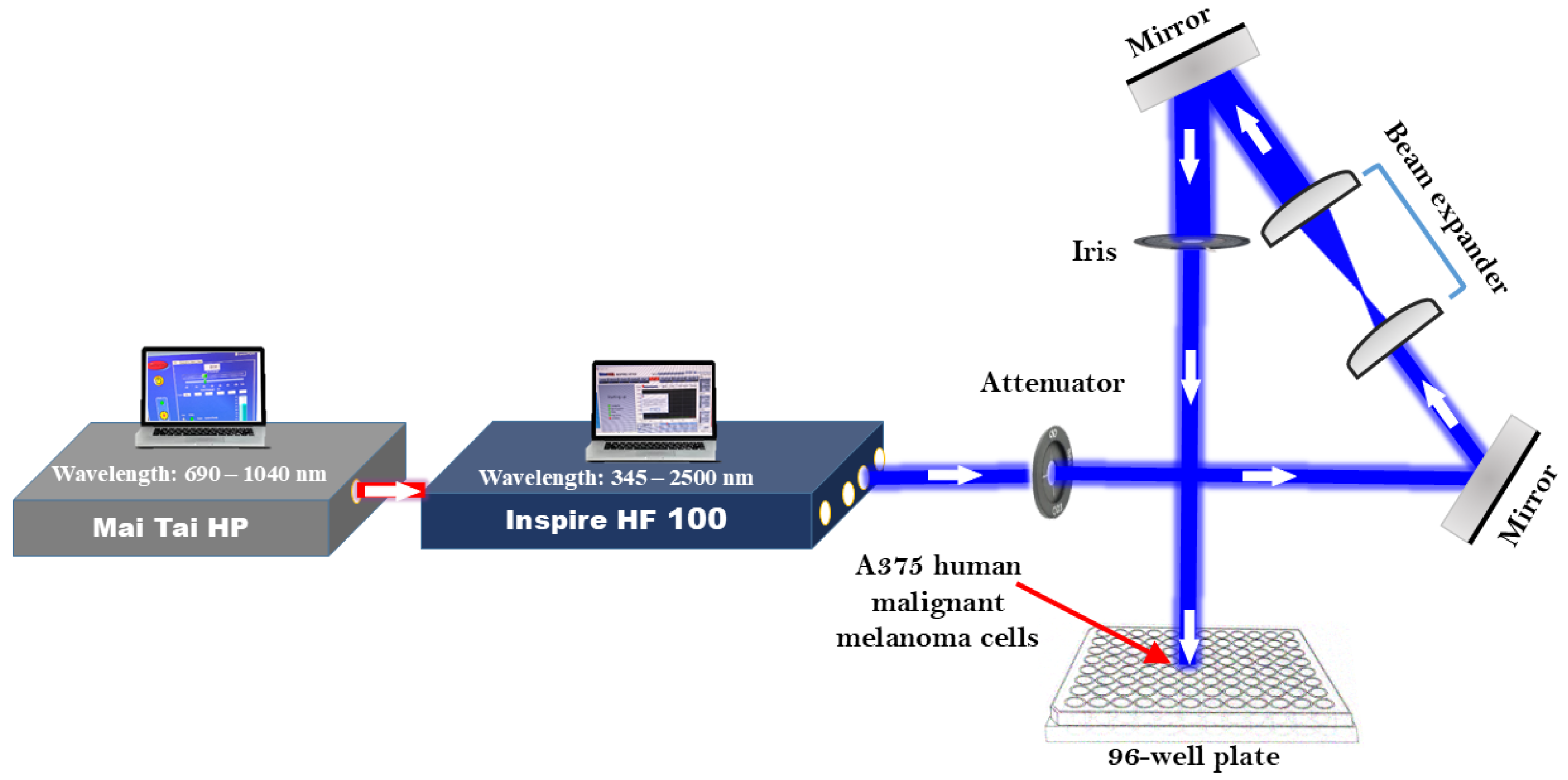
2.2. Setting up and Configuring Laser Systems

To investigate the effects of tunable femtosecond laser light on A375 human malignant melanoma cells, we used laser pulses produced by an INSPIRE HF100 laser system (Spectra-Physics, Milpitas, CA, USA). This system operates with a mode-locked femtosecond Ti: sapphire MAI TAI HP laser (Spectra-Physics), offering an average power output of approximately 1.5–2.9 W, an 80 MHz repetition rate, and a wavelength range spanning 690 to 1040 nm. A notable feature of the INSPIRE HF100 laser system is its ability to tune across different wavelength ranges. Besides its primary infrared pump wavelengths, the system also functions as a second harmonic generator (SHG) and an optical parametric oscillator (OPO). Tuning the Ti: sapphire laser and rotating the second harmonic nonlinear crystal (potassium dihydrogen phosphate) (KDP) enables the SHG mode to produce output wavelengths in the 345–520 nm range. Adjusting the lithium niobate (LN) crystals in the OPO mode provides two additional outputs: a signal from 490 to 750 nm and an idler from 930 to 2500 nm. These modes allow for wavelength tuning across a 345 to 2500 nm range, accessible through four distinct exit apertures. As illustrated in Figure 1, the laser beam was positioned approximately 10 cm above each well in a covered 96-well plate for irradiation, ensuring the cells were free from environmental contaminants. An initial laser beam diameter of approximately 2 mm was increased to 20 mm with a beam expander made up of two converging lenses. An iris was used to adjust the beam diameter, while a laser beam attenuator controlled the intensity of the laser light delivered to the cells. The laser beam was directed toward the cells in a covered 96-well plate using highly reflective mirrors, allowing femtosecond laser light at specified wavelengths to be applied to the seeded cells.
To accurately quantify the power received by the cells, we utilized a Newport 843R power meter. During the experiment, an initial irradiation power ranging from 114 to 116 mW was applied, yet only 100 mW of the laser power effectively reached the cells. This discrepancy is due to a 14% loss of the laser’s power as it passed through the cover of the 96-well plate. The experiment was conducted with a laser beam of a radius of r = 3 mm, an average power of Pave = 100 mW, a pulse duration of ∆t = 100 fs, and a repetition rate of f = 80 MHz. Based on these laser parameters and the experimental conditions, the energy per pulse E = Pave/f = 1.25 × 10⁻⁹ Joule, the peak power (power per pulse) Ppeak = E/∆t = 1.25 × 10⁴ Watts, the laser beam intensity (power density) I = Ppeak/π r2 = 4.5 × 10⁴ Watts/cm2, and the laser beam fluence (dose) for one-second exposure time F = E/π r2 = 8.8 × 10⁻⁹ Joule/cm2. In the cases of 3, 5, and 10 min durations, the doses were 1584 × 10−9 Joule/cm2, 2640 × 10−9 Joule/cm2, and 5280 × 10−9 Joule/cm2, respectively.

2.3. Femtosecond Laser Irradiation of A375 Cells

At a density of 10,000 cells per 200 µL, A375 cells were plated in 96-well plates. After an overnight incubation, the cells were exposed to various wavelengths of femtosecond laser light spanning the UV, visible, and infrared spectra (Figure 2). The exposure times used in the experiment were 3, 5, and 10 min for the selected wavelengths. The femtosecond laser beam diameter used for irradiation was 6 mm. The irradiation was performed in the dark to eliminate influences from other light sources. During all laser exposure experiments, the well temperature was monitored using a thermal microprobe. The temperature increase during irradiation was consistently below 0.5 °C, confirming the minimal thermal contribution. Twenty-four hours after femtosecond laser irradiation, the viability of the exposed cells was determined for each irradiation dose. For every dose, the experiments were run in triplicate.

2.4. Cell Viability Evaluation via MTT Assay

The impact of femtosecond laser irradiation on cell growth was examined through a 3-[4,5-dimethylthiazol-2-yl]-2,5-diphenyl tetrazolium bromide (MTT) assay. This technique depends on the activity of mitochondrial enzymes in living cells, which convert yellow MTT dye into purple formazan crystals. This technique is commonly employed for measuring cell proliferation in vitro because it correlates with both total mitochondrial activity and the number of viable cells. Before use, 5 mg/mL MTT stock solution (Life Technologies Corporation, M6494, Thermo Fisher Scientific, USA) was made by dissolving MTT powder in PBS in the dark, after which the solution was stored at 4 °C. Following 24 h of radiation, the medium was discarded and each well received 110 μL of MTT solution mixed with DMEM at a 1:10 ratio. The wells were incubated for three hours at 37 °C. After the MTT-containing medium was removed, 100 µL of DMSO was added to dissolve the remaining purple formazan and the plates were shaken for 12 min. Subsequently, the plates were incubated for an additional 15 min in a CO2 incubator. Finally, after shaking for 3 min on a plate shaker, the absorbance was measured at 570 nm using an ELISA reader (Agilent, Santa Clara, CA, USA).

2.5. Statistical Analysis

All experiments were performed in triplicate and the results are expressed as mean ± standard deviation. Statistical significance was assessed using one-way ANOVA followed by Tukey’s post hoc test.

3. Results

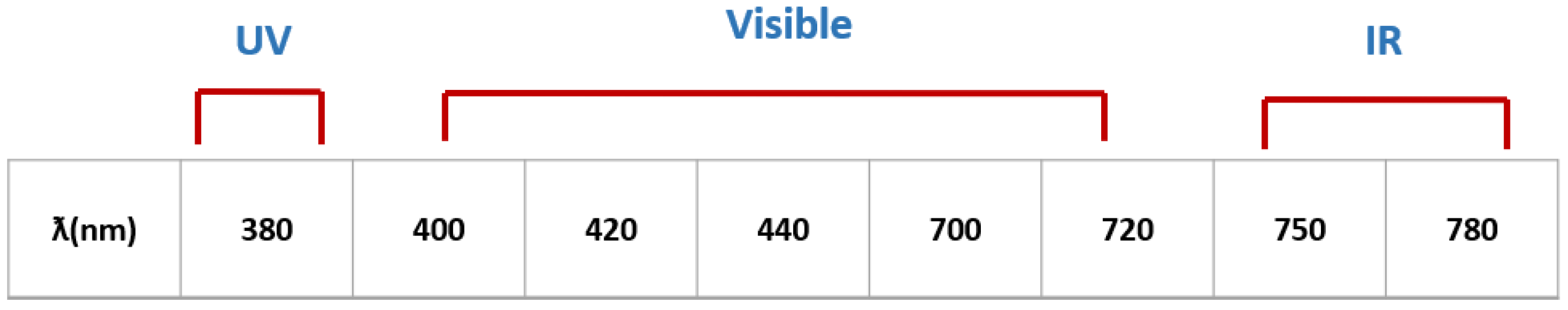
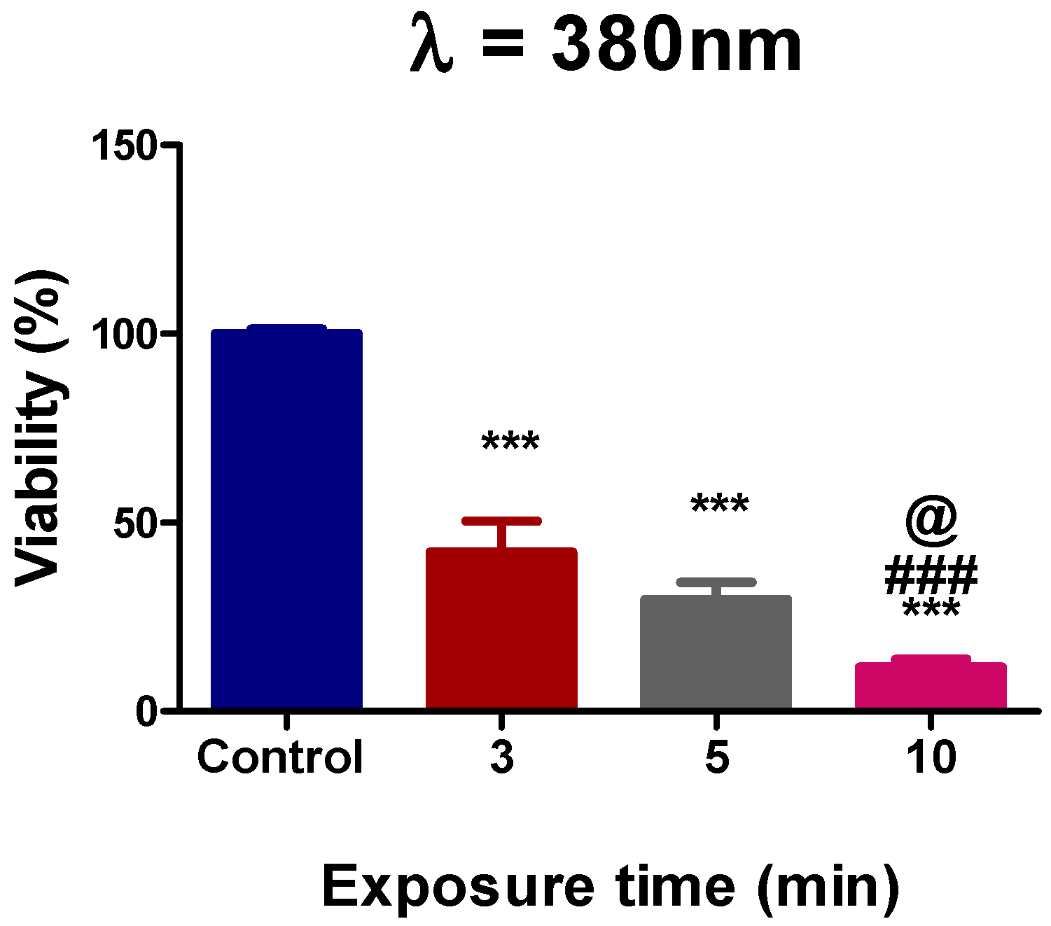
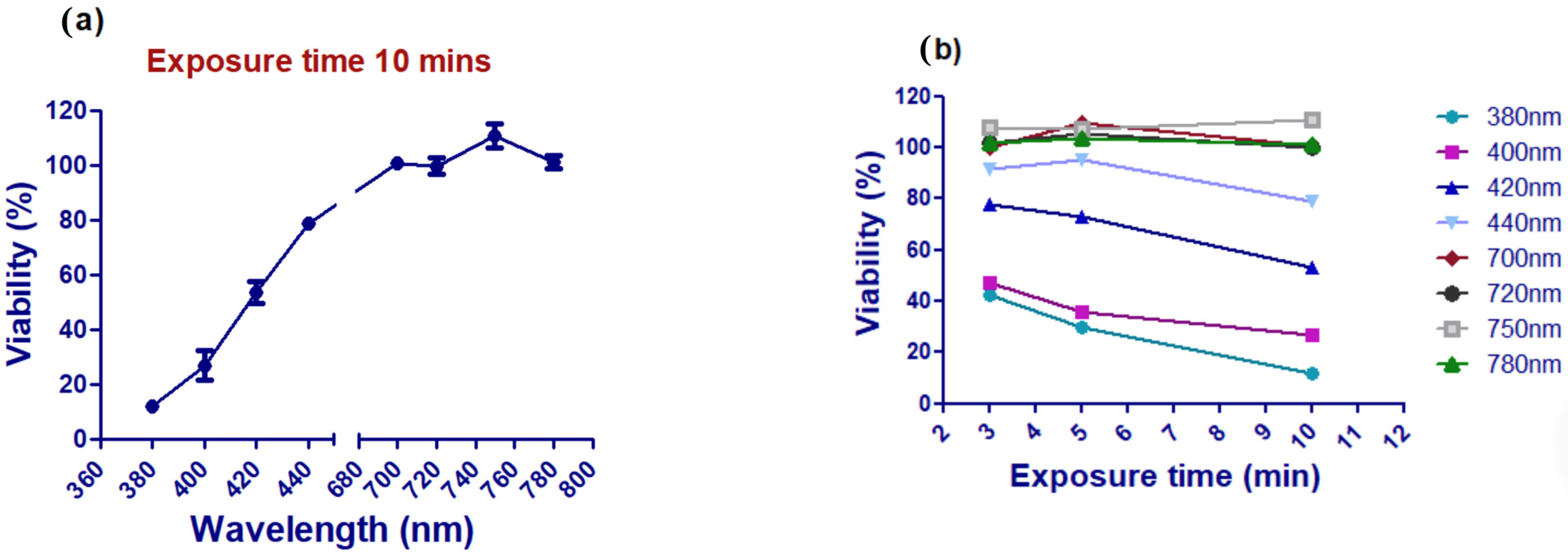
3.1. Assessment of Ultraviolet Femtosecond Laser Light Effects on A375 Cell Viability

Figure 3 shows that compared with the control, femtosecond laser light at a wavelength of 380 nm significantly reduced cell viability 24 h after 3, 5, and 10 min of irradiation (p < 0.0001). Furthermore, as shown in Figure 3, 10 min of exposure dramatically decreased the cell viability (p < 0.0001) compared to 3 or 5 min of exposure and the control. Compared to the control, the proportion of live cells at 380 nm for 10 min was 10%.

3.2. Assessment of A375 Cell Viability Following Visible Femtosecond Laser Light Exposure

Femtosecond laser light was tuned for visible wavelength emissions (400, 420, 440, 700, and 720 nm) to irradiate A375 cells. The findings demonstrated a decrease in the cell viability at 400 nm and 420 nm for various exposure durations when compared to the control group (Figure 4a,b), as well as at 440 nm, specifically after 10 min of exposure compared to the control (Figure 4c). The data for the various exposure times indicated that an increase in the exposure time significantly decreased the viability of the cells, with 10 min significantly decreasing the cell viability compared to 3 and 5 min. During the 10 min exposure period, the percentage of viable cells compared to the control was recorded as 26.9% for 400 nm, 52.3% for 420 nm, and 82.3% for 440 nm. This indicates that 400 nm is more effective at decreasing the cell viability than both 420 nm and 440 nm. In contrast, no significant differences in the cell viability were noted with femtosecond laser irradiation at 700 nm or 720 nm when compared to the control across different exposure durations (Figure 4d,e).

3.3. Assessment of A375 Cell Viability Following Near-Infrared Femtosecond Laser Exposure

Femtosecond laser light with wavelengths of 750 and 780 nm was used to irradiate A375 cells, as shown in Figure 5. The viability of the A375 cells remained unchanged compared to that of the control cells.
Figure 6 summarizes the results shown in Figure 3, Figure 4 and Figure 5. From the abovementioned results, it was obvious that the most effective wavelength for inhibiting cell viability was 380 nm and the most effective exposure time was 10 min, as shown in Figure 6a,b.

4. Discussion

This study investigated the reaction of the melanoma cancer cell line A375 to femtosecond laser light across various wavelengths, including visible (400, 420, 440, 700, and 720 nm), near-infrared (750 and 780 nm), and ultraviolet (380 nm). Additionally, this study explored various exposure durations (3, 5, or 10 min) at a power density of 4.5 × 10⁴ W/cm2, marking the first investigation of its kind. The results showed that, whereas wavelengths of 700, 720, 750, and 780 nm had no discernible effect on the cell viability, wavelengths of 380, 400, 420, and 440 nm significantly decreased the cell viability. This study provides a novel, photosensitizer-free approach for wavelength-specific melanoma cell inhibition using femtosecond laser light. The comprehensive spectrum assessment enables the identification of peak bioactive ranges, especially in the 380–420 nm region, thus informing future targeted laser therapy designs. The wavelengths that proved most effective were 400 nm and 380 nm, with the highest level of inhibition observed after 10 min of exposure, compared to 3 and 5 min for all effective wavelengths.
The wavelength-specific inhibition effects observed during the experiments are explained through the nonlinear absorption interactions between ultrashort laser pulses and intracellular chromophores [23]. The ultraviolet and blue visible spectrum (380–450 nm) contains photon energies which match the absorption spectra of important mitochondrial enzymes including cytochrome c oxidase (CCO) to initiate substantial ROS production, which disrupts mitochondrial operations [24]. The femtosecond pulses amplify these effects through multiphoton absorption and localized energy deposition to initiate apoptotic cascades [25]. The reduced biological impact of near-infrared light results from its lower photon energy which produces minimal interactions with these chromophores at longer wavelengths [24]. NIR light demonstrates its ability to penetrate deep into tissues while minimizing photothermal and photochemical effects on monolayer cell cultures. The photon energy levels at these specific wavelengths do not possess enough energy to start nonlinear reactions or cause significant harm to mitochondria, especially when photosensitizers are not present [24].
For the UV spectrum, 380 nm was the most effective wavelength for inhibiting cell viability. Laura and her research team utilized 380 nm light to create photoswitchable inhibitors designed for disrupting protein–protein interactions. Subsequently, they applied these inhibitors to live cell lines, including HeLa, HEK293, and MCF7 cells, effectively inhibiting their biological activities [26].
In comparison to the control cells, the femtosecond laser’s wavelengths in the visible spectrum at 400 and 420 nm notably reduced the cell viability. The results are consistent with earlier studies conducted by Wang et al., which showed that exposure to blue LED light (415 nm and 16 mW/cm2) resulted in a reduction in the intracellular ATP levels, a decline in the mitochondrial membrane potential, a decrease in the intracellular pH, and an elevation in the number of intracellular ROS in human adipose stem cells [27]. The observed effects may result from ROS generation, mitochondrial membrane potential disruption, and apoptosis induction, as reported in similar studies using blue and UV light. According to a previous study, lung cancer cells exposed to a 405 nm blue laser produced large amounts of intracellular ROS, while 664 nm or NIR (808 nm) had no effect on ROS levels [28]. Furthermore, the distinctive spectral characteristics of cytochrome c oxidase (CCO), which exhibit prominent peaks at wavelengths ranging from 418 to 420 nm and 598 to 600 nm, alongside minimal absorption in the red to near-infrared range, may be attributed to its absorption properties [29]. Previous studies have shown that different laser wavelengths interact uniquely with cellular components. Shorter wavelengths, such as UV and blue light, are more effective at inducing ROS generation and disrupting mitochondrial function, leading to apoptosis. In contrast, longer wavelengths in the near-infrared range penetrate deeper into tissues, but exhibit weaker effects on cell viability due to reduced absorption by intracellular chromophores [30]. While the current study did not directly measure intracellular ROS or apoptotic markers, the observed wavelength-dependent reduction in viability, particularly in the blue and UV regions, is consistent with previously reported ROS-mediated cytotoxic effects. These effects are often linked to mitochondrial dysfunction and oxidative stress [23,24,25]. Future studies will incorporate ROS assays and caspase activation profiling to validate these mechanisms. Moreover, a previous investigation revealed that the absorbance of cytochrome c oxidase (CCO) at wavelengths of 415 nm (blue) and 540 nm (green) is about 20 times and 3 times greater, respectively, compared to the absorbance at 670 nm [31]. This finding is also consistent with that of another study in which blue LEDs were shown to mediate the activation of the mitochondrial apoptotic pathway in murine melanoma cells [32,33,34]. According to the current findings, cells are affected by 440 nm laser irradiation for 10 min only. Apparently, 440 nm exhibited a weaker impact on cell viability compared to wavelengths of 400 nm and 420 nm. This could be attributed to the fact that 440 nm falls outside the absorption spectrum range of CCO [19]. In addition, a prior study showed that visible light (405–470 nm) has been applied in clinical settings to successfully treat infections caused by pathogens such as H. pylori and P. aeruginosa [35,36]. According to the results of the present study, the longest exposure time (10 min) resulted in the greatest inhibition of cancer cell growth. These findings are in agreement with the research by Husna et al., which demonstrated that extending the irradiation time to 900 s with a 532 nm laser significantly inhibited MCF-7 cell proliferation [37,38].
Based on the current data, the viability of A375 cells was not significantly affected by wavelengths of 700, 720, 750, or 780 nm. This result is consistent with an earlier study that used a 660 nm 50 mW CW laser, beam spot size of 2 mm2, irradiance of 2.5 W/cm2, and an irradiation time of 60 s (dose 150 J/cm2) [39]. Also, another study used a Gallium–Aluminum–Arsenide diode laser (830 nm, 150 mW) with energy densities of 1 and 2 J/cm2 for head and neck cancers and showed that laser irradiation with 1 J/cm2 increased the cell proliferation, whereas no significant increase was seen after laser irradiation with 2 J/cm2 [40].
This might occur because of the present study’s different laser light characteristics compared to the earlier research, which is in line with laser therapy’s biphasic dose–response curve. According to this theory, a particular disease benefits from optimal parameters and if these parameters are changed, the benefits disappear, and they may even have adverse consequences if the dosage is high. As a result, the effects of the same light source on the same tissue can change depending on the parameters used [21,41].
The biological response to laser exposure is governed by a combination of factors, including the wavelength, power density, exposure duration, and the spectral characteristics of the target tissue. While wavelength-specific absorption is a major determinant of photochemical interactions, the overall energy dose (fluence) is equally critical in defining the cellular outcome. Our calculated fluences, ranging from 1584 to 5280 × 10−9 J/cm2, fall within the ranges reported to induce inhibitory responses in cancer cells, consistent with the biphasic dose–response model described in prior studies [42,43].
Femtosecond pulses enable highly localized energy deposition due to nonlinear absorption mechanisms, leading to enhanced biological effects such as ROS generation and mitochondrial damage while minimizing thermal diffusion [44]. In contrast to nanosecond pulses, femtosecond lasers induce precise photodisruption with minimal collateral damage, making them particularly suitable for ophthalmic applications [45].
Further investigation through in vivo studies is necessary to explore the molecular mechanisms associated with femtosecond laser radiation in greater detail. Clinical research is also necessary to prove the effectiveness of femtosecond lasers and determine the treatment plan that may be used for melanoma patients. Compared to conventional melanoma treatments like chemotherapy and surgery, femtosecond laser therapy offers a non-invasive, targeted approach with minimal side effects. However, challenges remain in optimizing laser parameters for clinical applications, ensuring the selective targeting of melanoma cells without damaging healthy tissue, and achieving sufficient tissue penetration in deep-seated tumors [46]. It is important to note that this study utilized a 2D in vitro cell culture model, which does not fully replicate the structural and microenvironmental complexity of melanoma in vivo. Future work should incorporate 3D culture systems or animal models to evaluate the penetration depth, energy dispersion, and biological responses in a more physiologically relevant context. These models would better account for the optical scattering, absorption, and heterogeneous responses seen in tissue.

5. Conclusions

The effects of femtosecond laser exposure on the A375 melanoma cell line were illustrated in this study. Cell viability was significantly decreased by femtosecond laser irradiation at wavelengths of 380, 400, 420, and 440 nm, with 380 nm having the greatest impact. Furthermore, compared to that at 3 and 5 min, the inhibitory effect of 10 min on the cells was greater. However, there was no noticeable effect of the 700, 720, 750, or 780 nm wavelength on the viability of the cells, either stimulative or inhibitive. Consequently, 10 min of femtosecond laser irradiation at 380 nm can be effectively utilized in the treatment of melanoma. Further in vivo studies are required to investigate the molecular mechanisms underlying femtosecond laser exposure, including ROS generation, apoptosis induction, and DNA damage. Additionally, long-term studies will assess the safety and therapeutic efficacy of femtosecond laser irradiation, paving the way for potential clinical applications in melanoma treatment.

Author Contributions

Conceptualization, S.T., K.T.N., A.O.E.-G. and T.M.; Formal analysis, S.T., K.T.N. and A.O.E.-G.; Investigation, S.T., K.T.N., A.O.E.-G. and T.M.; Methodology, S.T., K.T.N., H.M.R., A.O.E.-G. and T.M.; Resources, H.M.R.; Software, H.M.R.; Supervision, A.O.E.-G. and T.M.; Validation, A.O.E.-G.; Visualization, H.M.R.; Writing—original draft, S.T. and K.T.N.; Writing—review and editing, A.O.E.-G. and T.M. All authors have read and agreed to the published version of the manuscript.

Funding

This research received no external funding.

Institutional Review Board Statement

Not applicable.

Informed Consent Statement

Not applicable.

Data Availability Statement

Data are contained within the article.

Conflicts of Interest

The authors declare no conflicts of interest.

References

  1. Jassim, A.; Rahrmann, E.P.; Simons, B.D.; Gilbertson, R.J. Cancers make their own luck: Theories of cancer origins. Nat. Rev. Cancer 2023, 23, 710–724. [Google Scholar] [CrossRef]
  2. Shakhova, O. Neural crest stem cells in melanoma development. Curr. Opin. Oncol. 2014, 26, 215–221. [Google Scholar] [CrossRef]
  3. Couto, G.K.; Junior, J.C.R.; Pacheco, B.S.; Simoes, L.D.; Paschoal, J.D.; Seixas, F.K.; Acunha, T.V.; Iglesias, B.A.; Collares, T. Zinc (II), copper (II) and nickel (II) ions improve the selectivity of tetra-cationic platinum (II) porphyrins in photodynamic therapy and stimulate antioxidant defenses in the metastatic melanoma lineage (A375). Photodiagnosis Photodyn. Ther. 2020, 31, 101942. [Google Scholar] [CrossRef]
  4. Schadendorf, D.; Fisher, D.E.; Garbe, C.; Gershenwald, J.E.; Grob, J.-J.; Halpern, A.; Herlyn, M.; Marchetti, M.A.; McArthur, G.; Ribas, A.; et al. Melanoma. Nat. Rev. Dis. Primers 2015, 1, 1–20. [Google Scholar] [CrossRef]
  5. Mundra, V.; Peng, Y.; Rana, S.; Natarajan, A.; Mahato, R.I. Micellar formulation of indocyanine green for phototherapy of melanoma. J. Control. Release 2015, 220, 130–140. [Google Scholar] [CrossRef]
  6. Najafabad, B.K.; Attaran, N.; Mahmoudi, M.; Sazgarnia, A. Effect of photothermal and photodynamic therapy with cobalt ferrite superparamagnetic nanoparticles loaded with ICG and PpIX on cancer stem cells in MDA-MB-231 and A375 cell lines. Photodiagnosis Photodyn. Ther. 2023, 43, 103648. [Google Scholar] [CrossRef]
  7. Szlasa, W.; Supplitt, S.; Drąg-Zalesińska, M.; Przystupski, D.; Kotowski, K.; Szewczyk, A.; Kasperkiewicz, P.; Saczko, J.; Kulbacka, J. Effects of curcumin based PDT on the viability and the organization of actin in melanotic (A375) and amelanotic melanoma (C32)–in vitro studies. Biomed. Pharmacother. 2020, 132, 110883. [Google Scholar] [CrossRef]
  8. Eggermont, A.M.; Gore, M. Randomized adjuvant therapy trials in melanoma: Surgical and systemic. Semin. Oncol. 2007, 34, 509–515. [Google Scholar] [CrossRef]
  9. Khelladi, M. Femtosecond Laser Pulses: Generation, Measurement and Propagation. In Recent Advances in Numerical Simulations; IntechOpen: London, UK, 2021. [Google Scholar]
  10. Niemz, M.H. Laser-Tissue Interactions; Springer: Cham, Switzerland, 2007; Volume 322. [Google Scholar]
  11. Krüger, J.; Kautek, W.; Newesely, H. Femtosecond-pulse laser ablation of dental hydroxyapatite and single-crystalline fluoroapatite. Appl. Phys. A 1999, 69, S403–S407. [Google Scholar] [CrossRef]
  12. Ryu, S.; Jun, I.; Kim, T.-I.; Seo, K.Y.; Kim, E.K. Prediction accuracy of conventional and total keratometry for intraocular lens power calculation in femtosecond laser-assisted cataract surgery. Sci. Rep. 2021, 11, 12869. [Google Scholar] [CrossRef]
  13. Yoon, J.; Ryu, S.-w.; Lee, S.; Choi, C. Cytosolic irradiation of femtosecond laser induces mitochondria-dependent apoptosis-like cell death via intrinsic reactive oxygen cascades. Sci. Rep. 2015, 5, 8231. [Google Scholar] [CrossRef] [PubMed]
  14. Thøgersen, J.; Knudsen, C.S.; Maetzke, A.; Knak Jensen, S.J.; Keiding, S.R.; Alsner, J.; Overgaard, J. Reproductive death of cancer cells induced by femtosecond laser pulses. Int. J. Radiat. Biol. 2007, 83, 289–299. [Google Scholar] [CrossRef]
  15. Tirlapur, U.K.; König, K. Femtosecond near-infrared laser pulse induced strand breaks in mammalian cells. Cell. Mol. Biol. 2001, 47, OL131–OL134. [Google Scholar]
  16. Gabel, C.V. Femtosecond lasers in biology: Nanoscale surgery with ultrafast optics. Contemp. Phys. 2008, 49, 391–411. [Google Scholar] [CrossRef]
  17. Ahmed, E.; El-Gendy, A.O.; Moniem Radi, N.A.; Mohamed, T. The bactericidal efficacy of femtosecond laser-based therapy on the most common infectious bacterial pathogens in chronic wounds: An in vitro study. Lasers Med Sci. 2021, 36, 641–647. [Google Scholar] [CrossRef]
  18. El-Gendy, A.O.; Samir, A.; Ahmed, E.; Enwemeka, C.S.; Mohamed, T. The antimicrobial effect of 400 nm femtosecond laser and silver nanoparticles on gram-positive and gram-negative bacteria. J. Photochem. Photobiol. B Biol. 2021, 223, 112300. [Google Scholar] [CrossRef]
  19. Zecha, J.A.; Raber-Durlacher, J.E.; Nair, R.G.; Epstein, J.B.; Sonis, S.T.; Elad, S.; Hamblin, M.R.; Barasch, A.; Migliorati, C.A.; Milstein, D.M.J.; et al. Low level laser therapy/photobiomodulation in the management of side effects of chemoradiation therapy in head and neck cancer: Part 1: Mechanisms of action, dosimetric, and safety considerations. Support. Care Cancer 2016, 24, 2781–2792. [Google Scholar] [CrossRef] [PubMed]
  20. Kars, M.D.; Yildirim, G.; Gundogdu, Y.; Gonce, F.; Ayan, E.; Kilic, H.S. Revealing the therapeutic effects of aminolevulinate mediated femtosecond laser induced photo-chemotherapy in different cancer cells. EuroBiotech J. 2020, 4, 207–215. [Google Scholar] [CrossRef]
  21. Huang, Y.-Y.; Sharma, S.K.; Carroll, J.; Hamblin, M.R. Biphasic dose response in low level light therapy—An update. Dose-Response 2011, 9, 602–618. [Google Scholar] [CrossRef]
  22. Taha, S.; Mohamed, W.R.; Elhemely, M.A.; El-Gendy, A.O.; Mohamed, T. Breast cancer and laser therapy. Laser Innov. Res. Appl. 2024, 1, 43–66. [Google Scholar] [CrossRef]
  23. Ilina, I.V.; Sitnikov, D.S. Application of Ultrashort Lasers in Developmental Biology: A Review. Photonics 2022, 9, 914. [Google Scholar] [CrossRef]
  24. Golovynska, I.; Golovynskyi, S.; Qu, J. Comparing the impact of NIR, visible and UV light on ROS upregulation via photoacceptors of mitochondrial complexes in normal, immune and cancer cells. Photochem. Photobiol. 2023, 99, 106–119. [Google Scholar] [CrossRef]
  25. Algorri, J.F.; Ochoa, M.; Roldan-Varona, P.; Rodriguez-Cobo, L.; López-Higuera, J.M. Light technology for efficient and effective photodynamic therapy: A critical review. Cancers 2021, 13, 3484. [Google Scholar] [CrossRef] [PubMed]
  26. Nevola, L.; Martín-Quirós, A.; Eckelt, K.; Camarero, N.; Tosi, S.; Llobet, A.; Giralt, E.; Gorostiza, P. Light-regulated stapled peptides to inhibit protein–protein interactions involved in clathrin-mediated endocytosis. Angew. Chem. Int. Ed. Engl. 2013, 52, 7704–7708. [Google Scholar] [CrossRef]
  27. Wang, Y.; Huang, Y.-Y.; Wang, Y.; Lyu, P.; Hamblin, M.R. Red (660 nm) or near-infrared (810 nm) photobiomodulation stimulates, while blue (415 nm), green (540 nm) light inhibits proliferation in human adipose-derived stem cells. Sci. Rep. 2017, 7, 7781. [Google Scholar] [CrossRef]
  28. Kushibiki, T.; Hirasawa, T.; Okawa, S.; Ishihara, M.J. Blue laser irradiation generates intracellular reactive oxygen species in various types of cells. Photomed. Laser Surg. 2013, 31, 95–104. [Google Scholar] [CrossRef] [PubMed]
  29. Malatesta, F.; Antonini, G.; Sarti, P.; Brunori, M. Structure and function of a molecular machine: Cytochrome c oxidase. Biophys. Chem. 1995, 54, 1–33. [Google Scholar] [CrossRef]
  30. Ushenko, V.A.; Hogan, B.T.; Dubolazov, A.; Piavchenko, G.; Kuznetsov, S.L.; Ushenko, A.G.; Ushenko, Y.O.; Gorsky, M.; Bykov, A.; Meglinski, I. 3D Mueller matrix mapping of layered distributions of depolarisation degree for analysis of prostate adenoma and carcinoma diffuse tissues. Sci. Rep. 2021, 11, 5162. [Google Scholar] [CrossRef]
  31. Mason, M.G.; Nicholls, P.; Cooper, C.E. Re-evaluation of the near infrared spectra of mitochondrial cytochrome c oxidase: Implications for non invasive in vivo monitoring of tissues. Biochim. Biophys. Acta Bioenerg. 2014, 1837, 1882–1891. [Google Scholar] [CrossRef]
  32. Oh, P.-S.; Hwang, H.; Jeong, H.-S.; Kwon, J.; Kim, H.-S.; Kim, M.; Lim, S.; Sohn, M.-H.; Jeong, H.-J. Blue light emitting diode induces apoptosis in lymphoid cells by stimulating autophagy. Int. J. Biochem. Cell Biol. 2016, 70, 13–22. [Google Scholar] [CrossRef]
  33. Zhuang, J.; Liu, Y.; Yuan, Q.; Liu, J.; Liu, Y.; Li, H.; Wang, D. Blue light-induced apoptosis of human promyelocytic leukemia cells via the mitochondrial-mediated signaling pathway. Oncol. Lett. 2018, 15, 6291–6296. [Google Scholar] [CrossRef]
  34. Oh, P.-S.; Na, K.S.; Hwang, H.; Jeong, H.-S.; Lim, S.; Sohn, M.-H.; Jeong, H.-J. Effect of blue light emitting diodes on melanoma cells: Involvement of apoptotic signaling. J. Photochem. Photobiol. B Biol. 2015, 142, 197–203. [Google Scholar] [CrossRef] [PubMed]
  35. Dai, T.; Gupta, A.; Huang, Y.-Y.; Yin, R.; Murray, C.K.; Vrahas, M.S.; Sherwood, M.E.; Tegos, G.P.; Hamblin, M.R. Blue light rescues mice from potentially fatal Pseudomonas aeruginosa burn infection: Efficacy, safety, and mechanism of action. Antimicrob. Agents Chemother. 2013, 57, 1238–1245. [Google Scholar] [CrossRef]
  36. Dai, T.; Gupta, A.; Murray, C.K.; Vrahas, M.S.; Tegos, G.P.; Hamblin, M.R. Blue light for infectious diseases: Propionibacterium acnes, Helicobacter pylori, and beyond? Drug Resist. Updat. 2012, 15, 223–236. [Google Scholar] [CrossRef] [PubMed]
  37. Habit, H.A.H.; Suardi, N.; Mahmud, S.; Mydin, R.B.S.; Bakhori, S.K.M. In vitro toxicity of low-level green laser irradiation effects on human breast cancer cell lines. Indian J. Biochem. Biophys. 2020, 57, 627–633. [Google Scholar]
  38. Taha, S.; Mohamed, W.R.; Elhemely, M.A.; El-Gendy, A.O.; Mohamed, T. Tunable femtosecond laser suppresses the proliferation of breast cancer in vitro. J. Photochem. Photobiol. B Biol. 2023, 240, 112665. [Google Scholar] [CrossRef] [PubMed]
  39. Frigo, L.; Luppi, J.S.; Favero, G.M.; Maria, D.A.; Penna, S.C.; Bjordal, J.M.; Bensadoun, R.J.; Lopes-Martins, R.A. The effect of low-level laser irradiation (In-Ga-Al-AsP-660 nm) on melanoma in vitro and in vivo. BMC Cancer 2009, 9, 1–8. [Google Scholar] [CrossRef]
  40. Bamps, M.; Dok, R.; Nuyts, S. Low-level laser therapy stimulates proliferation in head and neck squamous cell carcinoma cells. Front. Oncol. 2018, 8, 343. [Google Scholar] [CrossRef]
  41. Hamblin, M.R.; Nelson, S.T.; Strahan, J.R. Photobiomodulation and cancer: What is the truth? Photomed. Laser Surg. 2018, 36, 241–245. [Google Scholar] [CrossRef]
  42. Sommer, A.P.; Pinheiro, A.L.; Mester, A.R.; Franke, R.-P.; Whelan, H.T. Biostimulatory windows in low-intensity laser activation: Lasers, scanners, and NASA’s light-emitting diode array system. J. Clin. Laser Med. Surg. 2001, 19, 29–33. [Google Scholar] [CrossRef]
  43. Chung, H.; Dai, T.; Sharma, S.K.; Huang, Y.-Y.; Carroll, J.D.; Hamblin, M.R. The nuts and bolts of low-level laser (light) therapy. Ann. Biomed. Eng. 2012, 40, 516–533. [Google Scholar] [CrossRef] [PubMed]
  44. Ho, C.M.B.; Mishra, A.; Hu, K.; An, J.; Kim, Y.-J.; Yoon, Y.-J. Femtosecond-laser-based 3D printing for tissue engineering and cell biology applications. ACS Biomater. Sci. Eng. 2017, 3, 2198–2214. [Google Scholar] [CrossRef] [PubMed]
  45. Kurtz, R.M.; Spooner, G.J.; Sletten, K.R.; Yen, K.G.; Sayegh, S.I.; Loesel, F.H.; Horvath, C.; Liu, H.; Elner, V.; Cabrera, D. Ophthalmic applications of femtosecond lasers. In Commercial and Biomedical Applications of Ultrafast Lasers; SPIE: Bellingham, WA, USA, 1999. [Google Scholar]
  46. Malviya, R.; Meenakshi, D.U.; Goyal, P. Laser Therapy in Healthcare: Advances in Diagnosis and Treatment; John Wiley & Sons: Hoboken, NJ, USA, 2024. [Google Scholar]
Figure 1. The experimental setup used for irradiating A375 cells is depicted in a diagrammatic representation.
Photonics 12 00694 g001
Figure 2. Various wavelength ranges for irradiation of A375 cells.
Photonics 12 00694 g002
Figure 3. Assessment of A375 cell viability 24 h post-exposure to 380 nm femtosecond laser irradiation. *** indicates a significant difference compared to the control with p < 0.001; ### denotes a significant difference observed compared to 3 min treatment with p < 0.001; @ signifies a significant difference observed compared to 5 min treatment with p < 0.05.
Photonics 12 00694 g003
Figure 4. The viability of A375 cells was assessed 24 h after exposure to visible femtosecond laser irradiation. (a) Effect of 400 nm wavelength at different exposure times. (b) Effect of 420 nm wavelength at different exposure times. (c) Effect of 440 nm wavelength at different exposure times. (d) Effect of 700 nm wavelength at different exposure times. (e) Effect of 720 nm wavelength at different exposure times. Significant differences are indicated by symbols: *** indicates a significant difference compared to the control with p < 0.001; # denotes a significant difference observed compared to 3 min treatment with p < 0.05; ### denotes a significant difference observed compared to 3 min treatment with p < 0.001; @@@ signifies a significant difference observed compared to 5 min treatment with p < 0.001.
Photonics 12 00694 g004
Figure 5. An evaluation of the effect of infrared femtosecond laser exposure on the viability of A375 cells 24 h after irradiation. (a) Effect of 750 nm wavelength at different exposure times. (b) Effect of 780 nm wavelength at different exposure times.
Photonics 12 00694 g005
Figure 6. The optimum parameters of femtosecond laser irradiation for A375 cell inhibition. (a) Effect of different wavelengths at 10 min exposure time. (b) Effect of different wavelengths at different exposure times.
Photonics 12 00694 g006
Disclaimer/Publisher’s Note: The statements, opinions and data contained in all publications are solely those of the individual author(s) and contributor(s) and not of MDPI and/or the editor(s). MDPI and/or the editor(s) disclaim responsibility for any injury to people or property resulting from any ideas, methods, instructions or products referred to in the content.

