Article

Comparison of Dynamic Visual Acuity in Spectacles Prescribed to 0.05D Versus 0.25D Steps: A Self-Control, Randomized Study

Department of Ophthalmology, Peking University Third Hospital, Beijing 100191, China
*
Author to whom correspondence should be addressed.
Photonics 2025, 12(7), 692; https://doi.org/10.3390/photonics12070692
Submission received: 9 June 2025 / Revised: 28 June 2025 / Accepted: 30 June 2025 / Published: 9 July 2025

Abstract

The research aims to compare the dynamic visual acuity (DVA) in myopic adults wearing spectacles prescribed to 0.05D and 0.25D steps. This double-blind, randomized, self-control study included 40 myopic participants aged 18–40. The participants were randomly assigned to receive spectacles with one 0.05D step lens and the contralateral lens of 0.25D step. The monocular horizontal and vertical motion DVA at 20 and 40 degrees per second (dps) was measured. The DVA was compared between eyes with 0.25D and 0.05D step lenses and further analyzed by eye dominance and test sequence. The result demonstrated no significant difference in DVA between two eyes with 0.25D or 0.05D step lenses at 20 and 40 dps horizontal and vertical motion test (p > 0.05, respectively). When the eye with a 0.25D step lens was the dominant eye (p = 0.004) or measured secondly (p = 0.002), it outperformed the contralateral eye with a 0.05D step lens in the 40 dps horizontal motion test. In conclusion, the horizontal and vertical motion DVA of the eye with 0.05D step lens spectacles was comparable to that of contralateral eyes corrected with 0.25D step lens.

1. Introduction

Myopia is the prevalent refractive error that affects visual acuity in children and adults. A meta-analysis predicts that by 2050, myopia will affect nearly half of the global population [1]. Spectacles are a simple and safe method for correcting myopia, and most spectacles are currently manufactured with a 0.25 diopter (D) step. Determining the proper prescription of spectacles relies on accurate refraction, including automatic and subjective refraction. However, during subjective refraction, the 0.25D-step refraction may result in patients failing to achieve red-green balance in the Duochrome test, leading to either overcorrection or undercorrection [2,3]. Studies have shown that the resolution limit of the human eye to spherical lens change was about 0.05D, indicating demand for more precise prescriptions and lens manufacturing [2]. With advancements in refraction technology, higher-precision myopia prescriptions can be obtained using a new-generation refraction device with a 0.05D step [4,5].
Recent research has compared the difference in visual performance of spectacles obtained with 0.05D and 0.25D step refraction techniques [2,3,4,5]. However, these studies primarily focused on static visual performance, including contrast sensitivity and best-corrected visual acuity (BCVA). In daily life, individuals frequently need to observe moving objects, and static vision may not adequately reflect visual function in real-world scenarios [6,7]. Dynamic visual acuity (DVA) is the ability to discriminate details of an object when relative movement exists between the object and the observer [7]. Measuring visual acuity in dynamic situations and the detail resolution of moving objects can better reflect the visual function in real-life scenes [8]. Therefore, DVA has gradually become an important evaluation metric for functional vision [9,10,11,12,13,14]. The previous study demonstrated that myopia significantly affects DVA when fully corrected with eyeglasses, and worse DVA was associated with more significant myopia [15]. However, the impact of 0.05D versus 0.25D step spectacles on DVA remains unexplored.
The present study aims to compare the DVA with spectacles prescribed using 0.05D and 0.25D step refraction in a self-control design and to explore the associated factors for DVA. The findings may enhance the understanding of how these refraction methods influence dynamic vision and support the inclusion of DVA as an important metric to guide the selection of refraction and prescription methods based on individual needs.

2. Materials and Methods

2.1. Participants

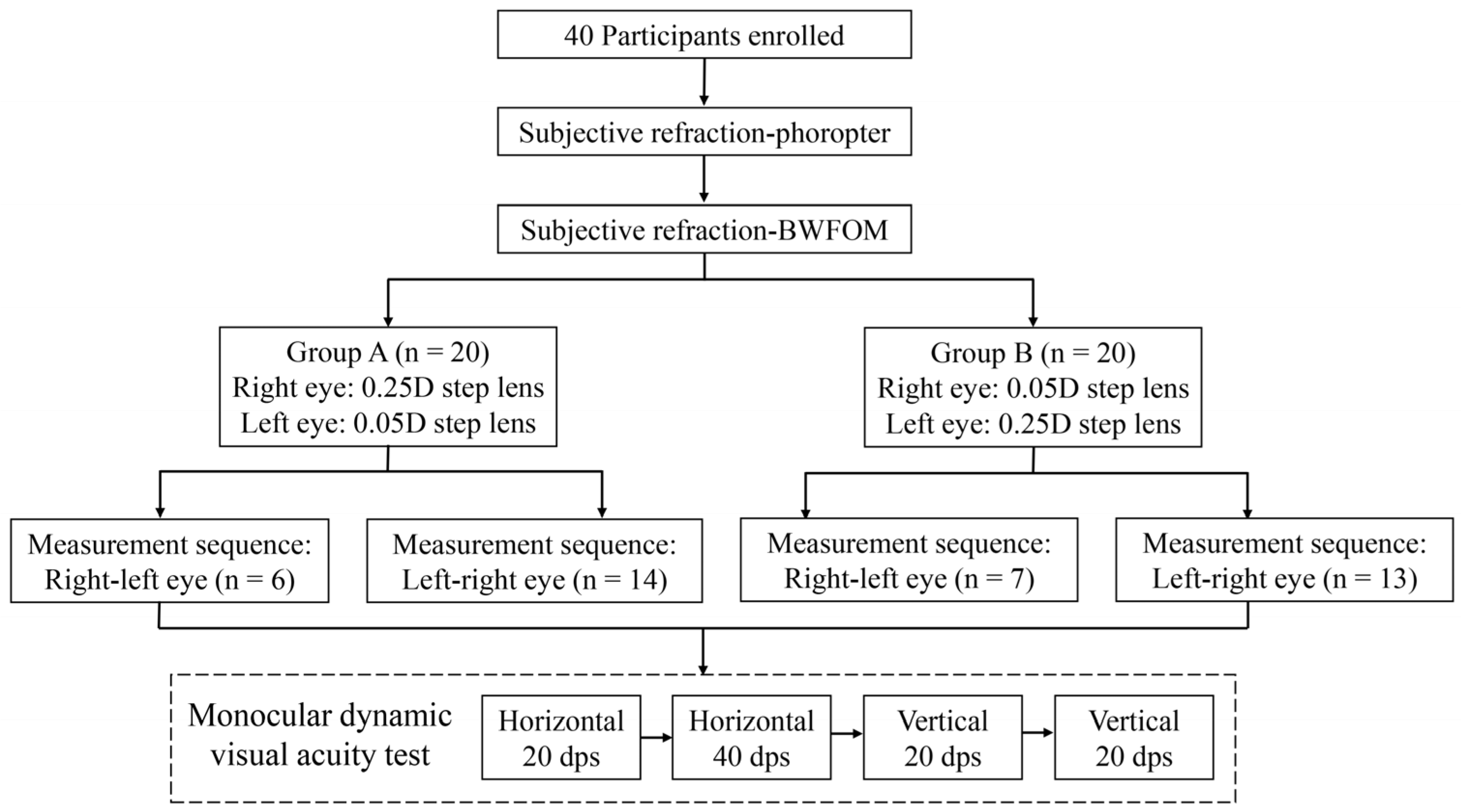
The present study was a double-blind, randomized, self-control observational study to assess the DVA with 0.05D step spectacles compared with 0.25D step spectacles in myopic patients. The study adhered to the tenets of the Declaration of Helsinki, and the protocol was approved by the ethics committee of Peking University Third Hospital. Informed consent was obtained from all participants before being included in the study.
Participants aged between 18 and 40 years with myopia were included in the study. Exclusion criteria were as follows: (1) Monocular corrected distance visual acuity (CDVA) ≤ 0 LogMAR; (2) severe myopia (sphere < −10D) or high astigmatism (> 3D); (3) significant anisometropia with a difference in spherical equivalent > 2D between eyes; (4) the presence of ocular diseases affecting eye structure or static vision, such as glaucoma, corneal disease, or retinal disorders; (5) systemic diseases impacting static vision; (6) vestibular dysfunction or cognitive impairment; and (7) inability to track dynamic visual targets or follow instructions during testing.

2.2. Randomization and Grouping

The study design and participant allocation process are shown in Figure 1. The participants were randomly assigned 1:1 to groups A and B with block randomization. The block size was set at 2, with a total of 20 blocks. A random number table was employed to sort the participants based on their identification numbers and sequential assignment to blocks was performed according to the sorting results. In group A, participants wore spectacles with a 0.25D step lens for the right eye and a 0.05D step lens for the left eye. In contrast, participants in group B used spectacles with lenses in 0.05D step for the right eye and 0.25D for the left eye. The participants were further randomly divided into two groups with different test sequences for the right and left eye because the test sequence may affect the result due to the perceptual learning effect during the DVA test [10]. The right eye was examined first in one group, followed by the left, while the other group reversed the test order. The lens allocation was masked to the DVA examiner and the participants.

2.3. Testing Procedure

All enrolled participants underwent slit-lamp and fundus examinations to rule out severe ocular diseases. Subsequently, subjective refraction was performed at 0.25D step using a phoropter, followed by subjective refraction at 0.05D step using a Binocular Wavefront Optometer (BWFOM, Aizhitong Medical Technology Co., Ltd., Huzhou, China). Next, based on the group assignment, the corresponding spectacles were fitted, the CDVA was measured with a LogMar visual chart, and the dominant eye was examined with the card hole method. The 0.25D step lens was prescribed with phoropter-based subjective refraction, and the 0.05D step lens was prescribed with optometer-based subjective refraction. Finally, the DVA test was performed sequentially for each eye according to the allocation wearing the prescribed spectacles.
Subjective refraction with a phoropter was performed monocularly based on automatic refraction (KR-8100, Topcon, Medical System, Tokyo, Japan). Fogging with a +0.75 to +1.50D lens was applied to relax accommodation. The negative lens was gradually increased in a 0.25D step, and we applied a duochrome test to achieve the first maximum plus to maximum visual acuity (MPMVA). Next, the cylinder axis was refined, followed by the power with the Jackson-cross cylinder. Then, the second MPMVA was obtained with a duochrome test to adjust the sphere. Then, the other eye was examined using the same procedure, followed by the binocular balance test.
The BWFOM is an innovative device that integrates objective and subjective refraction. Objective refraction utilizes Hartmann–Shack wavefront sensor and adaptive optics technology and separates higher- from lower-order aberrations, converting the lower-order aberration data into objective refractive values used as the starting point for subjective refraction [4,16]. The subjective refraction procedure with BWFOM is similar to phoropter-based refraction, including monocular spherical endpoints determination with a duochrome test, cylinder fine tune, and binocular balance test. Subjective refraction is facilitated by a series of optical modulators that simulate optical lenses. These modulators move linearly, achieving an adjustment precision of 0.05D. Leveraging this precision, the BWFOM enables examiners to prescribe lenses in the 0.05D step, delivering highly accurate refraction results for objective and subjective methods [4,5].

2.4. DVA Test

The DVA was evaluated with the previously reported test system [13,15,17]. The DVA tests were conducted monocularly with refractive error corrected using spectacles according to the group allocation. The test was arranged at a 2.5 m distance in a quiet room, and the optotypes were presented on a 24-inch 480 Hz screen. The test software was programmed with MATLAB 2017b (MathWorks, Natick, MA, USA), which could demonstrate moving the letter E with a certain size and speed. The optotype E was configured according to the LogMAR visual chart.
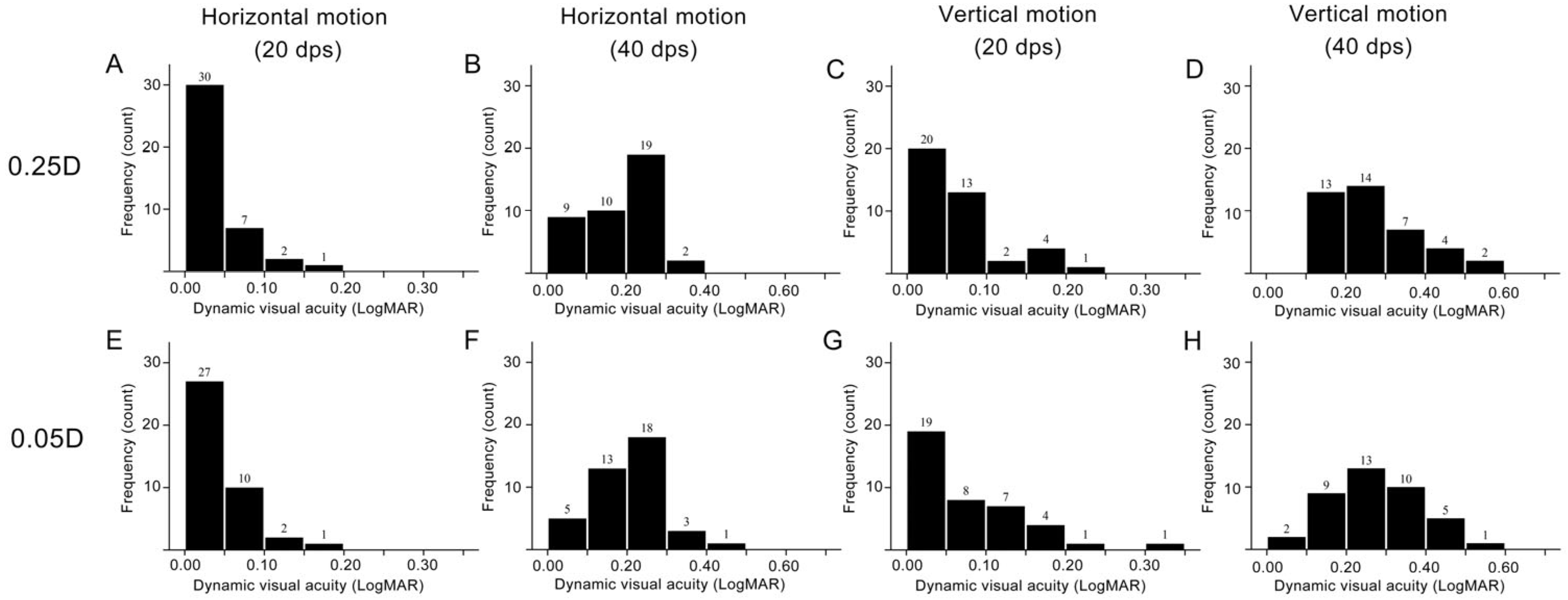
Two repeated sessions were performed for the right and left eyes at 5-min intervals. Each session contained four DVA tests with different motion types and velocities, with a 1-min interval between each test. The test sequence was the same during the two sessions, in the following order: horizontal motion of 20 degrees per second (dps), vertical motion of 20 dps, horizontal motion of 40 dps, and vertical motion of 40 dps. During the horizontal motion test, the optotype appeared in the middle of the screen’s left side and horizontally moved to the right side. For the vertical motion test, the optotype appeared in the middle of the screen on the upper side and vertically moved to the lower side.
During the test, the patient was required to discern the opening direction of the letter E in a four alternative forced-choice mode. The initial optotype was four sizes bigger than the CDVA. For each size, we presented eight optotypes one by one with a random opening direction. The letter E would be switched to one size smaller if the patient could correctly identify five out of eight optotypes. The minimum size the participant could recognize was recorded, and the result was calculated using the previously reported formula [17].

2.5. Statistical Analysis

The statistical analysis was conducted using SPSS 26.0 (IBM, Armonk, NY, USA). The spherical equivalent (SE) was calculated as the sphere plus half of the cylinder diopter. The Kolmogorov-Smirnov test was performed to determine the normality of the distribution. Descriptive statistics were reported as mean and standard deviation (SD) for continuous variables, whereas number and percentage were for categorical variables. Considering the correlation between two eyes, a linear mixed model was used to compare the DVA between 0.25D and 0.05D step spectacles, and the random intercept for the subject was included in the model. Subgroup analysis was performed by the dominant eye and test sequence. A p-value less than 0.05 was considered statistically significant.

3. Results

A total of 40 patients were included in the study. The mean age was 22.3 ± 3.4 years (19 to 33 years), and males accounted for 45% of the participants.

3.1. Differences in Subjective Refraction Results

The results of subjective refraction with phoropter and BWFOM are summarized in Table 1. The sphere measured by the BWFOM (−4.27 ± 1.86 D) was significantly less myopic than the phoropter (−4.36 ± 1.90 D, p = 0.013). More cylinder was measured with a phoropter (−0.80 ± 0.61 D) than optometer (−0.68 ± 0.52 D, p = 0.001). No significant differences were found in CDVA between the two refraction methods (p = 0.821).

3.2. Difference in DVA with Different Refraction Methods

The histogram of DVA for the eyes with spectacles prescribed to 0.05D and 0.25D step lenses is demonstrated in Figure 2, and the results are summarized in Table 2. The participants wore spectacles with one eye prescribed with a phoropter and the contralateral eye with BWFOM. For the horizontal motion test at 20 dps, 92.5% of eyes with 0.25D step and 0.05D step lens achieved DVA better than 0.1 LogMAR. For the 20 dps vertical motion test, 82.5% with 0.25D step lens and 67.5% of eyes with 0.05D step lens had better than 0.1 LogMAR DVA. The eyes with DVA better than 0.2 LogMAR were 47.5% and 45% in 0.25D and 0.05D step lens in horizontal motion at 40 dps, and the number was 32.5% and 27.5% in vertical motion at 40 dps. The self-controlled analysis demonstrated that there was no significant difference in DVA between the eyes wearing spectacles fitting with 0.05D and 0.25D lenses in 20 dps horizontal motion test (p = 0.634), 40 dps horizontal motion test (p = 0.112), 20 dps vertical motion test (p = 0.148) or 40 dps vertical motion test (p = 1.000).

3.3. Subgroup Analysis for the DVA by Eye Dominance and Measurement Sequence

The results of a comparison in DVA between dominant and nondominant eyes are demonstrated in Table A1. The dominant eye (0.1844 ± 0.0815) significantly outperformed the nondominant eye (0.2122 ± 0.0904, p = 0.025) in horizontal motion DVA at 40 dps. In other test patterns, no significant differences were found between dominant and nondominant eyes (p > 0.05, respectively).
The results of subgroup analysis by the dominant eye for DVA with different lenses are demonstrated in Table 3. When the dominant eye wore spectacles fitted with 0.25D step lens, the DVA of the dominant eye was 0.1766 ± 0.0761, significantly better than the nondominant eye (0.2185 ± 0.0871, p = 0.004) with 0.05D step lens in the horizontal motion test at 40 dps. No significant differences were observed in DVA between two eyes with 0.25D step lens and 0.05D step lens in other test patterns (p > 0.05, respectively). When the dominant eye was fitted with a 0.05D step lens, there was no significant difference in DVA between the dominant and nondominant eye (p > 0.05 for all test patterns).
The results of a comparison in DVA between first- and second-measured eyes are demonstrated in Table A2. On average, the DVA of the second-measured eye (0.1844 ± 0.0897) significantly outperformed the first-measured eye (0.2122 ± 0.0822, p = 0.025) in the horizontal motion test at 40 dps. No significant differences were observed in other test patterns (p > 0.05, respectively). The results of subgroup analysis by measurement sequence for DVA with different lenses are demonstrated in Table 4. When the first-measured eye was corrected with a 0.25D step lens, there was no significant difference in DVA between the first- and the second-measured eye (p > 0.05 for all test patterns, respectively). In contrast, when the first-measured eye was corrected with a 0.05D step lens, the DVA of the first-measured eye was 0.2171 ± 0.0770, which was significantly worse than that of the second-measured eye corrected with a 0.25D step lens (0.1664 ± 0.0784, p = 0.002) during horizontal motion test at 40 dps. No significant differences in DVA were found in other test patterns, including horizontal motion at 20 dps or vertical motion at 20 and 40 dps (p > 0.05, respectively).

4. Discussion

Accurate refraction is crucial for spectacle prescription and refractive surgery design. Benefiting from wavefront aberration and adaptive optics technology, 0.05D step refraction has become feasible in routine clinical practice. However, the clinical effectiveness of higher-precision spectacles correction for refractive errors remains insufficiently validated by research. This study aimed to investigate the differences in DVA between two eyes with spectacles prescribed with 0.05D and 0.25D step lenses in a self-control design. We found no significant difference in DVA performance in myopic eyes corrected with 0.05D and 0.25D step lenses in most DVA test patterns.
As for the subjective refraction results, statistically significant differences in sphere and cylinder exist between the outcomes obtained using phoropter and BWFOM, but the difference is not clinically significant since the difference was minimal. The phoropter measured more myopia and astigmatism than BWFOM in the present research. The result was consistent with previous research in myopic children that also showed a myopic tendency in cycloplegic subjective refraction measured with retinoscopy compared with BWFOM [4]. In contrast, Cheng et al. study demonstrated that BWFOM measured more myopic than conventional subjective refraction in noncycloplegic conditions but less myopic in cycloplegic conditions [5]. The contradicting results might be attributed to the difference in subjective refraction device and procedure since the fogging and cylinder fine-tuning procedures were omitted during the subjective refraction in previous research [5].
Accurate refraction is crucial for spectacle prescription to provide satisfactory visual performance. In the present research, despite the significant difference observed between phoropter and BWFOM measured sphere and cylinder, the spectacle-corrected static visual acuity and DVA were comparable between eyes with 025D and 0.05D lenses. The result was inconsistent with previous research, which demonstrated that eyes achieved better high contrast and low contrast visual acuity and contrast sensitivity with 0.05D step lens than with 0.25D step lens, but the difference between lenses was minimal [3]. In the previous research, the subjective refraction was performed with a trial lens rather than a phoropter, and the cylinder remained unchanged during lens fitting, which may cause inadequate precision in refraction prescription and less optimal static vision [3]. Theoretically, visual performance may benefit from precision refraction of 0.05D step because studies have shown that the resolution limit of the human eye to spherical lens change was about 0.05D [2]. The insignificant difference in DVA between eyes corrected with 0.05D and 0.25D step lenses might be attributed to the reason that DVA was influenced by multiple factors, including age, refractive error severity, continuous vision, and eye movement pattern [14,15,18]. Additionally, unlike static visual signals, mainly transmitted with the parvocellular pathway, the magnocellular pathway was applied to transmit high temporal frequency signals [7,19]. Thus, the advantage of a 0.05D step lens on visual performance may be inadequate to offset the impact of other influential factors on DVA. Considering the limitation of prescribing 0.05D step lenses, including availability of lenses and cost-effectiveness, the spectacles prescribed to 0.05D step do not show an advantage over 0.25D step to correct DVA in myopic patients.
For the horizontal motion test at 40 dps, the dominant eye showed significantly better DVA than the nondominant eye. The DVA test result is associated with eye movement tracking and visual information processing in the brain. Previous studies have found that, compared to the nondominant eye, the target seen by the dominant eye appears more prominent and is more easily detected, which may lead to faster processing of visual information from the dominant eye [20]. Another possibility is that more cortical neurons in the brain process information from the dominant eye [21]. However, the specific mechanisms behind the superior performance of the dominant eye and the difference in motion direction and speed remain to be further studied. When the dominant eye was corrected with 0.25D lens, the DVA was better in eyes with 0.25D than 0.05D lens in the 40 dps horizontal motion test, but the difference was not shown when the 0.05D lens was fitted to the dominant eye. The result may indicate that the dominant eye was more adapted to refractive correction with a phoropter than an optometer. The optometer applies a set of optical modulators that simulate optical lenses, while the phoropter uses tangible lenses. The participant wore spectacles prescribed directly from the optometer during the DVA test, without the procedure adaption process with trial lens tunning. The lack of trial lens adaption may cause the difference, and further research is required to illustrate the mechanism.
Furthermore, during the 40 dps horizontal motion test, the second-tested eye exhibited better DVA than the first, suggesting a learning effect. The result is consistent with previous studies, which found significant short-term learning effects in DVA tests, especially in horizontal motion and high-speed tests [10,22]. Although the DVA test included a brief pre-training stage before the formal test, perceptual learning may also affect the test result in the examination, and the test sequence was randomized to avoid the learning effect as much as possible in the present research. When the first-measured eye was corrected with a 0.05D step lens, the 40 dps horizontal motion DVA was significantly better in eyes with 0.25D than with a 0.05D step lens. However, the difference was not shown when the first-measured eye was corrected with a 0.25D step lens. The categorical analysis demonstrated that when the first-measured eye wore 0.25D lens-fitted spectacles, 61.9% was the dominant eye. The advantage of eye dominance on dynamic object identification may offset the inferiority during the first measurement.
This study has several limitations. Firstly, the sample size was relatively small, which may limit the generalizability of the findings. Secondly, this study only included young adults aged 18–40 with corrected normal vision. Previous research has shown that DVA is affected by age [15]. Therefore, future studies should investigate the visual performance of 0.05D refraction in other age groups. Thirdly, the subjective refraction was only performed by one experienced optometrist. The experience and procedure of subjective refraction may influence the result. Fourth, eye dominance was not considered during allocation, which caused the dominant eye proportion to be different among different groups. Further research may apply stratified randomization in the allocation that considers the impact of eye dominance and measurement sequence to solve the problem.

5. Conclusions

The present research compared the DVA in myopic eyes wearing spectacles prescribed to 0.05D step lens and 0.25D step lens in a self-control design and found that the horizontal and vertical motion DVA of the eye with 0.05D step lens spectacles was comparable to that of contralateral eyes corrected with 0.25D step lens. When the eye corrected with 0.25D step lens was the dominant eye or measured secondly, it showed a better DVA than the contralateral eye with 0.05D step lens in the 40 dps horizontal motion test.

Author Contributions

Conceptualization, Y.W.; methodology, Y.W.; software, Y.W.; validation, Z.D., N.T. and Y.W.; formal analysis, Z.D. and Y.W.; investigation, Z.D., N.T. and Y.W.; writing—original draft preparation, Z.D.; writing—review and editing, Z.D., N.T. and Y.W. All authors have read and agreed to the published version of the manuscript.

Funding

This work was supported by National Natural Science Foundation of China (82201243).

Institutional Review Board Statement

The study was conducted in accordance with the Declaration of Helsinki, and approved by the Ethics Committee of Peking University Third Hospital (code: S2023481; date: 14 August 2023).

Informed Consent Statement

Informed consent was obtained from all subjects involved in the study and written informed consent has been obtained from the subjects to publish this paper.

Data Availability Statement

The datasets generated during the current study are available from the corresponding author upon reasonable request.

Conflicts of Interest

The authors declare no conflicts of interest.

Abbreviations

The following abbreviations are used in this manuscript:
DVAdynamic visual acuity
dpsdegree per second
SEspherical equivalent
CDVAcorrected distance visual acuity
SDstandard deviation
Ddiopter
BCVAbest-corrected visual acuity
MPMVAmaximum plus to maximum visual acuity
BWFOMBinocular wavefront optometer

Appendix A

Table A1. Comparison of DVA between dominant eye and nondominant eye.
Test PatternDominant EyeNondominant Eyep
Horizontal (20 dps)0.0349 ± 0.03520.0384 ± 0.03510.563
Horizontal (40 dps)0.1844 ± 0.08150.2122 ± 0.09040.025
Vertical (20 dps)0.0600 ± 0.04920.0700 ± 0.07220.390
Vertical (40 dps)0.2653 ± 0.11520.2991 ± 0.10840.098
Abbreviation: dps, degree per second; DVA, dynamic visual acuity.
Table A2. Comparison of DVA between the first- and second-measured eye.
Test PatternFirst-Measured EyeSecond-Measured Eyep
Horizontal (20 dps)0.0413 ± 0.03890.0319 ± 0.03020.143
Horizontal (40 dps)0.2122 ± 0.08220.1844 ± 0.08970.025
Vertical (20 dps)0.0581 ± 0.05590.0719 ± 0.06670.235
Vertical (40 dps)0.2988 ± 0.10770.2656 ± 0.11600.104
Abbreviation: dps, degree per second; DVA, dynamic visual acuity.

References

  1. Holden, B.A.; Fricke, T.R.; Wilson, D.A.; Jong, M.; Naidoo, K.S.; Sankaridurg, P.; Wong, T.Y.; Naduvilath, T.J.; Resnikoff, S. Global Prevalence of Myopia and High Myopia and Temporal Trends from 2000 through 2050. Ophthalmology 2016, 123, 1036–1042. [Google Scholar] [CrossRef] [PubMed]
  2. Yi, Z.; Jie, G.; Kai, C.; Jing, S.; Wei, Z.; Yun, D. Theoretical Derivation and Clinical Validation of the Resolution Limit of Human Eye to Spherical Lens Change: A Self-Controlled Study. Int. Ophthalmol. 2023, 43, 1387–1394. [Google Scholar] [CrossRef] [PubMed]
  3. Jia, T.; Tilia, D.; Papas, E.; Stapleton, F.; Zhen, Y.; Conrad, F.; Tan, J. Comparison of Vision Performance of Spectacles Prescribed to 0.05D versus 0.25D Steps. Clin. Exp. Optom. 2024, 107, 66–74. [Google Scholar] [CrossRef] [PubMed]
  4. Lei, Y.; Chen, X.; Cheng, M.; Li, B.; Jiang, Y.; Xu, Y.; Wang, X. Comparisons of Objective and Subjective Refraction with and without Cycloplegia Using Binocular Wavefront Optometer with Autorefraction and Retinoscopy in School-Age Children. Graefes Arch. Clin. Exp. Ophthalmol. 2023, 261, 1465–1472. [Google Scholar] [CrossRef]
  5. Cheng, M.; Chen, X.; Lei, Y.; Li, B.; Jiang, Y.; Xu, Y.; Zhou, X.; Wang, X. Clinical Evaluation of a 0.05 D-Step Binocular Wavefront Optometer in Young Adults in China. Clin. Exp. Optom. 2024, 107, 395–401. [Google Scholar] [CrossRef]
  6. Chen, G.; Zhang, J.; Qiao, Q.; Zhou, L.; Li, Y.; Yang, J.; Wu, J.; Huangfu, H. Advances in Dynamic Visual Acuity Test Research. Front. Neurol. 2022, 13, 1047876. [Google Scholar] [CrossRef]
  7. Quevedo, L.; Aznar-Casanova, J.A.; da Silva, J.A. Dynamic Visual Acuity. Trends Psychol. 2018, 26, 1267–1281. [Google Scholar]
  8. Long, G.M.; Zavod, M.J. Contrast Sensitivity in a Dynamic Environment: Effects of Target Conditions and Visual Impairment. Hum. Factors J. Hum. Factors Ergon. Soc. 2002, 44, 120–132. [Google Scholar] [CrossRef]
  9. Kaido, M. Functional Visual Acuity. Investig. Ophthalmol. Vis. Sci. 2018, 59, DES29–DES35. [Google Scholar] [CrossRef]
  10. Wang, X.; Yan, M.; Li, J.; Wang, Y. The Short- and Long-Term Perceptual Learning of Clinical Dynamic Visual Acuity Test. Investig. Ophthalmol. Vis. Sci. 2024, 65, 43. [Google Scholar] [CrossRef]
  11. Wu, T.; Wang, Y.X.; Li, X.M. Applications of Dynamic Visual Acuity Test in Clinical Ophthalmology. Int. J. Ophthalmol. 2021, 14, 1771–1778. [Google Scholar] [CrossRef] [PubMed]
  12. Ren, X.; Wang, Y.; Wu, T.; Jing, D.; Li, X. Binocular Dynamic Visual Acuity in Dry Eye Disease Patients. Front. Neurosci. 2023, 17, 1108549. [Google Scholar] [CrossRef] [PubMed]
  13. Wang, Y.; Guo, Y.; Li, Y.; Zhang, Y.; Yuan, Y.; Wu, T.; Chen, Y.; Li, X. The Impact of Different Corneal Refractive Surgeries on Binocular Dynamic Visual Acuity. Front. Neurosci. 2023, 17, 1142339. [Google Scholar] [CrossRef]
  14. Ren, X.; Wang, Y.; Wang, D.; Wu, B.; Wu, L.; Xu, Y.; Yang, J.; Chen, Z.; Li, X. A Novel Standardized Test System to Evaluate Dynamic Visual Acuity Post Trifocal or Monofocal Intraocular Lens Implantation: A Multicenter Study. Eye 2020, 34, 2235–2241. [Google Scholar] [CrossRef]
  15. Wang, Y.; Guo, Y.; Wei, S.; Yuan, Y.; Wu, T.; Zhang, Y.; Chen, Y.; Li, X. Binocular Dynamic Visual Acuity in Eyeglass-Corrected Myopic Patients. JoVE 2022, 63864. [Google Scholar] [CrossRef]
  16. Vohnsen, B.; Martins, A.C.; Qaysi, S.; Sharmin, N. Hartmann–Shack Wavefront Sensing without a Lenslet Array Using a Digital Micromirror Device. Appl. Opt. 2018, 57, E199–E204. [Google Scholar] [CrossRef]
  17. Wang, Y.; Zhang, Y.; Wu, T.; Ren, X.; Yuan, Y.; Li, X.; Chen, Y. The Impact of Patient-Reported Visual Disturbance on Dynamic Visual Acuity in Myopic Patients after Corneal Refractive Surgery. Front. Neurosci. 2023, 17, 1278626. [Google Scholar] [CrossRef]
  18. Palidis, D.J.; Wyder-Hodge, P.A.; Fooken, J.; Spering, M. Distinct Eye Movement Patterns Enhance Dynamic Visual Acuity. PLoS ONE 2017, 12, e0172061. [Google Scholar] [CrossRef]
  19. Schiller, P.H.; Logothetis, N.K.; Charles, E.R. Functions of the Colour-Opponent and Broad-Band Channels of the Visual System. Nature 1990, 343, 68–70. [Google Scholar] [CrossRef]
  20. Shneor, E.; Hochstein, S. Effects of Eye Dominance in Visual Perception. Int. Congr. Ser. 2005, 1282, 719–723. [Google Scholar] [CrossRef]
  21. Coren, S.; Porac, C. Monocular Asymmetries in Visual Latency as a Function of Sighting Dominance. Optom. Vis. Sci. 1982, 59, 987. [Google Scholar] [CrossRef]
  22. Long, G.M.; Rourke, D.A. Training Effects on the Resolution of Moving Targets--Dynamic Visual Acuity. Hum. Factors 1989, 31, 443–451. [Google Scholar] [CrossRef]
Figure 1. Flow chart figure. BWFOM, binocular wavefront optometer; D, diopter; dps, degree per second.
Photonics 12 00692 g001
Figure 2. Histogram for DVA with spectacles prescribed to 0.05D and 0.25D step lenses in different DVA test patterns. (A) 0.25D step lens in 20 dps horizontal motion test; (B) 0.25D step lens in 40 dps horizontal motion test; (C) 0.25D step lens in 20 dps vertical motion test; (D) 0.25D step lens in 40 dps vertical motion test; (E) 0.05D step lens in 20 dps horizontal motion test; (F) 0.05D step lens in 40 dps horizontal motion test; (G) 0.05D step lens in 20 dps vertical motion test; (H) 0.05D step lens in 40 dps vertical motion test.
Photonics 12 00692 g002
Table 1. Refractive results using different methods.
Subjective Refraction with PhoropterSubjective Refraction with BWFOMp
Sphere (D)−4.36 ± 1.90−4.27 ± 1.860.013
Cylinder (d)−0.80 ± 0.61−0.68 ± 0.520.001
Axis102.16 ± 65.8594.46 ± 67.010.209
SE (D)−4.76 ± 1.98−4.61 ± 1.95<0.001
CDVA (LogMar)−0.135 ± 0.061−0.137 ± 0.0530.821
Abbreviation: BWFOM, binocular wavefront optometer; CDVA, corrected distance visual acuity; D, diopter; SE, spherical equivalent.
Table 2. Comparison of DVA with spectacles fitting using different step lenses.
Test Pattern0.25D Lens0.05D Lensp
Horizontal (20 dps)0.0350 ± 0.03660.0381 ± 0.03360.634
Horizontal (40 dps)0.1881 ± 0.08530.0208 ± 0.08790.112
Vertical (20 dps)0.0566 ± 0.05440.0734 ± 0.06760.148
Vertical (40 dps)0.2822 ± 0.11310.2822 ± 0.11331.000
Abbreviation: dps, degree per second; DVA, dynamic visual acuity.
Table 3. Comparison of CDVA and DVA with spectacles fitting using different step lenses in subgroup analysis by dominant eye #.
0.25D Lens0.05D Lensp
Dominant eye spectacle fitting with 0.25D lens (n = 23)
CDVA −0.1310 ± 0.064−0.1223 ± 0.0530.504
DVAHorizontal (20 dps)0.0342 ± 0.03990.0402 ± 0.03730.505
Horizontal (40 dps)0.1766 ± 0.07610.2185 ± 0.08710.004
Vertical (20 dps)0.0560 ± 0.05180.0793 ± 0.08030.179
Vertical (40 dps)0.2799 ± 0.11470.3092 ± 0.10530.264
Dominant eye spectacle fitting with 0.05D lens (n = 17)
CDVA −0.1404 ± 0.058−0.1574 ± 0.0470.218
DVAHorizontal (20 dps)0.0360 ± 0.03270.0353 ± 0.02870.937
Horizontal (40 dps)0.2037 ± 0.09670.1949 ± 0.08970.682
Vertical (20 dps)0.0574 ± 0.05950.0654 ± 0.04650.554
Vertical (40 dps)0.2853 ± 0.11430.2456 ± 0.11640.224
# Subgroup analysis was performed according to the lens applied in the dominant eye. Abbreviation: CDVA, corrected distance visual acuity; dps, degree per second; D, diopter; DVA, dynamic visual acuity.
Table 4. Comparison of CDVA and DVA with spectacles fitting using different step lenses in subgroup analysis by measurement sequence #.
0.25D Lens0.05D Lensp
First-measured the eye fitting spectacle with 0.25D lens (n = 21)
CDVA −0.1387 ± 0.0630−0.1446 ± 0.05530.504
DVAHorizontal (20 dps)0.0387 ± 0.04130.0327 ± 0.02300.505
Horizontal (40 dps)0.2077 ± 0.08840.2006 ± 0.09790.687
Vertical (20 dps)0.0601 ± 0.06360.0893 ± 0.07940.179
Vertical (40 dps)0.3083 ± 0.12070.2769 ± 0.13080.301
First-measured the eye fitting spectacle with 0.05D lens (n = 19)
CDVA −0.1309 ± 0.061−0.1289 ± 0.0370.875
DVAHorizontal (20 dps)0.0309 ± 0.03130.0441 ± 0.03710.156
Horizontal (40 dps)0.1664 ± 0.07840.2171 ± 0.07700.002
Vertical (20 dps)0.0526 ± 0.04360.0559 ± 0.04780.808
Vertical (40 dps)0.2533 ± 0.09920.2882 ± 0.09340.197
# Subgroup analysis was performed according to the lens applied in the first-measured eye. Abbreviation: CDVA, corrected distance visual acuity; dps, degree per second; DVA, dynamic visual acuity.
Disclaimer/Publisher’s Note: The statements, opinions and data contained in all publications are solely those of the individual author(s) and contributor(s) and not of MDPI and/or the editor(s). MDPI and/or the editor(s) disclaim responsibility for any injury to people or property resulting from any ideas, methods, instructions or products referred to in the content.

