1. Introduction
The advent of femtosecond laser technology has revolutionized ultrafast laser–matter interactions by enabling an unprecedented temporal resolution in probing non-equilibrium dynamics such as plasma formation and ionization processes [
1,
2,
3,
4,
5,
6]. This capability not only drives transformative applications in precision manufacturing—including laser micro/nanofabrication [
7,
8,
9,
10], laser-induced modification of phase-change materials [
11,
12,
13], and biomedical engineering [
14]—but also uncovers novel physical mechanisms, such as non-thermal phase transitions [
15] and dense electron-hole plasma generation in semiconductors [
16].
Among these advancements, femtosecond laser filamentation—a self-sustained plasma channel governed by the interplay of Kerr self-focusing and plasma defocusing—has emerged as a key phenomenon bridging fundamental nonlinear optics and practical applications [
17,
18,
19]. Within filaments, a plethora of nonlinear optical effects occur, including intensity clamping [
20] and supercontinuum generation [
21,
22], predominantly characterized in single-material systems [
17,
19].
Recent efforts have extended femtosecond laser plasma studies to multi-material systems [
23,
24,
25,
26,
27], where interfacial plasma interactions play a pivotal role in determining material responses. For instance, femtosecond laser welding of dissimilar materials (e.g., glass–graphite [
28] and fused silica–aluminum [
29]) and the plasma-enhanced gas solubility in liquids [
30] underscore the growing significance of interfacial effects. However, while these investigations provide critical insights into post-ionization outcomes, the transient dynamics of plasma initiation and propagation across interfaces remain underexplored.
Here, we address this gap by investigating the temporal sequencing of plasma formation at air–silicon interfaces under tightly focused femtosecond pulses. Using time-resolved pump-probe shadowgraphy—a well-established technique for capturing femtosecond-scale plasma morphologies [
31,
32,
33,
34,
35,
36]—we directly quantify the temporal offset between air and silicon plasma formation. Our measurements reveal a counterintuitive inversion of plasma chronology: silicon plasma precedes air filamentation by approximately 3 ps, governed by its 137.5-fold lower ionization threshold compared to air [
17,
37]. The combined influence of the laser temporal contrast and tight focusing geometry modulates this lead time from femtosecond to picosecond scales, directly linking material-dependent thresholds to spatiotemporal plasma control. These insights not only deepen our understanding of ultrafast laser–matter interactions but also establish a threshold-governed plasma chronology mechanism—a new paradigm for controlling laser–material interactions, with direct implications for precision manufacturing of layered composites, depth-resolved optical diagnostics, phase-change material characterization, and 3D material architectures.
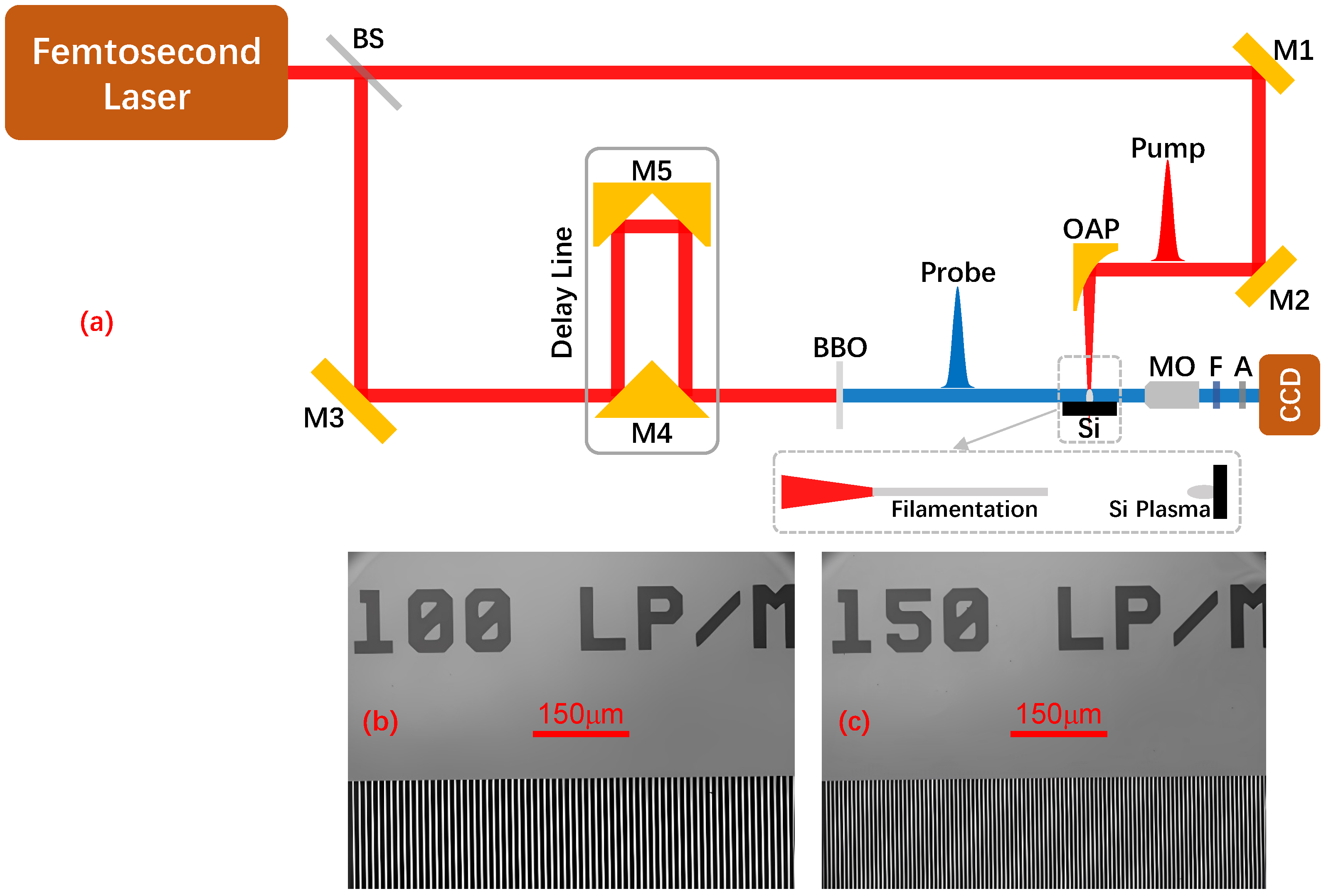
2. Experiment
To resolve the threshold-governed plasma chronology at air–silicon interfaces, we established a time-resolved shadowgraphy platform [
33,
36]. This experimental setup allowed us to capture the spatiotemporal evolution of both air filaments and silicon plasma, revealing an unexpected reversal in their excitation sequence. The experimental configuration is schematically illustrated in
Figure 1.
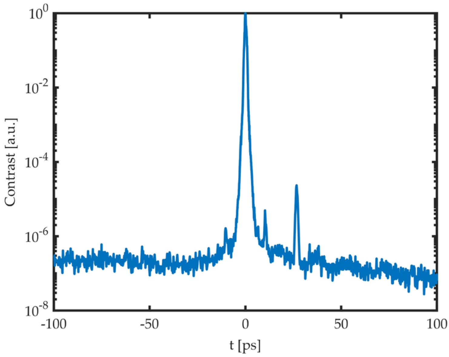
The experiments were conducted using a chirped pulse amplification (CPA) Ti:sapphire femtosecond laser system (HEFEI 50mJ, Amplititude Technologies S.A., Evry, France), which generated pulses with a central wavelength of 800 nm and a full width at half maximum (FWHM) pulse duration of 550 fs. The temporal contrast measurement data, as provided by the laser manufacturer, are shown in
Figure 2.
The fundamental laser beam was divided into pump (55% throughput) and probe (45% reflection) beams using an ultra-thin film beam splitter (BP245B2, Thorlabs Inc., Newton, MA, USA). The pump beam was utilized to initiate plasma formation, while the probe beam was employed to investigate transient morphological changes with femtosecond precision. The pump beam was tightly focused onto the silicon surface using an off-axis parabolic mirror (OAP) (RFL = 50.8 mm, MPD129-G01, Thorlabs Inc., Newton, MA, USA), achieving a calculated focused spot diameter of 4.7 μm. This tight focusing was crucial for reaching the necessary intensity threshold for both air filamentation and silicon plasma excitation. The peak intensity scales inversely with the square of the focal spot size, creating an axial intensity gradient with spatiotemporally engineered modulation across the air–silicon interface. This configuration establishes the optimal spatiotemporal overlap prerequisites for threshold-governed ionization competition. Given that all reflective components in the pump path are gold-coated mirrors, the group velocity dispersion (GDD) contribution from these mirrors is negligible.
The probe beam was steered through an optical delay line comprising a stepper motor stage (Daheng GCD-302002M, Daheng New Epoch Technology Inc., Beijing, China) and two precision reflectors (M4: 90° Specialty Mirror, Edmund #47-239, Edmund Optics Inc., Barrington, IL, USA; M5: Gold Retroreflector, Edmund #49-676, Edmund Optics Inc., Barrington, IL, USA), achieving a scanning range of 4 μs. Then, the probe beam underwent frequency doubling through a thin β-BBO crystal (thickness: 1 mm, CASTECH Inc., Fuzhou, China) to 400 nm, followed by coupling with a 10 nm bandpass filter (FBH400-10, Thorlabs Inc., Newton, MA, USA) that suppressed residual 800 nm light by >106:1. This spectral purification eliminated pump-induced artifacts, ensuring shadowgraphic fidelity at the air–silicon interface—a critical advancement over conventional single-wavelength diagnostics. The system’s temporal resolution of 550 fs was determined by the laser pulse width. Effective management of GDD was crucial in maintaining temporal resolution. By employing low-dispersion optics, including metallic mirrors, an ultra-thin beamsplitter, and a thin β-BBO crystal, the probe pulse duration broadening was also negligible.
Shadow images of the air filament and silicon plasma were captured using an imaging system composed of a long-working-distance microscope objective (10× Mitutoyo Plan Apo, Mitutoyo Corporation, Kawasaki, Japan), a narrow bandpass filter, several neutral density filters, and a CCD camera (pixel size: 3.69 μm, Point Grey GS3-U3-28S4M-C, FLIR Systems Inc., Richmond, BC, Canada). The spatial resolution of the imaging system was rigorously quantified using a USAF-1951 resolution target (R1L3S5P, Thorlabs Inc., Newton, MA, USA), confirming a resolution of 0.9 μm/pixel through modulation transfer function analysis. Representative shadowgraph images of the resolution test target are shown in
Figure 1b,c.
The intrinsic undoped silicon target with a polished (100)-oriented surface was precisely aligned at the focal plane of the off-axis parabolic mirror (OAP) and irradiated with 8.9 mJ pump pulses. Single-frame transient imaging was achieved by synchronizing the imaging system with the probe laser pulses through a triggered synchronization mechanism, ensuring precise temporal overlap between the probe pulses and the CCD camera’s integration window. To enable single-pulse irradiation mode in the CPA-based laser system, a shutter (SH1, Thorlabs Inc., Newton, MA, USA) was integrated into the input port of the main amplifier. Time-resolved image sequences were generated using a high-precision optical delay line with a mechanical displacement of 1.25 μm per step. Each retroreflector displacement (Δx) introduced a probe delay calculated as Δt = 2Δx/c (c: speed of light), achieving a minimum scan step of 8.3 fs. This deterministic optical path control enabled programmable acquisition of ionization dynamics across predefined delay intervals. Key parameters of the time-resolved shadowgraphy experimental setup are summarized in
Table 1.
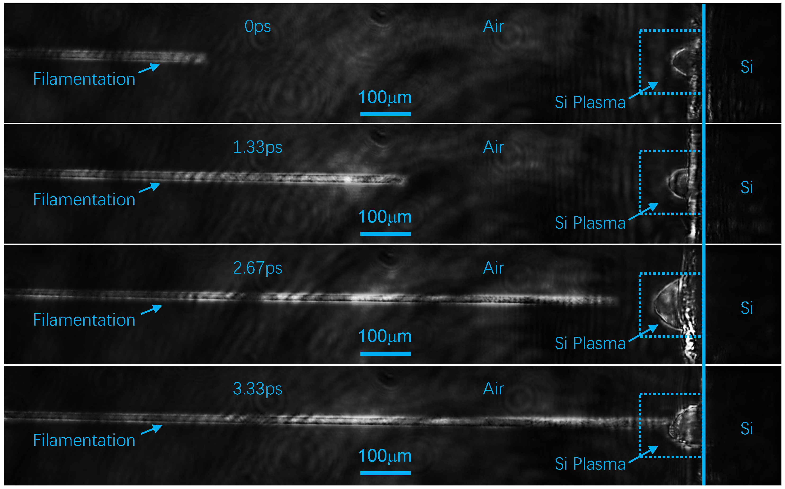
The temporal reference (t
0) was defined as the initial detection of plasma luminescence corresponding to the first observable shadowgraph image, establishing a self-consistent framework for analyzing the relative timing between air and silicon ionization events. The experimental setup successfully captured transient shadow images of air filaments and silicon plasmas at various delay times, as illustrated in
Figure 3.
In
Figure 3, the air–silicon interface is marked by a blue reference line, with the left dark region corresponding to air and the right black region representing the silicon target. Arrow-annotated filamentary structures are identified as air filaments, while semicircular morphological features within a dashed box (similarly arrow-labeled) align with characteristic silicon plasma signatures. Alternating bright–dark striations adjacent to the target surface manifest edge diffraction effects at the silicon interface.
Typically, in femtosecond laser–matter interactions, the conventional sequence of events is expected to begin with the excitation of an air filament, which propagates alongside the femtosecond laser pulse toward the silicon target surface, culminating in silicon plasma excitation. However, our spatiotemporally resolved measurements reveal a counterintuitive reversal in the plasma chronology: silicon plasma was excited prior to the arrival of the air filament at the silicon surface. This unexpected observation indicates that the ionization of the silicon target occurs earlier than that of the adjacent air layer, particularly in the region between the trailing edge of the air filament and the silicon surface. This finding not only provides new insights into the complex interactions between the laser and the material but also calls into question the established understanding of excitation sequences in tightly focused femtosecond laser fields.
To quantify this phenomenon, we conducted a meticulous temporal analysis of the air filament’s propagation. The thickness of the air layer is defined as the spatial distance between the silicon target surface and the trailing edge of the filamentation. Our measurements revealed that the maximum air layer thickness was 900 ± 32 μm, with uncertainty stemming from the gradual intensity transition at the filament’s trailing edges. This corresponds to a temporal delay of 3.0 ± 0.1 picoseconds (ps) between the trailing edge of the air filament and the onset of silicon target ionization. This significant temporal separation underscores the intricate coupling between material ionization mechanisms and laser energy deposition, thus challenging traditional views on excitation sequences in such laser fields.
3. Discussion
3.1. Exclusion of Alternative Mechanisms
The observed temporal inversion of the plasma chronology fundamentally reorients our understanding of energy deposition in multi-material systems under tight femtosecond focusing. Our experimental results demonstrate that silicon plasma nucleation precedes air filamentation by 3.0 ± 0.1 ps at air–silicon interfaces under tight femtosecond focusing. To elucidate this counterintuitive phenomenon, we systematically evaluated three potential mechanisms: flying focus effects, cascade ionization processes, and threshold-governed spatiotemporal ionization dynamics.
The initial analysis considered flying focus effects as a plausible explanation. Conventional flying focus mechanisms predict a consistent directional movement of the focal position (either forward or backward propagation) along the laser axis [
38]. However, in the present experiment, the initial ionization of silicon plasma precedes the subsequent ionization of air near the silicon surface, suggesting a backward shift of the focal region relative to the laser propagation direction. This contradicts the forward-directed nature of filamentation, which would require the focal movement to align with the pulse’s propagation. The temporal and spatial independence of the two ionization events further indicates that silicon and air plasma formation are decoupled processes. This directional paradox invalidates flying focus as the primary mechanism governing the observed temporal inversion.
Cascade ionization mechanisms were subsequently investigated. However, it is important to note that in gases at 1 atm under femtosecond laser conditions, there is generally a minimal occurrence of inverse Bremsstrahlung and cascade ionization. This is mainly due to the fact that the mean free time of a free electron that is released from an atom or molecule through multiphoton/tunnel ionization is longer than the duration of the femtosecond laser pulse itself [
17]. These constraints necessitated the exploration of alternative mechanisms.
3.2. Threshold-Governed Ionization Dynamics
The threshold-governed mechanism emerges as the dominant explanation for the observed plasma chronology. The ionization threshold for air under femtosecond laser irradiation has been well-established at 50 TW/cm
2 [
17]. In contrast, experimental data demonstrate that silicon exhibits a significantly lower ionization threshold of approximately 0.36 TW/cm
2 (equivalent to 0.2 J/cm
2 for a 550 fs pulse [
37]), derived from a fitted curve across varied pulse durations. This results in a 137.5-fold disparity in the thresholds between air and silicon, which critically governs the sequence of plasma formation. To quantify this threshold-driven process, we analyzed the spatiotemporal intensity distribution of the tightly focused femtosecond field and compared it with the material-specific ionization thresholds.
The intensity of a tightly focused femtosecond laser can be articulated through the following equation:
Herein,
signifies the pulse energy,
characterizes the temporal profile, and
delineates the spatial distribution of the laser. The temporal and spatial profiles are detailed as follows:
and
In Equation (2),
denotes the pulse duration,
c is the speed of light, and
t represents time. In Equation (3),
r is the transverse spatial coordinate,
z is the longitudinal spatial coordinate, and
is the radius of the laser beam at position
z, which can be determined by
where
λ is the laser wavelength, and
is the radius of the beam waist, calculated as follows:
Here,
is beam quality factor,
F is the focal length of the OAP, and
is the radius of the laser beam at the front surface of the OAP.
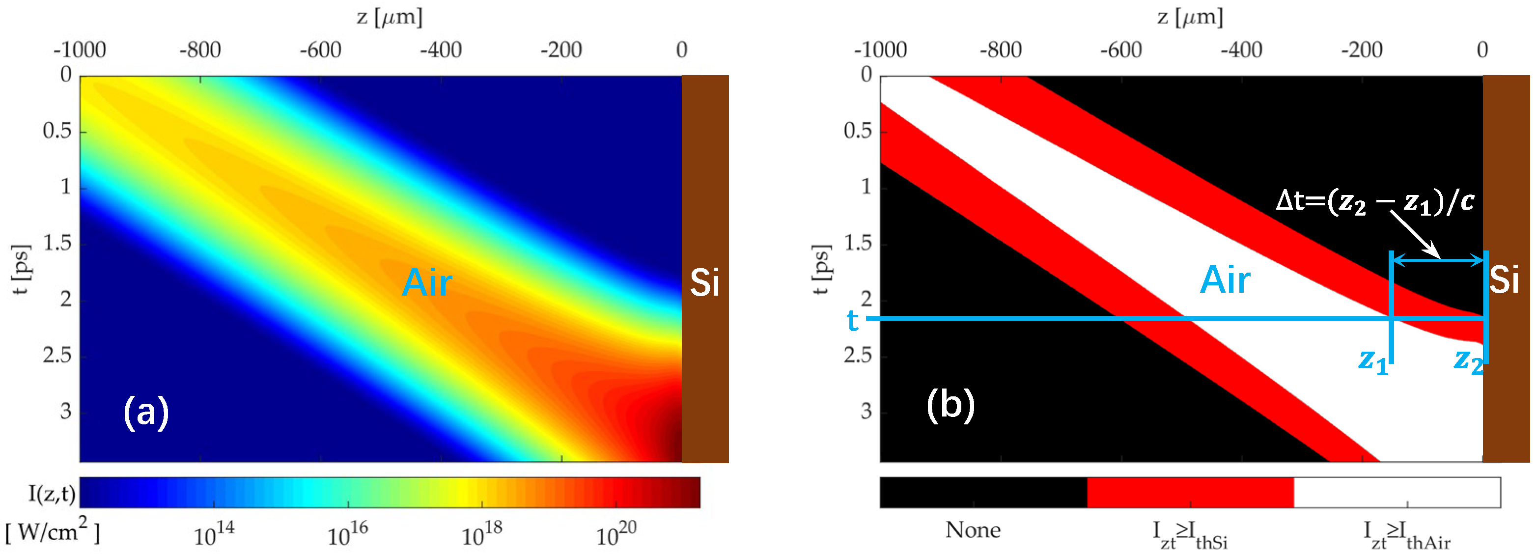
A femtosecond laser system operating at 800 nm wavelength with a 550 fs pulse duration, 8.9 mJ pulse energy, and a beam radius of 10 mm (
= 1.8) was employed to characterize the spatiotemporal intensity distribution. The OAP has a focal length of 50.8 mm. The laser propagates along the positive
z-axis in the laboratory coordinate system, with the OAP focal plane set at z = 0 μm to align with experimental measurements. Equations (1)–(5) were used to calculate the temporal intensity profile along the optical axis at discrete axial positions (z-values), as visualized in
Figure 4a.
Figure 4a illustrates the progressive propagation of the femtosecond laser pulse along the positive
z-axis, approaching the Si target at z = 0 μm. A dramatic enhancement in light intensity is observed at the OAP focal plane (z = 0 μm), with sustained high-intensity levels over an extended temporal window. The spatiotemporal intensity distribution is compared with the ionization thresholds of silicon (0.36 TW/cm
2) and air (50 TW/cm
2) in
Figure 4b. Regions exceeding silicon’s threshold are highlighted in red, while those surpassing air’s threshold are marked in white. Due to the significantly lower ionization threshold of silicon, the red region exhibits broader spatiotemporal coverage that overlaps with the central white region but extends both axially and radially.
At t = 2.29 ps, the femtosecond laser pulse induces air filamentation at z1 = –82.93 μm, while its leading edge reaches the Si surface at z2 = 0 μm, satisfying the ionization threshold of silicon. Critically, the leading edge—despite its initially lower power—achieves intensity levels that are comparable to the main pulse due to spatiotemporal coupling. This phenomenon arises from the following mechanism: the leading edge arrives at the focal region earlier, where geometric focusing amplifies its intensity, whereas the delayed main pulse remains in the pre-focal zone, dominated by beam divergence. This counterintuitive intensity enhancement is governed by spatiotemporal coupling, a process involving the intricate interplay between the pulse’s temporal profile and spatial focusing dynamics under tight focusing conditions.
This results in a calculated temporal precedence of Δt = (z2 − z1)/c = 276.4 fs for Si plasma formation relative to air filamentation. The pronounced disparity in ionization thresholds (137.5-fold) directly governs this temporal inversion, as the lower threshold enables Si plasma generation at an earlier axial position and time compared to air filamentation. However, this computed lead time (~276 fs) is significantly shorter than the experimentally observed ~3 ps temporal precedence. The discrepancy suggests the involvement of additional factors that modulate the actual temporal sequence of plasma generation, which will be further analyzed in subsequent discussions.
3.3. Temporal Contrast Effects on Plasma Chronology
This discrepancy is likely attributed to the temporal contrast characteristics of chirped-pulse-amplified (CPA) lasers, which inherently generate sub-threshold pre-pulses and amplified spontaneous emission (ASE) [
39]. To quantify this effect, we employed the temporal contrast data from the femtosecond laser system (
Figure 2) to correct the temporal distribution modeled in Equation (2), yielding a revised spatiotemporal intensity profile
, defined as follows:
Here,
represents the combined contribution of temporal contrast data fitting from
Figure 2 and the propagation delay associated with laser propagation. By applying Equation (6), the modified spatiotemporal optical field distribution and ionization region simulations are derived, as illustrated in
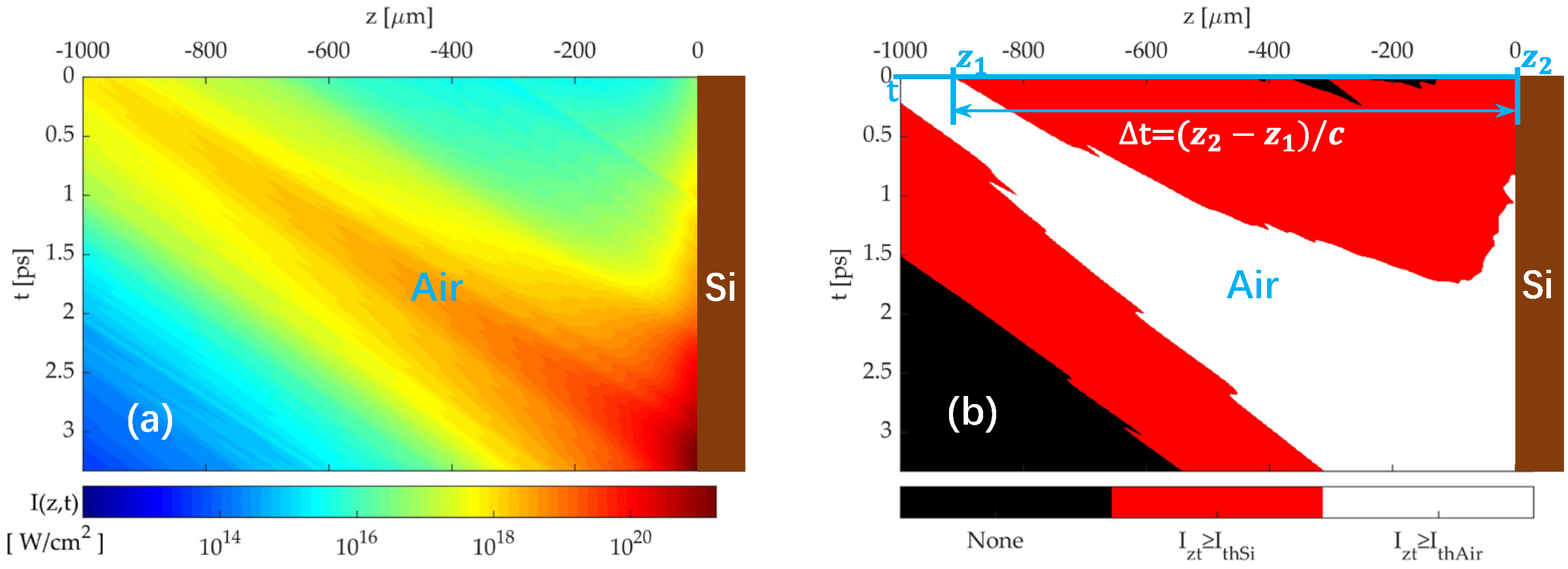
Figure 5.
Figure 5a demonstrates that due to the presence of pre-pulses and ASE, the light intensity at the focal plane (z = 0 μm) remains elevated over an extended temporal window compared to
Figure 4a. Adopting an analogous methodology to the previous analysis, the spatiotemporal intensity distribution in
Figure 5a is compared with the ionization thresholds of silicon (0.36 TW/cm
2) and air (50 TW/cm
2) in
Figure 5b. The resulting ionization regions for silicon and air exhibit significantly larger spatiotemporal coverage than those in
Figure 4b.
At t = 0 ps, both the air at z1 = −913.3 μm and the Si target at z2 = 0 μm are simultaneously ionized. However, since the femtosecond laser propagates along the positive z-axis, a finite time Δt is required for the pulse to travel from z1 to z2. This results in a temporal precedence of Δt = (z2 − z1)/c = 3.044 ps for Si plasma formation relative to air filamentation, which shows reasonable agreement with the experimentally observed temporal precedence (~3 ps).
Through systematic spatiotemporal analysis, we establish a fundamental re-ordering of plasma formation dynamics at air–silicon interfaces under femtosecond laser irradiation, driven by the interplay between material-specific ionization thresholds and spatiotemporally engineered laser parameters. The 137.5-fold lower ionization threshold of silicon (0.36 TW/cm2) compared to air (50 TW/cm2) enables silicon to nucleate plasma during the laser pulse’s rising edge, while air ionization remains confined to the peak intensity region. This threshold hierarchy dominates the observed ~3 ps precedence of silicon plasma, a phenomenon that is further modulated by the laser’s temporal contrast—pre-pulse and ASE components extend the effective ionization window, while tight focusing sharpens intensity gradients to sub-wavelength scales. Together, these factors establish a universal framework for predicting ionization sequences in multi-material systems, particularly those combining wide-bandgap dielectrics with narrow-bandgap semiconductors or metals.
These findings redefine conventional paradigms of laser–material interaction by emphasizing threshold-dominated chronology over purely intensity-driven dynamics. From a practical perspective, the ability to spatially and temporally decouple ionization events across heterogeneous interfaces opens transformative opportunities.
4. Conclusions
This study redefines the principles governing plasma chronology in multi-material systems under femtosecond laser irradiation. By integrating time-resolved shadowgraphy with threshold-driven analysis, we establish that ionization thresholds—not peak intensity—dictate the temporal sequence of plasma formation. The 137.5-fold threshold disparity between silicon and air enables silicon plasma nucleation during the pulse’s rising edge, while air ionization requires peak intensity. Laser temporal contrast and tight focusing geometries further modulate this sequence, enabling tunable delays from femtosecond to picosecond scales.
These findings offer new opportunities for advancing ultrafast laser–matter interaction research. In precision manufacturing, threshold-selective plasma initiation can potentially reduce collateral damage during micromachining of layered composites. By tailoring the laser parameters to match the ionization thresholds of individual layers, energy deposition can be spatially restricted to target regions, minimizing thermal diffusion into adjacent materials. For optical diagnostics, the picosecond-scale control over plasma delays enhances time-gated LIBS, enabling depth-resolved compositional analysis of multi-layered films with sub-μm resolutions. Beyond these applications, the demonstrated mechanism provides a foundation for exploring ultrafast interfacial phenomena in phase-change materials, where spatially and temporally resolved plasma dynamics could probe phase transition kinetics and metastable states.
Future work will extend this framework to 3D material architectures and explore machine learning-driven optimization of laser parameters for adaptive plasma control. While current studies focus on planar interfaces, extending this framework to 3D architectures requires addressing challenges such as non-uniform energy distribution and complex interfacial coupling. By coupling real-time plasma diagnostics with predictive models, we aim to unlock deterministic spatiotemporal engineering of laser–matter interactions across scales—from micro/nanofabrication to macroscopic device integration.


