Review

Advances in Distal-Scanning Two-Photon Endomicroscopy for Biomedical Imaging

by
Conghao Wang
1,
Biao Yan
1,
Siyuan Ma
2,
Haijun Li
3,
Tianxuan Feng
2,
Xiulei Zhang
4,
Dawei Li
1,
Lishuang Feng
2,5,6,* and
Aimin Wang
7,*
1
College of Future Technology, Peking University, Beijing 100871, China
2
School of Instrumentation and Optoelectronic Engineering, Beihang University, Beijing 100191, China
3
Jingjinji National Center of Technology Innovation, Beijing 100192, China
4
School of Automation Science and Electrical Engineering, Beihang University, Beijing 100191, China
5
Key Laboratory of Precision Opto-Mechatronics Technology, Ministry of Education, Beijing 100191, China
6
Beijing Laboratory of Biomedical Imaging, Beijing Municipal Education Commission, Beijing 100085, China
7
State Key Laboratory of Advanced Optical Communication System and Networks, School of Electronics, Peking University, Beijing 100871, China
*
Authors to whom correspondence should be addressed.
Photonics 2025, 12(6), 546; https://doi.org/10.3390/photonics12060546
Submission received: 8 April 2025 / Revised: 11 May 2025 / Accepted: 24 May 2025 / Published: 29 May 2025
(This article belongs to the Special Issue Emerging Trends in Multi-photon Microscopy)

Abstract

Two-photon endomicroscopy (2PEM), an endomicroscopic imaging technique based on the two-photon excitation effect, provides several technical benefits, including high spatiotemporal resolution, label-free structural and metabolic imaging, and optical sectioning. These characteristics make it extremely promising for biomedical imaging applications. This paper classifies distal-scanning 2PEMs based on their actuation mechanism (PZT or MEMS) and excitation–collection optical path configuration (common or separate path). Recent representative advancements are reviewed. Furthermore, we introduce its biomedical applications in tissue, organ, and brain imaging with free-behaving mice. Finally, future development directions for distal-scanning 2PEM are discussed.

1. Introduction

Two-photon endomicroscopy (2PEM), a significant step forward in miniature two-photon microscopy (m2PM), has emerged as a promising optical biopsy tool for clinical translation [1,2]. Its fundamental principle is to integrate miniature scanning mechanisms and micro-objective lenses into a millimeter-scale endoscopic probe, allowing for nonlinear excitation of two-photon fluorescence (2PF) and second-harmonic generation (SHG). In contrast to wide-field and confocal endomicroscopy, it offers the distinct advantages of high spatiotemporal resolution, depth-resolved optical sectioning, and label-free structural and metabolic imaging [3,4,5,6]. These technological advancements confirm its potential as a transformative platform for addressing biomedical challenges such as early-stage cancer screening and real-time intraoperative margin assessment.
After two decades of technological advancement, 2PEM has two primary configurations: proximal-scanning [7,8] and distal-scanning [9,10,11]. Proximal-scanning configurations describe systems where the scanning mechanism remains close to the light source, eliminating electrical components within the endoscopic probe. This method typically uses galvanometer scanners to scan the beam, with the pre-scanned laser beam transmitted via coherent fiber bundles. Distal-scanning configurations incorporate miniature scanners at the probe tip, enabled by advances in piezoelectric tube (PZT) fiber scanners and microelectromechanical systems (MEMS) scanners. While proximal-scanning 2PEM achieves simplified endomicroscopic probe design and enhanced reliability through elimination of internal electronics, its reliance on coherent fiber bundles inherently constrains spatial resolution due to fiber core spacing [12,13]. In contrast, distal-scanning 2PEM demonstrates enhanced spatiotemporal resolution that enables superior imaging quality. While requiring more sophisticated probe engineering, distal-scanning 2PEM has been successfully implemented in label-free tissue imaging and brain imaging of freely behaving mice, demonstrating translation potential in the biomedical imaging field [14,15].
This paper discusses recent advancements in distal-scanning 2PEM for biomedical imaging. The paper is organized as follows. Section 2 introduces a four-quadrant technical classification framework that categorizes systems based on their actuation mechanism (PZT and MEMS) and excitation–collection optical path configuration (common and separate paths). Section 3 examines the notable advancements of 2PEM in these categories. Section 4 discusses 2PEM’s biomedical applications, such as high-resolution tissue, organ imaging, and brain imaging in freely moving mice. Section 5 proposes future development directions for distal-scanning 2PEM from our perspective.

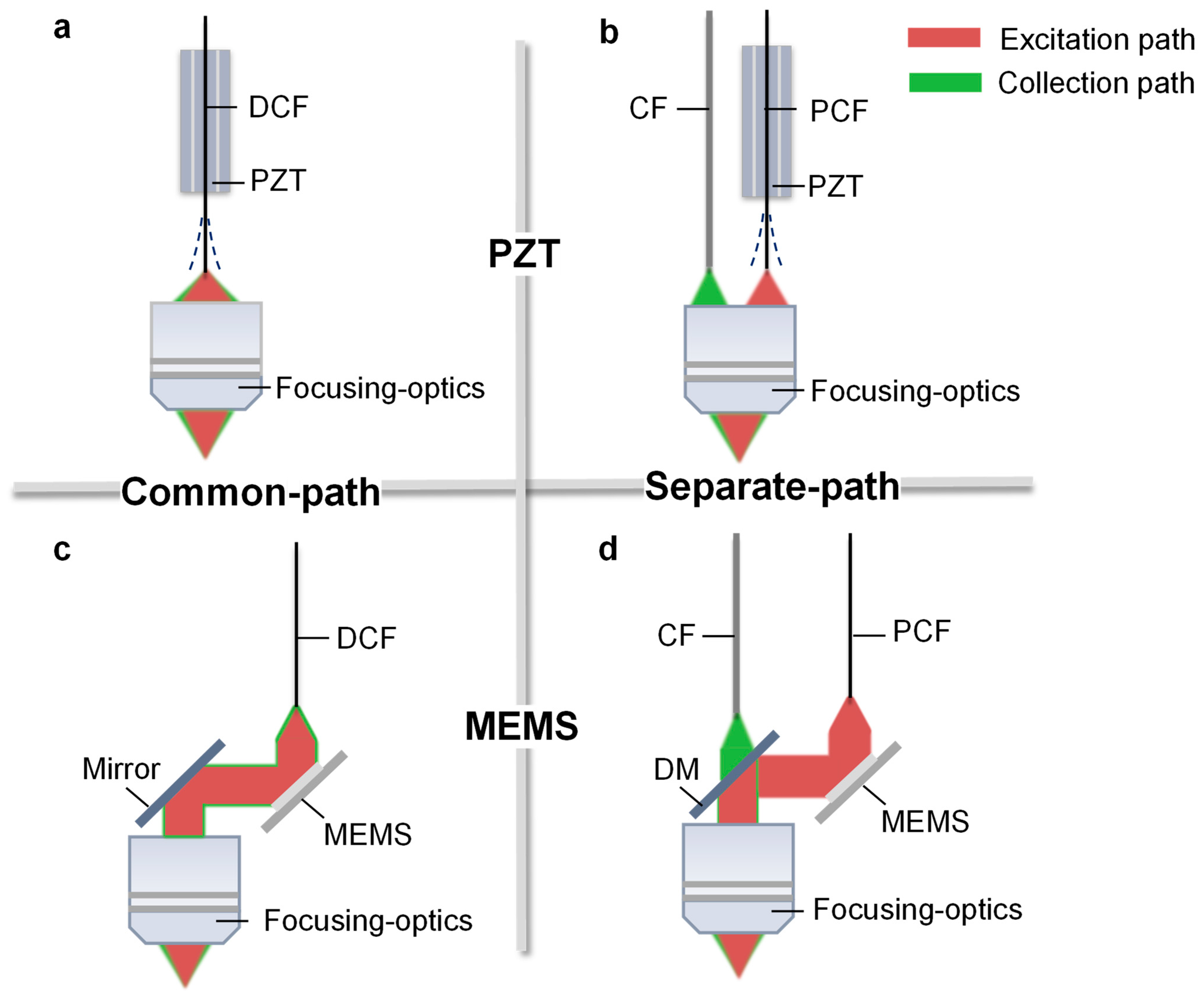
2. Classifications of Distal-Scanning 2PEM

Distal-scanning 2PEM can be divided into two types based on the actuation mechanism: the PZT fiber scanning scheme (hereafter referred to as the PZT scanning scheme) and the MEMS scanning scheme. The PZT scanning scheme uses the inverse piezoelectric effect for electromechanical energy conversion, with beam deflection caused by the resonance of the fiber cantilever. While achieving high resonant frequency comparable to the MEMS scanning scheme, this resonant-driven mechanism inherently elongates the scanner length due to the required fiber cantilever. The MEMS scanning scheme allows for beam scanning via precisely controlled rotation of the micromirror, with actuation methods including electrothermal and electrostatic drives that dominate in endomicroscopic applications [16,17,18]. PZT fiber scanners and MEMS scanning mirrors can generate various two-dimensional (2D) scanning patterns using different driving signals. The patterns are spiral [19,20,21], raster [22,23], and Lissajous [24,25,26].
Distal-scanning 2PEM can also be divided into two types based on the excitation–collection optical path configuration: common-path and separate-path. The common-path configuration uses a double-cladding fiber (DCF) for simultaneous propagation, with the core transmitting the femtosecond pulse and the outer cladding guiding the backscattered fluorescence. The refractive index hierarchy (fiber coating < fiber outer cladding) allows continuous fluorescence collection through total internal reflection. The primary advantage of the common-path configuration is the DCF’s dual functionality, which simplifies the optical path design. However, its miniature objective lens requires chromatic aberration compensation to maintain high fluorescence collection efficiency. In contrast, the separate-path configuration uses a photonic crystal fiber (PCF) to transmit femtosecond pulses and a collection fiber (CF) to collect fluorescence. The separate-path configuration physically separates the excitation and collection paths, thereby reducing optical design complexity. However, the probe requires additional optical beam-splitting components for beam-splitting and reflection (e.g., dichroic mirror, reflective mirror/prism), which directly contributes to the critical challenge of increased packaging dimensions. Note that recent advances in PCFs, which are distinguished by low loss, low dispersion, and broadband transmission, have sparked significant research interest [27,28]. Photonic bandgap fibers (PBFs), Kagome fibers, and anti-resonant fibers are examples of flexible delivery solutions for distal-scanning 2PEM that are currently being investigated [29,30].
Researchers developed four distal-scanning 2PEM probe schemes based on the aforementioned actuation mechanisms and optical path configurations, as illustrated in Figure 1a–d. These schemes include the PZT-based common-path scheme, the PZT-based separate-path scheme, the MEMS-based common-path scheme, and the MEMS-based separate-path scheme. Section 2 summarizes the representative progress made with these schemes.

3. Research Progress in Distal-Scanning 2PEM

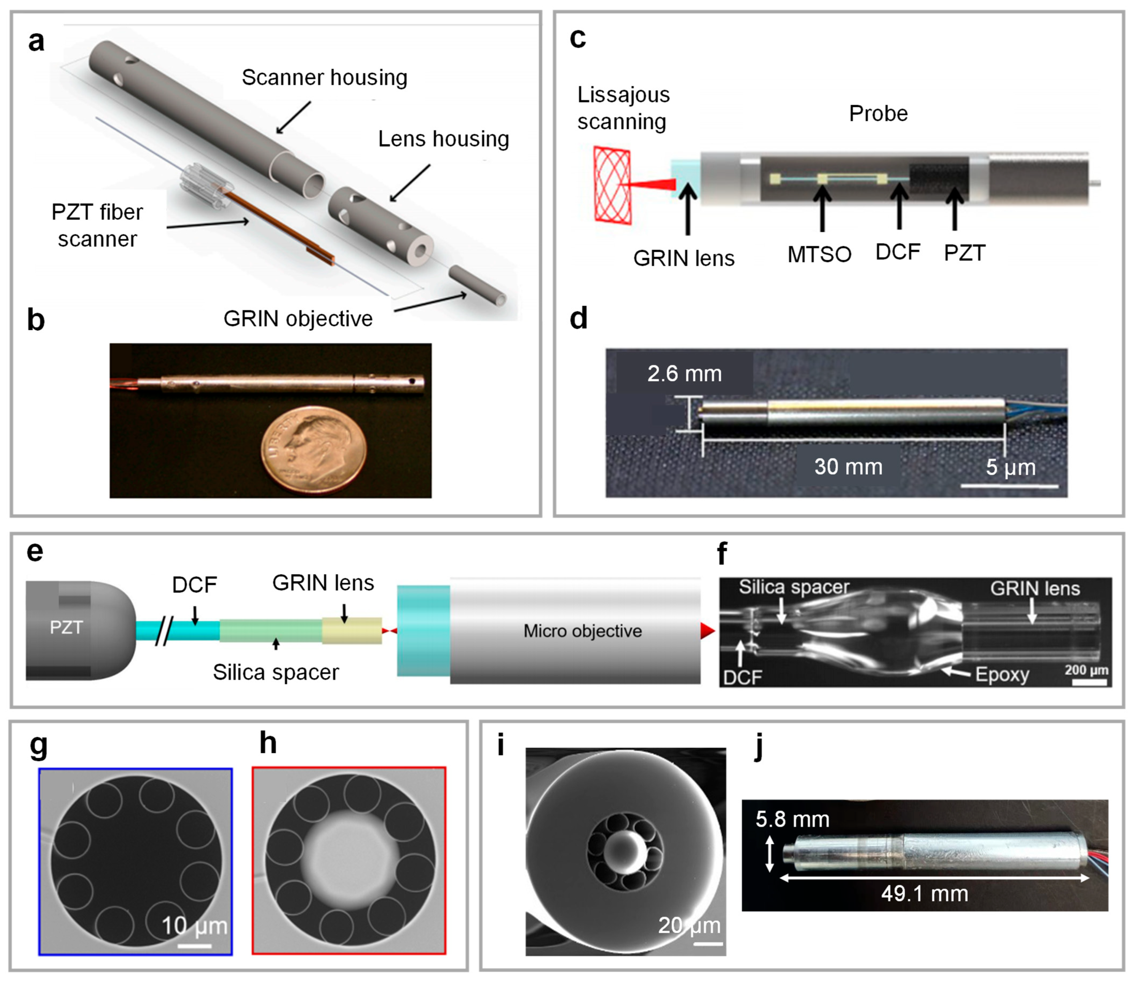
3.1. PZT-Based Common-Path 2PEM

In 2006, Myaing et al. demonstrated the technical feasibility of a PZT-based common-path 2PEM [9]. A spiral scanning pattern was created using ±x and ±y amplitude-modulated sinusoidal waveforms on the PZT fiber scanner. This platform used a commercially available DCF for excitation pulse and fluorescence multiplexed guidance, along with a gradient refractive index (GRIN) lens for focusing and imaging, to establish the fundamental paradigm for future technological development. Raster and Lissajous scanning patterns offer superior scanning uniformity relative to spiral patterns. Building on this advantage, Rivera et al. developed a raster scanning 2PEM probe in 2011, as depicted in the schematic and photograph in Figure 2a,b [31]. The probe used a dual-layer piezoelectric actuator for raster scanning that provided resonant and non-resonant actuation. A commercial DCF (SM-9/105/125-20A, NuFern) was glued to the scanner and had a 9 mm fiber cantilever with a resonant frequency of 1.05 kHz. Using a high numerical aperture (NA = 0.8) GRIN lens assembly, the system achieved an imaging resolution of 0.8 μm, a field of view (FOV) of 110 μm × 110 μm, and a frame rate of 4.1 frames per second (fps). The integrated probe had a 3 mm outer diameter and a rigid length of 40 mm, allowing depth-resolved 2PF and SHG imaging of mouse lung and colon tissues.
Clinical translation of 2PEM requires overcoming the critical challenge of low-dispersion transmission for femtosecond pulses through extended optical delivery. Addressing this limitation, Ducourthial et al. introduced a large FOV distal-scanning 2PEM through a long optical fiber in 2015 [32]. The system used a customized solid-core double-cladding photonic crystal fiber (DC-PCF) as the delivery fiber, with a group velocity dispersion (GVD) of 67 ps/km/nm. Through advanced spectro-temporal pulse shaping techniques, the system successfully achieved delivery of infrared excitation pulses below 40 fs at the output terminal of a 5-m fiber. The platform maintained an imaging resolution of 0.8 μm while extending the FOV to 450 μm × 450 μm and increasing the frame rate to 8 fps. The integrated probe was reduced to outer dimensions of 2.2 mm in diameter and a rigid length of 37 mm. In 2016, Wang et al. fabricated a square-tube PZT fiber scanner and validated its performance for two-photon endomicroscopic imaging [33]. This system achieved an imaging resolution of 2.2 μm and a FOV of 200 μm × 200 μm at 1.25 fps, housed within a probe with an outer diameter of 3.5 mm and a rigid length of 53 mm. In 2019, Kim et al. reported a Lissajous scanning 2PEM with high uniformity, as shown in Figure 2c,d [34]. By incorporating a micro-mass block and micro-spring structure onto the fiber cantilever, the scanner’s resonant frequencies were separated to 885 Hz and 1160 Hz, resulting in a 99% fill factor. The system’s imaging resolution was 0.7 μm, with a FOV of approximately 84 μm × 95 μm at an applied voltage of 20 V and a frame rate of 5 fps. The integrated probe had an outer diameter of 2.6 mm and a rigid length of 30 mm.
Recent advances in fiber-tip engineering techniques for PZT-based 2PEM have significantly improved imaging performance, particularly regarding resolution and FOV. Miniature optical lenses can be integrated directly onto fiber tips using advanced photonic processing techniques like precision fiber splicing, controlled adhesive gluing, and femtosecond laser 3D printing. It allows for modulation of the fiber’s output NA. This technological advancement opens up new possibilities for improving the imaging performance of distal-scanning 2PEM.
In 2017, Akhoundi et al. demonstrated three-photon endomicroscopic imaging by gluing a miniature objective lens to the fiber tip, resulting in an extended FOV exceeding 600 μm × 600 μm [35]. However, using lenses larger than 2 mm resulted in significant resonant frequency degradation of the PZT fiber scanner, limiting the frame rate to 15 s per frame (512 pixels × 512 pixels). In 2021, Liang et al. reported a PZT-based 2PEM with a GRIN lens-attached DCF [36]. As detailed in Figure 2e,f, gluing the GRIN lens to the DCF tip increased the fiber’s output NA from 0.12 to 0.35. The increased fiber NA reduced the constraints on objective lens magnification, allowing for a larger imaging FOV while maintaining resolution, thereby increasing imaging throughput. One prototype of the probe has an imaging resolution of 0.61 μm and a FOV of 300 μm × 300 μm.
There is a critical need for broadband optical fibers in multimodal nonlinear endomicroscopy, where DCFs operating across 700–1100 nm are essential for efficient nonlinear probe development. In 2020, Kudlinski et al. developed a double-cladding anti-resonant fiber (DC-ARF) for nonlinear endomicroscopy with ultra-low dispersion (GVD < 1 ps/km/nm) across a 700–1000 nm wavelength range. Using a CO2 laser to fabricate a microsphere-spliced DC-ARF increased the fiber’s output NA nearly ninefold—from 0.023 to 0.21 [37]. SEM images of DC-ARF and microsphere-spliced DC-ARF are presented in Figure 2g,h. Building on this approach, in 2022, Wang et al. reported the development of an integrated 2PEM probe using a microsphere-spliced DC-ARF [38,39,40]. Figure 2i depicts a functionalized fiber’s integrated probe and scanning electron microscope (SEM) image. Using an electric-arc technique for precise microsphere splicing, the fiber’s output NA was increased from 0.034 to 0.31. The final platform had an imaging resolution of 1.3 µm, a FOV of 210 μm × 210 μm, and a frame rate of 0.7 fps. As illustrated in Figure 2j, the integrated probe had dimensions (outer diameter × rigid length) of 5.8 mm × 49.1 mm.

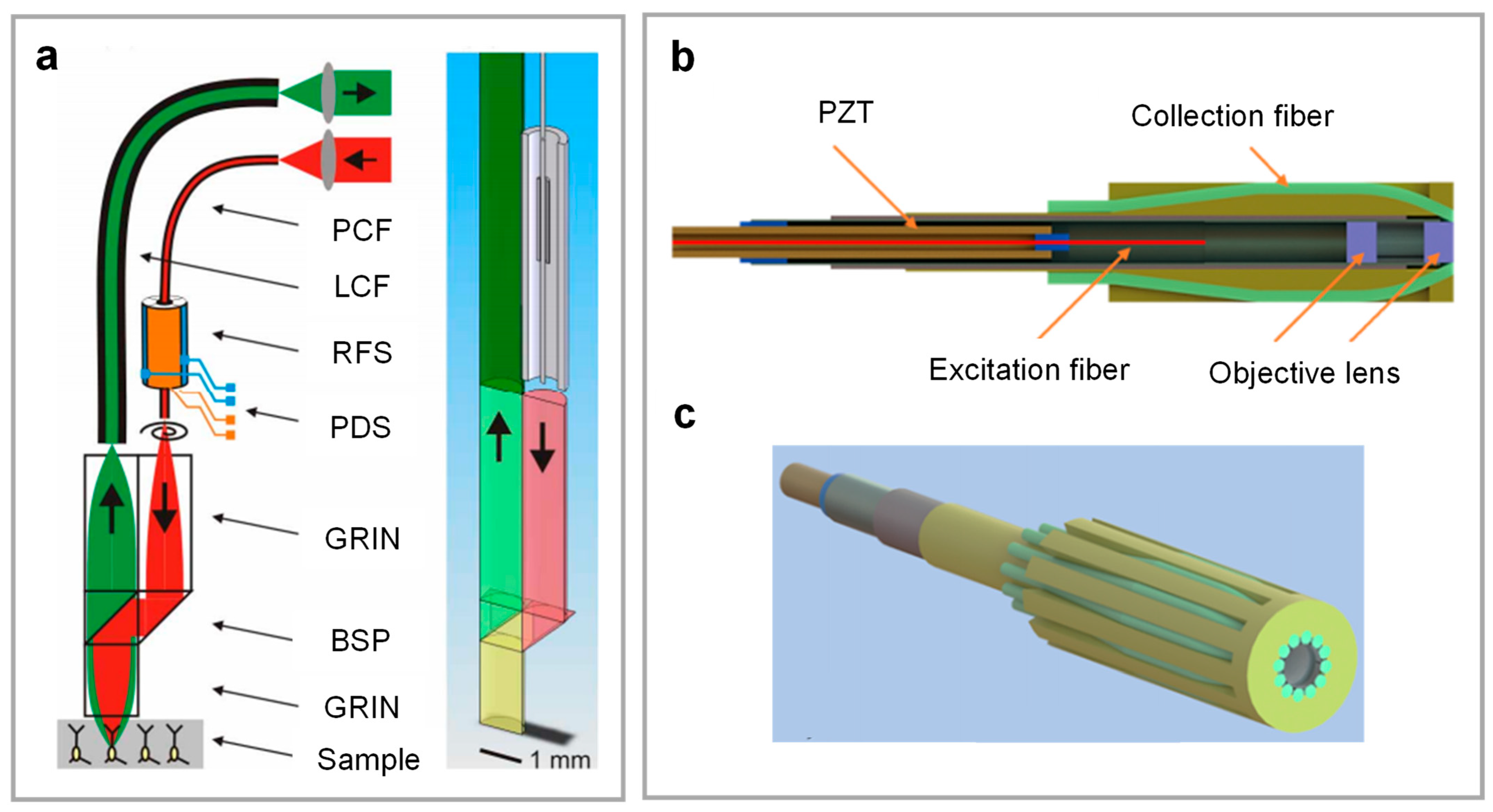
3.2. PZT-Based Separate-Path 2PEM

In 2008, Engelbrecht et al. proposed a m2PM using a PZT-based separate-path scheme [41]. The probe schematic is shown in Figure 3a. The system used PBF to deliver excitation pulses and a large-core multimode fiber to collect fluorescence. The integrated probe, weighing only 0.6 g, achieved micron-scale spatial resolution at 25 fps with a FOV of up to 200 μm× 200 μm. In vivo imaging results confirmed the functional imaging ability of cerebellar Purkinje cell dendrites in anesthetized rats. In 2009, Harzic et al. developed a large-FOV 2PEM [42]. In this work, PCF transmitted 790–830 nm femtosecond pulses while a multimode fiber collected fluorescence. For raster scanning, the scanner used orthogonally arranged piezoelectric trimorph actuators synchronously driven by micro-optical components such as fiber and a GRIN lens. The probe had an imaging resolution of 0.6 μm and extended the maximum FOV to 420 μm × 420 μm. However, the scanner’s operational frequency (<100 Hz) limited the frame rate to 0.77 fps (128 pixels × 128 pixels).
In 2024, Camli et al. proposed a PZT-based 2PEM with 12 multimode fibers for improved fluorescence collection [43]. The schematic and photograph of the 2PEM probe are depicted in Figure 3b,c, respectively. This design optimized the fiber tip angulation to increase the efficiency of collecting scattered fluorescence photons. Nevertheless, the minimum bending radius of the collection fibers requires a probe diameter exceeding 10 mm. The system had an imaging resolution of 0.66 μm, a FOV of 120 μm × 120 μm, and a frame rate of 2 fps. A direct current micro-motor to actuate the probe resulted in a Z-axis imaging depth of over 300 μm.

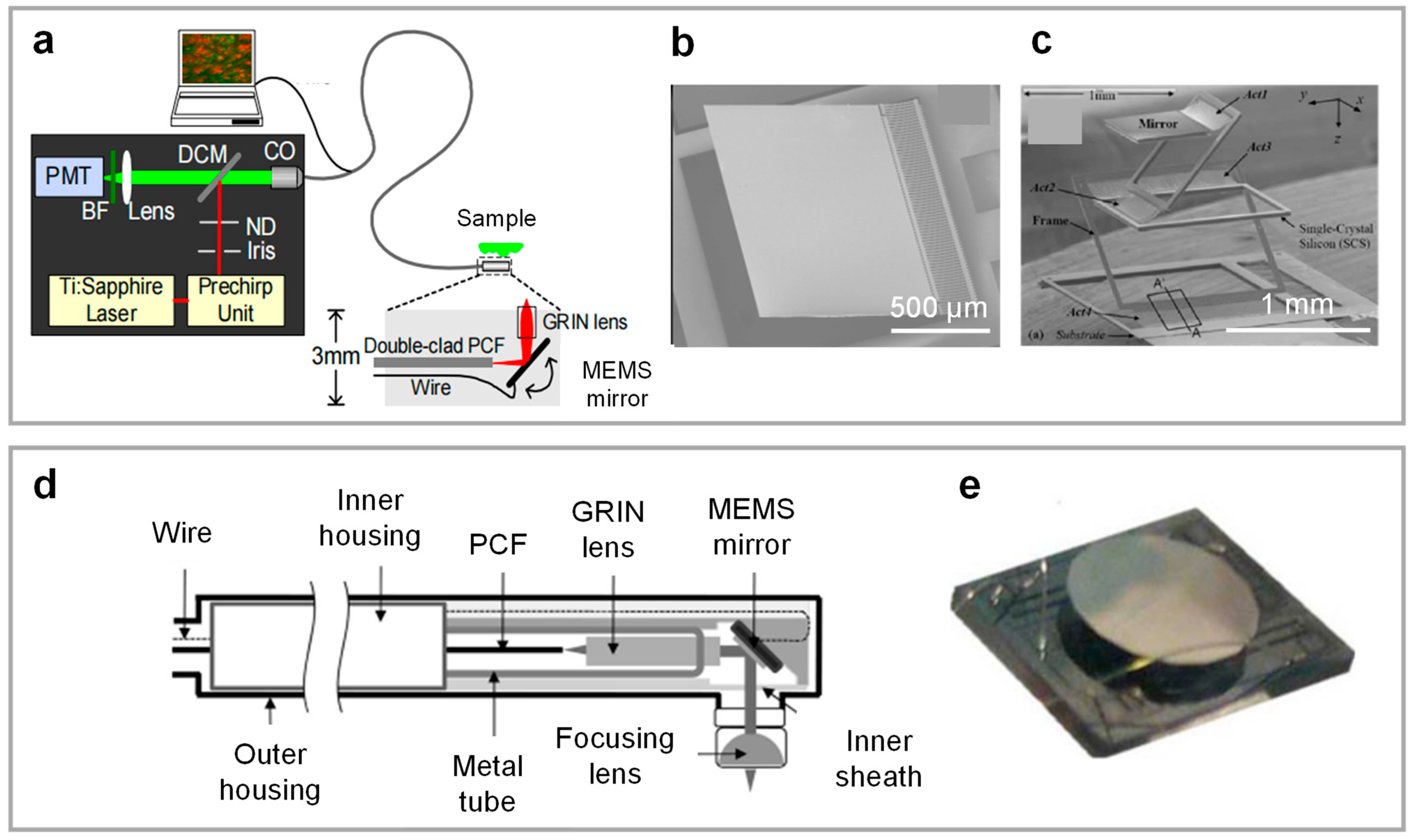
3.3. MEMS-Based Common-Path 2PEM

In 2006, Fu et al. proposed the first MEMS-based common-path 2PEM [44], as illustrated in Figure 4a. This configuration used a DC-PCF to transmit the excitation pulse and the fluorescence. The side-view probe had an outer diameter of 3 mm and included an electrothermal bimorph-actuated one-dimensional (1D) MEMS mirror. Figure 4b depicts a photograph of a MEMS mirror with a 1 mm mirror plate and a resonant frequency of 165 Hz. The coordinated operation of the 1D MEMS mirror and a 1D translation stage enables two-dimensional scanning. To demonstrate 2D scanning capability, Fu et al. developed a side-view 2PEM using a 2D electrothermal MEMS mirror in 2007 [45], with its structure depicted in Figure 4c. Nevertheless, the MEMS mirror’s low operational frequency limited the frame rate to approximately seven lines per second.
In 2008, Jung et al. proposed a common-path 2PEM using an electrostatic MEMS mirror [46], as shown schematically in Figure 4d. The integrated probe was 10 mm in outer diameter and 140 mm in rigid length. Figure 4e describes the electrostatic MEMS mirror, which includes a 2 mm circular mirror with dual-axis resonance at 1.26 kHz and 780 Hz [47]. This configuration achieved an imaging resolution of 2 μm, a FOV of 200 μm × 200 μm, and a frame rate of 0.25 fps. In 2020, Mehidine et al. demonstrated a side-view 2PEM probe employing a miniature electrothermal MEMS mirror [48]. The thermoelectric MEMS mirror measured 1.3 mm × 1.5 mm and had a mirror surface size of 0.52 mm. It had a symmetrical two-level ladder of dual S-shaped bimorph actuators, resulting in a first-order resonant frequency of around 1.4 kHz. A large scanning angle could be achieved at low driving voltages while maintaining the endoscopic integrated probe’s compact design.

3.4. MEMS-Based Separate-Path 2PEM

In 2008, Hoy et al. developed a MEMS-based separate-path m2PM [49], as illustrated in Figure 5a,b. A compact electrostatic MEMS mirror produced a 2D Lissajous scanning pattern with resonant frequencies of 1.54 kHz and 2.73 kHz (Figure 5c). A PBF delivered an excitation pulse, and fluorescence was collected using a 2 mm plastic optical fiber. Moreover, the system demonstrated femtosecond laser microsurgery capabilities using laser repetition rate modulation. The probe had an imaging resolution of 1.64 μm, a FOV of 310 μm × 310 μm, and a frame rate of 10 fps (256 × 256 pixels). The packaged probe dimensions (height × width × length) were 10 × 15 × 40 mm3.
In 2015, Duan et al. reported a 2PEM platform with a rigid probe [50], as depicted in Figure 5d. This device employed a parametric resonant electrostatic MEMS mirror with a mirror diameter of 1.8 mm; the schematic and photograph are illustrated in Figure 5e,f. A PBF delivered femtosecond pulses, and multimode fibers collected fluorescence signals. The system used a Lissajous scanning pattern with an imaging resolution of 2.03 µm, a FOV of 300 µm × 300 μm, and a frame rate of 5 fps (400 pixels × 400 pixels). The rigid distal end of the 2PEM had an outer diameter of 3 mm.

4. Biomedical Imaging

4.1. Label-Free Tissue and Organ Imaging

Endogenous fluorophores such as melanin, elastin, nicotinamide adenine dinucleotide (NADH), and flavin adenine dinucleotide (FAD) act as intrinsic biomarkers for 2PEM. NADH and FAD, in particular, act as metabolic coenzymes with concentration ratios that quantitatively indicate cellular redox states. In 2017, Liang et al. presented a PZT-based common-path 2PEM for functional imaging, as illustrated in Figure 6a [6]. To deliver femtosecond pulses and fluorescence efficiently, the platform used a custom-designed pure-silica DCF. The optical objective design included a hybrid objective lens that combined GRIN lenses, plano-convex lenses, and a phase-diffraction grating (Figure 6b), resulting in chromatic aberration correction and increased fluorescence collection efficiency. The imaging resolution was 0.7 μm, with a FOV of 110 μm × 110 μm and a frame rate of 3 fps. The integrated probe had a 2 mm outer diameter and a rigid length of 35 mm.
The dynamic changes in redox ratios during ischemia–reperfusion in the renal cortical tubules of living mice were measured, as illustrated in Figure 6c–e. The experiment revealed a significant increase in the red region with low signals during ischemia (reduced redox ratio), but the signal returned to baseline levels following reperfusion. These findings demonstrated 2PEM’s capability for label-free structural and metabolic imaging of tissues and organs. 2PEM offers a novel solution for optical biopsy in vivo, considering temperature sensitivity, sterilization protocols, and bioethical requirements.

4.2. Brain Imaging in Freely Behaving Mice

Developing a lightweight m2PM capable of stable and continuous monitoring of neuronal dynamic changes in freely behaving animals remains a critical challenge in neuroscience. In 2021, Li et al. reported a PZT-based common-path 2PEM for brain imaging in freely behaving mice, as shown in Figure 7a. Figure 7b,c depict the imaging results of GCaMP6m-expressing neuronal dendrites [51]. This system included an optoelectrical commutator, enabling stable calcium dynamics recording across over 50 motor cortex neurons. This advancement broadened the applicability of 2PEM for studying behavior-correlated neural activity.
Meanwhile, the MEMS-based separate-path scheme has emerged as the most popular technical framework for m2PM. In 2017, Zong et al. pioneered this approach using a 2.15 g head-mounted probe to image the brains of freely behaving mice [52]. Using a PCF with a 920 nm wavelength to efficiently excite the biomarkers GCaMP6s and a high NA miniature objective lens for imaging, the system achieved imaging quality comparable to a benchtop 2PM. The system had an imaging resolution of 0.65 μm, a FOV of 150 μm × 150 μm, and a frame rate of 40 fps. Three-dimensional (3D) imaging capability allows for the acquisition of more comprehensive spatial information, serving as a critical developmental direction for deciphering intricate physiological mechanisms as well as advancing neuroscience research. In 2021, Zong et al. developed an enlarged-FOV 3D m2PM, as illustrated in Figure 7d. An electrically tunable lens (ETL) performed rapid depth-resolved axial scanning (160 μm). The two-plane imaging results of GCaMP6s in the cortical layer are shown in Figure 7e,f [53]. This advancement also enabled the long-term observation of neural network dynamics within consistent imaging volumes. In 2023, Zong et al. further integrated a microtunable lens-based Z-axis scanning mechanism into mTPM, achieving an extended depth scanning range (240 μm) while significantly reducing the weight of the z-scanning module [54].
To summarize, distal-scanning 2PEM has made technological advances primarily through innovative and miniature endomicroscopic probe designs. Table 1 lists the technical specifications of recent representative platforms. Four distinct 2PEM probe schemes (PZT-based common paths, PZT-based separate paths, MEMS-based common paths, and MEMS-based separate paths) have reached implementation goals. The PZT-based common-path 2PEM scheme has emerged as the most popular design paradigm due to its simplified optical path design and superior integration capabilities. Furthermore, the continued advancement of novel PZT/MEMS scanning techniques and flexible PCF techniques is expected to significantly enhance the imaging performance of 2PEM and accelerate its clinical translation applications.

5. Future Directions

5.1. Multimodal Imaging

Multimodal endomicroscopy has advanced rapidly by combining complementary signals such as 2PF, SHG, coherent anti-Stokes Raman scattering (CARS), three-photon fluorescence (3PF), and third-harmonic generation (THG). This method allows the simultaneous acquisition of structure, metabolic, and molecular information from biological tissues. In 2018, Lombardini et al. presented a PZT-based common-path nonlinear multimodal endomicroscopy platform, as illustrated in Figure 8a [55]. The system used a double-cladding hollow-core Kagome fiber to simultaneously transmit Stokes pulses, pump pulses, and back-collection fluorescence. The system achieved an imaging resolution of 0.83 μm within a 320 μm × 320 μm FOV at 0.8 fps. The integrated probe had an outer diameter of 4.2 mm and a rigid length of 71 mm. Figure 8b,c present the respective CARS (red) and SHG (green) multimodal imaging results of fresh human colon tissues. In 2021, Pshenay-Severin et al. implemented a dual-core DCF design to simultaneously transmit Stokes pulses, pump pulses, and fluorescence, allowing for multimodal CARS, SHG, and 2PF imaging [56]. In 2022, Septier et al. utilized DC-ARF for PZT-based common-path 2PEM to achieve multimodal imaging of 2PF, SHG, CARS, 3PF, and THG while maintaining imaging resolution [57]. In 2023, Lai et al. integrated multimodal imaging (CARS, 2PF, SHG) with femtosecond laser ablation in a single rigid nonlinear endomicroscopy, demonstrating the potential for real-time imaging-guided intraoperative therapy [58]. Furthermore, the integration of 2PEM with optical coherence tomography (OCT) and photoacoustic imaging (PAI) demonstrates significant potential, where OCT provides depth-resolved structural mapping and PAI captures functional hemodynamic profiles, synergistically complementing TPEM’s specificity to enable cross-scale, multi-parametric imaging within a single platform [59,60,61].

5.2. AI-Powered Intelligent Imaging

Incorporating artificial intelligence (AI) technology has opened up new avenues for precise optical biopsies, potentially transforming cancer classification and treatment decision-making in endoscopic microscopy. In image enhancement, in 2022, Guan et al. employed deep learning (DL) and downsampling strategies to enhance images, employing conditional generative adversarial networks with two-stage learning [62]. This method achieved a frame rate of 26 fps in PZT-based common-path 2PEM while retaining a high signal-to-noise ratio (SNR) and image resolution. This advancement allowed for real-time visualization of neural activity in freely behaving mice, demonstrating the intelligent ability to overcome speed–SNR tradeoffs. In 2024, Hassan et al. reported that AI-driven cross-modal transformation using diffusion models [63] could reduce the need for invasive sample manipulation while producing super-resolution images, overcoming hardware limitations and enabling new approaches to multi-scale biological imaging. In intelligent diagnostic applications, Calvarese et al. used deep learning-based semantic segmentation to process nonlinear endomicroscopic data (CARS/2PEF/SHG) in 2024, achieving 88% sensitivity and 96% specificity in tumor identification [64]. Additionally, AI has improved the quality of multiphoton images through denoising, super-resolution reconstruction, and virtual staining [65]. Multiphoton images’ label-free, high-resolution characteristics allow for the comprehensive capture of tissue microstructures and metabolic information, providing strong support for accurate cancer diagnosis and treatment.

6. Conclusions

Distal-scanning 2PEM is a subcellular-resolution optical biopsy technique with transformative potential for biomedical imaging. This paper proposes a four-quadrant technical classification framework of distal-scanning 2PEM based on actuation mechanism (PZT and MEMS) and optical path configuration (common and separate excitation–collection paths). Representative advances in these four schemes are also discussed. Furthermore, the applications of 2PEM in tissue and organ imaging and brain imaging in freely behaving mice are introduced. Finally, future directions for multimodal imaging and AI-powered intelligent imaging are discussed. These technological advancements position distal-scanning 2PEM as a promising candidate for pioneering new optical biopsy paradigms in early-stage cancer detection.

Author Contributions

Conceptualization, C.W.; writing—original draft preparation, C.W., B.Y., S.M., H.L., T.F.; writing—review and editing, C.W., H.L., X.Z., D.L., L.F., A.W.; supervision, L.F., A.W. All authors have read and agreed to the published version of the manuscript.

Funding

This work was supported in part by National Natural Science Foundation of China (32327802 and 62475008), Beijing Nova Program (20220484045), Postdoctoral Fellowship of Peking-Tsinghua Center for Life Sciences.

Institutional Review Board Statement

Not applicable.

Informed Consent Statement

Not applicable.

Data Availability Statement

Not applicable.

Conflicts of Interest

The authors declare no conflicts of interest.

References

  1. Denk, W.; Strickler, J.H.; Webb, W.W. Two-photon laser scanning fluorescence microscopy. Science 1990, 248, 73–76. [Google Scholar] [CrossRef]
  2. Aharoni, D.; Khakh, B.S.; Silva, A.J.; Golshani, P. All the light that we can see: A new era in miniaturized microscopy. Nat. Methods 2019, 16, 11–13. [Google Scholar] [CrossRef]
  3. Zipfel, W.R.; Williams, R.M.; Webb, W.W. Nonlinear magic: Multiphoton microscopy in the biosciences. Nat. Biotechnol. 2003, 21, 1369–1377. [Google Scholar] [CrossRef]
  4. Helmchen, F.; Denk, W. Deep tissue two-photon microscopy. Nat. Methods 2005, 2, 932–940. [Google Scholar] [CrossRef]
  5. Shaked, N.T.; Boppart, S.A.; Wang, L.V.; Popp, J. Label-free biomedical optical imaging. Nat. Photonics 2023, 17, 1031–1041. [Google Scholar] [CrossRef]
  6. Liang, W.; Hall, G.; Messerschmidt, B.; Li, M.J.; Li, X. Nonlinear optical endomicroscopy for label-free functional histology in vivo. Light Sci. Appl. 2017, 6, e17082. [Google Scholar] [CrossRef]
  7. Göbel, W.; Kerr, J.N.D.; Nimmerjahn, A.; Helmchen, F. Miniaturized two-photon microscope based on a flexible coherent fiber bundle and a gradient-index lens objective. Opt. Lett. 2004, 29, 2521–2523. [Google Scholar] [CrossRef]
  8. Lukic, A.; Dochow, S.; Bae, H.; Matz, G.; Latka, I.; Messerschmidt, B.; Schmitt, M.; Popp, J. Endoscopic fiber probe for nonlinear spectroscopic imaging. Optica 2017, 4, 496–501. [Google Scholar] [CrossRef]
  9. Myaing, M.T.; MacDonald, D.J.; Li, X. Fiber-optic scanning two-photon fluorescence endoscope. Opt. Lett. 2006, 31, 1076–1078. [Google Scholar] [CrossRef]
  10. Flusberg, B.A.; Cocker, E.D.; Piyawattanametha, W.; Jung, J.C.; Cheung, E.L.; Schnitzer, M.J. Fiber-optic fluorescence imaging. Nat. Methods 2005, 2, 941–950. [Google Scholar] [CrossRef]
  11. Lee, C.M.; Engelbrecht, C.J.; Soper, T.D.; Helmchen, F.; Seibel, E.J. Scanning fiber endoscopy with highly flexible, 1 mm catheterscopes for wide-field, full-color imaging. J. Biophotonics 2010, 3, 385–407. [Google Scholar] [CrossRef]
  12. Ozbay, B.N.; Futia, G.L.; Ma, M.; Bright, V.M.; Gopinath, J.T.; Hughes, E.G.; Restrepo, D.; Gibson, E.A. Three dimensional two-photon brain imaging in freely moving mice using a miniature fiber coupled microscope with active axial-scanning. Sci. Rep. 2018, 8, 8108. [Google Scholar] [CrossRef]
  13. Accanto, N.; Blot, F.G.C.; Lorca-Cámara, A.; Zampini, V.; Bui, F.; Tourain, C.; Badt, N.; Katz, O.; Emiliani, V. A flexible two-photon fiberscope for fast activity imaging and precise optogenetic photostimulation of neurons in freely moving mice. Neuron 2023, 111, 176–189. [Google Scholar] [CrossRef]
  14. Stanciu, S.G.; König, K.; Song, Y.M.; Wolf, L.; Charitidis, C.A.; Bianchini, P.; Goetz, M. Toward next-generation endoscopes integrating biomimetic video systems, nonlinear optical microscopy, and deep learning. Biophys. Rev. 2023, 4, 021307. [Google Scholar] [CrossRef]
  15. Kučikas, V.; Werner, M.P.; Schmitz-Rode, T.; Louradour, F.; van Zandvoort, M.A.M.J. Two-photon endoscopy: State of the art and perspectives. Mol. Imaging Biol. 2021, 25, 3–17. [Google Scholar] [CrossRef]
  16. Qiu, Z.; Piyawattanametha, W. MEMS actuators for optical microendoscopy. Micromachines 2019, 10, 85. [Google Scholar] [CrossRef]
  17. Milanovic, V.; Matus, G.A.; McCormick, D.T. Gimbal-less monolithic silicon actuators for tip-tilt-piston micromirror applications. IEEE J. Sel. Top. Quantum Electron. 2004, 10, 462–471. [Google Scholar] [CrossRef]
  18. Holmström, S.T.S.; Baran, U.; Urey, H. MEMS laser scanners: A review. J. Microelectromech. Syst. 2014, 23, 259–275. [Google Scholar] [CrossRef]
  19. Zhao, Y.B.; Nakamura, H.; Gordon, R.J. Development of a versatile two-photon endoscope for biological imaging. Biomed. Opt. Express 2010, 1, 1159–1172. [Google Scholar] [CrossRef]
  20. Guan, H.; Liang, W.; Li, A.; Gau, Y.T.A.; Chen, D.; Li, M.J.; Li, X. Multicolor fiber-optic two-photon endomicroscopy for brain imaging. Opt. Lett. 2021, 46, 1093–1096. [Google Scholar] [CrossRef]
  21. Li, L.; Liang, X.; Qin, W.; Guo, H.; Qi, W.; Jin, T.; Xi, L. Double spiral resonant MEMS scanning for ultra-high-speed miniaturized optical microscopy. Optica 2023, 10, 1195–1202. [Google Scholar] [CrossRef]
  22. Do, D.; Yoo, H.; Gweon, D.G. Fiber-optic raster scanning two-photon endomicroscope using a tubular piezoelectric actuator. J. Biomed. Opt. 2014, 19, 066010. [Google Scholar] [CrossRef]
  23. Pan, T.; Gao, X.; Yang, H.; Cao, Y.; Zhao, H.; Chen, Q.; Xie, H. A MEMS mirror-based confocal laser endomicroscope with image distortion correction. IEEE Photonics J. 2023, 15, 3900408. [Google Scholar] [CrossRef]
  24. Park, H.C.; Seo, Y.H.; Jeong, K.H. Lissajous fiber scanning for forward viewing optical endomicroscopy using asymmetric stiffness modulation. Opt. Express 2014, 22, 5818–5825. [Google Scholar] [CrossRef]
  25. Li, G.; Duan, X.; Lee, M.; Birla, M.; Chen, J.; Oldham, K.R.; Wang, T.D.; Li, H. Ultra-compact microsystems-based confocal endomicroscope. IEEE Trans. Med. Imaging 2020, 39, 2406–2414. [Google Scholar] [CrossRef]
  26. Zhang, X.; Han, Y.; Liu, H.; Xiao, X.; Hu, Y.; Fu, Q.; Feng, L.; Hu, X.; Wang, C.; Wang, J.; et al. MEMS-based two-photon microscopy with Lissajous scanning and image reconstruction under a feed-forward control strategy. Opt. Express 2024, 32, 1421–1437. [Google Scholar] [CrossRef]
  27. Poletti, F.; Petrovich, M.N.; Richardson, D.J. Hollow-core photonic bandgap fibers: Technology and applications. Nanophotonics 2013, 2, 315–340. [Google Scholar] [CrossRef]
  28. Ding, W.; Wang, Y.; Gao, S.; Wang, M.; Wang, P. Recent progress in low-loss hollow-core anti-resonant fibers and their applications. IEEE J. Sel. Top. Quantum Electron. 2019, 26, 4400312. [Google Scholar] [CrossRef]
  29. Yu, J.; Zeng, H.; Lui, H.; Skibina, J.S.; Steinmeyer, G.; Tang, S. Characterization and application of chirped photonic crystal fiber in multiphoton imaging. Opt. Express 2014, 22, 10366–10379. [Google Scholar] [CrossRef]
  30. Andreana, M.; Le, T.; Drexler, W.; Unterhuber, A. Ultrashort pulse Kagome hollow-core photonic crystal fiber delivery for nonlinear optical imaging. Opt. Lett. 2019, 44, 1588–1591. [Google Scholar] [CrossRef]
  31. Rivera, D.R.; Brown, C.M.; Ouzounov, D.G.; Pavlova, I.; Kobat, D.; Webb, W.W.; Xu, C. Compact and flexible raster scanning multiphoton endoscope capable of imaging unstained tissue. Proc. Natl. Acad. Sci. USA 2011, 108, 17598–17603. [Google Scholar] [CrossRef]
  32. Ducourthial, G.; Leclerc, P.; Mansuryan, T.; Fabert, M.; Brevier, J.; Habert, R.; Braud, F.; Batrin, R.; Vever-Bizet, C.; Bourg-Heckly, G. Development of a real-time flexible multiphoton microendoscope for label-free imaging in a live animal. Sci. Rep. 2015, 5, 18303. [Google Scholar] [CrossRef]
  33. Wang, Y.; Li, Z.; Liang, X.; Fu, L. Four-plate piezoelectric actuator driving a large-diameter special optical fiber for nonlinear optical microendoscopy. Opt. Express 2016, 24, 19949–19960. [Google Scholar] [CrossRef]
  34. Kim, D.Y.; Hwang, K.; Ahn, J.; Seo, Y.H.; Kim, J.B.; Lee, S.; Yoon, J.H.; Kong, F.; Jeong, Y.; Jon, S.; et al. Lissajous scanning two-photon endomicroscope for in vivo tissue imaging. Sci. Rep. 2019, 9, 3560. [Google Scholar] [CrossRef]
  35. Akhoundi, F.; Qin, Y.; Peyghambarian, N.; Barton, J.K.; Kieu, K. Compact fiber-based multi-photon endoscope working at 1700 nm. Biomed. Opt. Express 2018, 9, 2326–2335. [Google Scholar] [CrossRef]
  36. Liang, W.; Park, H.C.; Li, K.; Li, A.; Chen, D.; Guan, H.; Yue, Y.; Gau, Y.T.A.; Bergles, D.E.; Li, M.J.; et al. Throughput-speed product augmentation for scanning fiber-optic two-photon endomicroscopy. IEEE Trans. Med. Imaging 2020, 39, 3779–3787. [Google Scholar] [CrossRef]
  37. Kudlinski, A.; Cassez, A.; Vanvincq, O.; Septier, D.; Pastre, A.; Habert, R.; Baudelle, K.; Douay, M.; Mytskaniuk, V.; Tsvirkun, V.; et al. Double clad tubular anti-resonant hollow core fiber for nonlinear microendoscopy. Opt. Express 2020, 28, 15062–15070. [Google Scholar] [CrossRef]
  38. Wang, C.; Liu, H.; Cui, H.; Ma, J.; Li, Y.; Tian, J.; Jin, C.; Chen, Y.; Gao, Y.; Fu, Q.; et al. Two-photon endomicroscopy with microsphere-spliced double-cladding antiresonant fiber for resolution enhancement. Opt. Express 2022, 30, 26090–26101. [Google Scholar] [CrossRef]
  39. Wang, C.; Liu, H.; Ma, J.; Cui, H.; Li, Y.; Wu, D.; Hu, Y.; Wu, D.; Fu, Q.; Liang, L.; et al. Spiral scanning fiber-optic two-photon endomicroscopy with a double-cladding antiresonant fiber. Opt. Express 2021, 29, 43124–43135. [Google Scholar] [CrossRef]
  40. Wang, C.; Liu, H.; Ma, J.; Fu, Q.; Li, Y.; Chen, Y.; Gao, Y.; Tian, J.; Luo, X.; Yu, F.; et al. Lensed fiber-optic two-photon endomicroscopy for field-of-view enhancement. Photonics 2023, 10, 342. [Google Scholar] [CrossRef]
  41. Engelbrecht, C.J.; Johnston, R.S.; Seibel, E.J.; Helmchen, F. Ultra-compact fiber-optic two-photon microscope for functional fluorescence imaging in vivo. Opt. Express 2008, 16, 5556–5564. [Google Scholar] [CrossRef]
  42. Harzic, R.L.; Weinigel, M.; Riemann, I.; König, K.; Messerschmidt, B. Nonlinear optical endoscope based on a compact two axes piezo scanner and a miniature objective lens. Opt. Express 2008, 16, 20588–20596. [Google Scholar] [CrossRef]
  43. Camli, B.; Andrus, L.; Roy, A.; Mishra, B.; Xu, C.; Georgakoudi, I.; Tkaczyk, T.; Ben-Yakar, A. Two photon imaging probe with highly efficient autofluorescence collection at high scattering and deep imaging conditions. Biomed. Opt. Express 2024, 15, 3163–3182. [Google Scholar] [CrossRef]
  44. Fu, L.; Jain, A.; Xie, H.; Cranfield, C.; Gu, M. Nonlinear optical endoscopy based on a double-clad photonic crystal fiber and a MEMS mirror. Opt. Express 2006, 14, 1027–1032. [Google Scholar] [CrossRef]
  45. Fu, L.; Jain, A.; Cranfield, C.; Xie, H.; Gu, M. Three-dimensional nonlinear optical endoscopy. J. Biomed. Opt. 2007, 12, 040501. [Google Scholar] [CrossRef]
  46. Jung, W.; Tang, S.; McCormic, D.T.; Xie, T.; Ahn, Y.C.; Su, J.; Tomov, I.V.; Krasieva, T.B.; Tromberg, B.J.; Chen, Z. Miniaturized probe based on a microelectromechanical system mirror for multiphoton microscopy. Opt. Lett. 2008, 33, 1324–1326. [Google Scholar] [CrossRef]
  47. Tang, S.; Jung, W.; McCormick, D.; Xie, T.; Ahn, Y.C.; Su, J.; Tomov, I.V.; Krasieva, T.B.; Tromberg, B.J.; Chen, Z. Design and implementation of fiber-based multiphoton endoscopy with microelectromechanical systems scanning. J. Biomed. Opt. 2009, 14, 034005. [Google Scholar] [CrossRef]
  48. Mehidine, H.; Li, M.; Lendresse, J.-F.; Bouvet, F.; Xie, H.; Abi Haidar, D. A customized two photon fluorescence imaging probe based on 2D scanning MEMS mirror including electrothermal two-level-ladder dual S-shaped actuators. Micromachines 2020, 11, 704. [Google Scholar] [CrossRef]
  49. Hoy, C.L.; Durr, N.J.; Chen, P.; Piyawattanametha, W.; Ra, H.; Solgaard, O.; Ben-Yakar, A. Miniaturized probe for femtosecond laser microsurgery and two-photon imaging. Opt. Express 2008, 16, 9996–10005. [Google Scholar] [CrossRef]
  50. Duan, X.; Li, H.; Qiu, Z.; Joshi, B.P.; Pant, A.; Smith, A.; Kurabayashi, K.; Oldham, K.R.; Wang, T.D. MEMS-based multiphoton endomicroscope for repetitive imaging of mouse colon. Biomed. Opt. Express 2015, 6, 3074–3083. [Google Scholar] [CrossRef]
  51. Li, A.; Guan, H.; Park, H.C.; Yue, Y.; Chen, D.; Liang, W.; Li, M.J.; Lu, H.; Li, X. Twist-free ultralight two-photon fiberscope enabling neuroimaging on freely rotating/walking mice. Optica 2021, 8, 870–879. [Google Scholar] [CrossRef]
  52. Zong, W.; Wu, R.; Li, M.; Hu, Y.; Li, Y.; Li, J.; Rong, H.; Wu, H.; Xu, Y.; Lu, Y.; et al. Fast high-resolution miniature two-photon microscopy for brain imaging in freely behaving mice. Nat. Methods 2017, 14, 713–719. [Google Scholar] [CrossRef]
  53. Zong, W.; Wu, R.; Chen, S.; Wu, J.; Wang, H.; Zhao, Z.; Chen, G.; Tu, R.; Wu, D.; Hu, Y.; et al. Miniature two-photon microscopy for enlarged field-of-view, multi-plane and long-term brain imaging. Nat. Methods 2021, 18, 46–49. [Google Scholar] [CrossRef]
  54. Zong, W.; Obenhaus, H.A.; Skytøen, E.R.; Eneqvist, H.; de Jong, N.L.; Vale, R.; Jorge, M.R.; Moser, M.B.; Moser, E.I. Large-scale two-photon calcium imaging in freely moving mice. Cell 2022, 185, 1240–1256. [Google Scholar] [CrossRef] [PubMed]
  55. Lombardini, A.; Mytskaniuk, V.; Sivankutty, S.; Andresen, E.R.; Chen, X.; Wenger, J.; Fabert, M.; Joly, N.; Louradour, F.; Kudlinski, A.; et al. High-resolution multimodal flexible coherent Raman endoscope. Light Sci. Appl. 2018, 7, 10. [Google Scholar] [CrossRef] [PubMed]
  56. Pshenay-Severin, E.; Bae, H.; Reichwald, K.; Matz, G.; Bierlich, J.; Kobelke, J.; Lorenz, A.; Schwuchow, A.; Meyer-Zedler, T.; Schmitt, M.; et al. Multimodal nonlinear endomicroscopic imaging probe using a double-core double-clad fiber and focus-combining micro-optical concept. Light Sci. Appl. 2021, 10, 207. [Google Scholar] [CrossRef]
  57. Septier, D.; Mytskaniuk, V.; Habert, R.; Labat, D.; Baudelle, K.; Cassez, A.; Brévalle-Wasilewski, G.; Conforti, M.; Bouwmans, G.; Rigneault, H.; et al. Label-free highly multimodal nonlinear endoscope. Opt. Express 2022, 30, 25020–25033. [Google Scholar] [CrossRef]
  58. Lai, C.; Calvarese, M.; Reichwald, K.; Bae, H.; Vafaeinezhad, M.; Meyer-Zedler, T.; Hoffmann, F.; Mühlig, A.; Eidam, T.; Stutzki, F.; et al. Design and test of a rigid endomicroscopic system for multimodal imaging and femtosecond laser ablation. J. Biomed. Opt. 2023, 28, 066004. [Google Scholar] [CrossRef]
  59. Yang, J.M.; Favazza, C.; Chen, R.; Yao, J.; Cai, X.; Maslov, K.; Zhou, Q.; Shung, K.K.; Wang, L.V. Simultaneous functional photoacoustic and ultrasonic endoscopy of internal organs in vivo. Nat. Med. 2012, 18, 1297–1302. [Google Scholar] [CrossRef] [PubMed]
  60. Li, Y.; Lu, G.; Zhou, Q.; Chen, Z. Advances in endoscopic photoacoustic imaging. Photonics 2021, 8, 281. [Google Scholar] [CrossRef]
  61. Xi, J.; Chen, Y.; Zhang, Y.; Murari, K.; Li, M.J.; Li, X. Integrated multimodal endomicroscopy platform for simultaneous en face optical coherence and two-photon fluorescence imaging. Opt. Lett. 2012, 37, 362. [Google Scholar] [CrossRef]
  62. Guan, H.; Li, D.; Park, H.C.; Li, A.; Yue, Y.; Gau, Y.T.A.; Li, M.J.; Bergles, D.E.; Lu, H.; Li, X. Deep-learning two-photon fiberscopy for video-rate brain imaging in freely-behaving mice. Nat. Commun. 2022, 13, 1534. [Google Scholar] [CrossRef]
  63. Hassan, D.; Domínguez, J.; Midtvedt, B.; Klein Moberg, H.; Pineda, J.; Langhammer, C.; Volpe, G.; Corbera, A.H.; Adiels, C.B. Cross-modality transformations in biological microscopy enabled by deep learning. Adv. Photonics 2024, 6, 064001. [Google Scholar] [CrossRef]
  64. Calvarese, M.; Corbetta, E.; Contreras, J.; Bae, H.; Lai, C.; Reichwald, K.; Meyer-Zedler, T.; Pertzborn, D.; Mühlig, A.; Hoffmann, F.; et al. Endomicroscopic AI-driven morphochemical imaging and fs-laser ablation for selective tumor identification and selective tissue removal. Sci. Adv. 2024, 10, eado9721. [Google Scholar] [CrossRef]
  65. Wang, S.; Pan, J.; Zhang, X.; Li, Y.; Liu, W.; Lin, R.; Wang, X.; Kang, D.; Li, Z.; Huang, F.; et al. Towards next-generation diagnostic pathology: AI-empowered label-free multiphoton microscopy. Light Sci. Appl. 2024, 13, 254. [Google Scholar] [CrossRef]
Figure 1. Classifications of distal-scanning 2PEM probe. (a) PZT-based common-path scheme [6,31,32,33,34,35,36,37,38,39,40]. (b) PZT-based separate-path scheme [41,42,43]. (c) MEMS-based common-path scheme [44,45,46,47,48]. (d) MEMS-based separate-path scheme [49,50]. Brown line: excitation path; green line: collection path. DCF: double-cladding fiber; PZT: piezoelectric ceramic tube; PCF: photonic crystal fiber; CF: collection fiber; MEMS: microelectromechanical systems mirror.
Photonics 12 00546 g001
Figure 2. PZT-based common-path 2PEM. (a,b) A schematic and photograph of the raster scanning 2PEM. Reproduced with permission from [31]. Copyright 2011, National Academy of Sciences. (c,d) A schematic and photograph of the Lissajous scanning 2PEM. Reproduced with permission from [34]. Copyright 2019, The Author(s). (e,f) A schematic and photograph of the 2PEM probe based on a GRIN lens-attached DCF. Reproduced with permission from [36]. Copyright 2020, IEEE. (g,h) SEM images of DC-ARF and microsphere-spliced DC-ARF. Reproduced with permission from [37]. Copyright 2020, Optica Publishing Group. (i,j) An SEM image of the microsphere-spliced DC-ARF and a physical diagram of the 2PEM with microsphere-spliced DC-ARF. Reproduced with permission from [38]. Copyright 2022, Optica Publishing Group.
Photonics 12 00546 g002
Figure 3. PZT-based separate-path 2PEM. (a) A schematic of m2PM. PCF: photonic crystal fiber; LCF: large core fiber; PDS: piezo drive signals; RFS: resonant fiber scanner; GRIN: gradient-index lens; BSP: beamsplitter prism. Reproduced with permission from [41]. Copyright 2008, Optica Publishing Group. (b,c) A schematic of the 2PEM incorporating 12 multimode fibers. Reproduced with permission from [43]. Copyright 2024, Optica Publishing Group.
Photonics 12 00546 g003
Figure 4. MEMS-based common-path 2PEM. (a) A schematic of the raster scanning 2PEM with an electrothermal MEMS mirror. Reproduced with permission from [44]. Copyright 2006, Optica Publishing Group. (b) A photograph of the 1D electrothermal MEMS mirror. Reproduced with permission from [44]. Copyright 2006, Optica Publishing Group. (c) A photograph of the 1D electrothermal MEMS mirror. Reproduced with permission from [45]. Copyright 2007, Society of Photo-Optical Instrumentation Engineers. (d) A schematic of the raster scanning 2PEM with electrostatic MEMS mirror. Reproduced with permission from [46]. Copyright 2008, Optica Publishing Group. (e) A photograph of the electrostatic MEMS mirror on a 3.3 mm × 2.6 mm die. Reproduced with permission from [47]. Copyright 2009, Society of Photo-Optical Instrumentation Engineers.
Photonics 12 00546 g004
Figure 5. MEMS-based separate-path 2PEM. (a,b) A schematic diagram and photograph of the m2PM. 1: PCF and GRIN collimating lens; 2: MEMS scanner; 3: aspheric relay lenses; 4: mirror; 5: dichroic mirror; 6: 0.46-NA GRIN objective lens; 7: plastic optical fiber. Reproduced with permission from [49]. Copyright 2008, Optica Publishing Group. (c) A photograph of the electrostatic MEMS mirror. Reproduced with permission from [49]. Copyright 2008, Optica Publishing Group. (d) A schematic diagram and photograph of the Lissajous scanning 2PEM. PBF: photonic bandgap fiber; MMF: multimode fiber. Reproduced with permission from [50]. Copyright 2015, Optica Publishing Group. (e,f) A schematic diagram and photograph of the electrostatic MEMS mirror. Reproduced with permission from [50]. Copyright 2015, Optica Publishing Group.
Photonics 12 00546 g005
Figure 6. Distal-scanning 2PEM for label-free tissue and organ imaging. Reproduced with permission from [6]. Copyright 2017, Springer Nature. (a) A schematic of 2PEM for functional imaging. (b) A schematic of the hybrid achromatic objective lens. (ce) Redox ratio imaging results before ischemia, post ischemia, and post reperfusion.
Photonics 12 00546 g006
Figure 7. Distal-scanning 2PEM and m2PM for brain imaging in freely behaving mice. (ac) 2PEM and dendritic imaging results of GCaMP6m-expressing mice. Reproduced with permission from [51]. Copyright 2021, Optica Publishing Group. (df) 3D m2PM and neuronal imaging results of GCaMP6s-expressing mice. Reproduced with permission from [52]. Copyright 2017, Springer Nature.
Photonics 12 00546 g007
Figure 8. PZT-based common-path nonlinear multimodal endomicroscopy. Reproduced with permission from [55]. Copyright 2018, Springer Nature. (a) A schematic of the platform and photograph of the integrated probe. (b,c) CARS (red) and SHG (green) imaging results of the fresh human colon tissues.
Photonics 12 00546 g008
Table 1. Specifications of distal-scanning 2PEM (including the m2PM) reported in recent years.
ClassificationReferenceScanning PatternScanning
Frequency
Delivery FiberResolution (μm)FOV/(μm2)Frame Rate (fps)Outer Diameter (mm)Rigid Length (mm)
PZT-based common-path[31]RasterFast: 1.05 kHz;
Slow: 2 Hz
DCF0.8110 × 1104.1340
[32]Spiral1.41 kHzDC-PCF0.8450 × 45082.237
[33]Spiral520 HzDC-PCF2.2200 × 2001.253.553
[6]Spiral~1.4 kHzDCF0.7110 × 1103235
[34]Lissajous885 Hz and
1160 Hz
DCF0.784 × 9552.630
[36]SpiralScanner 1: ~1 kHz;
Scanner 2: ~3.1 kHz
DCF0.61300 × 300;
160 × 160;
~1.9
6
//
[38]Spiral716 HzDC-ARF1.3210 × 2100.75.849.1
PZT-based
separate-path
[41]Spiral~5 kHzPCF + LCF0.98200 × 20025//
[43]Spiral~1 kHzPCF + multimode fibers0.66120 × 120214.8/
MEMS-based common-path[46]RasterFast: 64 Hz;
Slow: 0.25 Hz
DC-PCF2200 × 2000.2510140
MEMS-based separate-path[49]Lissajous1.54 kHz and 2.73 kHzPCF + plastic optical fiber1.64310 × 31010/40
[50]Lissajous~2.91 kHz and
~805 Hz
PCF + MMF2.03300 × 30053/
[52]Raster~6 kHzPCF + fiber bundles0.65150 × 15040>10/
[53]Raster~2.8 kHzPCF + fiber bundles1.1420 × 42010>10/
Disclaimer/Publisher’s Note: The statements, opinions and data contained in all publications are solely those of the individual author(s) and contributor(s) and not of MDPI and/or the editor(s). MDPI and/or the editor(s) disclaim responsibility for any injury to people or property resulting from any ideas, methods, instructions or products referred to in the content.

