Article

Optimization of Tungsten Anode Target Design for High-Energy Microfocus X-Ray Sources via Geant4 Monte Carlo Simulation

1
State Key Laboratory of Ultrafast Optical Science and Technology, Xi’an Institute of Optics and Precision Mechanics (XIOPM), Chinese Academy of Sciences (CAS), Xi’an 710119, China
2
School of Physics and Information Technology, Shaanxi Normal University, Xi’an 710062, China
*
Authors to whom correspondence should be addressed.
Photonics 2025, 12(11), 1062; https://doi.org/10.3390/photonics12111062
Submission received: 24 September 2025 / Revised: 21 October 2025 / Accepted: 23 October 2025 / Published: 27 October 2025
(This article belongs to the Special Issue Ultrafast Dynamics Probed by Photonics and Electron-Based Techniques)

Abstract

High-energy microfocus X-ray sources are increasingly applied in non-destructive testing, high-resolution imaging, and additive manufacturing. The design and optimization of the anode target critically determine source efficiency, spectral characteristics, and imaging performance. In this study, Monte Carlo simulations using the Geant4 toolkit were conducted to systematically evaluate transmission and reflection tungsten targets with varied thicknesses and incidence angles under electron beam energies ranging from 100 to 300 keV. The results reveal that, for a microfocus X-ray source operating at a maximum tube voltage of 225 kV, the optimal transmission tungsten target exhibits a thickness of 18 μm, whereas the optimal reflection tungsten target achieves maximum efficiency at a 30 μm thickness with a 25° incidence angle. A nearly linear relationship between electron energy and optimal transmission target thickness is established within the 100–300 keV range. Additionally, the influence of beryllium window thickness and filter materials on the emergent X-ray spectrum is analyzed, demonstrating pathways for spectral hardening and transmission optimization. This study further elucidates the angular–intensity distribution of emitted X-rays, providing critical insights into beam spatial characteristics. Collectively, these findings establish a theoretical foundation for target optimization, enabling enhanced X-ray source performance in high-resolution imaging and supporting applications in detector calibration, flatness correction, beam hardening correction, and radiation shielding design.

1. Introduction

X-ray computed tomography (XCT) has emerged as a powerful imaging technique for visualizing both external and internal structures of objects through multi-angle projections reconstructed into three-dimensional models [1]. Initially developed for biomedical applications [2], XCT has been widely adopted across industrial sectors for materials characterization, quality control, and non-destructive testing [3]. Industrial XCT systems, particularly those requiring high spatial resolution and penetration depth, demand higher X-ray energies to probe dense or large-volume samples [4].
The performance of XCT systems is fundamentally constrained by the properties of their X-ray sources, including acceleration voltage, focal spot size, and tube current. While conventional XCT employs focal spot sizes larger than 0.1 mm, microfocus XCT systems (μCT) achieve spot sizes on the order of microns, enabling significantly enhanced spatial resolution [5]. This capability has led to their extensive use in advanced manufacturing, materials science, electronics inspection, and biomedical imaging [6]. However, microfocus sources typically operate at very low beam currents, often in the range of several hundred microamperes, resulting in reduced X-ray intensity and prolonged exposure times [7].
Since the anode target is the critical component for converting electron beam energy into X-rays, its optimization is central to improving source brightness and efficiency under these current-limited conditions. Tungsten (W) remains the preferred target material due to its high atomic number, melting point, thermal conductivity, and X-ray emission efficiency [8]. Nonetheless, the interplay between target geometry, thickness, incidence angle, and electron beam energy strongly affects the bremsstrahlung yield and spectral characteristics. Analytical approaches to capture these coupled interactions are limited, motivating the application of advanced numerical methods.
Monte Carlo (MC) simulations provide a robust framework for modeling electron–matter interactions and bremsstrahlung generation with high accuracy. Codes such as MCNP [9], PENELOPE [10], FLUKA [11], EGSnrc [12], and Geant4 [13,14,15] have been widely applied to investigate X-ray tube targets. For instance, Mohammad et al. simulated the parameters of transmission tungsten targets with voltages ranging from 30 to 110 keV [16] and 50 kV transmission molybdenum targets [17]. Praneeth et al. found that the optimal thickness of tungsten targets with an electron energy of 60–140 keV is linearly related to the electron energy [18]. Junbiao Liu et al. used Geant4 to simulate the relationship between the target material and the inclination angle between the target and the electron beam action surface, finding that the optimal X-ray emission rate occurs at a 60° inclination angle [19]. They also observed that the X-ray yield is highest when the energy deposition of the electron beam in the target reaches about 60% of the total energy [20]; Lei Zheng et al. used the MCNP program to simulate the interaction between 20 and 160 keV electron beams and targets, identifying the optimal target thickness for the highest light extraction efficiency [21]. However, these studies primarily focus on X-ray tubes with a voltage below 160 kV. Additionally, there is a lack of comprehensive research on the bremsstrahlung characteristics of both transmission and reflection targets.
This work addresses that gap by employing Geant4-based simulations to optimize tungsten anode targets for high-energy microfocus X-ray sources operating in the 100–300 keV range. This study systematically evaluates the relationship between target thickness and electron energy, the optimal incidence angle for reflection targets, and the effects of beryllium windows and spectral filters on X-ray output. The insights gained establish a theoretical and practical foundation for advancing the design of high-efficiency microfocus X-ray sources, with implications for imaging quality, detector calibration, and radiation safety.

2. Materials and Methods

2.1. Simulation Framework

All simulations were performed using the Geant4 Monte Carlo toolkit (version 11.0.3), which provides a comprehensive platform for modeling the interactions of charged particles and photons with matter [13,14,15], and we used the G4EmStandardPhysics_option4 package, which provides detailed low-energy electromagnetic models suitable for electron–photon transport in the 100–300 keV range. Geant4 includes physics models for electromagnetic interactions spanning the keV to GeV range, enabling accurate descriptions of electron scattering, bremsstrahlung generation, and photon transport [22]. For this study, a custom simulation geometry was constructed to replicate the key components of a microfocus X-ray tube, including the electron beam source, tungsten anode target, beryllium window, and optional spectral filters.
The primary electron beam was modeled with energies between 100 and 300 keV, corresponding to the operational range of high-energy microfocus X-ray sources. The beam was assumed to have a uniformly distributed circular profile with a diameter of 10 μm, consistent with the characteristics of microfocus electron beams. To simplify the simulation, the beam was treated as perfectly monochromatic in energy and strictly collimated along the +Z axis, with zero angular divergence. The influence of finite beam divergence and energy spread is expected to slightly smooth the spectra but not alter the presented optimization trends. Each simulation run involves 108 sampled electrons. The relative statistical uncertainty in each pixel is estimated to be less than 1%, which is within the typically accepted range for Monte Carlo simulations in radiological and imaging physics [23,24].

2.2. Target Geometry and Materials

The tungsten target was modeled as a rectangular parallelepiped with square faces of 10 mm × 10 mm. Target thickness was varied from several microns to a few millimeters, depending on the specific research parameters. Both transmission- and reflection-type configurations were considered, as illustrated in Figure 1b,c. The target angle was defined as the angle between the surface normal and the incident electron beam axis. Material properties for tungsten (density 19.3 g/cm3) were obtained from the Geant4 materials database to ensure accurate treatment of electron stopping power, bremsstrahlung generation, and X-ray attenuation.

2.3. Beryllium Window and Filters

Unless otherwise specified, all simulations included a 0.5 mm Be exit window and a 1 mm Cu filter placed in front of the detector. This configuration reflects typical microfocus X-ray tube designs, where Be serves as the protective window and Cu is commonly employed for spectral filtering. Incorporating these components ensures that the simulated spectra closely represent realistic operating conditions. Alternative filter materials were considered only in the dedicated section on filter selection.

2.4. Scoring and Data Collection

An annular detector was placed coaxially with the beam axis, centered at the incident point of the tungsten target, with a radius of 30 mm. It comprised 90 uniformly distributed rectangular pixel elements, each 2 mm wide and 40 mm high. The detector was modeled with ideal response, recording the energy of each transmitted X-ray photon with 100% efficiency and without additional physical effects.
Although flat-panel detectors are widely used in practice, an annular configuration was adopted in the simulation to ensure uniform angular sampling and facilitate intensity–angle-resolved statistical analysis.

3. Results and Discussion

3.1. Transmission-Anode Tungsten Target

The X-ray anode target significantly impacts the performance of an X-ray source. In a transmission target structure, if the target material is too thin, a considerable proportion of electrons will penetrate the target, reducing the probability of X-ray generation through interactions with target atoms. Conversely, as the target thickness increases, the target material absorbs more X-rays. To maximize X-ray output behind the transmission target, both X-ray generation and attenuation must be considered.
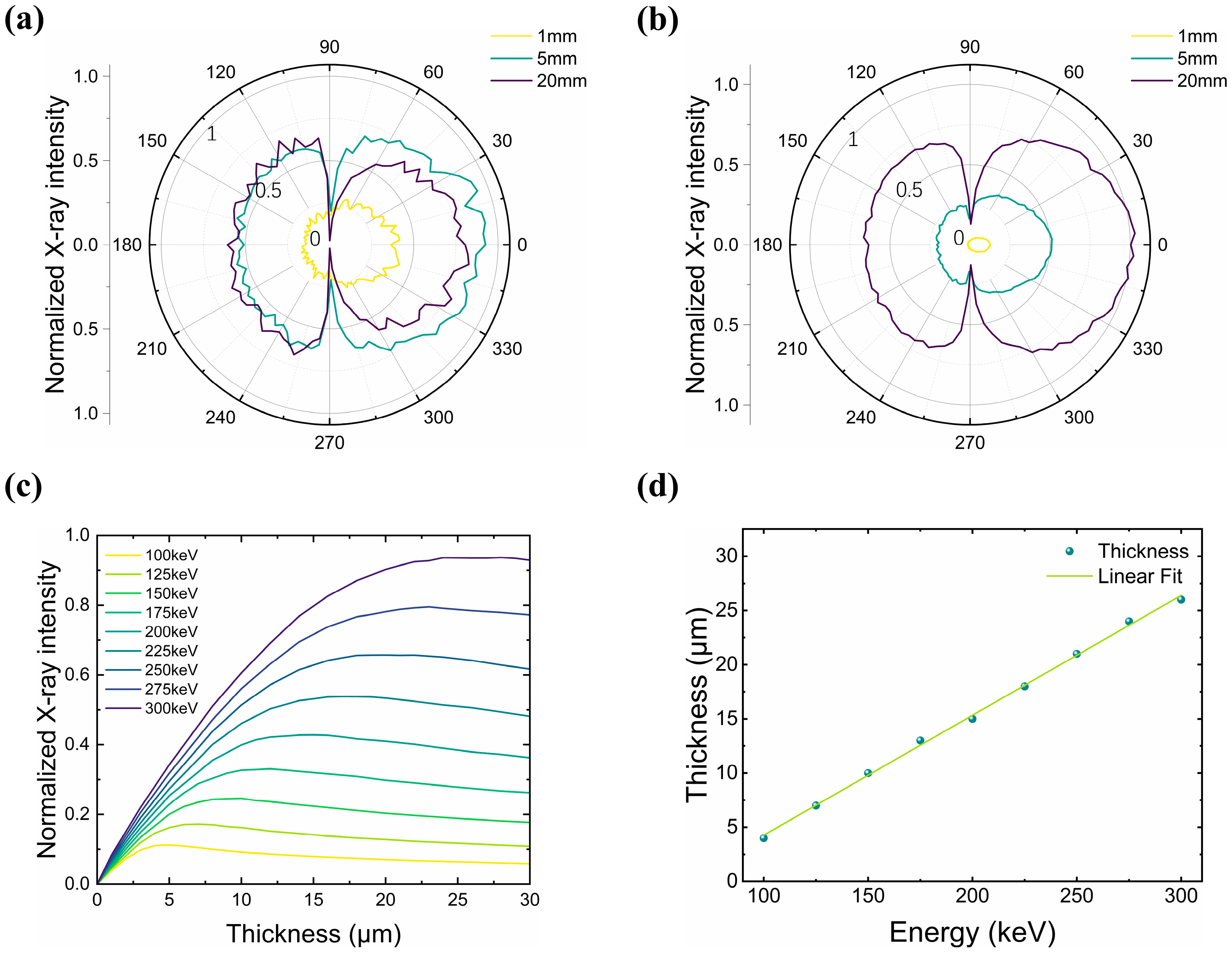
The angular distribution of X-rays of transmission tungsten targets with incident electron energies of 100 keV and 225 keV and target thicknesses of 1 μm, 5 μm, and 20 μm are shown in Figure 2a,b. It can be observed that the emitted X-rays have similar distribution patterns, that is, the density of X-rays emitted near 0° in the forward direction (away from the electron source) is the highest, and as the emission angle increases, the density of X-rays gradually decreases, and the density of X-rays is the lowest near the tungsten target plane. While the density of backward X-rays (close to the electron source) is basically uniformly distributed within the 30° emission angle. For incident electron energy of 100 keV and target thickness of 1 μm, both forward and backward X-ray energies are minimum. When the target thickness increases to 5 μm, the forward X-ray intensity reaches the maximum. When the target thickness continues to increase to 20 μm, the forward X-ray intensity is less than that of 5 μm, indicating that the target is too thick at this time and more X-rays are absorbed by the target itself. For the transmission target with incident electron energy of 225 keV, the distribution rules of 1 μm and 5 μm are similar to those of 100 keV. The difference is that when the target thickness is 20 μm, the forward X-ray intensity is higher than that of 5 μm, indicating that the optimal target thickness increases with the increase in incident electron energy.
To determine the optimal target thickness, the X-ray intensity recorded at eight pixels (0°, 4°, 8°, 12°, 16°, 20°, 24°, and 28°) was used to represent the forward transmission yield. As shown in Figure 2c, for different incident electron energies, the total X-ray intensity initially increases rapidly with target thickness, then slowly decreases after reaching a maximum. The thickness corresponding to this peak, therefore, defines the optimal transmission tungsten target thickness for a given electron energy. As the incident electron energy increases, the optimal target thickness also increases. Figure 2d plots the optimal target thickness as a function of incident electron energy. Owing to the model configuration, the thickness values are determined with a precision of 1 μm. The results clearly show that the optimal thickness increases almost linearly with electron energy in the 100–300 keV range. This trend reflects the balance between two competing processes: (i) the increasing penetration depth of incident electrons, which can be described by the Kanaya–Okayama relation, R c m = 0.0276 A E 0 1.67 ρ Z 0.89 [25] and (ii) the attenuation of X-rays in tungsten, which follows the Beer–Lambert law, I x = I 0 · e μ x [26], the energy-dependent attenuation coefficient μ exhibits sharp increases at tungsten’s absorption edges (K: 69.5 keV; L: 12.1 keV) [27]. leading to stronger self-absorption when the photon spectrum overlaps these regions.
Within the studied range, however, the electron penetration depth dominates, and the interplay between electron range and X-ray absorption gives rise to an approximately linear relationship between optimal thickness and electron energy. A linear fit to the data yields, t * μ m = 0.11 E k e V 6.8 , with a coefficient of determination R2 = 0.999, confirming the strong linear correlation. Since the transmitted X-ray output decreases only gradually beyond the maximum, for X-ray sources with adjustable tube voltage, it is recommended to select the optimal thickness corresponding to the highest operating voltage to ensure efficient performance across the entire range. It is noteworthy that for 225 keV incident electrons, the corresponding optimal thickness is 18 μm. In practice, the final selection for a 225 kV microfocus X-ray source, aiming to balance the X-ray generation efficiency, focal spot size (resolution), and target lifetime/heat dissipation, typically results in a tungsten target thickness within the range of 5–20 μm, often skewed toward thinner values to better manage focal spot size and thermal load [28].

3.2. Reflection-Anode Tungsten Target

The thickness of the reflection tungsten target is closely related to its anode angle. To determine the optimal target thickness, the optimal target angle must first be established. If the target angle is too small, X-rays generated on the anode side must penetrate thicker metal to reach the detector, resulting in a significant anode heel effect and reduced X-ray reception [29]. As the target angle increases, the thickness of the anode target absorbed by photons along the beam direction decreases. However, as the incident angle increases, more electrons are directly scattered, reducing bremsstrahlung X-ray generation. Therefore, there is an optimal target angle for the reflective anode that maximizes X-ray yield in the direction perpendicular to the electron beam. To ensure saturation of the reflected X-ray yield and eliminate the influence of transmitted X-rays, the target thickness is set to 4 mm when studying the optimal target angle.
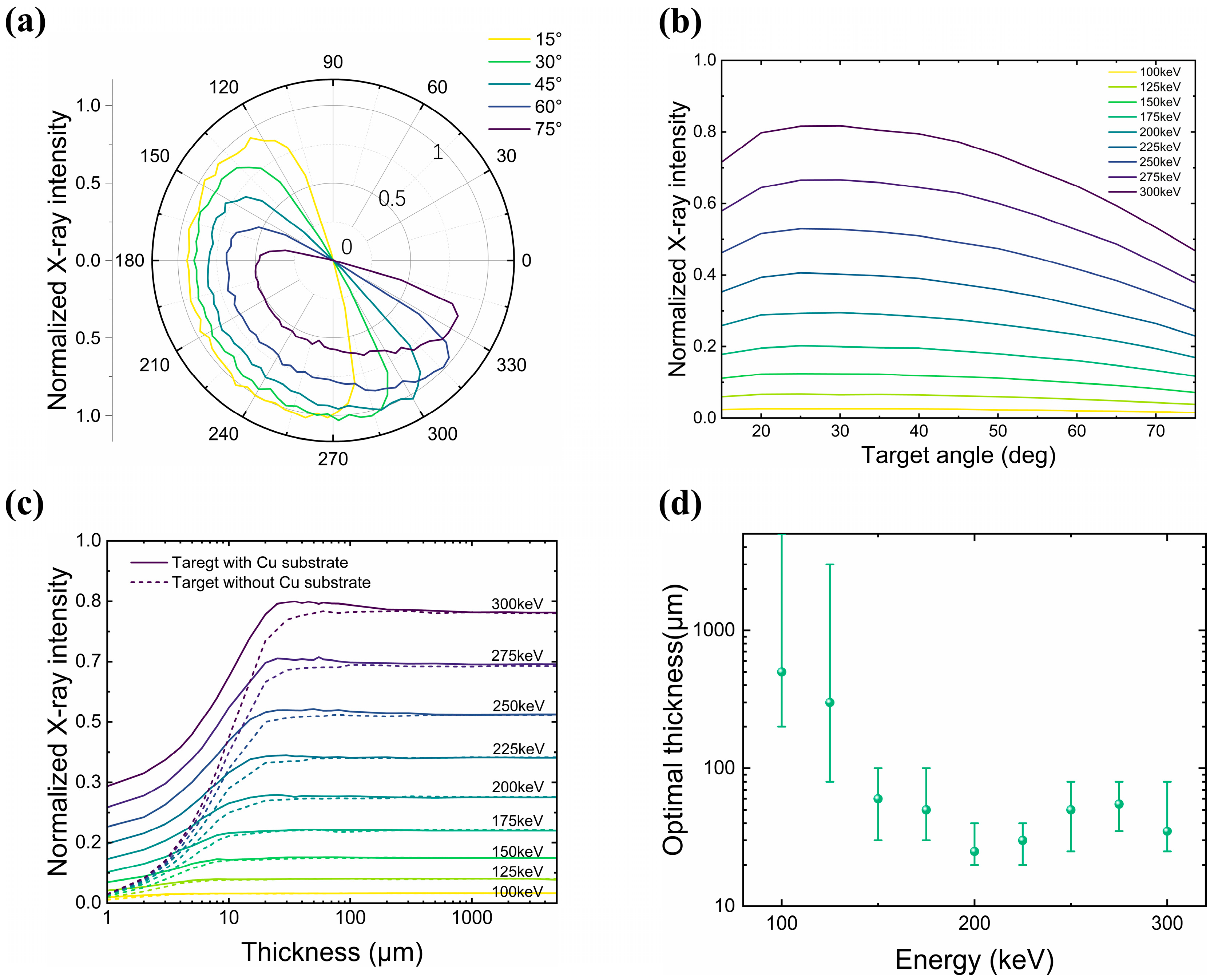
Figure 3a shows the angular distribution of the X-ray intensity for target inclination angles of 15°, 30°, 45°, 60°, and 75° at an incident electron energy of 225 keV. The observation angle, corresponding to the detector position, is defined with respect to the incident electron beam axis (0° = forward, 90° = perpendicular). The X-ray intensity is highest at a 15° target angle, but the intensity distribution is asymmetric, exhibiting a noticeable heel effect [30]. At a 30° target angle, the X-ray density is highest near 90°, with a relatively uniform distribution between 75° and 105°. As the target angle increases, the X-ray output gradually decreases, and the point of highest X-ray density shifts toward the anode.
To determine the X-ray intensity received by the detector perpendicular to the electron beam direction, eight pixels centered at 76°, 80°, 84°, 88°, 92°, 96°, 100°, and 104° are selected, and their total X-ray intensity is recorded. Figure 3b shows the variation in the total X-ray intensity recorded by these pixels with the target angle for incident electron energies of 100–300 keV. The target angle corresponding to the peak total X-ray intensity is around 25°. Therefore, for a 100–300 keV reflection anode, the optimal target angle is 25° when considering the X-ray intensity received by the detector.
In practice, X-ray tubes need to consider the effective focus size to improve the spatial resolution; because of the line focusing principle, many X-ray anodes often present a very shallow beveled angle (6–15°) [31]. For micro-focus X-ray sources, the focus obtained by electron beam focusing has reached the micron to sub-micron level. In order to avoid high-temperature damage to the anode, it is necessary to obtain a stronger X-ray intensity at a lower tube current, so a target angle of 25° might be a better choice.
Based on the determination of the optimal angle for the reflection tungsten target, we further investigated its optimal thickness. Because tungsten has a lower thermal conductivity (~173 W·m−1·K−1) compared with the copper substrate (~401 W·m−1·K−1), target heating and thermal damage must be considered. In conventional fixed-anode reflective X-ray tubes, the tungsten layer embedded in a copper rod is typically 2–3 mm thick [32]. In this study, we considered two configurations: a tungsten target backed by a 4 mm copper substrate and a freestanding tungsten target without substrate.
Figure 3c shows the dependence of the transmitted X-ray intensity on tungsten thickness at an incident angle of 25°. For the case with a copper substrate (solid lines), the X-ray output initially increases with target thickness, reaches a maximum, and then gradually decreases to a saturation value. This behavior can be explained by two factors: (i) when the tungsten thickness is less than the electron continuous-slowing-down approximation (CSDA) range [25], part of the incident electrons penetrate the tungsten layer and generate X-rays in the copper substrate; as the tungsten thickness increases, the number of transmitted electrons decreases, thereby reducing the Cu contribution; (ii) with increasing tungsten thickness, X-rays generated in the copper must pass through a thicker tungsten layer, leading to stronger attenuation.
In the case without the copper substrate (dashed lines), the transmitted X-ray intensity increases with tungsten thickness and then saturates. The saturation level coincides with that of the copper-backed case, indicating that for sufficiently thick tungsten layers, nearly all X-rays originate from tungsten, and the copper contribution becomes negligible. Therefore, the difference between the two cases in the thin-target regime quantifies the contribution of the copper substrate to the forward X-ray output.
Figure 3d shows the dependence of the optimal reflection tungsten target thickness on the incident electron energy. Contrary to the monotonic increase observed in transmission targets, the reflective configuration exhibits an opposite trend: the optimal tungsten thickness decreases as the incident electron energy rises, falling from several hundred micrometers at 100–125 keV to only a few tens of micrometers in the 200–300 keV range. While minor fluctuations appear at higher energies, the overall tendency is clearly downward.
We interpret this inverse correlation as resulting from a competition between electron penetration depth and photon self-absorption, specific to the reflection geometry. This interpretation is based on the following physical reasoning: at higher incident energies, electrons penetrate deeper into the tungsten, which likely shifts the bremsstrahlung generation region further below the surface. It follows that in a reflection target, X-rays produced at greater depths would need to traverse a longer path through the material to escape, potentially leading to stronger attenuation. Therefore, the observed preference for thinner targets at higher energies could be a mechanism to minimize this path length and the associated self-absorption, while still accommodating sufficient electron stopping power. This proposed interplay offers a plausible explanation for the overall downward trend, although the precise contribution of each factor has not been isolated in the present simulation.
At lower incident electron energies (100–125 keV), the forward X-ray yield as a function of tungsten thickness shows a broad plateau rather than a sharp maximum. As a result, even a small tolerance (e.g., 1%) in defining the output leads to a large spread in the “optimal” thickness, such as the wide interval of 200–4500 μm observed at 100 keV. This means that at low energies the notion of an optimal reflective thickness is less precise, since the yield changes only weakly with thickness over this range. In practice, the reported values should therefore be regarded as guidelines rather than strict limits.
While the optimal thickness is ambiguous at lower energies, the trend at higher energies is clearer: thinner tungsten layers are preferable for reflective X-ray sources. Such designs not only reduce unnecessary self-absorption but also help limit heat accumulation in the anode, thereby enabling more efficient photon extraction and improving source stability under high-power operation.
It is acknowledged that temperature rise and associated stress at the tungsten–copper interface are paramount for operational stability and lifetime. Future work will incorporate coupled thermo-mechanical analysis to quantitatively evaluate these effects under realistic operating conditions.

3.3. Influence of Beryllium Window Thickness and Filter

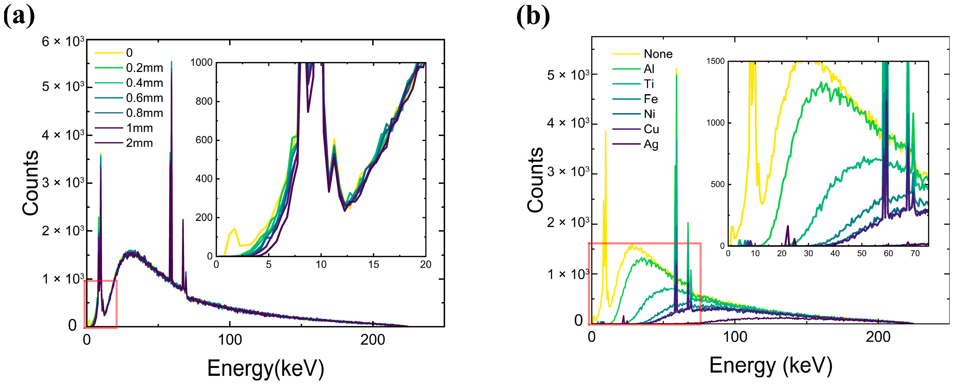
To further evaluate the influence of window and filter materials on the transmitted X-ray spectrum, simulations were performed by varying the thickness of the beryllium window and by replacing the copper filter in the model with alternative materials of identical thickness (e.g., aluminum). Based on the results presented in Section 3.1, the optimal transmission tungsten target thickness at an incident electron energy of 225 keV is 18 μm. Under these conditions, the effect of a 0–2 mm Be window on the emergent X-ray spectrum was examined. Figure 4a shows the spectra recorded by eight pixels (0–28°) with an energy resolution of 0.1 keV, consistent with the Doane rule [33]. The results demonstrate that Be windows strongly attenuate low-energy photons (E < 10 keV), whereas their effect on higher-energy photons is negligible. For example, at 225 keV with an 18 μm transmission tungsten target, the total transmittance through a 1 mm Be window remains as high as 99%.
Beyond the effect of the Be window, spectral shaping is further achieved by introducing filters of different materials into the beam path. Conventional X-ray tubes typically employ aluminum filters for spectral hardening [34], while high-performance CT systems often require much thicker filters (≥10 mm) to ensure adequate beam quality [3].
Here, a 0.5 mm Be window together with 1 mm filters of different materials was simulated under 225 keV electron irradiation. As summarized in Figure 4b and Table 1, the total X-ray transmittance decreases significantly with increasing atomic number (Z) of the filter material, dropping from 0.82 for aluminum (Z = 13) to only 0.11 for silver (Z = 47). At the same time, the mean photon energy shows a strong hardening effect, rising from 62.6 keV without filter to 140.0 keV with silver. These results clearly demonstrate the trade-off between photon flux and spectral hardening: low-Z filters provide higher transmittance but weaker hardening, whereas high-Z filters achieve stronger hardening at the expense of photon output. This emphasizes the importance of selecting appropriate filter materials to balance dose efficiency and spectral quality in high-energy microfocus X-ray and industrial CT systems.

4. Conclusions

Using the Geant4 toolkit, this study systematically investigates the performance of tungsten anode targets in high-energy microfocus X-ray sources exposed to electron beams between 100 and 300 keV. By analyzing both transmission and reflection tungsten targets with various thicknesses and angles, this study derives the intensity-angular distribution of the emitted X-rays and optimizes the tungsten target parameters. The results reveal that, for a microfocus X-ray source operating at a maximum tube voltage of 225 kV, the optimal transmission tungsten target thickness is 18 μm, while the optimal reflection tungsten target features a thickness of 30 μm at a 25° incidence angle. Additionally, the optimal transmission target thickness exhibits a linear relationship with the incident electron energy. Furthermore, the investigation explores the impact of beryllium windows and different filter materials on the X-ray spectrum. The findings indicate that the thickness of the beryllium window significantly attenuates low-energy X-rays, whereas high-energy X-rays are minimally affected. The choice of filter materials effectively modulates the transmittance and energy spectrum of the X-rays, thereby facilitating the optimization of the X-ray source design. Future work will prioritize the experimental validation of these simulation results through the fabrication of optimized anode targets and the characterization of their emission properties. Overall, this research provides a crucial theoretical foundation for the design optimization of microfocus X-ray sources and offers technical support for applications in related fields.

Author Contributions

Conceptualization, D.L. and Y.L. (Yuetian Liu); methodology, Y.L. (Yuetian Liu); software, Y.L. (Yuetian Liu) and Y.L. (Yiheng Liu); validation, Y.L. (Yuetian Liu), L.L., L.X. and X.Z.; formal analysis, Y.L. (Yuetian Liu); investigation, Y.L. (Yuetian Liu), L.L., L.X. and X.Z.; resources, D.L.; data curation, Y.L. (Yuetian Liu); writing—original draft preparation, Y.L. (Yuetian Liu); writing—review and editing, all authors; visualization, Y.L. (Yuetian Liu), L.L., L.X. and X.Z.; supervision, D.L.; project administration, D.L.; funding acquisition, D.L. All authors have read and agreed to the published version of the manuscript.

Funding

This research was funded by the National Key Research and Development Program of China (Grant Nos. 2024YFF0727300 and 2024YFF0727303) and the Shaanxi Province Natural Science Foundation (Grant No. 2025JC-YBQN-067).

Data Availability Statement

Data is available on reasonable request.

Acknowledgments

The authors gratefully acknowledge the computational support provided by the Streak Camera Engineering Center, Xi’an Institute of Optics and Precision Mechanics (XIOPM), Chinese Academy of Sciences (CAS).

Conflicts of Interest

The authors declare no conflicts of interest.

References

  1. De Chiffre, L.; Carmignato, S.; Kruth, J.-P.; Schmitt, R.; Weckenmann, A. Industrial applications of computed tomography. CIRP Ann. 2014, 63, 655–677. [Google Scholar] [CrossRef]
  2. Kak, A.C.; Slaney, M.; Wang, G. Principles of Computerized Tomographic Imaging. Phys. Med. 2002, 29, 107. [Google Scholar] [CrossRef]
  3. Carmignato, S.; Dewulf, W.; Leach, R. Industrial X-Ray Computed Tomography; Springer: Cham, Switzerland, 2018; Volume 10. [Google Scholar]
  4. Withers, P.J.; Bouman, C.; Carmignato, S.; Cnudde, V.; Grimaldi, D.; Hagen, C.K.; Maire, E.; Manley, M.; Plessis, A.D.; Stock, S.R. X-ray computed tomography. Nat. Rev. Methods Primers 2021, 1, 18. [Google Scholar] [CrossRef]
  5. Hendee, W.R.; Ritenour, E.R. Medical Imaging Physics; John Wiley & Sons: Hoboken, NJ, USA, 2003. [Google Scholar]
  6. Cierniak, R. X-Ray Computed Tomography in Biomedical Engineering; Springer Science & Business Media: Berlin/Heidelberg, Germany, 2011. [Google Scholar]
  7. Lassner, E.; Schubert, W.-D. Tungsten: Properties; Chemistry, Technology of the Element, Alloys, and Chemical Compounds; Springer Science & Business Media: Berlin/Heidelberg, Germany, 2012. [Google Scholar]
  8. Poludniowski, G.G.; Evans, P.M. Calculation of X-ray spectra emerging from an X-ray tube. Part I. Electron penetration characteristics in X-ray targets. Med. Phys. 2007, 34, 2164–2174. [Google Scholar] [CrossRef]
  9. Goorley, T.; James, M.; Booth, T.; Brown, F.; Bull, J.; Cox, L.J.; Durkee, J.; Elson, J.; Fensin, M.; Forster, R.A.; et al. Initial MCNP6 Release Overview. Nucl. Technol. 2012, 180, 298–315. [Google Scholar] [CrossRef]
  10. Baró, J.; Sempau, J.; Fernández-Varea, J.M.; Salvat, F. PENELOPE: An algorithm for Monte Carlo simulation of the penetration and energy loss of electrons and positrons in matter. Nucl. Instrum. Methods Phys. Res. Sect. B Beam Interact. Mater. At. 1995, 100, 31–46. [Google Scholar] [CrossRef]
  11. Böhlen, T.; Cerutti, F.; Chin, M.; Fassò, A.; Ferrari, A.; Ortega, P.; Mairani, A.; Sala, P.; Smirnov, G.; Vlachoudis, V. The FLUKA Code: Developments and Challenges for High Energy and Medical Applications. Nucl. Data Sheets 2014, 120, 211–214. [Google Scholar] [CrossRef]
  12. Kawrakow, I. Accurate condensed history Monte Carlo simulation of electron transport. I. EGSnrc, the new EGS4 version. Med. Phys. 2000, 27, 485–498. [Google Scholar] [CrossRef]
  13. Agostinelli, S.; Allison, J.; Amako, K.; Apostolakis, J.; Araujo, H.; Arce, P.; Asai, M.; Axen, D.; Barrand, G.; Behner, F.; et al. Geant4—A simulation toolkit. Nucl. Instrum. Methods Phys. Res. Sect. A Accel. Spectrometers Detect. Assoc. Equip. 2003, 506, 250–303. [Google Scholar] [CrossRef]
  14. Allison, J.; Amako, K.; Apostolakis, J.; Araujo, H.; Arce Dubois, P.; Asai, M.; Barrand, G.; Capra, R.; Chauvie, S.; Chytracek, R.; et al. Geant4 developments and applications. IEEE Trans. Nucl. Sci. 2006, 53, 270–278. [Google Scholar] [CrossRef]
  15. Allison, J.; Amako, K.; Apostolakis, J.; Arce, P.; Asai, M.; Aso, T.; Bagli, E.; Bagulya, A.; Banerjee, S.; Barrand, G.; et al. Recent developments in Geant4. Nucl. Instrum. Methods Phys. Res. Sect. A Accel. Spectrometers Detect. Assoc. Equip. 2016, 835, 186–225. [Google Scholar] [CrossRef]
  16. Nasseri, M.M. Determination of Tungsten Target Parameters for Transmission X-ray Tube: A Simulation Study Using Geant4. Nucl. Eng. Technol. 2016, 48, 795–798. [Google Scholar] [CrossRef]
  17. Nasseri, M.M. Determination of molybdenum target parameters for transmission X-ray tube: A Geant4 simulation study. Pramana 2019, 92, 54. [Google Scholar] [CrossRef]
  18. Kandlakunta, P.; Thomas, A.; Tan, Y.; Khan, R.; Zhang, T. Design and numerical simulations of W-diamond transmission target for distributed x-ray sources. Biomed. Phys. Eng. Express 2019, 5, 025030. [Google Scholar] [CrossRef]
  19. Geng, N.; Wei-xia, Z.; Jun-biao, L.; Li, H.; Yu-tian, M. Optimization and design of target of low energy X-ray microscopy. J. Chin. Electron Microsc. Soc. 2019, 38, 384–388. [Google Scholar]
  20. Niu, G.; Liu, J.; Zhao, W.; Han, L.; Ma, Y. Effect of Focused Bombarding Electron Beam on Transmission Microfocus X-Ray Source. Acta Opt. Sin. 2019, 39, 0634001. [Google Scholar] [CrossRef]
  21. Lei, Z.; Huarong, L.; Buliang, S.; Junting, W.; Tingting, L.; Yijing, L. Optimization of Transmission-Type Anode Target in Microfocus X-Ray Tube. Chin. J. Vaccum Sci. Technol. 2015, 35, 1443–1448. [Google Scholar]
  22. Carrier, J.F.; Archambault, L.; Beaulieu, L.; Roy, R. Validation of GEANT4, an object-oriented Monte Carlo toolkit, for simulations in medical physics. Med. Phys. 2004, 31, 484–492. [Google Scholar] [CrossRef]
  23. Seco, J.; Verhaegen, F. Monte Carlo Techniques in Radiation Therapy; CRC Press: Boca Raton, FL, USA, 2013. [Google Scholar]
  24. Keall, P.J.; Siebers, J.V.; Libby, B.; Mohan, R. Determining the incident electron fluence for Monte Carlo-based photon treatment planning using a standard measured data set. Med. Phys. 2003, 30, 574–582. [Google Scholar] [CrossRef]
  25. Kanaya, K.; Okayama, S. Penetration and energy-loss theory of electrons in solid targets. J. Phys. D Appl. Phys. 1972, 5, 43. [Google Scholar] [CrossRef]
  26. Bakri, F.; Gareso, P.L.; Tahir, D. Advancing radiation shielding: A review the role of Bismuth in X-ray protection. Radiat. Phys. Chem. 2024, 217, 111510. [Google Scholar] [CrossRef]
  27. National Institute of Standards and Technology(NIST). XCOM: Photon Cross Sections Database. Available online: https://physics.nist.gov/PhysRefData/Xcom/html/xcom1.html (accessed on 1 October 2025).
  28. X-Ray Worx [EB/OL]. Available online: https://www.x-ray-worx.com/ (accessed on 20 October 2025).
  29. Behling, R. Modern Diagnostic X-Ray Sources: Technology, Manufacturing, Reliability; CRC Press: Boca Raton, FL, USA, 2021. [Google Scholar]
  30. Oostveen, L.J.; Tunissen, S.; Sechopoulos, I. Comparing organ and effective dose of various CT localizer acquisition strategies: A Monte Carlo study. Med. Phys. 2025, 52, 576–584. [Google Scholar] [CrossRef]
  31. Behling, R. X-ray sources: 125 years of developments of this intriguing technology. Phys. Medica 2020, 79, 162–187. [Google Scholar] [CrossRef]
  32. Paul, S. Biomedical Engineering and Its Applications in Healthcare; Springer: Cham, Switzerland, 2019. [Google Scholar]
  33. Doane, D.P. Aesthetic frequency classifications. Am. Stat. 1976, 30, 181–183. [Google Scholar] [CrossRef]
  34. Hsieh, J. Computed Tomography: Principles; Design; Artifacts; Recent Advances; SPIE: Washington, DC, USA, 2003. [Google Scholar]
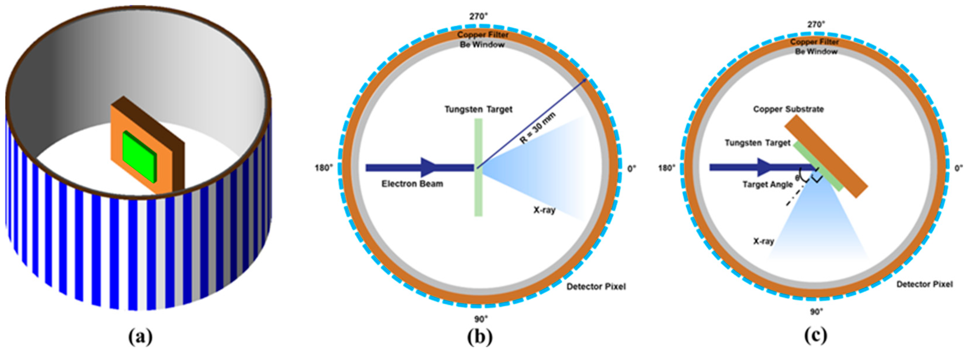
Figure 1. Tungsten target and detector model established in Geant4. (a) 3D model, where alternating blue and white stripes indicate individual detector pixels (colors are for positional distinction only). (b) Transmission anode target model. (c) Reflection anode target model. In all cases, the model assumes a parallel electron beam with zero half-angle divergence.
Photonics 12 01062 g001
Figure 2. The optimal thickness of the transmission tungsten target under different electron beam energies. (a) Distribution of outgoing X-rays intensity at target thicknesses of 1 μm, 5 μm, and 20 μm with incident electron energy of 100 keV; (b) Distribution of outgoing X-rays intensity at target thicknesses of 1 μm, 5 μm, and 20 μm with incident electron energy of 225 keV; (c) Variation in outgoing X-ray intensity with tungsten target thickness for different incident electron energies; (d) Variation in optimal tungsten target thickness with incident electron energy.
Photonics 12 01062 g002
Figure 3. Study on reflection anode tungsten target under different incident electron energies. (a) Angular distribution of X-ray intensity at an incident electron energy of 225 keV for target inclination angles of 15°, 30°, 45°, 60°, and 75°. The polar angle (0–360°) represents the emission direction of X-rays relative to the incident electron beam axis (0° = forward, 90° = perpendicular). (b) Total X-ray intensity recorded by 76–104° pixels vs. target angle for different incident electron energies; (c) Total X-ray intensity perpendicular to the electron beam direction vs. target thickness for different incident electron energies, solid lines represent tungsten targets with a 4 mm copper substrate, dashed lines represent tungsten targets without a copper substrate; (d) Variation in optimal tungsten target thickness with incident electron energy (Error bars indicate a 1% uncertainty range).
Photonics 12 01062 g003
Figure 4. The influence of the Be window and filter on the outgoing X-ray. (a) Influence of 0.2–2 mm Be window on outgoing X-rays at incident electron energy of 225 keV; (b) Influence of 1 mm filters of different materials on outgoing X-rays at incident electron energy of 225 keV.
Photonics 12 01062 g004
Table 1. Effects of different filters on the transmitted X-ray spectrum at an incident electron energy of 225 keV.
Material (Z)No FilterAl(13)Ti(22)Fe(26)Ni(28)Cu(29)Ag(47)
transmittance1.000.820.530.350.290.290.11
Mean Energy/keV62.671.888.4102.5108.6109.8140.0
Disclaimer/Publisher’s Note: The statements, opinions and data contained in all publications are solely those of the individual author(s) and contributor(s) and not of MDPI and/or the editor(s). MDPI and/or the editor(s) disclaim responsibility for any injury to people or property resulting from any ideas, methods, instructions or products referred to in the content.