Article

Characterization of Nanocrystals of Eu-Doped GaN Powders Obtained via Pyrolysis, Followed by Their Nitridation

1
División de Sistemas Automotrices, Universidad Tecnológica de Puebla (UTP), Antiguo Camino a la Resurrección 1002-A, Zona Industrial, Puebla 72300, Puebla, Mexico
2
Departamento de Investigación en Física, Universidad de Sonora (UNISON), Rosales y Colosio, C. De la Sabiduría, Centro, Hermosillo 83000, Sonora, Mexico
3
Departamento de Ingeniería Eléctrica y Electrónica, Instituto Tecnológico de Puebla (ITP), Del Tecnológico 420, Corredor Industrial la Ciénega, Puebla 72220, Puebla, Mexico
4
Centro de Investigación en Dispositivos Semiconductores, Benemérita Universidad Autónoma de Puebla (BUAP), 14 Sur y Av. San Claudio, Puebla 72570, Puebla, Mexico
5
Centro de Nanociencias y Nanotecnología, Universidad Nacional Autónoma de Mexico (UNAM), Carr. Tijuana-Ensenada km107, C.I.C.E.S.E., Ensenada 22860, Baja California, Mexico
6
Departamento de Física, Universidad de Sonora (UNISON), Rosales y Colosio, C. De la Sabiduría, Centro, Hermosillo 83000, Sonora, Mexico
7
Departamento de Investigación y Desarrollo, Universidad Tecnológica de Puebla (UTP), Antiguo Camino a La Resurrección 1002-A, Zona Industrial, Puebla 72300, Puebla, Mexico
8
Departamento de Polímeros y Materiales, Universidad de Sonora (UNISON), Rosales y Colosio, C. De la Sabiduría S/N, Centro, Hermosillo 83000, Sonora, Mexico
*
Author to whom correspondence should be addressed.
Photonics 2025, 12(10), 982; https://doi.org/10.3390/photonics12100982
Submission received: 21 August 2025 / Revised: 16 September 2025 / Accepted: 30 September 2025 / Published: 2 October 2025
(This article belongs to the Special Issue Emerging Trends in Rare-Earth Doped Material for Photonics)

Abstract

Nanocrystals of Eu-doped GaN powders are produced via pyrolysis of a viscous compound made from europium and gallium nitrates. Furthermore, carbohydrazide is used as a fuel and toluene as a solvent; subsequently, a crucial nitridation process is carried out at 1000 °C for one hour. A slight shift of 0.04 degrees toward larger angles was observed for the X-ray diffraction patterns in the Eu-doped GaN powders regarding the undoped GaN powders, while Raman scattering also displayed a slight shift of 10.03 cm−1 toward lower frequencies regarding the undoped GaN powders for the vibration mode, E2(H), in both cases indicating the incorporation of europium atoms into the GaN crystal lattice. A scanning electron microscope micrograph demonstrated a surface morphology for the Eu-doped GaN with a shape similar to elongated platelets with a size of 3.77 µm in length. Energy-dispersive spectroscopy and X-ray photoelectron spectroscopy studies demonstrated the europium elemental contribution in the GaN. The X-ray photoelectron spectroscopy spectrum for gallium demonstrated the binding energies for Ga 2P3/2, Ga 2P1/2, and Eu 3d5/2, which could indicate the incorporation of europium into the GaN and the bonding between gallium and europium atoms. The transmission electron microscope micrograph showed the presence of nanocrystals with an average size of 9.03 nm in length. The photoluminescence spectrum showed the main Eu3+ transition at 2.02 eV (611.69 nm) for europium emission energy, corresponding to the 5D07F2 transition of the f shell, which is known as a laser transition.

1. Introduction

Gallium nitride (GaN) is among the most prominent III-V semiconductor compounds owing to its wide band gap (3.4 eV for the hexagonal structure and 3.2 eV for the cubic structure). GaN has applications in energy systems, photoelectronic devices, power devices, communication systems, and medicine [1]. Rare earth ions’ luminescence has been of interest to certain researchers (RE) (La, Nd, Eu, Tb, and Er) through the doping of III-nitride compounds, as is the case of Eu-doped GaN, which has important luminescent properties for GaN-based red-light-emitting diodes necessary in micro-display technology [2,3,4,5,6,7]. GaN is generally synthesized by metalorganic chemical vapor deposition (MOCVD), as well as molecular beam epitaxy (MBE). One of the reasons for synthesizing Eu-doped GaN is to produce sharp and stable red-light emissions for optoelectronic devices, such as LEDs and displays. This is because europium provides a distinct red-light wavelength through its 4f-4f electron transitions, offering a more efficient and reliable alternative to InGaN-based red LEDs, which struggle with low efficiency and broad emission. Furthermore, the most well-known dopants for GaN, such as Mg or Zn, can emit energy in the blue band. However, research works to obtain Eu-doped GaN have been previously carried out using different techniques, where authors such as Brown et al. [8] present the red emission properties of Eu-doped GaN powders made through a Na flux method. In another work, Zhu et al. [9] obtained Eu-doped GaN layers using the MOCVD technique, where trimethylgallium (Ga(CH3)3) and ammonia (NH3) were the sources of gallium atoms and nitrogen atoms, respectively, while bis-europium (EuCppm2) was used as the source of europium atoms; the layers were grown at 960 °C. Kudrawiec et al. [10] synthesized Eu-doped GaN nanocrystalline powders using 0.5 g of Ga2O3 and 7.2 mg of EuO2. They were combined and dissolved in hot concentrated nitric acid and evaporated to dryness, where, gradually, there was an increase in temperature from 70 °C to 200 °C.
On the other hand, Nyk et al. [11] prepared nanocrystals of Eu-doped GaN powders through the hydrothermal process using a microwave reactor and Ga2O3, as well as Eu2O3 and nitric acid as reagents with a rise in temperature from 70 °C to 200 °C, and calcined them at 600 °C for 6 h in an air flow. Inaba et al. [12] showed that Eu-doped GaN is an applicable material for red emissions for light-emitting diodes (LEDs), where the samples were grown on sapphire substrates by MOCVD using, as precursors, the metal-organic compounds trimethylgallium, ammonia, and EuCppm2. El-Himri et al. [13] obtained Eu-doped GaN nanoparticles prepared by ammonolysis of freeze-dried reagents, using ammoniumhexafluorometallate as a component, and where the distinctive features of the trivalent lanthanide ions are the result of the incomplete inner 4f shell, which gives rise to paramagnetic properties, and interactions with UV, VIS, or IR radiation. XiaoDan et al. [14] synthesized Eu-doped GaN powders prepared by a co-precipitation method using Ga(NO3)3, Eu2O3, nitric acid, ammonium hydroxide solution, and ammonia as reagents, where Ga(NO3)3 was dissolved in deionized water, while Eu2O3 was dissolved in nitric acid. Braun et al. [15] demonstrated the advantages of controlling the functionality and luminescence properties of modern energy-efficient white-light-emitting diodes through the doping of GaN with Eu, achieved through a successive combustion synthesis using, as precursors, the metal chloride EuCl3 or Eu(NO3)3, while ammonium nitrate (NH4NO3), urea, and H2O were included in the sample. This mixture was placed in a furnace at a range of 400 to 600 °C for 10 min. In another work, Wu et al. [16] also showed that Eu-doped GaN has applications in red-light-emitting diodes, obtained gallium nitride powders doped with europium by reacting a molten alloy of Ga and Eu, along with NH3, at 1000 °C using Bi as a wetting agent. Other studies have developed simulations based on density functional theory (DFT), which has been used to perform calculations, to study the behavior of the electronic and optical properties of nanostructured semiconductor materials [17,18].
The present work describes the nanocrystal synthesis of Eu-doped GaN powders via pyrolysis of a viscous compound made from europium and gallium nitrates, with carbohydrazide used as a precursor and toluene as a solvent (viscous compound), followed by a nitridation process (NH3) at 1000 °C for one hour. This method has been used by our research group for some years [19]. The method of pyrolysis or combustion is an interesting technique used to obtain GaN powders doped with some rare earth ions (REs). The synthesis is based on highly exothermic, self-sustaining, and self-propagating oxidation—reduction is an interaction that is involved in the reaction of fuel and metal nitrates in a liquid solution. The technique requires a fuel such as carbohydrazide (CH6N4O) or hydrazide (N2H4). The pyrolysis method, followed by nitridation, has the advantages of being an economical, safe, and easy synthesis process for obtaining III-nitride materials compared to other methods, such as MOCVD or MBE. A disadvantage of the combustion method is the low generation of bulk material due to the loss of material during the exothermic reaction. Another disadvantage of the combustion method is that, to deposit films, several synthesis processes must be performed to gather enough material to prepare a target and obtain films by magnetron RF sputtering or pulsed laser deposition (PLD). Thus, this paper is important in that the procurement of GaN powers doped with europium using the pyrolysis method has still not been reported upon.

2. Materials and Methods

Eu-doped GaN powders were prepared at atmospheric pressure (1012 hPa) via pyrolysis of a viscous compound in a nitrogen environment [20,21]; for the synthesis of Eu-doped GaN powders, gallium nitrate (1.7 g) (Ga(NO3)3), carbohydrazide (1.2 g) (CH6N4O), and europium nitrate (117.4 mg) (Eu(NO3)3) (3% europium to dope GaN) were used as precursors, all of ultra-high purity from Sigma-Aldrich (St. Louis, MI, USA), while HPLC-grade toluene (20 mL) (C6H5CH3) from Merck (St. Louis, MI, USA) was utilized to dissolve the nitrates and carbohydrazide. Furthermore, a flow of ultra-high-purity ammonia gas (NH3) was employed as a source of nitrogen atoms to obtain the GaN powders. The following reaction was proposed to produce the Eu-doped GaN powders:
10((Ga(NO3)3)(Eu(NO3)3)(CH6N4O))(s) ⟶ 10GaN:Eu(s) + 9H2O(g) + 10CO(g)+
+3N2O(g) + 42NO2(g) + 42HNO2(g)
After calculating and weighing them on an analytical balance, the nitrates and toluene were placed inside a Teflon beaker, which had been placed on a hot plate, and the hot plate was later placed in an extraction hood. Afterward, the temperature on the hot plate was elevated to 100 °C, and magnetic stirring was turned on at 800 rpm for 20 min; at this temperature, the carbohydrazide was aggregated inside the Teflon beaker. Then, the temperature was increased until reaching an internal temperature of 111 °C in the Teflon beaker, which was maintained. It took 35 min for the solvent to evaporate and the viscous compound to be obtained. Then, the Teflon beaker was taken from the hot plate to allow the viscous compound to cool to room temperature, while the hot plate was turned off.
When the viscous compound (2.5 g) was obtained, a sample of approximately 1 g was placed in an alumina crucible and placed inside a CVD furnace to realize pyrolysis. At this point, the CVD furnace was purged three times to remove residual impurities, and later a flow of N2 at 50 sccm was used as a carrier gas for the sub-products of the reaction. Afterward, the temperature was elevated to 250 °C, which is the pyrolysis temperature for synthesizing GaN [22]. Once the temperature reached 250 °C, white vapors were carried by nitrogen toward the outlet of the CVD system, indicating that the pyrolysis of the sample was carried out. When pyrolysis was achieved, the temperature was increased to 1000 °C. At this point, an ammonia flow (NH3) of 100 sccm was opened (NH3 was employed as the source of nitrogen atoms), while the nitrogen flow was closed. The sample was kept at 1000 °C in an ammonia environment for one hour, whereupon the temperature was decreased to 20 °C, the ammonia flow was closed, and the nitrogen flow at 50 sccm was turned on again until cooling to 20 °C; afterward, the nitrogen flow was closed, and the sample was removed from the CVD furnace. After the sample was removed, it was finely ground to carry out the planned characterizations.

Characterizations

Eu-doped GaN and undoped GaN powders were studied by X-ray diffraction (XRD) using D8 ADVANCE ECO BRUKER equipment (Bruker, Karlsruhe, Germany) with a wavelength (Cu Kα) of 1.5406 Å and a nickel filter, operated at 40 kV and 25 mA. XRD patterns were made in a range of 30° to 60°. The elemental contributions and the surface morphology (EDS/SEM) of the Eu-doped GaN and undoped GaN powders were measured using JEOL JSM-7800F Schottky Field Emission equipment (Pleasanton, CA, USA) with an ETD (Everhart–Thornley Detector) for secondary electron detection (SED). X-ray photoelectron spectroscopy (XPS) studies were performed by using Escalab 250Xi Brochure equipment (Thermo Scientific, East Grinstead, UK) on the Eu-doped GaN powders. Transmission electron microscopy (TEM) was performed using JEOL JEM-2010 equipment (Akishima, Tokyo, Japan) on the Eu-doped GaN. Photoluminescence spectra (PL) were created at room temperature with an excitation wavelength of 325 nm (UV), a power of 55 mW, and an accuracy of +/− 0.2 nm, using an FLS1000 Photoluminescence Spectrometer (Livingston Village, Livingston, UK) on the Eu-doped GaN powders. Finally, Raman studies for the Eu-doped GaN and undoped GaN powders were performed using a Horiba Jobin Yvon HR-800 Micro Raman spectrophotometer with an argon laser, power up to 50 mW, and a 488 nm filter (Edison, NJ, USA).

3. Results and Discussion

3.1. Structural Analysis

Undoped and Eu-doped GaN powders were made from the pyrolysis of a viscous compound made from europium and gallium nitrates; moreover, carbohydrazide was used as a precursor, and toluene as a solvent, to continue their nitridation in a NH3 flow at 1000 °C for one hour. Figure 1a demonstrates the diffraction patterns of the GaN for ICDD PDF card No. 00-050-0792, which indexed all the peaks of the undoped and Eu-doped GaN powders. Furthermore, Figure 1b indicates the undoped GaN powders annealed in an ammonia environment for one hour, while Figure 1c shows the peaks of the X-ray diffraction patterns for the Eu-doped GaN powders. The a peak for hexagonal GaN was located in plane (100) in the Miller index crystallographic notation, the b peak was found in plane (002), the c peak had the greatest intensity in plane (101), the e peak was found in plane (102), and the g peak was located in plane (110). Furthermore, in Figure 1c, two small peaks are located at positions 47.27° (d peak) and 56.01° (f peak), which could be related to gallium oxide (Ga2O3) that originated during the combustion process through the use of carbohydrazide as a reagent (exothermic reaction) and the strong electron affinity of oxygen with gallium. The d peak was detected in plane ( 2 ¯ 03 ), and the f peak was located in plane ( 1 ¯ 13 ), where both peaks were indexed in ICDD PDF card No. 00-041-1103 for Ga2O3. The calculations for the lattice constants of the hexagonal GaN (wurtzite) were a = 3.18 Å and c = 5.18 Å, with a c/a ratio of 1.62. Comparing the X-ray diffraction patterns in Figure 1b,c, a shift of 0.04 degrees toward larger angles for the Eu-doped GaN powders (Figure 1c) relative to the undoped GaN powders (Figure 1b) can be observed, which might be associated with the larger size of the europium atomic radius of 240 pm compared to the gallium atomic radius of 187 pm, indicating the possible incorporation of europium into the GaN lattice structure. FWHM, crystal size, and interplanar spacing measurements showed the possible existence of nanocrystals in the Eu-doped GaN powders due to the broadening of their X-ray diffraction patterns (Figure 1c). Table 1 shows the FWHM, interplanar spacing, and crystal size measurements, which were calculated using the Debye–Scherrer equation with a Scherrer constant for hexagonal crystal structures with a value of 0.9, showing crystal sizes as small as 5.38 nm for the b peak of the Eu-doped GaN powders (Figure 1c). A significant difference in atomic radii between europium (Eu) and gallium (Ga), with europium being much larger, would likely expand the lattice spacing in the GaN crystal lattice because the larger europium atoms take up more space and prevent the smaller gallium atoms from packing as tightly. However, when the dopant quantity is as small as 3%, as in this investigation, the GaN interplanar spacing does not show such a significant change quantitatively, as shown in the lattice spacing values in Table 1, confirming the importance of the slight shift of 0.04 degrees toward larger angles. On the other hand, the 0.04° shift toward larger angles in the X-ray diffraction pattern (Figure 1c), despite being a small shift, could indicate the possible incorporation of europium atoms into the GaN crystal lattice structure. However, this 0.04° shift toward larger angles could also be influenced by factors such as the presence of strain in the lattice structure due to the introduction of an atom with a larger atomic radius (Eu), as well as the introduction of defects such as vacancies or interstices into the crystal structure, which is very common in GaN due to the presence of non-intentional impurities, such as oxygen atoms or carbon atoms. Using the ICCD PDF-4+2022 software and the Debye–Scherrer equation, the crystal size was calculated, showing an average of 9.09 nm for the Eu-doped GaN powders, while for the undoped GaN powders, it was 22.52 nm [22]. The broadening of the peaks in Figure 1c could be associated with the nature of the pyrolysis of the viscous compound, which produces a nitrogen deficiency in GaN powders.

3.2. Electron Microscopy

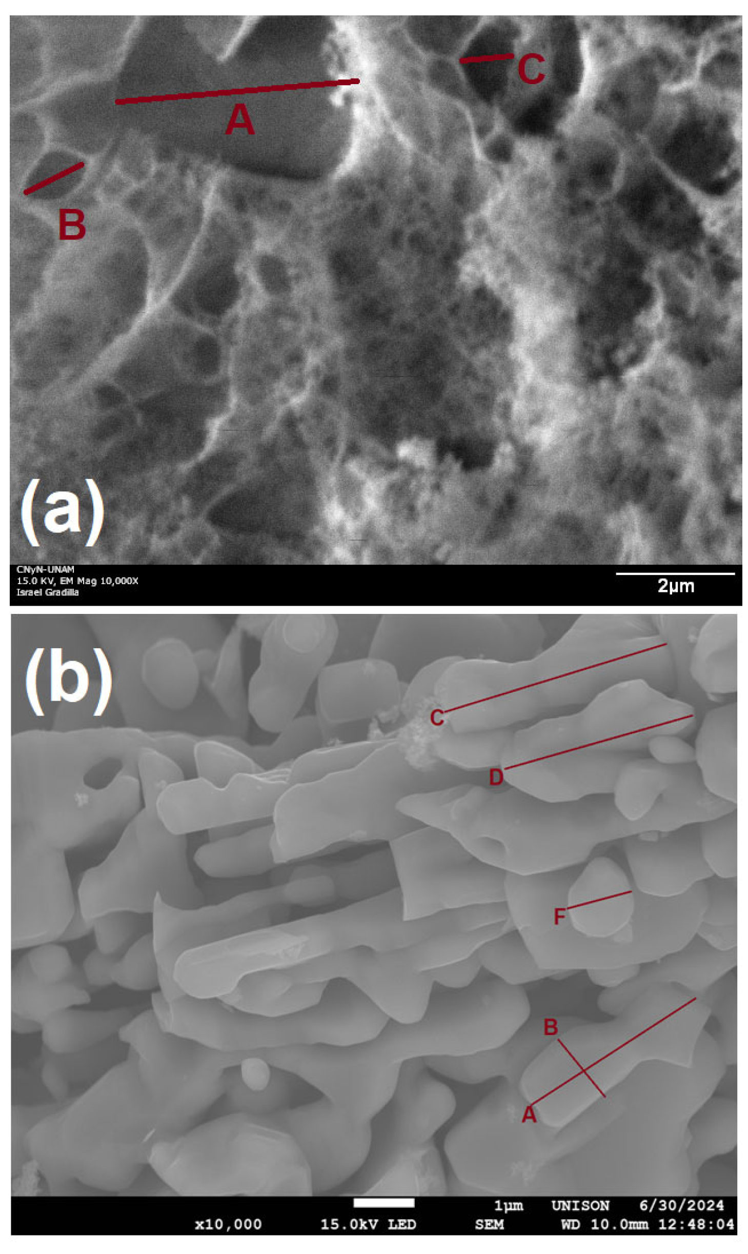
Figure 2a shows the undoped GaN powders, in which a porous surface morphology can be seen. This could be associated with the nitrogen deficiency of the pyrolysis and the existence of non-intentional impurities, such as oxygen, in the gallium nitrate and carbohydrazide. Figure 2a shows three measures of the length of the pores; the A pore was 3.71 µm in length, the B pore was 1.10 µm in length, and the C pore was 0.76 µm in length. Furthermore, Figure 2b shows the surface morphology of the Eu-doped GaN, which demonstrates a shape similar to elongated platelets. Figure 2b shows the A measure, which was 3.32 µm in length, while the B measure was 1.20 µm in width. Furthermore, the C measure was 3.77 µm in length; the D measure was 3.30 µm in length; and finally, the F measure was 1.06 µm in length. It is important to note that using the pyrolysis method to obtain III-nitride materials generally produces porous surface morphologies, as shown in the work of El-Himri et al. [13]. The surface morphology obtained in this work is particularly noteworthy in that elongated platelets were obtained using the pyrolysis method to synthesize Eu-doped GaN powders. It is important to mention that adding ions of Eu to GaN can affect its morphology, primarily by influencing the growth mode and introducing defects.
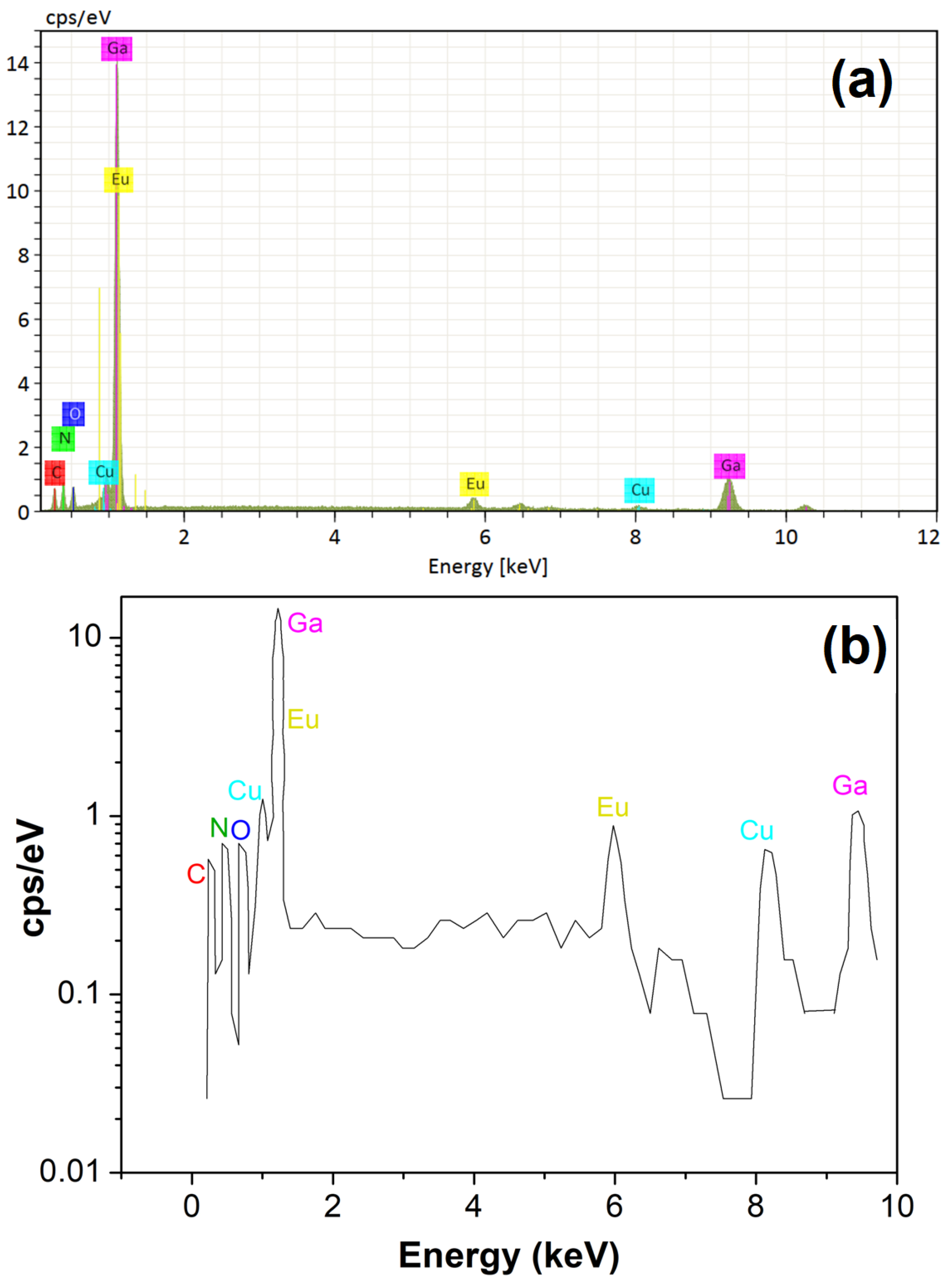
Figure 3a shows the EDS spectrum of the Eu-doped GaN powders and presents the compositional analysis. In the spectrum of Figure 3, it is possible to observe the elemental contributions of gallium (Kα 9.241 keV; Lα 1.098 keV), nitrogen (Kα 0.392 keV), europium (Lα 5.845 keV; M 1.131 keV), copper (Kα 8.040 keV; Lα 0.930 keV), oxygen (Kα 0.525 keV), and carbon (Kα 0.277 keV). It is crucial to emphasize that the copper contribution comes from the sample holder, while the oxygen and carbon were generated during the pyrolysis process by the gallium nitrate and carbohydrazide. The atomic percentages shown by the EDS analysis of Figure 3 are as follows: gallium (44.0%), nitrogen (21.8%), europium (2.6%), oxygen (7.9%), and carbon (23.5%). Also, it is important to mention that due to the nitridation time (one hour), the elemental contributions of oxygen and carbon were high; these elemental contributions could be reduced with a longer nitridation time (two hours). EDS analysis showed the presence of europium in the GaN samples. This agrees with the shift of 0.04 degrees toward larger angles in the Eu-doped GaN powders (Figure 1c) compared with the undoped GaN powders (Figure 1b). Figure 3b shows the spectrum from Figure 3a, plotted logarithmically regarding the Y-axis, in which the small peaks corresponding to carbon, nitrogen, oxygen, and copper can be observed more clearly, in addition to the europium and gallium peaks overlapping at low energies. Additionally, the SEM micrographs revealed a modification from a porous surface morphology to crystals with the shape of elongated platelets, possibly due to the europium incorporation into the GaN.
Figure 4a displays a TEM micrograph that presents the polycrystalline state for an Eu-doped GaN nanocrystal, where it is possible to observe different crystalline planes. It is important to mention that the polycrystalline state could confirm the procurement of hexagonal GaN, which would agree with the XRD analysis. Figure 4b shows the presence of nanocrystals of Eu-doped GaN, where the A nanocrystal is 7.58 nm in length, the B nanocrystal is 9.38 nm, the C nanocrystal is 8.24 nm, the D nanocrystal is 9.89 nm, the E nanocrystal is 7.78 nm, the F nanocrystal is 8.69 nm, and the G nanocrystal is 11.66 nm; this demonstrates an average size of 9.03 nm, which agrees with the average size of 9.09 nm obtained in the XRD analysis. It is important to mention that the above values for the nanocrystals are consistent with the c, e, and g X-ray diffraction peaks in Table 1. Finally, Figure 4c presents the electron diffraction pattern of the Eu-doped GaN, which confirms that the presence of symmetrical rings demonstrates the existence of nanocrystals in all of the material, where planes (002) and (101) were indexed. In the XRD analysis, the form factor used the Debye–Scherrer equation to estimate the crystallite size, with an average of 9.09 nm for a proportionality constant K of 0.9 for the hexagonal structure, using equipment with a maximum resolution of 1.5 Å. Conversely, in the SEM and TEM analyses, the form factor describes the morphology of the particles. In particular, in the SEM analysis, the shape of the sample surface was observed; in this case, they were elongated platelets with a size of 3.32 µm in length, with a maximum equipment resolution of 1 nm. In the TEM analysis, the internal structure of the particles was visualized; in this case, they were nanocrystals with a size of 9.03 nm, with a maximum equipment resolution of 0.05 nanometers (0.5 Å).
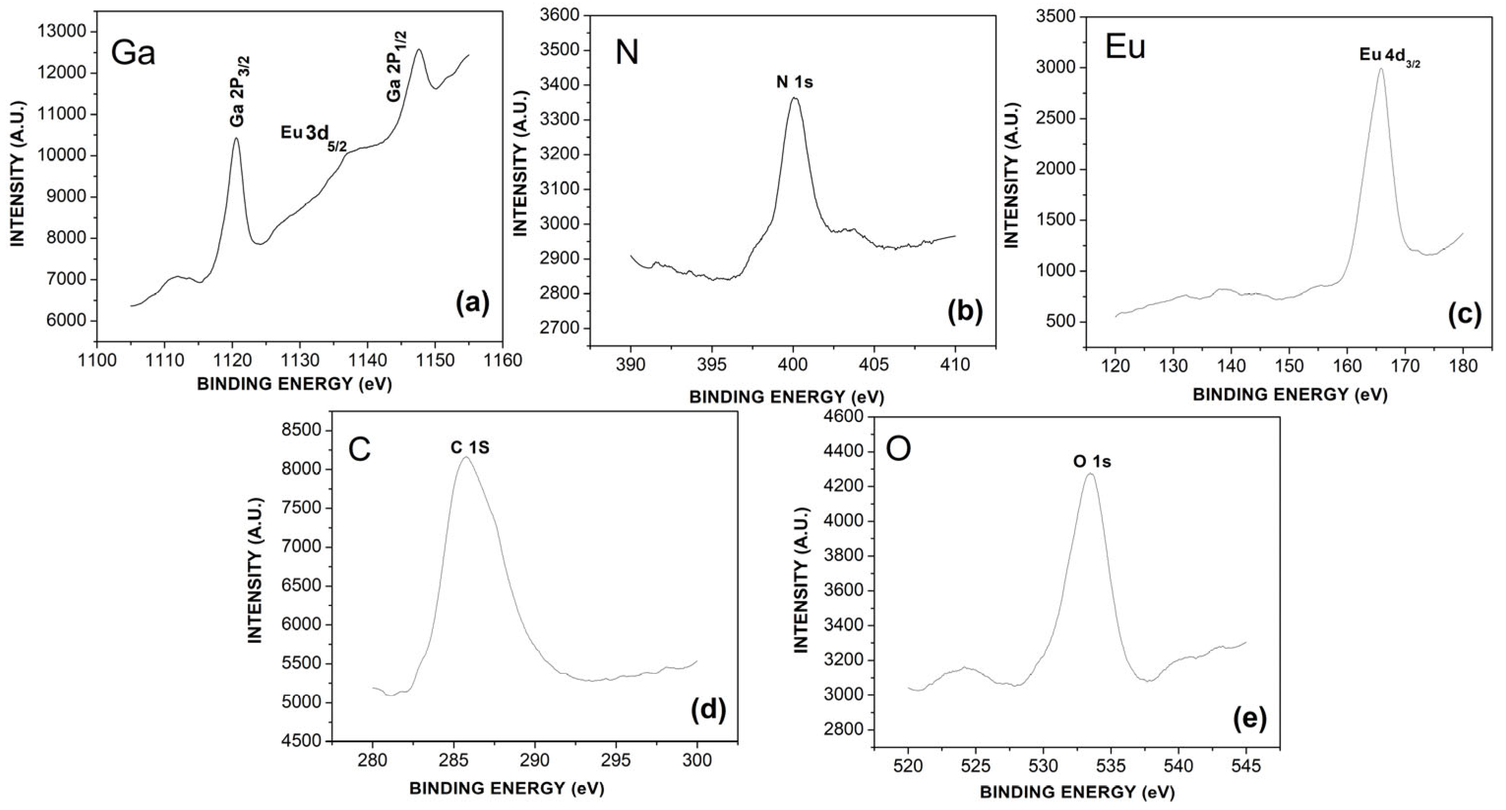
Figure 5 depicts the XPS spectra of the Eu-doped GaN powders. Figure 5a shows the peaks for high energies of Ga 2P1/2 and Ga 2P3/2, with values of 1147.6 eV and 1120.6 eV, respectively. Figure 5b shows a N 1s peak with an energy value of 400.1 eV. Figure 5c depicts the presence of an Eu 4d doping peak with an energy value of 165.8 eV, whose binding energy belongs to the europium core level. Figure 5a shows a small spectrum for the Eu 3d5/2 core energy level with a binding energy of 1136.9 eV, which confirms the binding of europium atoms with the gallium atoms. Xie et al. [23] present, in their research, the growth of Eu-doped GaN microcrystals using the ammonothermal method. In their XPS analysis of the element gallium, a small peak related to Eu 3d5/2 was located with a binding energy of 1133.35 eV. This is very similar to the behavior observed in the present work, where between the two main gallium peaks, a small peak for Eu 3d5/2 was also found with a binding energy of 1136.9 eV. This could indicate, as previously mentioned, the bonding of gallium atoms with europium. Figure 5d depicts the presence of the elemental contribution of C 1s with a value of 285.5 eV, while Figure 5e shows the elemental contribution of an O 1s peak with a binding energy of 533.4 eV, which demonstrates the presence of non-intentional impurities of oxygen and carbon, agreeing with the results obtained in the EDS analysis. These unintentional impurities could affect the optical properties of Eu-doped GaN powders. These impurities could have been generated during the pyrolysis process, as discussed in the EDS analysis.

3.3. Photoluminescence

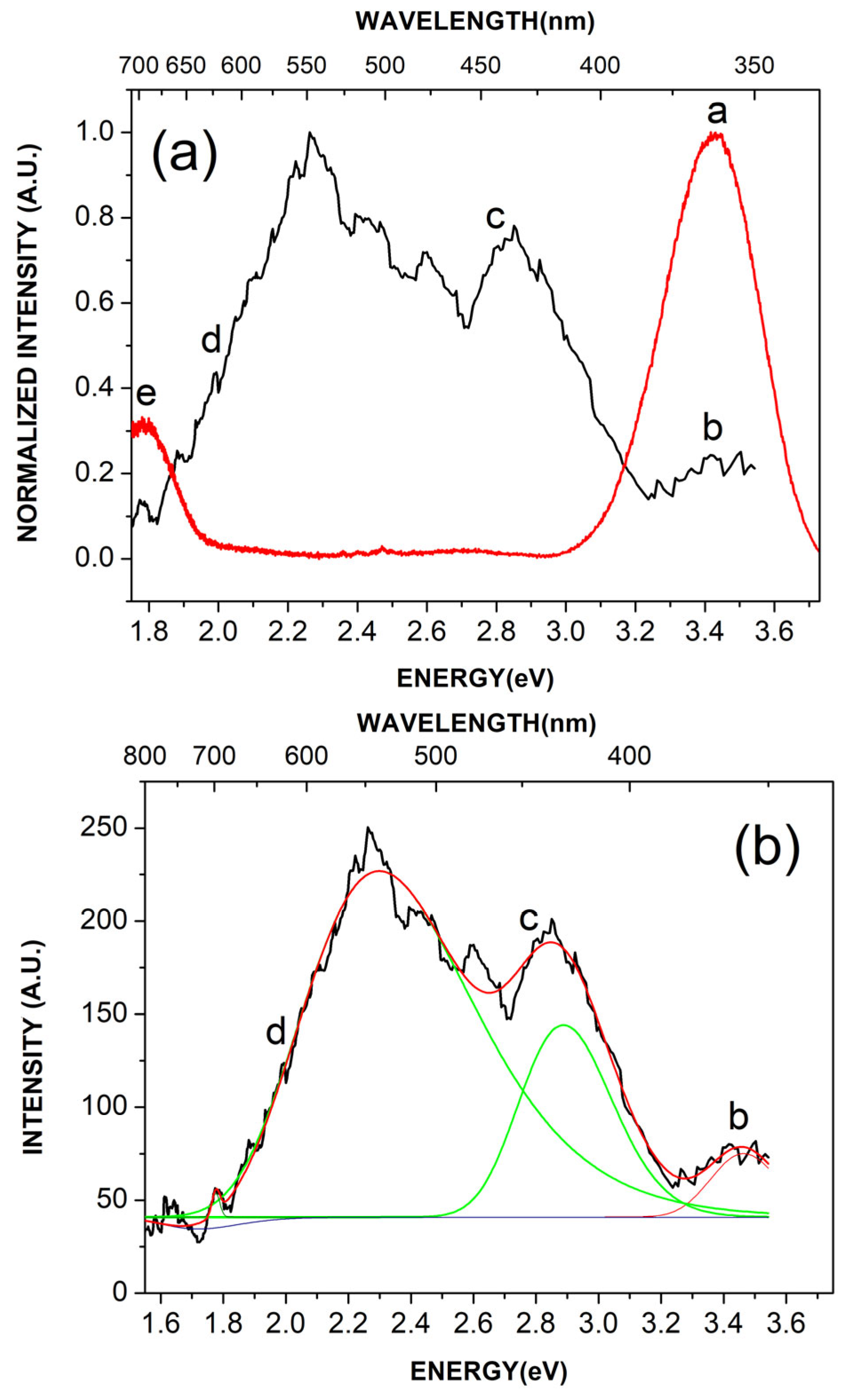
Figure 6a displays the photoluminescence spectra of the undoped GaN powders (red line) and Eu-doped GaN powders (black line), annealed in a NH3 environment at 1000 °C for one hour. Conversely, Figure 6b shows the decomposed photoluminescence spectrum of the Eu-doped GaN powders. The peak in Figure 6a shows the band-to-band transition of hexagonal GaN for the energy emission found at 3.43 eV (361.00 nm). The b peak in Figure 6a,b shows a small energy emission detected at 3.44 eV (359.69 nm) associated with the band-to-band transition of hexagonal GaN, which has been strongly reduced because of the incorporation of europium into the GaN crystal lattice. The c peak in Figure 6a,b shows an energy emission at 2.67eV (464.41 nm) and is related to surface defects in the GaN (blue luminescence BL); these surface defects can be observed in the TEM micrograph (Figure 4a). The emission in the blue band was accompanied by an enhancement of the yellow luminescence (YL) [24]. It is important to note that yellow luminescence (YL) in GaN is generally the result of gallium vacancies and oxygen substitutions. Hirata et al. [25] present various energy emissions for the yellow luminescence in their work. However, it is important to mention that their primary reagent for obtaining Eu-doped GaN was gallium oxide, which shows a significant oxygen signal in its EDS spectrum. In the presented research work, gallium and europium nitrates, as well as carbohydrazide, are used. In undoped GaN powders, oxygen traces decrease considerably. This behavior does not occur in Eu-doped GaN since more oxygen is added due to the europium nitrate. The d peak in Figure 6a,b shows a broad energy emission in a range of 2.52 eV to 1.60 eV; the main Eu3+ transition at 2.02 eV (611.69 nm) is found in this energy range, corresponding to the 5D07F2 transition of the f shell, which is a known laser transition [25,26]. Xie et al. [23] present, in their research, a 2 eV emission (620 nm) for the main Eu3+ emission corresponding to the 5D07F2 transition, which is very similar to the 2.02 eV (611.69 nm) reported in our work, also corresponding to the 5D07F2 transition. Furthermore, the behavior of the broad spectrum for an excitation wavelength of 365 nm is also very similar to that reported in this work. Some theoretical studies suggest that Eu doping can improve electrical conductivity in GaN by creating an n-type material. Regarding the d peak range, the energy emission situated at 2.52 eV (492.06 nm) is associated with oxygen impurities, while the energy emission associated with 1.60 eV (775 nm) is related to carbon impurities, agreeing with the non-intentional impurities found by the EDS and XPS analyses; these impurities could have been generated during the pyrolysis process, as was discussed in the EDS and XPS analyses [27,28,29,30,31]. Finally, the e peak in Figure 6a demonstrated an energy emission located at 1.58 eV (780.00 nm) for undoped GaN powders, associated with carbon impurities in red luminescence (RL) [23]. It is significant that the photoluminescence spectrum of the Eu-doped GaN powders in Figure 6a (black line) shows some noise, which could indicate that it has a low photoluminescence efficiency due to the incorporation of europium into the crystal structure compared with the photoluminescence spectrum of the undoped GaN powders (red line). Furthermore, in addition to the aforementioned results, the noise present in Figure 6a,b could be associated with the presence of impurities in the sample, such as oxygen and carbon. However, this could also be due to a low Raman signal intensity. If the Raman signal is weak, the detector noise may be more prominent, affecting the spectral quality. Zhang et al. [32,33] present studies of quantum yield in nanocrystals in their work. Krivolapchuk et al. [34] present a study of variations in the photoluminescence spectra of Eu-doped GaN, which revealed that the dopant can reside in the crystal in various charge states depending on the total defect concentration.

3.4. Raman Scattering

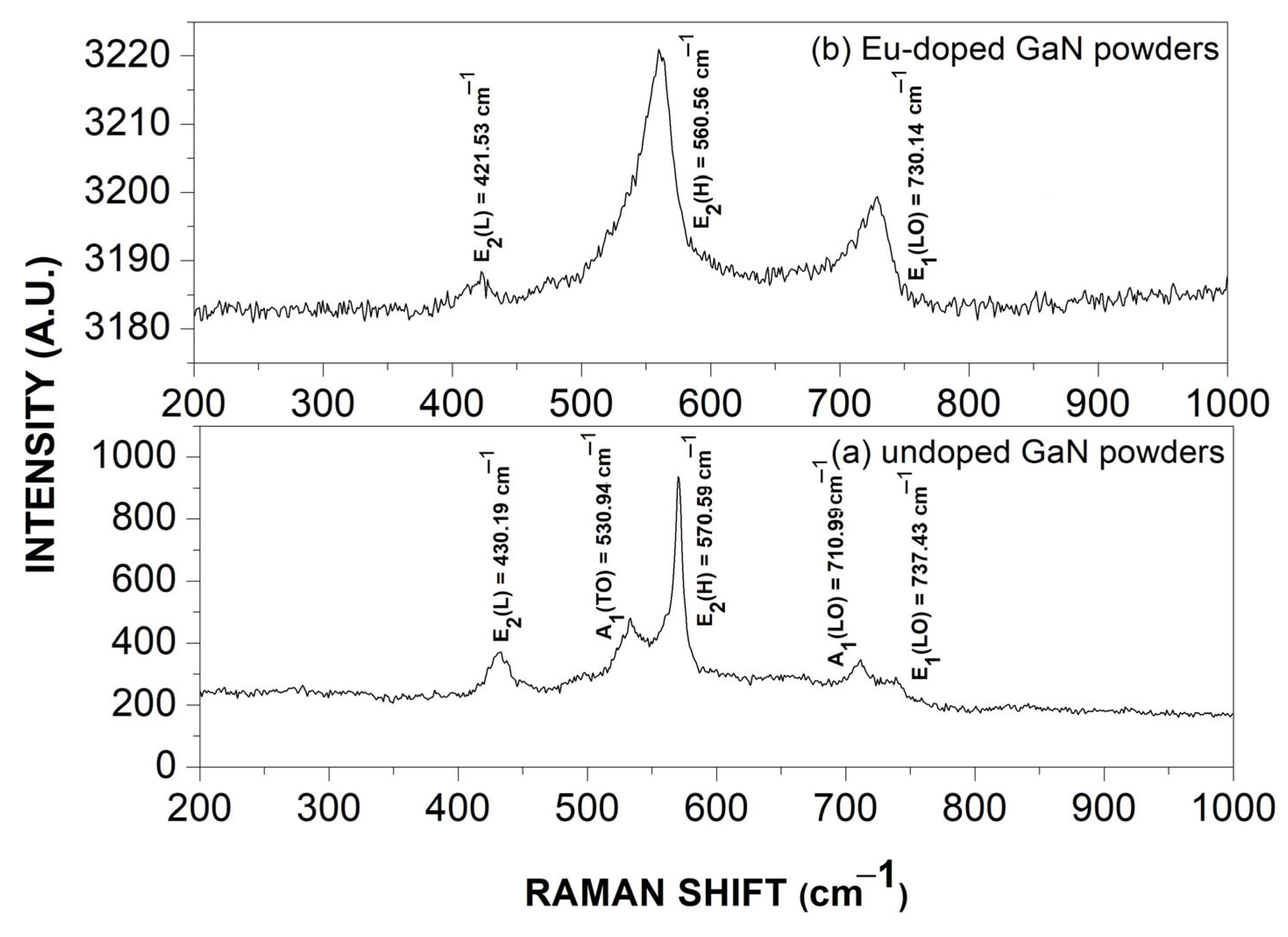
Figure 7 depicts the Raman scattering spectra for the undoped GaN powders (Figure 7a), along with the Eu-doped GaN powders (Figure 7b) obtained via pyrolysis of a viscous compound made from europium and gallium nitrates, with carbohydrazide used as a precursor and toluene as a solvent, with subsequent nitridation at 1000 °C for one hour. Figure 7a shows the characteristic vibration modes for the undoped GaN, where the more intense vibration mode is E2(H), located at 570.59 cm−1. Another vibration mode close to E2(H) was A1(TO) at 530.94 cm−1; other vibration modes belonging to hexagonal GaN are A1(LO) at 710.99 cm−1, E1(LO) at 737.43 cm−1, and E2(L) at 430.19 cm−1. Furthemore, Figure 7b shows the vibration modes obtained for the Eu-doped GaN powders, where the more intense vibration mode E2(H) overlapped with the A1(TO) vibration mode, which obtained a value of 560.56 cm−1; at the same time, the E1(LO) vibration mode overlaps with the A1(LO) vibration mode, which obtained a value of 730.14 cm−1, while the E2(L) vibration mode had a value of 421.53 cm−1. Comparing the Raman scattering spectrum in Figure 7a with the Raman scattering spectrum in Figure 7b, it is possible to observe a slight shift of 10.03 cm−1 toward lower frequencies in the E2(H) vibration mode, which could indicate that the europium atoms replaced gallium atoms (gallium interstitials) in the GaN crystal lattice, with smaller vibrations related to the difference in atomic weights between europium and gallium, as was demonstrated in the XRD analysis. It is important to mention that the main E2(H) peak for Eu-doped GaN powders in this work is 560.56 cm−1. Other research works present similar values for the main E2(H) peak compared with this study. Pan et al. [35] present a value of 568.00 cm−1, while Shi et al. [36] present a value of 567.00 cm−1, indicating that our value corresponds to others reported in the literature. Furthermore, Thomas et al. [37], Vajpeyi et al. [38], and Inaba et al. [12] present, in their research works, a shift in the main E2(H) peak toward lower frequencies for Eu-doped GaN, as presented in this work.

4. Conclusions

The fundamental question in this research work was whether it was possible to obtain Eu-doped GaN powders with the pyrolysis method, using gallium and europium nitrates, as well as carbohydrazide, as fuel and toluene as the reagent solvent, followed by nitridation at 1000 °C for 1 h. Using the aforementioned method, Eu-doped GaN nanocrystals were obtained, with the following most relevant results: XRD analysis of the Eu-doped GaN powders presented a nanocrystal size average of 9.09 nm. X-ray diffraction patterns for the Eu-doped GaN powders showed a slight shift of 0.04 degrees toward larger angles compared to the undoped GaN powders, which could indicate the incorporation of Eu into the GaN. SEM micrographs demonstrated a surface morphology for the Eu-doped GaN with a shape similar to elongated platelets, whereas the surface morphology of the undoped GaN powders was porous. EDS and XPS studies demonstrated the presence of a europium elemental contribution to the GaN crystal lattice. Furthermore, the XPS spectrum for gallium showed binding energies for Ga 2P3/2, Ga 2P1/2, and Eu3d5/2, which could indicate the incorporation of europium into the GaN and bonding between gallium and europium atoms, where Eu atoms generally substitute Ga sites in the GaN lattice structure. An electron diffraction pattern TEM micrograph of the Eu-doped GaN powders confirmed the presence of symmetrical rings, demonstrating the existence of nanocrystals in all the material. Photoluminescence showed the main Eu3+ transition at 2.02 eV (611.69 nm) for the europium emission corresponding to the 5D07F2 transition of the f shell, which is recognized as a laser transition. Finally, the Raman scattering spectrum of the Eu-doped GaN powders shows a slight shift of 10.03 cm−1 toward lower frequencies compared with the undoped GaN powders for the E2(H) vibration mode, which could indicate the incorporation of europium atoms into the GaN crystal lattice. The aforementioned results indicate that Eu-doped GaN powders were obtained by pyrolysis, which stands out as an economical, safe, and easy method for synthesizing III-nitride materials compared to other methods.

Author Contributions

Conceptualization, Formal Analysis, Investigation, Writing—Original Draft, E.G.; Resources, R.G.; Resources, Formal Analysis, Investigation, Writing—Original Draft, A.M.H.; Resources, A.R.; Resources, G.G.; Resources, G.A.H.; Resources, J.A.L.; Resources, R.C.C.; Writing—Original Draft, J.A.R.; Resources, R.R.; Validation, Y.D.R.; Resources, F.B.; Resources, A.C. All authors have read and agreed to the published version of the manuscript.

Funding

This research received no external funding.

Institutional Review Board Statement

Not applicable.

Informed Consent Statement

Not applicable.

Data Availability Statement

The data is contained within the article.

Acknowledgments

The authors gratefully acknowledge the technical support of CNyN-UNAM, particularly Eloísa Aparicio and Israel Granilla. This research was partially supported by SECIHTI (México) and DIFUS-UNISON.

Conflicts of Interest

The authors declare no conflicts of interest.

References

  1. Amir, H.A.A.A.; Fakhri, M.A.; Alwahib, A.A. Review of GaN optical device characteristics, applications, and optical analysis technology. Mater. Today Proc. 2021, 42, 2815–2821. [Google Scholar] [CrossRef]
  2. Podhorodecki, A.; Kudrawiec, R.; Nyk, M.; Misiewicz, J.; Strek, W. Surface- and volume-related excitation of Eu-doped nanocrystalline GaN powders. Opt. Mater. 2009, 31, 1252–1255. [Google Scholar] [CrossRef]
  3. Jadwisienczak, W.; Wisniewski, K.; Spencer, M.; Thomas, T.; Ingram, D. Optical properties, luminescence quenching mechanism and radiation hardness of Eu-doped GaN red powder phosphor. Radiat. Meas. 2010, 45, 500–502. [Google Scholar] [CrossRef]
  4. Murakami, F.; Takeo, A.; Mitchell, B.; Dierolf, V.; Fujiwara, Y.; Tonouchi, M. Enhanced luminescence efficiency in Eu-doped GaN superlattice structures revealed by terahertz emission spectroscopy. Commun. Mater. 2023, 4, 100. [Google Scholar] [CrossRef]
  5. Iwaya, T.; Ichikawa, S.; Murakami, M.; Tatebayashi, J.; Fujiwara, Y. Investigation on Suitable Structure for Laser Oscillation in Eu-doped GaN with Two-Dimensional Photonic Crystal Nanocavities. J. Soc. Mater. Sci. Jpn. 2020, 69, 721–726. [Google Scholar] [CrossRef]
  6. Fragkos, I.E.; Tan, C.K.; Dierolf, V.; Fujiwara, Y.; Tansu, N. Pathway Towards High-Efficiency Eu-doped GaN Light-Emitting Diodes. Sci. Rep. 2017, 7, 14648. [Google Scholar] [CrossRef]
  7. Angadi, V.C.; Benz, F.; Tischer, I.; Thonke, K.; Aoki, T.; Walther, T. Evidence of terbium and oxygen co-segregation in annealed AlN:Tb. Appl. Phys. Lett. 2017, 110, 222102. [Google Scholar] [CrossRef]
  8. Brown, E.; Hömmerich, U.; Yamada, T.; Yamane, H.; Zavada, J. Luminescence spectroscopy of europium doped gallium nitride powder prepared by a Na flux method. Phys. Status Solidi (A) 2011, 208, 156–160. [Google Scholar] [CrossRef]
  9. Zhu, W.; Mitchell, B.; Timmerman, D.; Koizumi, A.; Gregorkiewicz, T.; Fujiwara, Y. High-Power Eu-Doped GaN Red LED Based on a Multilayer Structure Grown at Lower Temperatures by Organometallic Vapor Phase Epitaxy. MRS Adv. 2017, 2, 159–164. [Google Scholar] [CrossRef]
  10. Kudrawiec, R.; Nyk, M.; Podhorodecki, A.; Misiewicz, J.; Strek, W.; Wołcyrz, M. Change in photoluminescence spectra of Eu-doped GaN powders due to the aggregation of nanosized grains into micrometer-sized conglomerations. Appl. Phys. Lett. 2006, 88, 061916. [Google Scholar] [CrossRef]
  11. Nyk, M.; Podhorodecki, A.; Kudrawiec, R.; Misiewicz, J.; Strek, W. Size Shrinkage of GaN Nanocrystalline Grains Induced by Eu Doping Electrochem. Solid-State Lett. 2007, 10, H203–H205. [Google Scholar] [CrossRef]
  12. Inaba, T.; Mitchell, B.; Koizumi, A.; Fujiwara, Y. Emission enhancement and its mechanism of Eu-doped GaN by strain engineering. Opt. Mater. Express 2017, 7, 1381–1387. [Google Scholar] [CrossRef]
  13. El-Himri, A.; Pérez-Coll, D.; Núñez, P.; Martín, I.R.; Lavín, V.; Rodríguez, V.D. Preparation and optical spectroscopy of Eu3+-doped GaN luminescent semiconductor from freeze-dried precursors. J. Solid State Chem. 2004, 177, 4213–4220. [Google Scholar] [CrossRef]
  14. Wang, X.; Zeng, X.; Xu, K.; Mao, H.; Ma, C. Luminescence mechanism and energy level structure of Eu-doped GaN powders investigated by cathodoluminescence spectroscopy. Sci. China Phys. Mech. Astron. 2014, 57, 628–631. [Google Scholar] [CrossRef]
  15. Braun, C.; Mereacre, L.; Chen, Z.; Slabon, A. Closing the yellow gap with Eu- and Tb-doped GaN: One luminescent host resulting in three colours. Sci. Rep. 2022, 12, 2503. [Google Scholar] [CrossRef] [PubMed]
  16. Wu, H.; Poitras, C.B.; Lipson, M.; Spencer, M.G.; Hunting, J.; DiSalvo, F.J. Photoluminescence and cathodoluminescence analyses of GaN powder doped with Eu. Appl. Phys. Lett. 2006, 88, 011921. [Google Scholar] [CrossRef]
  17. Pela, R.R.; Hsiao, C.L.; Hultman, L.; Birch, J.; Gueorguiev, G.K. Electronic and optical properties of core–shell InAlN nanorods: A comparative study via LDA, LDA-1/2, mBJ, HSE06, G0W0 and BSE methods. Phys. Chem. Chem. Phys. 2024, 26, 7504–7514. [Google Scholar] [CrossRef]
  18. Filho, M.A.M.; Farmer, W.; Hsiao, C.-L.; dos Santos, R.B.; Hultman, L.; Birch, J.; Ankit, K.; Gueorguiev, G.K. Density Functional Theory-Fed Phase Field Model for Semiconductor Nanostructures: The Case of Self-Induced Core–Shell InAlN Nanorods. Cryst. Growth Des. 2024, 24, 4717–4727. [Google Scholar] [CrossRef]
  19. Gastellóu, E.; Morales, C.; García, R.; García, G.; Hirata, G.A.; Galeazzi, R.; Herrera, A.M.; Rosendo, E.; Díaz, T.; Ramos, J.R.; et al. Enhanced crystalline size of undoped GaN powders obtained by nitridation of metallic gallium. Opt. Mater. 2018, 83, 220–224. [Google Scholar] [CrossRef]
  20. Herrera, A.M.; Ramos, A.; Gastellóu, E.; García, R.; García, G.; Carrillo, R.C.; Santos, I.; Brown, F.; Mora, R.; Hirata, G.A. Effect of the Pyrolysis Environment in a Complex Compound in the Synthesis of In0.6Ga0.4N Powders and their Characterization. Phys. Status Solidi (A) 2024, 222, 2400593. [Google Scholar] [CrossRef]
  21. Herrera, A.M.; García, R.; García, G.; Gastellóu, E.; Nieto, F.; Hirata, G.A.; Contreras, O.E.; Morales, C.; Rosendo, E.; Díaz, T. Experimental determination of the pyrolysis temperatures of an organometallic complex to obtain AlxGa1-xN powders. J. Alloys Compd. 2019, 775, 109–115. [Google Scholar] [CrossRef]
  22. Garcia, R.; Hirata, G.A.; Thomas, A.C.; Ponce, F.A. Structure and luminescence of nanocrystalline gallium nitride synthesized by a novel polymer pyrolysis route. Opt. Mater. 2006, 29, 19–23. [Google Scholar] [CrossRef]
  23. Xie, K.; Li, T.; Ren, G.; Wang, L.; Lu, W.; Shen, L.; Zhou, H.; Xu, K. Optical characterization of GaN:Eu microcrystals grown by the ammonothermal method. J. Alloys Compd. 2024, 1008, 176776. [Google Scholar] [CrossRef]
  24. Reshchikov, M.A.; Morkoç, H. Luminescence properties of defects in GaN. J. Appl. Phys. 2005, 97, 061301. [Google Scholar] [CrossRef]
  25. Hirata, G.A.; Ramos, F.; Garcia, R.; Bosze, E.J.; McKittrick, J.; Ponce, F.A. A New Combustion Synthesis Method for GaN:Eu3+ and Ga2O3:Eu3+ Luminescent Powders. Phys. Status Solidi (A) 2001, 188, 179–182. [Google Scholar] [CrossRef]
  26. Fujiwara, Y.; Dierolf, V. Present understanding of Eu luminescent centers in Eu-doped GaN grown by organometallic vapor phase epitaxy. Jpn. J. Appl. Phys. 2014, 53, 05FA13. [Google Scholar] [CrossRef]
  27. Otabara, T.; Tatebayashi, J.; Hasegawa, S.; Timmerman, D.; Ichikawa, S.; Ichimiya, M.; Ashida, M.; Fujiwara, Y. Formation and optical characteristics of GaN:Eu/GaN core–shell nanowires grown by organometallic vapor phase epitaxy. Jpn. J. Appl. Phys. 2022, 61, SD1022. [Google Scholar] [CrossRef]
  28. Hoang, K. Tuning the valence and concentration of europium and luminescence centers in GaN through co-doping and defect association. Phys. Rev. Mater. 2021, 5, 034601. [Google Scholar] [CrossRef]
  29. Hoang, K. Rare-earth defects in GaN: A systematic investigation of the lanthanide series. Phys. Rev. Mater. 2022, 6, 044601. [Google Scholar] [CrossRef]
  30. Mohamed, H.E.A.; Hkiri, K.; Khenfouch, M.; Dhlamini, S.; Henini, M.; Maaza, M. Optical properties of biosynthesized nanoscaled Eu2O3 for red luminescence applications. J. Opt. Soc. Am. A 2020, 37, C73–C79. [Google Scholar] [CrossRef]
  31. Ohata, S.; Kawamura, T.; Akiyama, T.; Usami, S.; Imanishi, M.; Yoshimura, M.; Mori, Y.; Sumi, T.; Takino, J. Influence of oxy-gen-related defects on the electronic structure of GaN. Jpn. J. Appl. Phys. 2022, 61, 061004. [Google Scholar] [CrossRef]
  32. Zhang, L.; Zhou, H.; Chen, Y.; Zheng, Z.; Huang, L.; Wang, C.; Dong, K.; Hu, Z.; Ke, W.; Fang, G. Spontaneous crystallization of strongly confined CsSnxPb1-x I3 perovskite colloidal quantum dots at room temperature. Nat. Commun. 2024, 15, 1609. [Google Scholar] [CrossRef]
  33. Zhang, L.; Chen, Y.; Zheng, Z.; Zhou, Y.; Li, C.; Li, G.; Ren, B.; Hu, Z.; Zhou, H.; Ren, F.; et al. Rapid Room Temperature Entropy-Stabilized Synthesis Enabling Super-Stable Metal Halide Perovskite Semiconductor Colloidal Nanocrystals. Adv. Funct. Mater. 2025, 35, 2423450. [Google Scholar] [CrossRef]
  34. Krivolapchuk, V.V.; Mezdrogina, M.M.; Nasonov, A.V.; Rodin, S.N. Photoluminescence of Bulk Eu-Doped GaN Crystals. Phys. Solid State 2003, 45, 1634–1637. [Google Scholar] [CrossRef]
  35. Pan, X.; An, X.; Zhang, Z.; Zhou, J.; Xie, E. Structural and optical properties of GaN:Eu nanoparticles synthesized by simple ammonification method. J. Alloys Compd. 2012, 519, 67–71. [Google Scholar] [CrossRef]
  36. Shi, J.; Chandrashekhar, M.V.S.; Reiherzer, J.; Schaff, W.J.; Lu, J.; Disalvo, F.J.; Spencer, M.G. Effect of growth temperature on Eu incorporation in GaN powders. J. Cryst. Growth 2008, 310, 452–456. [Google Scholar] [CrossRef]
  37. Thomas, T.; Guo, X.; Chandrashekhar, M.; Poitras, C.B.; Shaff, W.; Dreibelbis, M.; Reiherzer, J.; Li, K.; DiSalvo, F.J.; Lipson, M.; et al. Purification and mechanical nanosizing of Eu-doped GaN. J. Cryst. Growth 2009, 311, 4402–4407. [Google Scholar] [CrossRef]
  38. Vajpeyi, A.P.; Tripathy, S.; Wang, L.S.; Foo, B.C.; Chua, S.J.; Fitzgerald, E.A.; Alves, E. Optical activation of Eu ions in nanoporous GaN films. J. Appl. Phys. 2006, 99, 104305. [Google Scholar] [CrossRef]
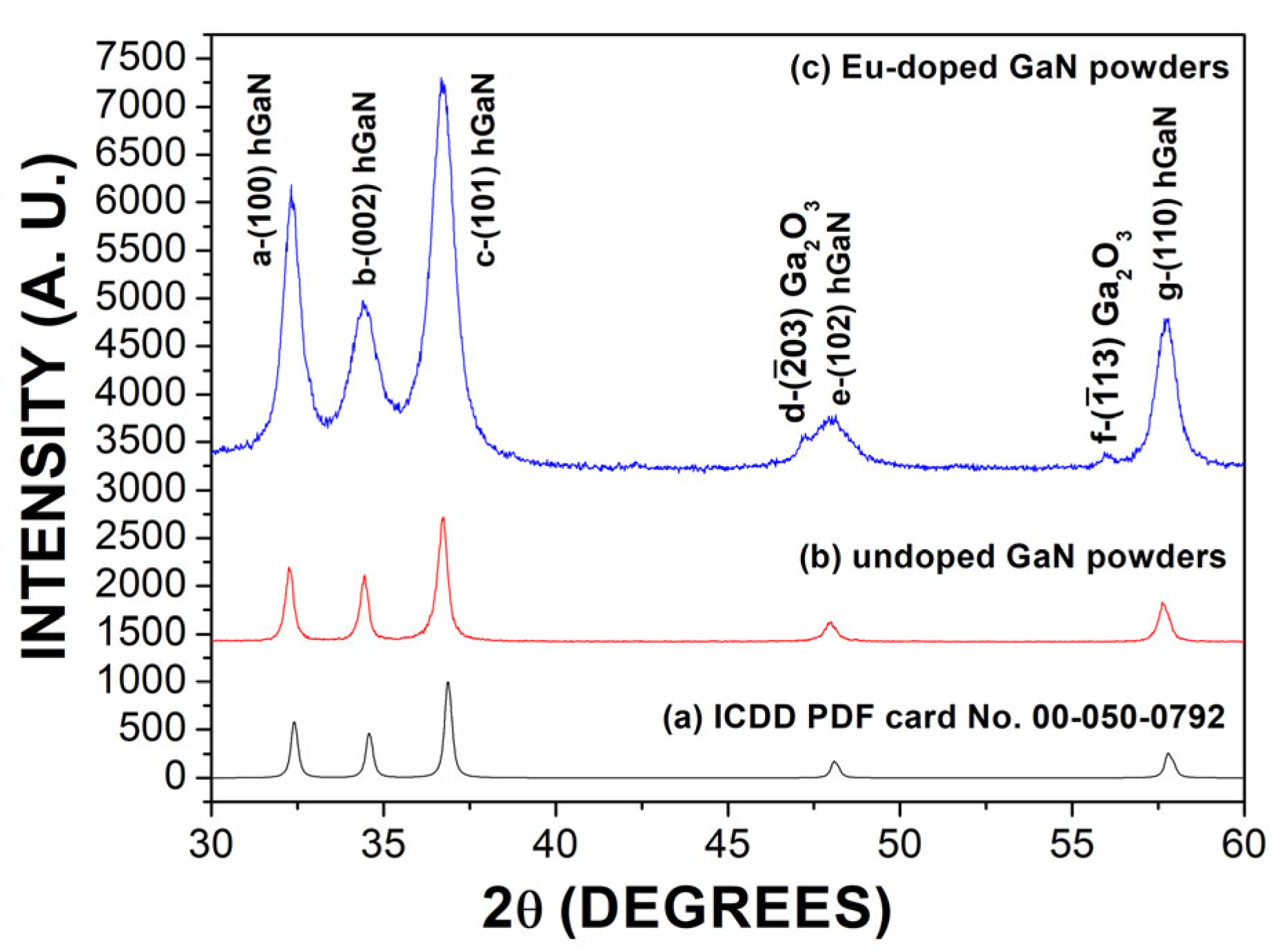
Figure 1. (a) ICDD PDF card No. 00-050-0792. (b) Undoped GaN powders. (c) Eu-doped GaN powders.
Photonics 12 00982 g001
Figure 2. (a) SEM micrograph of the undoped GaN powders; (b) SEM micrograph of the Eu-doped GaN powders.
Photonics 12 00982 g002
Figure 3. (a) EDS spectrum for the Eu-doped GaN powders. (b) Logarithmic plot of the EDS spectrum for the Eu-doped GaN powders.
Photonics 12 00982 g003
Figure 4. TEM micrographs: (a) polycrystalline state of Eu-doped GaN nanocrystal at 5 nm; (b) Eu-doped GaN nanocrystals at 100 nm; (c) electron diffraction pattern of Eu-doped GaN powders.
Photonics 12 00982 g004
Figure 5. XPS spectra of the Eu-doped GaN powders: (a) Ga 2P3/2, Ga 2P1/2, and Eu 3d5/2; (b) N 1s; (c) Eu 4d; (d) C 1s; and (e) O 1s peaks.
Photonics 12 00982 g005
Figure 6. Photoluminescence spectra for (a) undoped GaN powders (red line) and Eu-doped GaN powders (black line). (b) Decomposed photoluminescence spectrum for Eu-doped GaN powders.
Photonics 12 00982 g006
Figure 7. Raman scattering spectra for (a) undoped GaN powders and (b) Eu-doped GaN powders.
Photonics 12 00982 g007
Table 1. Values calculated for peak position, FWHM, crystal size, and interplanar spacing for undoped GaN and Eu-doped GaN powders.
Undoped GaN PowdersEu-Doped GaN Powders
PeakPeak Position (Degree)FWHM (Degree)Crystal Size (nm)Interplanar Spacing
(Å)
Peak Position (Degree)FWHM (Degree)Crystal Size (nm)Interplanar Spacing
(Å)
a32.270.3225.202.7732.340.7411.102.76
b34.430.3324.942.6034.481.545.382.59
c36.710.4020.582.4436.710.938.992.44
e47.970.4319.921.8947.941.217.171.89
g57.660.4121.941.5957.720.70812.791.59
Disclaimer/Publisher’s Note: The statements, opinions and data contained in all publications are solely those of the individual author(s) and contributor(s) and not of MDPI and/or the editor(s). MDPI and/or the editor(s) disclaim responsibility for any injury to people or property resulting from any ideas, methods, instructions or products referred to in the content.