Abstract
In this study, we systematically investigate theoretically and experimentally the plasmonic effect and roles of big and small gold nanoparticles (Au NPs) within a mixed solution. The polydisperse solution was initially prepared by mixing small (10, 30 nm) Au NPs with larger ones (50, 80 nm), followed by measuring the extinction using ultraviolet–visible (UV-vis) spectroscopy. The experimental results clearly showed that the extinction of the mixed solution is predominantly influenced by the presence of the larger NPs, even though their quantity is small. Subsequently, we conducted simulations to explore the plasmonic properties of Au NPs of different sizes as well as their mixings and to validate the experimental results. To explain the deviation of the extinction spectra between experimental observations and simulations, we elaborated a simulation model involving the mixture of spherical Au NPs with ellipsoidal NPs, thus showing agreement between the simulation and the experiment. By performing simulations of plasmonic near-field of NPs, our investigation revealed that the maximal electric field intensity does not occur precisely at the plasmonic resonant wavelength but rather at a nearby redder wavelength. The optimal size of the Au NP dispersed in water for achieving the highest field enhancement was found to be 60 nm, with an excitation wavelength of 553.7 nm. These interesting findings not only enrich our understanding of plasmonic NPs’ optical behavior but also guide researchers for potential applications in various domains.
1. Introduction
Over the past three decades, research on nanoparticles (NPs) has attracted increasing interest thanks to their unique properties, which are regarded as a bridge between individual atoms and bulk material [1]. Among them, noble metal NPs (10–100 nm) stand out for their promising, diverse, and intriguing physical and chemical properties, which can be finely tuned by adjusting their size, morphology, composition, and various preparation parameters [2,3]. The irradiation by an electromagnetic field induces collective oscillation of free electrons of metal NPs, reaching its maximum at a specific frequency known as surface plasmon resonance (SPR). This phenomenon possesses the ability to strongly absorb and scatter visible light. Since the NPs have a limited size and are well separated from each other, the plasmonic effect of a metal NP is localized near its surface, thus called localized SPR (LSPR). Thanks to this optical property, metal NPs find applications across various domains including nanocatalysis [4,5,6], solar cells [7,8], photothermal therapy [9], biosensing [10,11], and drug delivery [12,13]. While absorption and scattering at resonance wavelength can be considered as a drawback for many applications since it introduces a loss for light energy, the light intensity at resonance is strongly enhanced, as compared with that of the incident light beam, thus offering many interesting applications, such as renewable energy transducers [14], hyperthermia therapy [15], fluorescence enhancement [16,17], and lasers [18,19].
The methods for characterizing and measuring NPs play a crucial role in the advancement of nanotechnology, as the sizes, shapes, and structures of NPs influence their physicochemical properties, and consequently their applicative performances. Among the techniques used for characterization of NPs, UV-vis spectroscopy is commonly used, which is available in most laboratories. Indeed, UV–vis spectroscopy measures the extinction spectrum, which involves both absorption and scattering of NPs, despite being occasionally referred to as an absorption spectrum [20]. Researchers have used it as a simple, rapid, and cost-effective method for efficiently monitoring NP size, concentration, as well as particle shape. V. Amendola et al. [21] reported the evaluation of the average size of Au NPs based on the fitting of their UV-vis spectra by the Mie model for spherical NPs. By determining the position of the maximal SPR spectrum and fitting it with a theoretical model, the size of NPs is calculated [22,23]. However, this approach is only valid for monodisperse solutions, i.e., with mostly uniform and identical NPs. When the solution is highly polydispersed with a large range of particle sizes, their diameters become overestimated. E. Tomaszewska et al. [24], by analyzing the results from dynamic light scattering (DLS)/UV-Vis spectroscopy conducted on polydisperse colloids, observed that several percent of the volume content of larger NPs could completely obscure the presence of smaller ones. This is particularly beneficial in understanding experimental data, especially for those who work with DLS and/or UV-Vis spectroscopy.
Furthermore, the exploration of plasmonic near-field enhancement supported by noble metal NPs is currently a subject of intense research. Surface-enhanced Raman scattering (SERS) serves as a typical example, where the strong electromagnetic near-field leads to the enhancement factors of 5 to 6 orders of magnitude [25,26]. Other examples, such as photovoltaic enhancement or nonlinear optical processes, display linear [27] or nonlinear behaviors [28,29] with respect to the intensity of the plasmonic near-field, respectively. Therefore, it is necessary to design NPs showing the most intense near electric field (NEF) intensity. Indeed, extensive work has been performed to enhance the plasmonic near-field intensity by designing NPs with specific geometries, such as sharp tips [30,31], or by coupling NPs [32,33]. Moreover, it has been demonstrated that the maximum electric field intensity does not occur precisely at the plasmonic resonant wavelength, but rather at a nearby, redder wavelength [34,35]. Despite numerous reports on the field enhancement of Au NPs, a systematic study on the influence of size and excited wavelength on near-fields has not been fully carried out. Furthermore, most demonstrations have employed Au NPs with a size of about 50 nm, as a standard choice [36,37]. We wonder what will be the best Au NP size which allows the best field enhancement to be obtained while staying in the frame of plasmonic NPs.
In this work, we systematically investigate how large Au NPs obscure the presence of small NPs in polydispersed solution through both experiments using UV-vis spectroscopy and simulations using the FDTD method. For that, we propose to quantitatively mix small (10, 30 nm) Au NPs with larger ones (50, 80 nm) and characterize the SPR spectra. In order to clarify the deviation between the experimental SPR spectra and theoretical ones, we propose a model of mixing spherical Au NPs with ellipsoidal NPs to find out the best match. Furthermore, we perform simulations of the NEF around spherical Au NPs with varying sizes and wavelengths, aiming to determine optimal parameters, such as particle size and excited wavelength, for maximal electric field enhancement.
2. Experimental Study of Polydisperse Au NPs
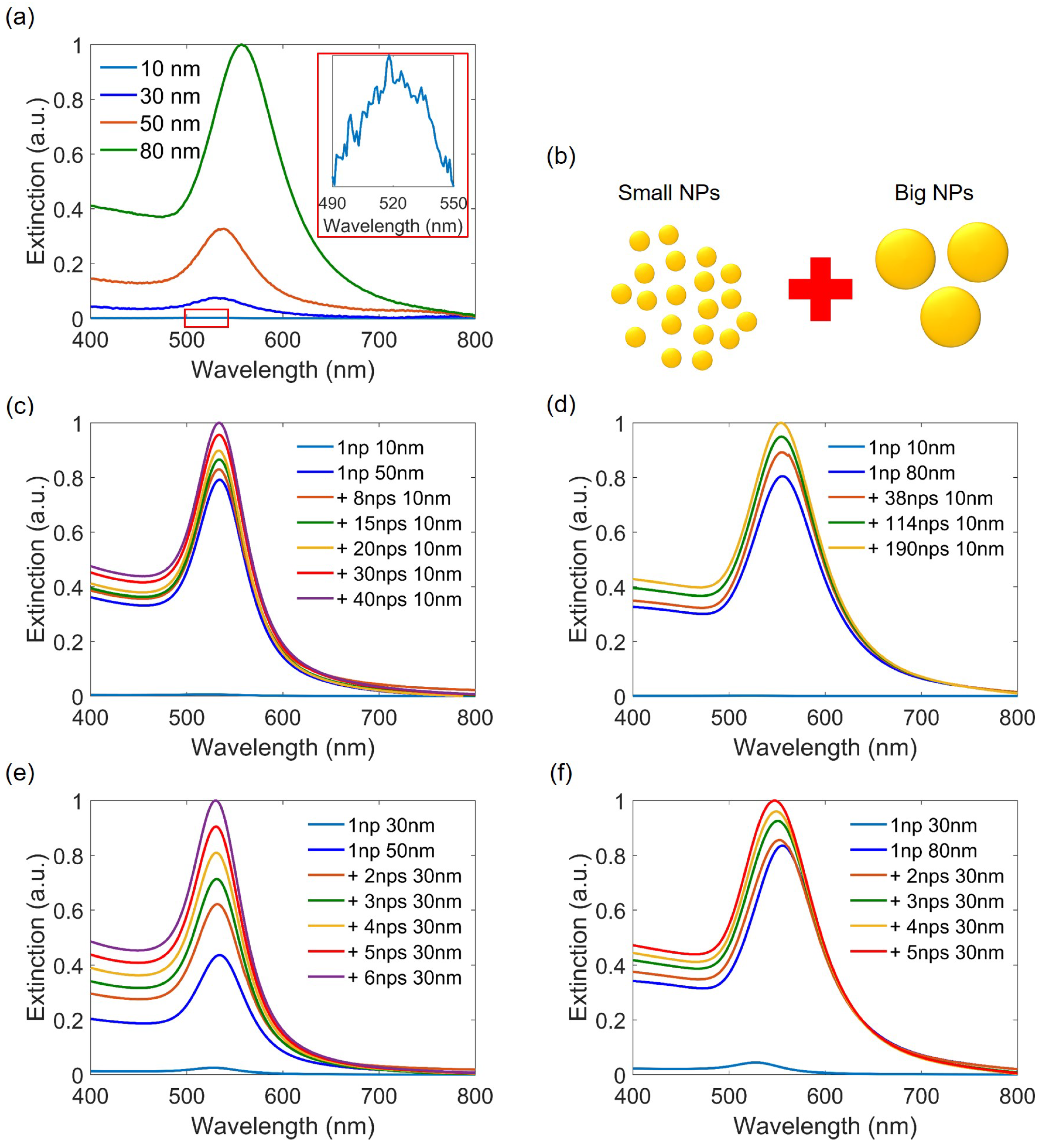
Spherical-like Au NP solutions of different sizes (10, 30, 50, and 80 nm) were purchased from Sigma Aldrich, with concentrations of particles/mL, particles/mL, particles/mL, and particles/mL, respectively. Initially, we diluted each type of Au NP solution with pure water to obtain the same concentrations, followed by extinction measurements using UV-vis spectroscopy. Figure 1a shows the extinction spectra of the prepared Au colloid solutions. It is clear that as the size of Au NPs increases, the resonant peak becomes higher and undergoes a redshift, ranging from about 520 nm for particles of 10 nm to 556 nm for particles of 80 nm in size. This occurs because large Au NPs possess larger absorption and scattering cross-sections, thereby shifting the SPR peak.
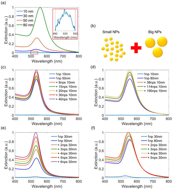
Figure 1.
Experimental extinction spectra of colloidal Au NPs measured by UV-Vis spectroscopy. (a) Extinction spectra of commercial spherical Au NPs of different sizes, obtained with the same NP concentration after diluting the original solution with pure water. (b) Illustration of a mixture of a high number of small Au NPs and a low number of large Au NPs. (c–f) Extinction spectra of the mixed solutions with different NP ratios: (c) 10 nm with 50 nm, (d) 10 nm with 80 nm, (e) 30 nm with 50 nm, and (f) 30 nm with 80 nm, respectively. Insert of (a) shows a zoom on the plasmon resonance of 10 nm Au NPs.
After that, we mixed the solutions of small Au NPs with large Au NPs with different NP number ratios to create a polydisperse colloidal solution, as illustrated in Figure 1b. In practice, from the concentration of the original solution, we calculated the number of NPs in a mixed solution, and normalized it to the number of large NPs, which is also normalized to one, for easier comparison. Figure 1c shows the experimental SPR spectra obtained when mixing a solution of 50 nm Au NPs with a 10 nm Au NP solution with different particle numbers ratios. It is clear that the extinction of large Au NPs dominates even when the number of small NPs is forty times higher. As more 10 nm Au NPs were added, the SPR peak originating from the 50 nm Au NPs increases and shifts to shorter wavelengths. This is because when the amount of small NPs is important, their influence becomes more apparent and the resultant SPR peak shifts to the one of small NPs, i.e., blueshift. Similar studies using UV-vis spectroscopy have been performed on polydisperse colloids obtained by mixing 80 nm with 10 nm Au NPs (Figure 1d), 50 nm with 30 nm Au NPs (Figure 1e), and 80 nm with 30 nm Au NPs (Figure 1f). All results indicate that the SPR of larger NPs dominates the mixed solution, although their number is very small. These findings suggest that obtaining precise information about the sizes and even the shapes of NPs is challenging, as it is impossible to distinguish peak separation among specific populations within these solutions. Despite this, some works have attempted to determine the size distribution of polydisperse solutions by analyzing their extinction spectra using Mie theory [38] or, more recently, by employing an advanced method like deep learning to analyze UV-vis spectroscopy, achieving a high accuracy down to 1.2% [39].
3. Numerical Investigation of Polydisperse Au NPs
3.1. Simulation Model
So far, we have experimentally characterized the optical properties of mixed solutions containing Au NPs with different sizes. Due to the highly dispersed size distributions of NPs, understanding the contribution of size effect to the resonance peaks requires numerical studies. While numerous investigations have explored the influence of individual metal NP sizes on the plasmonic effect using Mie theory [40,41,42,43,44], there is a lack of research on the role of particle size when multiple Au NPs of various sizes coexist in a solution. To address this gap, we conducted numerical simulations using a commercial three-dimensional FDTD solver (Ansys Lumerical software, https://www.ansys.com/products/optics/fdtd, accessed on 12 July 2024) to investigate the optical properties of Au NPs in polydisperse solutions.
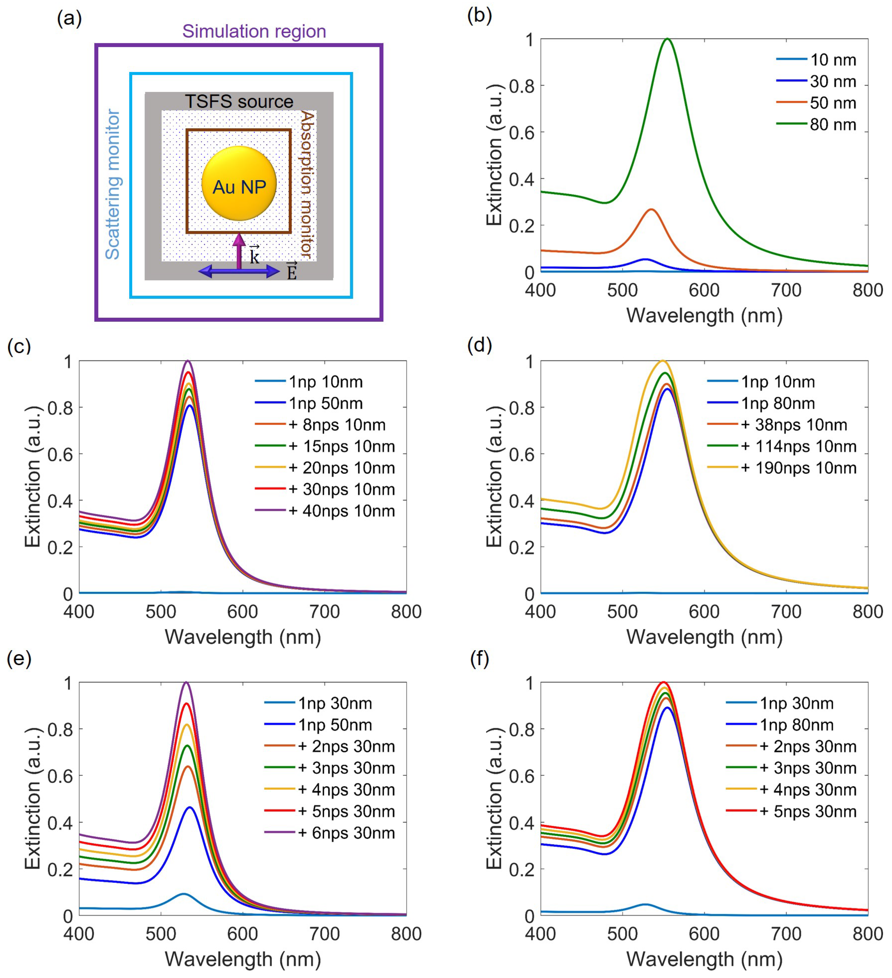
The top view of the FDTD model is depicted in Figure 2a. Since the Au NPs are sphere-like, the simulation shapes are selected as spheres, with the particle parameters obtained from the optical material database of [45] for Au. We note that the Au NPs are dispersed in a water solution. Therefore, we established that the background refractive index of the simulation is 1.33. The excitation light source is a total-field scattered-field (TFSF) with a wavelength range between 400 nm and 800 nm to match the operating bandwidth of the UV-Vis spectroscopy. After performing the convergence test, a mesh size of 0.3 nm was selected. The boundary conditions are perfectly matched layers, enabling outgoing waves from inside the computational region to be absorbed without reflection. Absorption and scattering were calculated using an analysis group, while extinction was determined by summing absorption and scattering. In simulations, we computed the extinction of individual NPs with different sizes and calculated their average to represent the extinction of the mixed solution, without affecting the physical meaning.
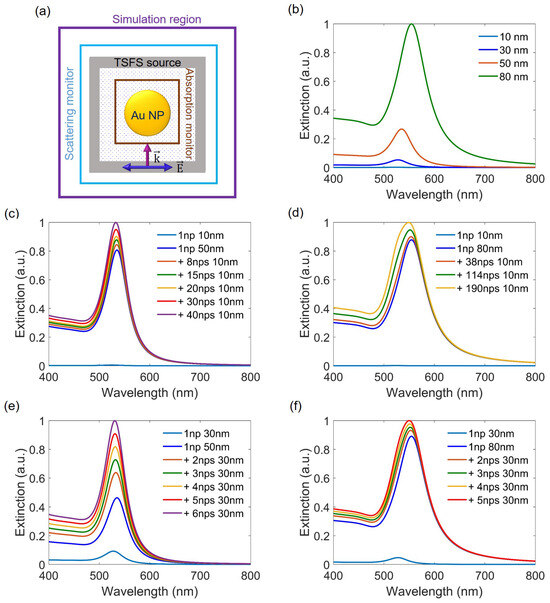
Figure 2.
(a) Structural model of the FDTD simulation method used to calculate the LSPR of the Au NPs. (b) Extinction spectra of a single Au NP as the size increases from 10 nm to 80 nm. (c–f) Extinction spectra of a mixture of small NPs and large NPs with different number ratios: (c) 10 nm with 50 nm, (d) 10 nm with 80 nm, (e) 30 nm with 50 nm, and (f) 30 nm with 80 nm, respectively.
3.2. Simulation Results
3.2.1. Optical Properties of Spherical Au NPs
Figure 2b shows the extinction spectra of spherical Au NPs when the sizes increase from 10 nm to 80 nm. It is obvious that the resonant peaks increase and shift to longer wavelengths with increasing sizes of NPs because of their greater absorption and scattering capacity. Subsequently, we calculated the extinction of mixed NPs by averaging the extinction sums of large NPs with small NPs (the number of small and large NPs is based on experimental data). Figure 2c illustrates that even when the number of 10 nm Au NPs is several dozen times greater than that of 50 nm Au NPs, the extinction of 50 nm Au NPs still dominates. This aligns with the experimental results, revealing that the influence of large NPs is significant and cannot be neglected in samples containing both small and large NPs, even when their number is minimal. Therefore, the determination of Au NP size based on the position/intensity of the SPR peaks must be performed with great care. By adding more small NPs (thirty 10 nm Au NPs), the peak becomes higher and experiences a blueshift, indicating that the contribution of the small particles becomes evident. Similar studies were conducted to calculate the extinction spectra for the mixed Au NPs of 10 nm with 80 nm (Figure 2d), 30 nm with 50 nm (Figure 2e), and 30 nm with 80 nm (Figure 2f). In all cases, we obtained the same trend indicating that the contribution of big Au NPs is much more significant compared to the small ones.
3.2.2. Optical Properties of Spherical Au NPs Mixed with Ellipsoidal Au NPs
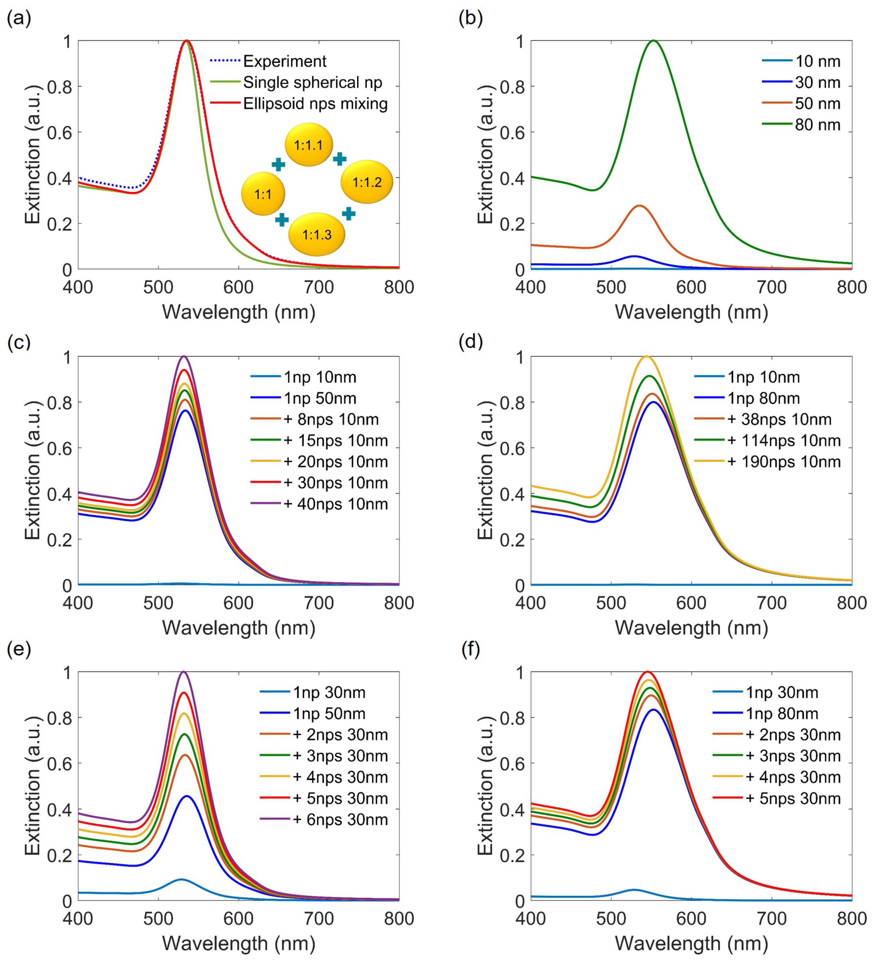
In the previous sections, the large Au NPs were demonstrated as the main contributors to the extinction peaks when mixed with a high number of small particles. The simulation was also performed to verify the experiment results. However, it is observed that the simulated curve is narrower than the experimental ones. To solve this problem, we proposed calculating mixtures of NPs with different shapes and averaging the total extinction. In fact, the shape of Au NPs is not perfectly spherical in reality, and the deformation of the NPs can lead to the broadening of the peak [46]. In a simple case, we assumed the deformed shape of NPs as ellipsoids. Figure 3a shows that the extinction of 50 nm spherical Au NPs in the simulation case is narrower compared to the experiment. However, when we consider the contribution of ellipsoidal NPs, the simulation peaks become broader and almost match the experimental ones. Indeed, ellipsoidal NPs exhibit two distinct modes (horizontal and vertical modes) corresponding to two resonant peaks. Nonetheless, as the aspect ratio is small (shape of spherical NPs deforms slightly), the two modes overlap, resulting in a broader single peak. This phenomenon explains why, when we mix spherical NPs with ellipsoidal NPs, the average extinction peaks become wider and closely approach the experimental results. Here, we mixed one spherical Au NP with ellipsoidal NPs having minor-to-major axis ratios ranging from 1:1.1 to 1:1.3, along with a coefficient distribution of 3:4:2. We note that these selected coefficients are already normalized to the number of spherical Au NPs. By averaging the linear combination of all the Au NP components, the extinction of the mixed sample was calculated, resulting in a close alignment with the experimental findings. It is worth noting that we selected this combination as an example to demonstrate the contribution of deformed particles to resonant peaks, and the coefficient distribution could be chosen and combined in various ways. By repeating calculations with other sizes of NPs, we can obtain the optimal absorption resonant peaks for NPs of 10 nm, 30 nm, and 80 nm. Figure 3b–f illustrate the extinction spectra of Au NPs with various sizes, followed by mixing small NPs (10 nm, 30 nm) with large NPs (50 nm, 80 nm). The theoretical results obtained now agree well with the experimental ones (see Figure 1).
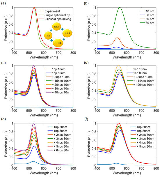
Figure 3.
(a) Comparison of extinction spectra obtained with Au NP of 50 nm: experiment, simulation with only spherical Au NP, and simulation incorporating a mixture of spherical and ellipsoidal Au NPs with minor-to-major axis ratios ranging from 1:1.1 to 1:1.3. (b) Average extinction spectra of a mixture of spherical Au NPs and ellipsoidal Au NPs, as the size of the minor axis increases from 10 nm to 80 nm. Repeated calculations of average extinction spectra of the mixture between spherical and non-spherical NPs: (c) 10 nm with 50 nm, (d) 10 nm with 80 nm, (e) 30 nm with 50 nm, and (f) 30 nm with 80 nm, respectively.
4. Numerical Investigation of NEF Enhancement of Single Spherical Au NP
As mentioned earlier, when considering metal NPs, the near-field enhancement at the nanoscale has attracted considerable interest. In this section, we will investigate the near-field region of Au NPs and determine their optimal size and wavelength to maximize field intensity.
4.1. Local Electric Field at Resonant Wavelength
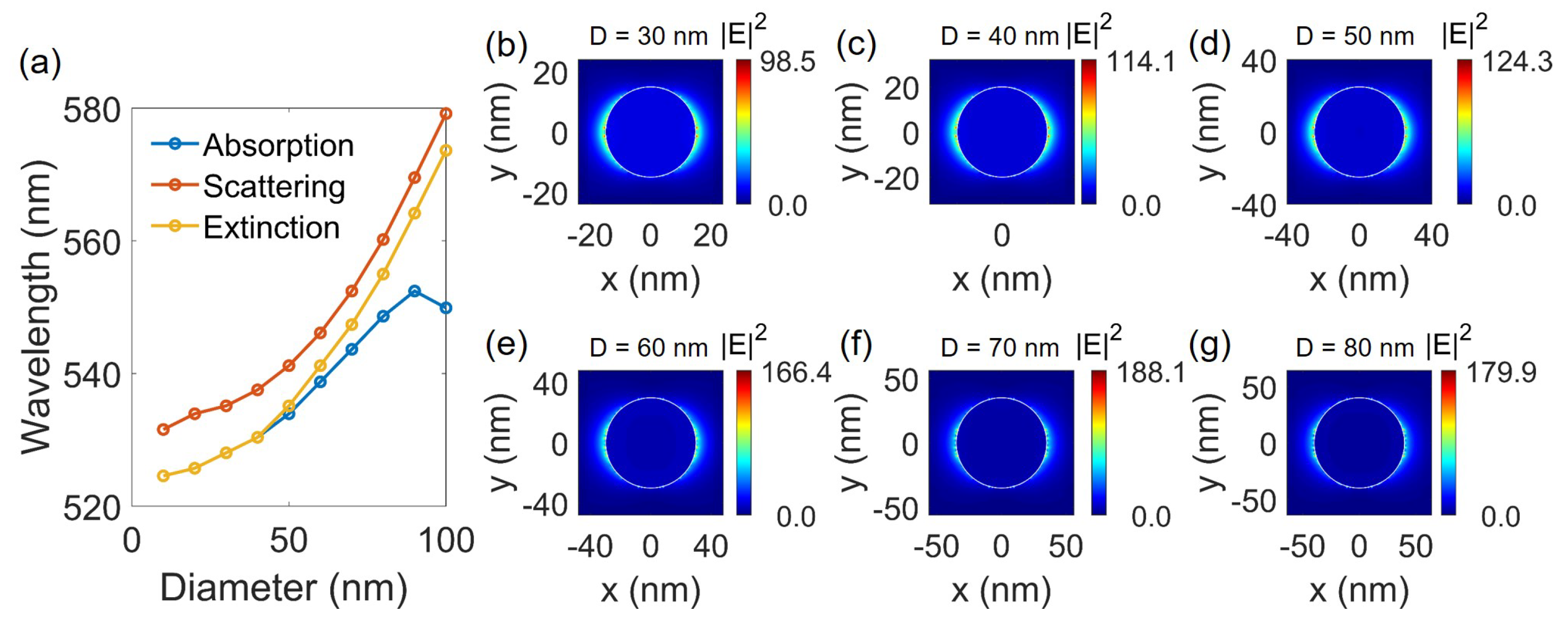
Figure 4a shows the wavelength at which absorption, scattering, and extinction reach their maximum values as the sizes of Au NPs increase from 10 nm to 100 nm. With the increment in size, a general trend is observed in shifting towards longer wavelengths. When the size of the Au NP is smaller than 50 nm, the absorption and extinction wavelengths overlap, contrasting with the divergence when the NP size surpasses 50 nm, resulting in significant differences, particularly when the size reaches 100 nm. This is because at small sizes (<50 nm), nonradiative absorption dominates due to electron collisions with the NP surface, while for larger sizes, radiative scattering prevails, with its damping rate increasing as NP sizes increase [47]. Therefore, the SPR wavelength is significantly influenced by the absorption of small Au NPs, while for larger NPs, the dominating effect is scattering.
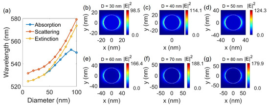
Figure 4.
(a) Absorption, scattering, and extinction wavelength as a function of NP sizes. (b–g) NEF intensity at the resonant wavelength when the sizes of Au NPs increase from 30 nm to 80 nm.
Figure 4b–g illustrate examples of the spatial distribution of the NEF intensity in the ()-plane for spherical NPs with sizes ranging from 30 nm to 80 nm, excited by the peak extinction wavelength, i.e., LSPR wavelength. Since the NEF corresponds to the dipole mode at the NP surface aligned parallel to the x-axis (perpendicular to the excitation direction), the unique p-orbital shape surrounding the sphere is observed. When the size of the NPs increases from 30 nm to 70 nm, the value of electric field intensity also increases, followed by a small decrease as the NP size reaches 80 nm. The initial increase in the near-field is attributed to the higher light absorption by bigger NPs [48], reaching a maximum for NPs with a diameter of 70 nm. The subsequent decrease in the near-field for larger NPs is due to their enhanced light scattering capabilities. While a substantial fraction of the light absorbed by small metal NPs is converted into an evanescent and non-propagating NEF [49], a significant portion of the absorbed light in larger NPs is reradiated to the far-field region as scattered (propagating) light. Along a distance r of a few nanometers above the surface, the electric field decays rapidly at a rate proportional to [50]. We note that similar results have been observed [37] when considering Au NPs in a polymer material (n = 1.485). In such high-refractive-index media, Au NPs have an optimum size of 50 nm for a highest NEF intensity. Furthermore, in our work, when considering the Au NPs in air medium (n = 1), the Au NP size providing maximum NEF intensity is about 110 nm. This allows us to confirm that the optimum NP size depends on the medium in which the Au NPs are immersed. A lower-refractive-index medium requires larger NPs. This information is very important for applications using LSPR-based field enhancement.
4.2. Maximal Local Electric Field at All Wavelengths
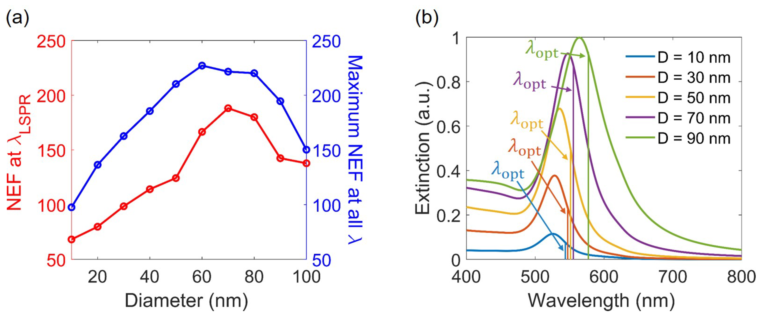
Furthermore, we compared the NEF intensity at the LSPR wavelength with the NEF intensity across all wavelengths from 400 nm to 800 nm, as illustrated in Figure 5a. Interestingly, we realized that the NEF intensity at the LSPR wavelength is not maximum. Indeed, for all investigated Au NP sizes, the maximum near-field intensity is located at a wavelength longer than the LSPR. This phenomenon was explained by the physics of a driven and damped harmonic oscillator [35]. As can be seen, a similar trend is witnessed for both cases of near-field at resonance wavelength and at all wavelengths, where the intensity increases to reach a maximum at the optimal NP size (70 nm for LSPR wavelength and 60 nm for all wavelengths) and then decreases. Although the same FDTD method has been used to investigate the plasmonic NEF of Au NPs [51], it does not provide a complete understanding of the dependence of the NEF enhancement on the particles’ size or the optimum wavelength at which the field is the best. This discrepancy is understandable because, in the previous work, the authors only simulated the NEF intensity of Au NPs with sizes ranging from 10 nm to 60 nm, and for only the wavelength corresponding to the LSPR. Moreover, it was suggested in another work [37] that the size dependence of NEF intensity originates from various factors: small NPs show weak fields due to surface damping; radiative damping results in lower field enhancements in larger NPs; and importantly, there is a dual role of electromagnetic retardation.
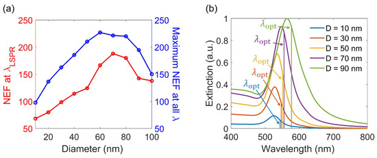
Figure 5.
(a) NEF intensity at the LSPR wavelength and at the optimum wavelength as a function of Au NP size. (b) Extinction spectra and corresponding to the maximum near-field of Au NPs ranging in size from 10 nm to 90 nm. The surrounding medium is water with a refractive index of 1.33.
We also note that the optimal size of Au NPs in water could be slightly different from 60 nm since we did not finely simulate all NP sizes near 60 nm, but this observation is enough to confirm that the best NEF intensity should be obtained at another wavelength, far from the LSPR peak wavelength. Figure 5b displays the LSPR wavelength, where the extinction is maximum, and the optimal wavelength, where maximum NEF intensity is achieved, plotted as a function of NP size spanning from 10 nm to 90 nm. It is clear that while the optimal wavelength is located within the resonance region, it consistently exhibits a redshift compared to the LSPR wavelength for all cases of Au NPs. Certainly, the optimal wavelength follows the same trend as the LSPR wavelength, shifting towards longer wavelengths as the size of the Au NPs increases.
Table 1 provides detailed information on the resonant wavelength, the optimal wavelength for maximal NEF, and the field enhancement factor as a function of NP size. To achieve the highest NEF, the LSPR peak needs to shift to redder wavelengths. When choosing an optimal wavelength, the electric field intensity can be enhanced from 1.09 times (100 nm) to 1.71 times (20 nm), as compared with that obtained at the LSPR wavelength. From this table, we can extract crucial information regarding maximal NEF intensity. When focusing solely on the LSPR wavelength, the optimal choice is 70 nm, with an excitation wavelength of 547.4 nm. However, if we consider all wavelengths ranging from 400 nm to 800 nm, the preferable option shifts to the 60 nm Au NP in water, corresponding to an excitation wavelength of 553.7 nm.

Table 1.
Resonant wavelength and corresponding light field intensity, optimum wavelength and corresponding light field intensity, and field enhancement factor (light intensity obtained at optimum wavelength over light intensity obtained at LSPR peak), as a function of the size of Au NPs in water.
5. Conclusions
In this work, we investigated the optical properties of colloidal polydisperse solutions using UV-vis spectroscopy. It is observed that detecting small NPs in the presence of several percent of large NPs, mixed together as polydisperse colloids, is indeed very challenging. We also conducted simulations to validate the experimental results. The agreement between the experiment and simulation reveals that the influence of large Au NPs is strong, and they can dominate the entire sample and obscure the effects of small NPs. Therefore, caution is required when drawing a conclusion on the size of Au NPs based on the absorption spectra of the polydisperse solution. Additionally, to match the width of the experimental curves with the simulation results, we proposed a simple model that mixes spherical NPs with ellipsoidal NPs, resulting in a broadened absorption curve that agrees well with the experiment. Moreover, we systematically studied the enhancement of the NEF around Au NPs. Interestingly, while the electric field is highly intensified at the SPR wavelength, the maximal electric field was observed to occur at a longer excitation wavelength. According to the simulation results, the optimal size and excited wavelength of Au NPs dispersed in water for the highest NEF intensity are 60 nm and 553.7 nm, respectively. By selecting the most optimized plasmonic NPs for near-field enhancement, numerous applications could be further advanced, particularly in non-linear optics, fluorescence enhancement, Raman spectroscopy, photocatalysis, and laser technologies. Indeed, by choosing a suitable nanoparticle size with an optimized excitation wavelength, we can achieve the highest NEF intensity, leading to the best improvement in the light–matter interaction, and thus the highest emitted signal. For example, considering the second-harmonic generation of Au NPs, which is quadratically proportional to the NEF intensity of Au NPs, a well-chosen wavelength allows the second-harmonic signal to be increased by a factor of 3–4, as compared to the case of choosing a wavelength located at LSPR.
Author Contributions
N.D.L. supervised the project. Q.T.P., G.L.N., C.T.N., I.L.-R. and N.D.L. participated in discussions and idea development. Q.T.P. performed the experiment, simulation, and data analysis. G.L.N. performed simulation. Q.T.P. drafted the initial version of the manuscript. Q.T.P., G.L.N., C.T.N., I.L.-R. and N.D.L. contributed to the manuscript revisions. All authors have read and agreed to the published version of the manuscript.
Funding
This research received no external funding.
Institutional Review Board Statement
Not applicable.
Informed Consent Statement
Not applicable.
Data Availability Statement
Data are contained within the article.
Acknowledgments
The authors acknowledge the facility of the Institut D’Alembert, and the financial support of the French Embassy in Egypt in the frame of the Imhotep project.
Conflicts of Interest
The authors declare no conflicts of interest.
References
- Schmid, G. Nanoparticles: From Theory to Application; John Wiley & Sons: Hoboken, NJ, USA, 2011. [Google Scholar]
- Schmid, G.; Corain, B. Nanoparticulated gold: Syntheses, structures, electronics, and reactivities. Eur. J. Inorg. Chem. 2003, 2003, 3081–3098. [Google Scholar] [CrossRef]
- Abdelhalim, M.A.K.; Mady, M.M.; Ghannam, M.M. Physical properties of different gold nanoparticles: Ultraviolet-visible and fluorescence measurements. J. Nanomed. Nanotechnol. 2012, 3, 178–194. [Google Scholar] [CrossRef]
- Rodrigues, T.S.; da Silva, A.G.; Camargo, P.H. Nanocatalysis by noble metal nanoparticles: Controlled synthesis for the optimization and understanding of activities. J. Mater. Chem. A 2019, 7, 5857–5874. [Google Scholar] [CrossRef]
- Mahmoud, M.A.; O’Neil, D.; El-Sayed, M.A. Hollow and solid metallic nanoparticles in sensing and in nanocatalysis. Chem. Mater. 2014, 26, 44–58. [Google Scholar] [CrossRef]
- Astruc, D. Introduction: Nanoparticles in catalysis. Chem. Rev 2020, 120, 461–463. [Google Scholar] [CrossRef]
- Mubeen, S.; Lee, J.; Lee, W.r.; Singh, N.; Stucky, G.D.; Moskovits, M. On the plasmonic photovoltaic. ACS Nano 2014, 8, 6066–6073. [Google Scholar] [CrossRef] [PubMed]
- Luo, Q.; Zhang, C.; Deng, X.; Zhu, H.; Li, Z.; Wang, Z.; Chen, X.; Huang, S. Plasmonic effects of metallic nanoparticles on enhancing performance of perovskite solar cells. ACS Appl. Mater. Interfaces 2017, 9, 34821–34832. [Google Scholar] [CrossRef] [PubMed]
- Huang, X.; Jain, P.K.; El-Sayed, I.H.; El-Sayed, M.A. Plasmonic photothermal therapy (PPTT) using gold nanoparticles. Lasers Med. Sci. 2008, 23, 217–228. [Google Scholar] [CrossRef]
- Doria, G.; Conde, J.; Veigas, B.; Giestas, L.; Almeida, C.; Assunção, M.; Rosa, J.; Baptista, P.V. Noble metal nanoparticles for biosensing applications. Sensors 2012, 12, 1657–1687. [Google Scholar] [CrossRef]
- Wang, J. Electrochemical biosensing based on noble metal nanoparticles. Microchim. Acta 2012, 177, 245–270. [Google Scholar] [CrossRef]
- Chandrakala, V.; Aruna, V.; Angajala, G. Review on metal nanoparticles as nanocarriers: Current challenges and perspectives in drug delivery systems. Emergent Mater. 2022, 5, 1593–1615. [Google Scholar] [CrossRef]
- Desai, N.; Momin, M.; Khan, T.; Gharat, S.; Ningthoujam, R.S.; Omri, A. Metallic nanoparticles as drug delivery system for the treatment of cancer. Expert Opin. Drug Deliv. 2021, 18, 1261–1290. [Google Scholar] [CrossRef] [PubMed]
- Enrichi, F.; Quandt, A.; Righini, G.C. Plasmonic enhanced solar cells: Summary of possible strategies and recent results. Renew. Sustain. Energy Rev. 2018, 82, 2433–2439. [Google Scholar] [CrossRef]
- Chatterjee, D.K.; Diagaradjane, P.; Krishnan, S. Nanoparticle-mediated hyperthermia in cancer therapy. Ther. Deliv. 2011, 2, 1001–1014. [Google Scholar] [CrossRef]
- Yaraki, M.T.; Tan, Y.N. Metal nanoparticles-enhanced biosensors: Synthesis, design and applications in fluorescence enhancement and surface-enhanced Raman scattering. Chem. Asian J. 2020, 15, 3180–3208. [Google Scholar] [CrossRef]
- Bek, A.; Jansen, R.; Ringler, M.; Mayilo, S.; Klar, T.A.; Feldmann, J. Fluorescence enhancement in hot spots of AFM-designed gold nanoparticle sandwiches. Nano Lett. 2008, 8, 485–490. [Google Scholar] [CrossRef]
- Wang, Z.; Meng, X.; Kildishev, A.V.; Boltasseva, A.; Shalaev, V.M. Nanolasers enabled by metallic nanoparticles: From spasers to random lasers. Laser Photonics Rev. 2017, 11, 1700212. [Google Scholar] [CrossRef]
- Long, L.; He, D.; Bao, W.; Feng, M.; Zhang, P.; Zhang, D.; Chen, S. Localized surface plasmon resonance improved lasing performance of Ag nanoparticles/organic dye random laser. J. Alloys Compd. 2017, 693, 876–881. [Google Scholar] [CrossRef]
- Grand, J.; Auguié, B.; Le Ru, E. Combined extinction and absorption UV–vis spectroscopy reveals shape imperfections of metallic nanoparticles. Anal. Chem. 2019, 91, 14639–14648. [Google Scholar] [CrossRef]
- Amendola, V.; Meneghetti, M. Size evaluation of gold nanoparticles by UV- vis spectroscopy. J. Phys. Chem. C 2009, 113, 4277–4285. [Google Scholar] [CrossRef]
- Gaikwad, A.V.; Verschuren, P.; Eiser, E.; Rothenberg, G. A simple method for measuring the size of metal nanoclusters in solution. J. Phys. Chem. B 2006, 110, 17437–17443. [Google Scholar] [CrossRef] [PubMed]
- Njoki, P.N.; Lim, I.I.S.; Mott, D.; Park, H.Y.; Khan, B.; Mishra, S.; Sujakumar, R.; Luo, J.; Zhong, C.J. Size correlation of optical and spectroscopic properties for gold nanoparticles. J. Phys. Chem. C 2007, 111, 14664–14669. [Google Scholar] [CrossRef]
- Tomaszewska, E.; Soliwoda, K.; Kadziola, K.; Tkacz-Szczesna, B.; Celichowski, G.; Cichomski, M.; Szmaja, W.; Grobelny, J. Detection limits of DLS and UV-Vis spectroscopy in characterization of polydisperse nanoparticles colloids. J. Nanomater. 2013, 2013, 313081. [Google Scholar] [CrossRef]
- Michaels, A.M.; Jiang, n.; Brus, L. Ag nanocrystal junctions as the site for surface-enhanced Raman scattering of single rhodamine 6G molecules. J. Phys. Chem. B 2000, 104, 11965–11971. [Google Scholar] [CrossRef]
- Schatz, G.C.; Young, M.A.; Van Duyne, R.P. Surface-Enhanced Raman Scattering: Physics and Applications; Springer: Berlin, Germany, 2006; Volume 103. [Google Scholar]
- Liu, Z.; Hou, W.; Pavaskar, P.; Aykol, M.; Cronin, S.B. Plasmon resonant enhancement of photocatalytic water splitting under visible illumination. Nano Lett. 2011, 11, 1111–1116. [Google Scholar] [CrossRef] [PubMed]
- Butet, J.; Brevet, P.F.; Martin, O.J. Optical second harmonic generation in plasmonic nanostructures: From fundamental principles to advanced applications. ACS Nano 2015, 9, 10545–10562. [Google Scholar] [CrossRef] [PubMed]
- Liu, T.M.; Tai, S.P.; Yu, C.H.; Wen, Y.C.; Chu, S.W.; Chen, L.J.; Prasad, M.R.; Lin, K.J.; Sun, C.K. Measuring plasmon-resonance enhanced third-harmonic χ (3) of Ag nanoparticles. Appl. Phys. Lett. 2006, 89, 043122. [Google Scholar] [CrossRef]
- Ma, W.; Yao, J.; Yang, H.; Liu, J.; Li, F.; Hilton, J.; Lin, Q. Effects of vertex truncation of polyhedral nanostructures on localized surface plasmon resonance. Opt. Express 2009, 17, 14967–14976. [Google Scholar] [CrossRef] [PubMed]
- Khlebtsov, B.N.; Burov, A.M.; Zarkov, S.V.; Khlebtsov, N.G. Surface-enhanced Raman scattering from Au nanorods, nanotriangles, and nanostars with tuned plasmon resonances. Phys. Chem. Chem. Phys. 2023, 25, 30903–30913. [Google Scholar] [CrossRef]
- Slablab, A.; Le Xuan, L.; Zielinski, M.; De Wilde, Y.; Jacques, V.; Chauvat, D.; Roch, J.F. Second-harmonic generation from coupled plasmon modes in a single dimer of gold nanospheres. Opt. Express 2012, 20, 220–227. [Google Scholar] [CrossRef]
- Huang, Y.; Ma, L.; Hou, M.; Li, J.; Xie, Z.; Zhang, Z. Hybridized plasmon modes and near-field enhancement of metallic nanoparticle-dimer on a mirror. Sci. Rep. 2016, 6, 30011. [Google Scholar] [CrossRef]
- Messinger, B.J.; Von Raben, K.U.; Chang, R.K.; Barber, P.W. Local fields at the surface of noble-metal microspheres. Phys. Rev. B 1981, 24, 649. [Google Scholar] [CrossRef]
- Zuloaga, J.; Nordlander, P. On the energy shift between near-field and far-field peak intensities in localized plasmon systems. Nano Lett. 2011, 11, 1280–1283. [Google Scholar] [CrossRef]
- Hong, S.; Li, X. Optimal size of gold nanoparticles for surface-enhanced Raman spectroscopy under different conditions. J. Nanomater. 2013, 2013, 790323. [Google Scholar] [CrossRef]
- Deeb, C.; Zhou, X.; Plain, J.; Wiederrecht, G.P.; Bachelot, R.; Russell, M.; Jain, P.K. Size dependence of the plasmonic near-field measured via single-nanoparticle photoimaging. J. Phys. Chem. C 2013, 117, 10669–10676. [Google Scholar] [CrossRef]
- Mansour, Y.; Battie, Y.; En Naciri, A.; Chaoui, N. Determination of the size distribution of metallic colloids from extinction spectroscopy. Nanomaterials 2021, 11, 2872. [Google Scholar] [CrossRef]
- Klinavičius, T.; Khinevich, N.; Tamulevičienė, A.; Vidal, L.; Tamulevičius, S.; Tamulevičius, T. Deep Learning Methods for Colloidal Silver Nanoparticle Concentration and Size Distribution Determination from UV–Vis Extinction Spectra. J. Phys. Chem. C 2024, 128, 9662–9675. [Google Scholar] [CrossRef]
- Liang, C.C.; Liao, M.Y.; Chen, W.Y.; Cheng, T.C.; Chang, W.H.; Lin, C.H. Plasmonic metallic nanostructures by direct nanoimprinting of gold nanoparticles. Opt. Express 2011, 19, 4768–4776. [Google Scholar] [CrossRef]
- Zhao, J.; Pinchuk, A.O.; McMahon, J.M.; Li, S.; Ausman, L.K.; Atkinson, A.L.; Schatz, G.C. Methods for describing the electromagnetic properties of silver and gold nanoparticles. Acc. Chem. Res. 2008, 41, 1710–1720. [Google Scholar] [CrossRef] [PubMed]
- Iqbal, M.; Usanase, G.; Oulmi, K.; Aberkane, F.; Bendaikha, T.; Fessi, H.; Zine, N.; Agusti, G.; Errachid, E.S.; Elaissari, A. Preparation of gold nanoparticles and determination of their particles size via different methods. Mater. Res. Bull. 2016, 79, 97–104. [Google Scholar] [CrossRef]
- Fu, Q.; Sun, W. Mie theory for light scattering by a spherical particle in an absorbing medium. Appl. Opt. 2001, 40, 1354–1361. [Google Scholar] [CrossRef] [PubMed]
- Wrigglesworth, E.G.; Johnston, J.H. Mie theory and the dichroic effect for spherical gold nanoparticles: An experimental approach. Nanoscale Adv. 2021, 3, 3530–3536. [Google Scholar] [CrossRef] [PubMed]
- Haynes, W.M. CRC Handbook of Chemistry and Physics; CRC press: Boca Raton, FL, USA, 2014. [Google Scholar]
- Kim, D.K.; Hwang, Y.J.; Yoon, C.; Yoon, H.O.; Chang, K.S.; Lee, G.; Lee, S.; Yi, G.R. Experimental approach to the fundamental limit of the extinction coefficients of ultra-smooth and highly spherical gold nanoparticles. Phys. Chem. Chem. Phys. 2015, 17, 20786–20794. [Google Scholar] [CrossRef] [PubMed]
- Shafiqa, A.; Abdul Aziz, A.; Mehrdel, B. Nanoparticle optical properties: Size dependence of a single gold spherical nanoparticle. J. Phys. Conf. Ser. 2018, 1083, 012040. [Google Scholar] [CrossRef]
- Montaño-Priede, J.L.; Pal, U. Estimating near electric field of polyhedral gold nanoparticles for plasmon-enhanced spectroscopies. J. Phys. Chem. C 2019, 123, 11833–11839. [Google Scholar] [CrossRef]
- Choi, K.W.; Zhong, X.L.; Li, Z.Y.; Im, S.H.; Park, O.O. Robust synthesis of gold rhombic dodecahedra with well-controlled sizes and their optical properties. CrystEngComm 2013, 15, 252–258. [Google Scholar] [CrossRef]
- Kelly, K.L.; Coronado, E.; Zhao, L.L.; Schatz, G.C. The optical properties of metal nanoparticles: The influence of size, shape, and dielectric environment. J. Phys. Chem. B 2003, 107, 668–677. [Google Scholar] [CrossRef]
- Cheng, L.; Zhu, G.; Liu, G.; Zhu, L. FDTD simulation of the optical properties for gold nanoparticles. Mater. Res. Express 2020, 7, 125009. [Google Scholar] [CrossRef]
Disclaimer/Publisher’s Note: The statements, opinions and data contained in all publications are solely those of the individual author(s) and contributor(s) and not of MDPI and/or the editor(s). MDPI and/or the editor(s) disclaim responsibility for any injury to people or property resulting from any ideas, methods, instructions or products referred to in the content. |
© 2024 by the authors. Licensee MDPI, Basel, Switzerland. This article is an open access article distributed under the terms and conditions of the Creative Commons Attribution (CC BY) license (https://creativecommons.org/licenses/by/4.0/).

