Real-Time Resolution Enhancement of Confocal Laser Scanning Microscopy via Deep Learning

 ,
,
Abstract
1. Introduction
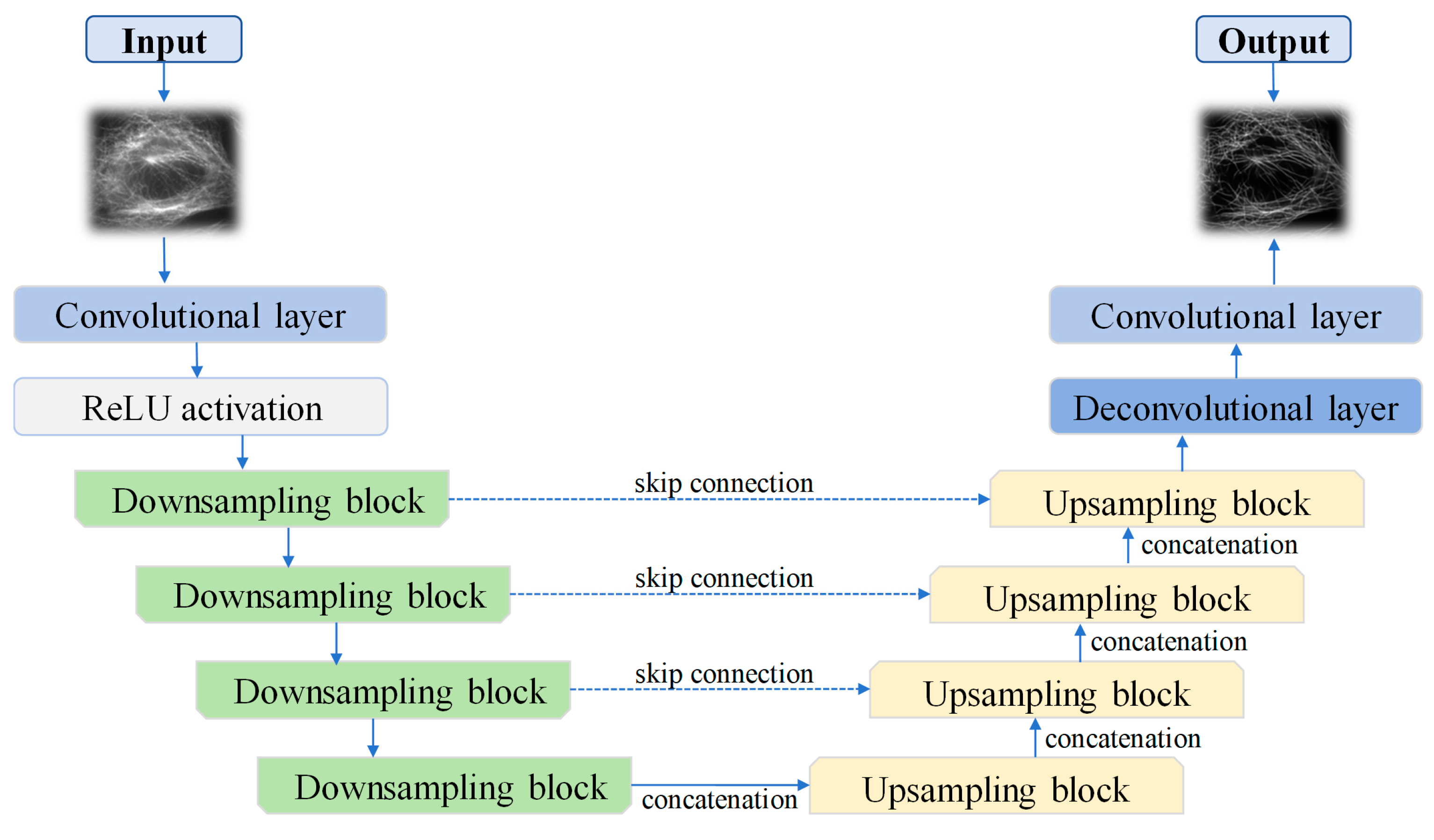
2. Materials and Methods
3. Results and Discussion
4. Conclusions
Supplementary Materials
Author Contributions
Funding
Data Availability Statement
Conflicts of Interest
References
- Heintzmann, R.; Sarafis, V.; Munroe, P.; Nailon, J.; Hanley, Q.S.; Jovin, T.M. Resolution enhancement by subtraction of confocal signals taken at different pinhole sizes. Micron 2003, 34, 293–300. [Google Scholar] [CrossRef] [PubMed]
- Di Franco, E.; Costantino, A.; Cerutti, E.; D’Amico, M.; Privitera, A.P.; Bianchini, P.; Vicidomini, G.; Gulisano, M.; Diaspro, A.; Lanzanò, L. SPLIT-PIN software enabling confocal and super-resolution imaging with a virtually closed pinhole. Sci. Rep. 2023, 13, 2741. [Google Scholar] [CrossRef] [PubMed]
- Kuang, C.; Li, S.; Liu, W.; Hao, X.; Gu, Z.; Wang, Y.; Ge, J.; Li, H.; Liu, X. Breaking the diffraction barrier using fluorescence emission difference microscopy. Sci. Rep. 2013, 3, 1441. [Google Scholar] [CrossRef] [PubMed]
- Dong, W.; Huang, Y.; Zhang, Z.; Xu, L.; Kuang, C.; Hao, X.; Cao, L.; Liu, X. Fluorescence emission difference microscopy based on polarization modulation. J. Innov. Opt. Health Sci. 2022, 15, 2250034. [Google Scholar] [CrossRef]
- Tanaami, T.; Sugiyama, Y.; Kosugi, Y.; Mikuriya, K.; Abe, M. High-speed confocal fluorescence microscopy using a nipkow scanner with microlenses for 3-d imaging of single fluorescent molecule in real time. Bioimages 1996, 4, 57–62. [Google Scholar]
- Hayashi, S.; Okada, Y. Ultrafast superresolution fluorescence imaging with spinning disk confocal microscope optics. Mol. Biol. Cell 2015, 26, 1743–1751. [Google Scholar] [CrossRef]
- Hayashi, S. Resolution doubling using confocal microscopy via analogy with structured illumination microscopy. Jpn. J. Appl. Phys. 2016, 55, 082501. [Google Scholar] [CrossRef]
- Huff, J. The Airyscan detector from ZEISS: Confocal imaging with improved signal-to-noise ratio and super-resolution. Nat. Methods 2015, 12, i–ii. [Google Scholar] [CrossRef]
- Huff, J.; Bergter, A.; Birkenbeil, J.; Kleppe, I.; Engelmann, R.; Krzic, U. The new 2D Superresolution mode for ZEISS Airyscan. Nat. Methods 2017, 14, 1223. [Google Scholar] [CrossRef]
- Dey, N.; Blanc-Feraud, L.; Zimmer, C.; Roux, P.; Kam, Z.; Olivo-Marin, J.-C.; Zerubia, J. Richardson–Lucy algorithm with total variation regularization for 3D confocal microscope deconvolution. Microsc. Res. Tech. 2006, 69, 260–266. [Google Scholar] [CrossRef]
- Dupé, F.X.; Fadili, M.J.; Starck, J.L. Deconvolution of confocal microscopy images using proximal iteration and sparse representations. In Proceedings of the 2008 5th IEEE International Symposium on Biomedical Imaging: From Nano to Macro, Paris, France, 14–17 May 2008; IEEE: Piscataway, NJ, USA, 2008; pp. 736–739. [Google Scholar]
- He, T.; Sun, Y.; Qi, J.; Hu, J.; Huang, H. Image deconvolution for confocal laser scanning microscopy using constrained total variation with a gradient field. Appl. Opt. 2019, 58, 3754–3766. [Google Scholar] [CrossRef] [PubMed]
- Stockbridge, C.; Lu, Y.; Moore, J.; Hoffman, S.; Paxman, R.; Toussaint, K.; Bifano, T. Focusing through dynamic scattering media. Opt. Express 2012, 20, 15086–15092. [Google Scholar] [CrossRef] [PubMed]
- Galaktionov, I.; Nikitin, A.; Sheldakova, J.; Toporovsky, V.; Kudryashov, A. Focusing of a laser beam passed through a moderately scattering medium using phase-only spatial light modulator. Photonics 2022, 9, 296. [Google Scholar] [CrossRef]
- Katz, O.; Small, E.; Guan, Y.; Silberberg, Y. Noninvasive nonlinear focusing and imaging through strongly scattering turbid layers. Optica 2014, 1, 170–174. [Google Scholar] [CrossRef]
- Hillman, T.R.; Yamauchi, T.; Choi, W.; Dasari, R.R.; Feld, M.S.; Park, Y.; Yaqoob, Z. Digital optical phase conjugation for delivering two-dimensional images through turbid media. Sci. Rep. 2013, 3, 1909. [Google Scholar] [CrossRef] [PubMed]
- Tao, X.; Fernandez, B.; Azucena, O.; Fu, M.; Garcia, D.; Zuo, Y.; Chen, D.C.; Kubby, J. Adaptive optics confocal microscopy using direct wavefront sensing. Opt. Lett. 2011, 36, 1062–1064. [Google Scholar] [CrossRef]
- Weigert, M.; Schmidt, U.; Boothe, T.; Müller, A.; Dibrov, A.; Jain, A.; Wilhelm, B.; Schmidt, D.; Broaddus, C.; Culley, S.; et al. Content-aware image restoration: Pushing the limits of fluorescence microscopy. Nat. Methods 2018, 15, 1090–1097. [Google Scholar] [CrossRef]
- Li, X.; Dong, J.; Li, B.; Zhang, Y.; Zhang, Y.; Veeraraghavan, A.; Ji, X. Fast confocal microscopy imaging based on deep learning. In Proceedings of the 2020 IEEE International Conference on Computational Photography (ICCP), St. Louis, MO, USA, 24–26 April 2020; IEEE: Piscataway, NJ, USA, 2020; pp. 1–12. [Google Scholar]
- Wang, W.; Wu, B.; Zhang, B.; Ma, J.; Tan, J. Deep learning enables confocal laser-scanning microscopy with enhanced resolution. Opt. Lett. 2021, 46, 4932–4935. [Google Scholar] [CrossRef]
- Fang, L.; Monroe, F.; Novak, S.W.; Kirk, L.; Schiavon, C.R.; Yu, S.B.; Zhang, T.; Wu, M.; Kastner, K.; Latif, A.A.; et al. Deep learning-based point-scanning super-resolution imaging. Nat. Methods 2021, 18, 406–416. [Google Scholar] [CrossRef]
- Huang, B.; Li, J.; Yao, B.; Yang, Z.; Lam, E.Y.; Zhang, J.; Yan, W.; Qu, J. Enhancing image resolution of confocal fluorescence microscopy with deep learning. PhotoniX 2023, 4, 2. [Google Scholar] [CrossRef]
- Ji, C.; Zhu, Y.; He, E.; Liu, Q.; Zhou, D.; Xie, S.; Wu, H.; Zhang, J.; Du, K.; Chen, Y.; et al. Full field-of-view hexagonal lattice structured illumination microscopy based on the phase shift of electro–optic modulators. Opt. Express 2024, 32, 1635–1649. [Google Scholar] [CrossRef] [PubMed]
- Qiao, C.; Li, D.; Guo, Y.; Liu, C.; Jiang, T.; Dai, Q.; Li, D. Evaluation and development of deep neural networks for image super-resolution in optical microscopy. Nat. Methods 2021, 18, 194–202. [Google Scholar] [CrossRef] [PubMed]
- Xiao, X.; Lian, S.; Luo, Z.; Li, S. Weighted res-unet for high-quality retina vessel segmentation. In Proceedings of the 2018 9th International Conference on Information Technology in Medicine and Education (ITME), Hangzhou, China, 19–21 October 2018; IEEE: Piscataway, NJ, USA, 2018; pp. 327–331. [Google Scholar]
- Qi, J.; Du, J.; Siniscalchi, S.M.; Ma, X.; Lee, C.-H. On mean absolute error for deep neural network based vector-to-vector regression. IEEE Signal Process. Lett. 2020, 27, 1485–1489. [Google Scholar] [CrossRef]
- Zhao, H.; Gallo, O.; Frosio, I.; Kautz, J. Loss functions for image restoration with neural networks. IEEE Trans. Comput. Imaging 2016, 3, 47–57. [Google Scholar] [CrossRef]
- Luebke, D. CUDA: Scalable parallel programming for high-performance scientific computing. In Proceedings of the 2008 5th IEEE International Symposium on Biomedical Imaging: From Nano to Macro, Paris, France, 14–17 May 2008; IEEE: Piscataway, NJ, USA, 2008; pp. 836–838. [Google Scholar]
- Abadi, M.; Agarwal, A.; Barham, P.; Brevdo, E.; Chen, Z.; Citro, C.; Corrado, G.S.; Davis, A.; Dean, J.; Devin, M.; et al. TensorFlow: Large-scale machine learning on heterogeneous systems. arXiv 2015, arXiv:1603.04467. [Google Scholar]
- Ketkar, N. Introduction to keras. In Deep Learning with Python: A Hands-On Introduction; Apress: Berkeley, CA, USA, 2017; pp. 97–111. [Google Scholar]
- Kingma, D.P. Adam: A method for stochastic optimization. arXiv 2014, arXiv:1412.6980. [Google Scholar]
- Qiao, C.; Zeng, Y.; Meng, Q.; Chen, X.; Chen, H.; Jiang, T.; Wei, R.; Guo, J.; Fu, W.; Lu, H.; et al. Zero-shot learning enables instant denoising and super-resolution in optical fluorescence microscopy. Nat. Commun. 2024, 15, 4180. [Google Scholar] [CrossRef]
- Ndajah, P.; Kikuchi, H.; Yukawa, M.; Watanabe, H.; Muramatsu, S. SSIM image quality metric for denoised images. In Proceedings of the 3rd WSEAS Conference International on Visualization, Imaging and Simulation, Faro, Portugal, 3–5 November 2010; pp. 53–58. [Google Scholar]


| Pixels | Ours (frame/s) | Reference [32] (frame/s) |
|---|---|---|
| 512 × 512 | ≈11.11 | ≈2.43 |
| 1024 × 1024 | ≈3.85 | ≈1.45 |
Disclaimer/Publisher’s Note: The statements, opinions and data contained in all publications are solely those of the individual author(s) and contributor(s) and not of MDPI and/or the editor(s). MDPI and/or the editor(s) disclaim responsibility for any injury to people or property resulting from any ideas, methods, instructions or products referred to in the content. |
© 2024 by the authors. Licensee MDPI, Basel, Switzerland. This article is an open access article distributed under the terms and conditions of the Creative Commons Attribution (CC BY) license (https://creativecommons.org/licenses/by/4.0/).
Share and Cite
Cui, Z.; Xing, Y.; Chen, Y.; Zheng, X.; Liu, W.; Kuang, C.; Chen, Y. Real-Time Resolution Enhancement of Confocal Laser Scanning Microscopy via Deep Learning. Photonics 2024, 11, 983. https://doi.org/10.3390/photonics11100983
Cui Z, Xing Y, Chen Y, Zheng X, Liu W, Kuang C, Chen Y. Real-Time Resolution Enhancement of Confocal Laser Scanning Microscopy via Deep Learning. Photonics. 2024; 11(10):983. https://doi.org/10.3390/photonics11100983
Chicago/Turabian StyleCui, Zhiying, Yi Xing, Yunbo Chen, Xiu Zheng, Wenjie Liu, Cuifang Kuang, and Youhua Chen. 2024. "Real-Time Resolution Enhancement of Confocal Laser Scanning Microscopy via Deep Learning" Photonics 11, no. 10: 983. https://doi.org/10.3390/photonics11100983
APA StyleCui, Z., Xing, Y., Chen, Y., Zheng, X., Liu, W., Kuang, C., & Chen, Y. (2024). Real-Time Resolution Enhancement of Confocal Laser Scanning Microscopy via Deep Learning. Photonics, 11(10), 983. https://doi.org/10.3390/photonics11100983