Article

High-Sensitivity Refractive Index Sensing Based on an SNPNS Composite Structure

1
Zhejiang Institute of Mechanical & Electrical Engineering, Hangzhou 310053, China
2
School of Marine Science and Technology, Tianjin University, Tianjin 300072, China
3
School of Precision Instruments and Optoelectronics Engineering, Tianjin University, Tianjin 300072, China
4
Key Laboratory of Optoelectronic Information Science and Technology (Ministry of Education), Tianjin University, Tianjin 300072, China
*
Authors to whom correspondence should be addressed.
Photonics 2024, 11(10), 941; https://doi.org/10.3390/photonics11100941
Submission received: 18 August 2024 / Revised: 24 September 2024 / Accepted: 5 October 2024 / Published: 7 October 2024
(This article belongs to the Section Lasers, Light Sources and Sensors)

Abstract

:
In this paper, we design and demonstrate an all-fiber-sensitive refractive index (RI) sensor based on the Mach–Zehnder interferometer (MZI). It is constructed by splicing two no-core fibers (NCFs) and a photonic crystal fiber (PCF) between two single-mode fibers (SMFs) to obtain an SMF–NCF–PCF–NCF–SMF composite structure (SNPNS). A study of the effect of varying PCF lengths on the RI reveals that the shorter the length, the higher the sensitivity. The maximum RI sensitivity of 176.9 nm/RIU is attained within the RI range of 1.3365–1.3767 when the PCF length in the SNPNS structure is 3 cm. Meanwhile, the sensor exhibits a high stability in water, with an RSD of only 0.0019% for the interference trough over a duration of two hours. This proposed sensing structure offers the advantages of a large extinction ratio, small size, low temperature sensitivity, and simple fabrication, exhibiting a great potential in RI measurements.

1. Introduction

All-fiber-sensitive refractive index (RI) sensors are extensively used in the domains of environmental monitoring and biomedicine [1,2,3,4], thanks to their benefits, including compact structure, electromagnetic interference compatibility, affordability, small size, and high repeatability. Various types of all-fiber RI sensors developed in recent years include fiber Bragg gratings (FBGs) [5,6], long-period fiber gratings (LPFGs) [7,8], tapered fibers [9,10], surface plasmon resonance (SPR) [11,12], and an assortment of optical fiber interferometers [13,14]. With its various configurations, the Mach–Zehnder interferometer (MZI), utilizing an all-optical fiber, is a compact sensor with one arm in the core mode and the other arm located in the cladding, avoiding the requirement for two inter-fused branches. This sensor is simple to manufacture and requires only a commercially made fusion splicer to achieve. Therefore, MZI-based fiber RI sensors have become a focal point of significant research and development.
Different MZI structures have been used to perform RI measurements, including core-offset splicing structures [15,16], micro-cavity structures [17], etc. For instance, Zhao et al. [15] presented a simple structure and a low-cost RI sensor utilizing the core-offset single-mode fiber (SMF) with a measured RI sensitivity of only 78.7 nm/RIU within an RI range of 1.333–1.374. Yu et al. [16] presented a large lateral core-offset in-line fiber RI sensor. They observed an enhancement in RI sensitivity, which increased from 43.97 nm/RIU to 123.40 nm/RIU as the core-offset displacement was adjusted from 6 μm to 40 μm. Wu [4] used the offset splicing standard communication SMFs to achieve an ultra-high sensitivity of −17,905 nm/RIU within the RI range of 1.333–1.338. Dong [17] proposed rectangular-shaped and V-shaped MZI sensors using the femtosecond laser inscription and chemical etching based on the SMF micro-cavity structure, respectively, where the former achieved a sensitivity of −17,503.73 nm/RIU. Cui [18] demonstrated RI sensors based on inter-mode interference (IMI) and inter-core-mode interference (ICMI). The IMI-based configuration consisted of a C-type fiber spliced between two multi-mode fibers (MMFs), obtaining an RI sensitivity of −7999.76 nm/RIU within the RI measurement range of 1.333–1.338. However, RI sensors based on core-offset splicing structures have inferior mechanical strength and suffer from encapsulation difficulties. Sensors based on micro-cavity structures are mostly etched using expensive equipment, and most of the preparation process requires corrosion that cannot be precisely controlled.
In recent years, different novel photonic crystal fiber (PCF)-based RI sensors have been designed and proposed, such as a D-shaped plasmonic PCF sensor [19], PCF tapered structure [20], polarization maintaining PCF (PM-PCF) structure [21], bi-tapered PCF structure [22], etc. In [20], we presented a tapered PCF-MZI-based RI sensor with a compact structure and good robustness, whose RI sensitivity was only 51.9 nm/RIU within the range of 1.3411–1.3737. In a study by Zhao [23], a method involving cascading a segment of PCF with half-taper collapse regions between two SMFs was proposed for RI sensing, which could provide a sensitivity of 181.96 nm/RIU within the 1.3333–1.3574 RI range. However, the process of manufacturing tapered PCF requires the use of in-line tapered-fused technology, which is relatively complex. Tapered PCFs are susceptible to fracture and have difficulties in encapsulation and preservation during practical applications. The linear sensor structures have considerably low RI sensitivity, e.g., a linear SMF–MMF–PCF–SMF structure was proposed that only achieved a sensitivity of 108 nm/RIU [24]. In conclusion, PCF-based RI sensors require further improvement in terms of RI sensitivity amelioration and mechanical strength of the device, as well as reliability enhancement and optimization of the fabrication process to provide more reliable measurement solutions for research in various fields.
In this work, an all-fiber RI fiber sensor with a large measurement range is designed and showed. The sensor consists of splicing a no-core fiber (NCF)–PCF–NCF structure between two SMFs to obtain an SMF–NCF–PCF–NCF–SMF composite structure (SNPNS). A maximum RI sensitivity of 176.9 nm/RIU is obtained within the RI range of 1.3365–1.3767 when the length of the PCF is 3 cm. This fiber sensor achieves an excellent linear fit and a high stability, exhibiting minimal fluctuations in its transmission spectrum in water over two hours. Furthermore, the temperature sensitivities of the SNPNS sensor are low at 1.18 × 10⁻³ nm/°C, 4.0 × 10⁻⁴ nm/°C, and 9.55 × 10⁻⁴ nm/°C, respectively, which indicates that the SNPNS sensor is suitable in environments with large temperature variations. In addition, the sensor can be fabricated easily, is more robust than the tapered PCF structure, and has a high RI sensitivity, which is promising for RI sensing applications.

2. Sensor Fabrication and Principle

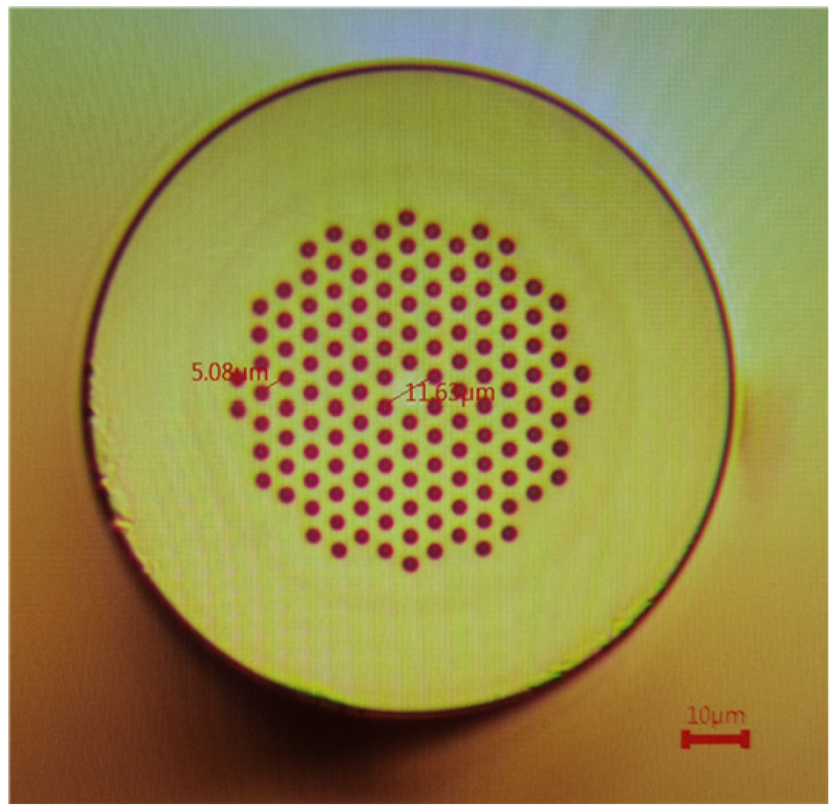
Figure 1 illustrates a schematic diagram of the MZI sensor that utilizes the SNPNS composite structure. The PCF used in this structure is LMA-8 with a core diameter of 8.3 μm and is manufactured by NKT Photonics, Denmark. Figure 2 shows its cross-section under the electronic microscope. The fiber cladding is composed of six layers of air holes, and its characteristic parameters are shown in Table 1. The PCF is spliced between two sections of NCF to enhance the sensor RI sensitivity. Compared to splicing the PCF directly with an SMF having a small core diameter, the mode-field mismatch between the NCF and the PCF is more significant than the mismatch between the SMF and the PCF. This allows for the coupling of more light to the PCF cladding through the collapsed region, improving the evanescent field between the PCF cladding and the surrounding environment to increase the RI sensitivity. The composite structure (NCF1–PCF–NCF2) is spliced into the middle of the two SMF segments (SMF-28e, Corning, Shanghai, China) for light transmission. Eventually, the SNPNS composite structure is formed.
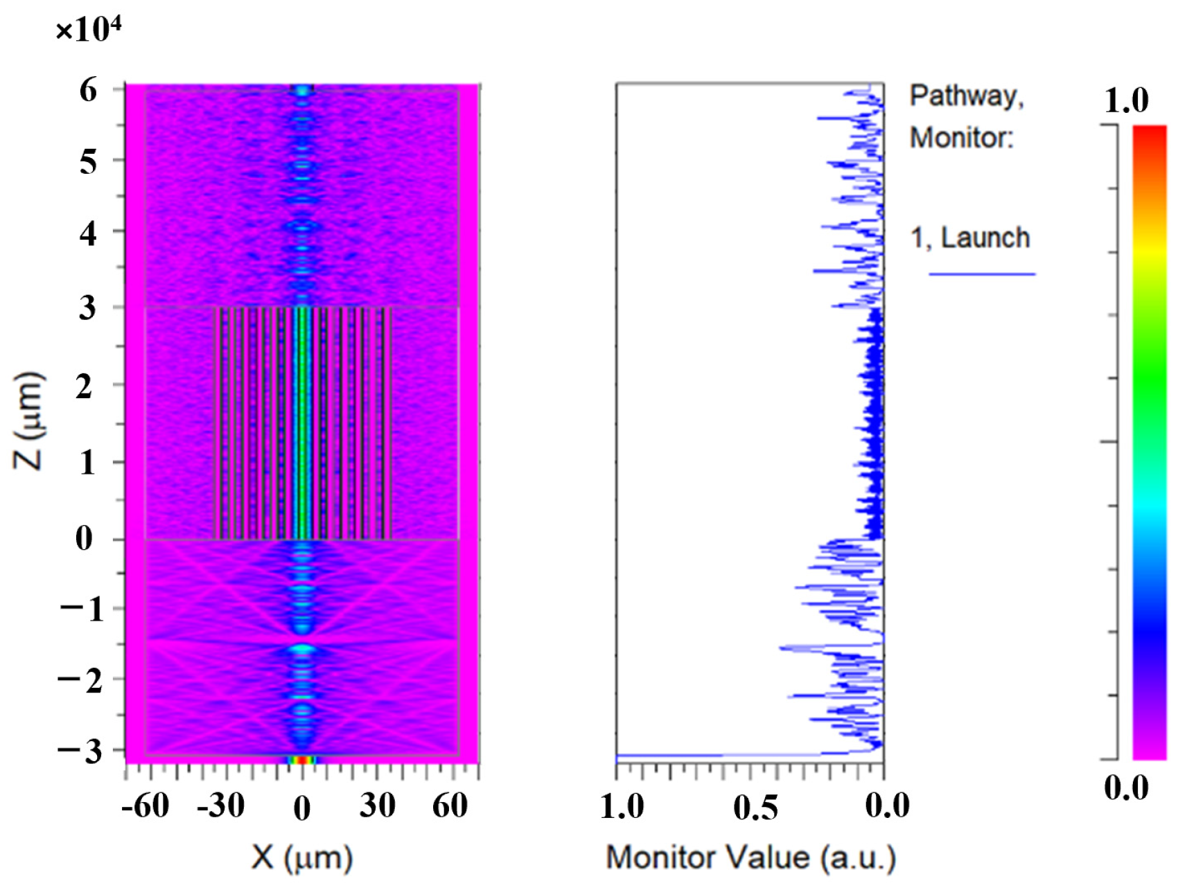
In the SNPNS structure, the transmission of light from the SMF to the NCF1 causes the excitation of multiple high-order modes within the NCF1. Subsequently, as the light enters the PCF, a portion of the light is coupled into the PCF cladding due to the mode-field mismatch, while the rest continues to propagate within the core of the PCF. The two light parts converge in the NCF2. When the two light parts satisfy a certain phase difference, an interference pattern is formed in the transmission spectrum. Figure 3 shows the field distribution of the SNPNS sensor obtained utilizing the beam propagation method (BPM) of Rsoft Software, with an incident wavelength of 1550 nm; a background RI of 1.34; and NCF1, PCF, and NCF2 lengths equal to 3 cm. The x- and z-axes in the left graph of Figure 3 represent the width and length of the fibers, respectively. It can be noted that the light field enhances as the light propagates through the fusion point of SMF and NCF1. The fundamental mode of SMF couples into NCF1, producing a number of high-order modes. In the collapsed region of NCF1 and PCF, a higher amount of light energy is transmitted to the core of the PCF and cladding of the PCF. The light is refracted from lighter to denser medium as the RI values of the surroundings rise, and the light energy decreases significantly while traversing the aperture ring in the PCF. The introduction of NCF1 enhances the evanescent field between the surroundings and the PCF cladding, which increases the RI sensitivity. The modes in the core and cladding of the PCF aggregate are again at NCF2 and are finally transmitted through the SMF. The right graph in Figure 3 shows the variation in the normalized optical power density after transmission in SMF, NCP, and PCF. The normalized power density of light transmitted in the core of SMF is set to 1. As the light enters the NCF1 from the SMF, there is no RI value difference in the NCF, which will cause the light to leak out and the optical power to decrease in the NCF1. In the collapsed region of the NCF1 and PCF, the light couples to the core and cladding of the PCF. Thus, the power density in the core of PCF is once again reduced. When the light passes through the PCF into the NCF2, the light in the core and cladding of the PCF converges. The power energy enhancement is shown in the NCF2.
The total interference intensity of the MZI formed by coupling the light from the core and cladding in the PCF into the NCF2 can be given as
I = I 1 + I 2 + 2 I 1 I 2 cos Δ φ
where I stands for the output light intensity and I1 and I2 represent the light intensities of the two interference beams, respectively. The phase difference between the two interfering beams is denoted by Δφ, which is calculated as follows:
Δ φ = 2 π n 2 n 1 L λ = 2 π Δ n e f f L λ = 2 π n e f f 1 n e f f 2 L λ
In (2), L denotes the total length of the interference region, which corresponds to the length of the PCF, λ is the center wavelength of the incident light in the vacuum, and Δneff stands for the effective RI difference between the core and cladding. When Δ φ = 2 m + 1 π , where m is an integer, the interference fringe has the maximum optimum visibility, where a low interference light intensity represents the valley of the interference spectrum. The corresponding resonance wavelength λm is
λ m = 2 π Δ n e f f L Δ φ = 2 π Δ n e f f L ( 2 m + 1 ) π = 2 Δ n e f f L 2 m + 1
The free spectral range (FSR) of an optical interference spectrum, which corresponds to the wavelength interval between the centers of two adjacent interference wave valleys, can be defined as
F S R = λ m 1 λ m = 2 Δ n e f f L 2 m 1 2 Δ n e f f L 2 m + 1 = 4 Δ n e f f L 2 m 1 2 m + 1 λ m 2 Δ n e f f L
The m-order wavelengths vary with the surrounding RI because the mode in the cladding is sensitive to it, whereas the core mode is RI insensitive. When the Δneff changes by Δn, the wavelength shift Δλm is given by (5). When the RI of the surroundings changes, the wavelength of the MZI shifts. Therefore, the variation in the wavelength can be utilized to display the RI measurements.
Δ λ m = 2 ( Δ n e f f + Δ n ) L 2 m + 1 2 Δ n e f f L 2 m + 1 = 2 Δ n L 2 m + 1

3. Experimental Results and Discussion

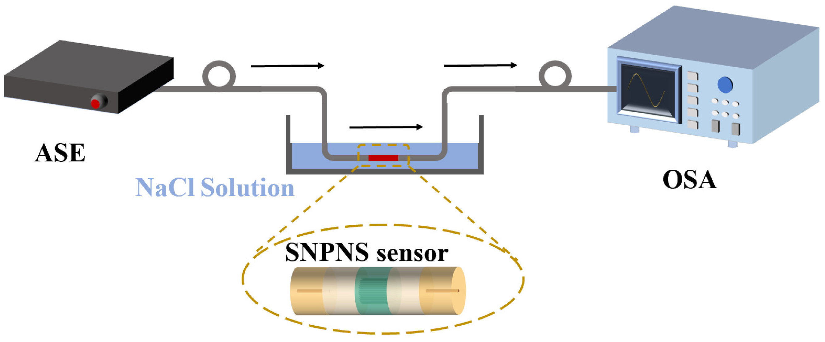
The SNPNS composite structure sensor is prepared using a fiber cutter (Fujikura CT-50) and a fiber fusion splicer (Fujikura FSM-88s). The length of the NCF is fixed at 3 cm. The fiber cutter is used to cut the fiber and obtain a flat end surface, and subsequently, the SMF and NCF are fused using the fiber fusion splicer. The above operation is repeated to prepare the SNPNS composite structure sensor with PCF lengths of 3 cm, 4 cm, and 5 cm, respectively. Figure 4 displays the experimental setup for RI sensing testing, which is composed of a broadband light source (ASE-CL-30-M), an SNPNS composite sensor, liquids to be measured with different RI values, and a spectrum analyzer (OSA, YOKOGAWA AQ6370D). The ASE light source, with a bandwidth of 100 nm, emits light with a wavelength from 1500 nm to 1600 nm, which carries the information about the surrounding environment through the sensing unit. It is collected and recorded by the spectrum analyzer, manifesting in the form of the interference spectrum drift. To ensure that the sensor is not affected by temperature, the RI sensing experiments are performed inside a constant temperature chamber, maintaining a temperature of 23 °C.
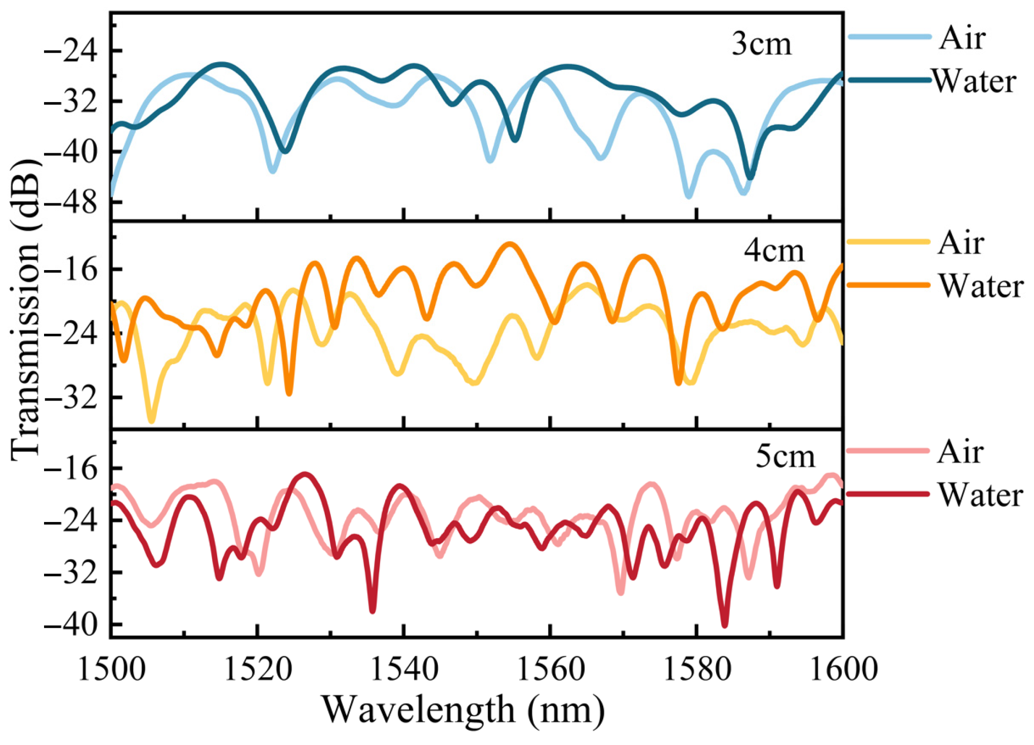
Figure 5 shows the transmission spectra of the SNPNS sensors for varying PCF lengths in the air and water when the NCF length is fixed at 3 cm. It is observed that the FSR of the transmission spectra reduces with increasing PCF length. When the PCF length is 3 cm, 4 cm, and 5 cm, respectively, the maximum extinction ratios of the transmission spectra of the SNPNS sensors in the air are 18.3 dB, 14.4 dB, and 16.7 dB, respectively, while those in the water are 13.8 dB, 12.9 dB, and 16.5 dB, respectively. The transmission spectra of the SNPNS sensor in the water corresponding to PCF lengths equal to 4 cm and 5 cm show multiple interference valleys within the observation area. If the sensor has a higher sensitivity, the problem of overlapping interference valleys will occur.
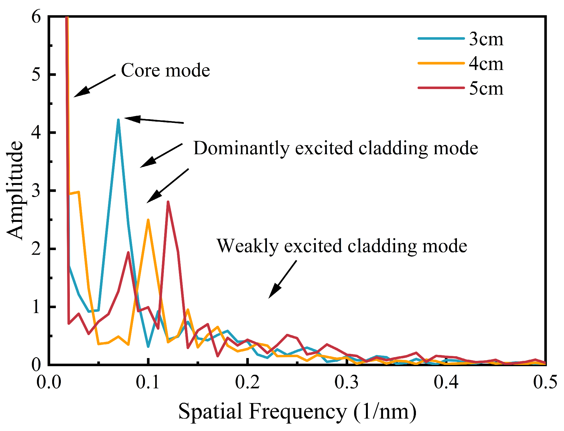
Fast Fourier transform (FFT) is a common way to analyze the interference spectrum of optical fibers, and it is often used to recognize the transmission modes in fiber sensing. The transmission spectra shown in Figure 5 are subjected to FFT to obtain the spatial spectra corresponding to the lengths of PCF of 3 cm, 4 cm, and 5 cm, as illustrated in Figure 6. The core modes interfere with the highest peak energy, and the rest of the cladding modes are divided into dominant cladding modes and weakly high-order cladding modes. The intensity of each peak represents the importance of the different modes in the intermodal interference. When the PCF length is 3 cm, the dominant peak is higher in the spatial spectrum compared with those obtained with other PCF lengths. Furthermore, there is an obvious difference between the core modes and the cladding modes, which indicates strong and highly stable intermodal interference in the fiber.
In the subsequent step, RI sensing experiments are conducted at room temperature using SNPNS composite structure sensors with PCF lengths of 3 cm, 4 cm, and 5 cm, respectively. The influence of fiber bending and vibration on the experimental results is avoided by placing these sensors horizontally in a container and securely fixing them to maintain the fiber structure stability. Deionized water is poured into the container in successive quantities of 100 mL until the sensor structure is completely submerged. Subsequently, NaCl solid (AR analytically pure, >99.5%) is added to the container, and the refractive index of the solution is measured using an Abbe refractometer once the NaCl solid dissolves completely. The average of ten samples of the transmission spectra at different RI values is recorded after the sensors reach stabilization. This process is repeated by adding an equal amount (1.5 g) of NaCl solid to the container each time, allowing for the recording of the transmission spectra of the sensors.
Initially, the SNPNS composite structure sensor with a PCF length of 3 cm is evaluated, and the transmission spectra responses of this sensor with different RI solutions are shown in Figure 7a–c. It can be observed that all three interferometric dips are shifted in the direction of the long wavelength as the RI increases. The interferometric dips at 1520 nm (dip A), 1545 nm (dip B), and 1575 nm (dip C) exhibit wavelength changes of 6.1 nm, 4.75 nm, and 6.5 nm, respectively, when the RI of the solutions varies from 1.3365 to 1.3767. As depicted in Figure 7d, it can be observed that the relationships between the interferometric dip shifts and the RI variation in the surrounding environment can be expressed as a perfect linear fit. The RI sensitivity responses of this sensor within the RI range of 1.3365 to 1.3767 are 176.90 nm/RIU, 84.3 nm/RIU, and 155.2 nm/RIU for dips A, B, and C, respectively, with coefficient of determination (R2) values of 0.990, 0.977, and 0.980, respectively.
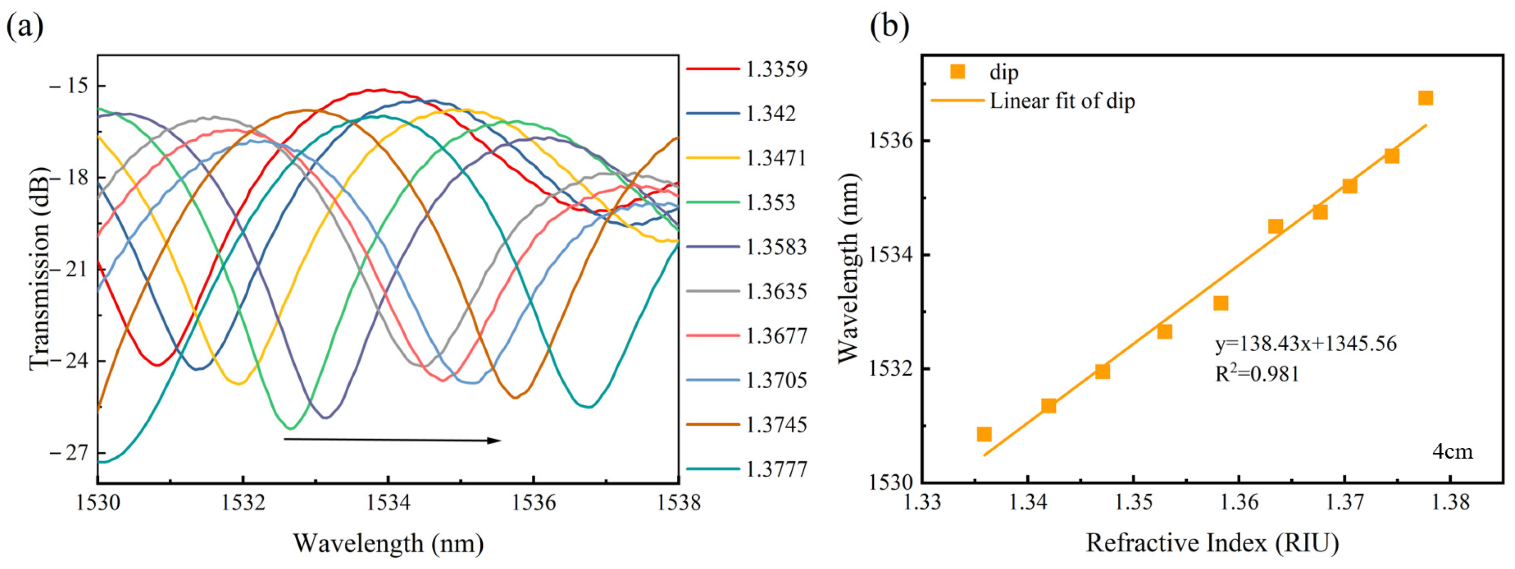
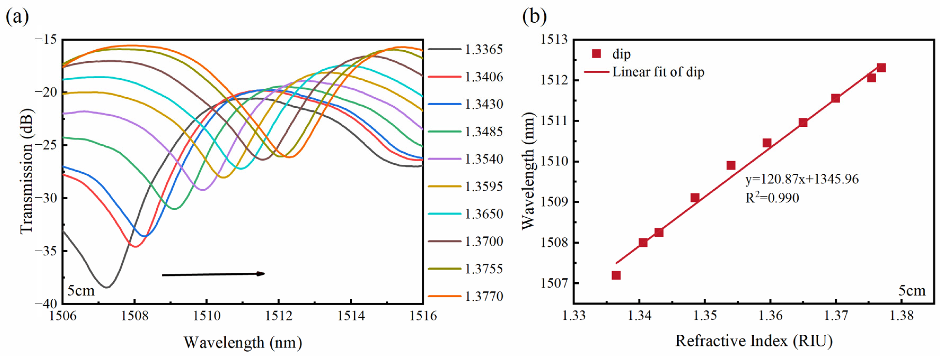
Subsequently, the RI sensing experiments are carried out for the sensors with PCF lengths of 4 cm and 5 cm. Figure 8 illustrates the linear fitting results between the dip shifts of the transmission spectra response and different RI sensitivity values of the sensor for the PCF length of 4 cm. It can be noted that, as the RI of the external environment increases, the interference dip is moved in the long wavelength direction. Figure 8b shows a good linear relationship between the interference dip and the RI values within the RI range of 1.3359–1.3777. The sensor RI sensitivity is determined as 138.43 nm/RIU, with an R2 value of 0.981. Figure 9 displays the transmission spectra response and fitting results for sensors with different RI values, where the PCF length is 5 cm. Similar to Figure 8, an increase in RI causes a red shift of wavelength in the transmission spectra. The sensor exhibits an RI sensitivity of 120.87 nm/RIU over 1.3365–1.3770 RI range, accompanied by a linearity coefficient of 0.990.
To ensure measurement stability, the SNPNS sensor with the PCF length of 3 cm is placed in the water over two hours. The transmission spectra are sampled at twenty-minute intervals. The drifts of the three dips (dip 1, dip 2, and dip 3) in the transmission spectrum shown in Figure 10a are monitored separately over two hours, as displayed in Figure 10b. The relative standard deviations (RSDs) of the throughs with wavelengths of 1523.8 nm (dip1), 1555.2 nm (dip2), and 1587.4 nm (dip3) are only 0.0022%, 0.0019%, and 0.0023% in water over two hours, respectively. It can be concluded that the sensor is highly stable. This small fluctuation may be caused by external environmental factors, such as temperature and pressure. For the effect of temperature on the stability of the sensors, it is mainly considered that the SNPNS sensors are placed in water with minor temperature fluctuations, which can impact the stability of the SNPNS sensor. To test the temperature characteristics of the sensing system, the variations in the transmission spectra of dip 1, dip 2, and dip 3 in the temperature range of 10 °C to 60 °C were measured, as shown in Figure 10c. It can be observed that, when the temperature is increased from 10 °C to 60 °C, the transmitted wavelengths of dip 1, wavelength 2, and wavelength 3 are varied from 1523.78 nm, 1555.2 nm, and 1587.45 nm to 1523.85 nm, 1555.22 nm, and 1587.5 nm, respectively. The temperature sensitivities of the SNPNS sensor are low at 1.18 × 10⁻³ nm/°C, 4.0 × 10⁻⁴ nm/°C, and 9.55 × 10⁻⁴ nm/°C, respectively, which indicates that the temperature has a slight impact on the stability of the SNPNS sensor. In the actual measurement process, packaging can be used to avoid the influence of temperature and pressure and to improve the stability of the sensing system.
The experimental analysis of the SNPNS composite structure sensors with three different lengths shows that the sensor we designed achieves a maximum RI sensitivity of 176.90 nm/RIU with a PCF of 3 cm at the RI values ranging from 1.3365 to 1.3767. The proposed sensor exhibits an excellent linear fit between the dips of the transmission spectrum and the RI. Table 2 compares the performance of our designed MZI sensor with other MZI-based PCF RI sensors. The RI sensitivity of our sensor is significantly higher than those of the sensors proposed in [20,24,25] but lower than those of the sensors proposed in [23,26,27]. However, the sensors in [23,26,27] adopted fiber-tapered structures, causing a thinning of fibers and a reduction in the mechanical strength of the device. In addition, some fiber RI sensors prepared based on other MZI structures also have very high sensitivities, such as long-period fiber grating (LPFG) structures [28,29,30,31], large core-offset structures [14,32,33], tapered structures [34,35], microcavity structures based on femtosecond laser inscription and chemical etching [17,36,37,38], as shown in Table 3. Similar to Refs. [23,36,37], these optical fiber sensors with high sensitivity require reprocessing of the optical fibers. The processed optical fibers, with their low mechanical strength, are susceptible to fracture and have difficulties in encapsulation and preservation during practical applications. The damage risk of the sensors is increased.

4. Conclusions

This paper proposed a composite fiber RI sensor with a simple structure, high sensitivity, and high stability, which was fabricated by splicing NCF–PCF–NCF between two SMFs. The experimental results showed that the maximum RI sensitivity of this structure was 176.90 nm/RIU in the measurement range of 1.3365–1.3767. This sensor could be fabricated conveniently and offers excellent mechanical durability. Notably, the SNPNS sensor with high stability and low temperature sensitivity makes it suitable for use in environments with significant temperature variations. Furthermore, this composite interferometer was formed by splicing different optical fibers, which offered a novel approach to simultaneously measure multiple parameters.

Author Contributions

Conceptualization, D.W. and J.Z.; methodology, J.Z. and Y.S.; software, J.Z. and X.Y.; validation, D.W. and Y.S.; formal analysis, J.Z.; investigation, Y.S.; resources, D.W.; data curation, J.Z.; writing—original draft preparation, D.W.; writing—review and editing, J.Z. and Y.S.; visualization, D.W. and Y.S.; supervision, D.W.; project administration, D.W. and Y.S.; funding acquisition, D.W. All authors have read and agreed to the published version of the manuscript.

Funding

This research was funded by the Zhejiang Provincial Natural Science Foundation of China under Grant No. LQ22F050012.

Institutional Review Board Statement

Not applicable.

Informed Consent Statement

Not applicable.

Data Availability Statement

Data are contained within the article.

Conflicts of Interest

The authors declare no conflicts of interest.

References

  1. Joe, H.E.; Yun, H.; Jo, S.H.; Jun, M.; Min, B. A review on optical fiber sensors for environmental monitoring. Int. J. Precis. Eng. Man. GT 2018, 5, 173–191. [Google Scholar] [CrossRef]
  2. Chaudhary, V.S.; Kumar, D.; Kumar, S. Gold-immobilized photonic crystal fiber-based SPR biosensor for detection of malaria disease in human body. IEEE Sen. J. 2021, 21, 17800–17807. [Google Scholar] [CrossRef]
  3. Chen, S.; Zhang, C.; Wang, J.; Li, N.; Song, Y.; Wu, H.; Liu, Y. A fiber Bragg grating sensor based on cladding mode resonance for label-free biosensing. Biosensors 2023, 13, 97. [Google Scholar] [CrossRef] [PubMed]
  4. Wu, Y.; Wang, G.; Yu, X.; Fan, Y.; Chen, X.; Liu, S. Label-free DNA hybridization detection using a highly sensitive fiber microcavity biosensor. Sensors 2024, 24, 278. [Google Scholar] [CrossRef] [PubMed]
  5. Zhao, Y.; He, T.; Chen, M.; Zhang, Z.; Tong, R. Ultra-short fiber Bragg grating composed of cascaded microchannels in a microprobe for refractive index measurement. J. Lightwave Technol. 2023, 41, 2555–2561. [Google Scholar] [CrossRef]
  6. Upadhyay, C.; Dhawan, D. Fiber Bragg grating refractive index sensor based on double D-shaped fiber. Opt. Quantum Electron. 2023, 55, 271. [Google Scholar] [CrossRef]
  7. Qi, L.; Zhao, C.L.; Yuan, J.; Ye, M.; Wang, J.; Zhang, Z.; Jin, S. Highly reflective long period fiber grating sensor and its application in refractive index sensing. Sens. Actuators B 2014, 193, 185–189. [Google Scholar] [CrossRef]
  8. Li, H.C.; Liu, J.; He, X.; Yuan, J.; Wu, Q.; Liu, B. Long-period fiber grating based on side-polished optical fiber and its sensing application. IEEE Trans. Instrum. Meas. 2023, 72, 7001109. [Google Scholar] [CrossRef]
  9. Liu, D.; Mallik, A.K.; Yuan, J.; Yu, C.; Farrell, G.; Semenova, Y.; Wu, Q. High-sensitivity refractive index sensor based on a tapered small core single-mode fiber structure. Opt. Lett. 2015, 40, 4166–4169. [Google Scholar] [CrossRef]
  10. Lu, Z.; Liu, C.; Ren, J.; Chen, L.; Li, C.; Zhou, W. Temperature-insensitive high-sensitivity refractive index sensor based on tapered no core fiber. Meas. Sci. Technol. 2023, 34, 084001. [Google Scholar] [CrossRef]
  11. Islam, N.; Arif, M.F.H.; Yousuf, M.A.; Asaduzzaman, S. Highly sensitive open channel-based PCF-SPR sensor for analyte refractive index sensing. Results Phys. 2023, 46, 106266. [Google Scholar] [CrossRef]
  12. Chaudhary, V.S.; Kumar, D.; Kumar, S. Au-TiO2 coated photonic crystal fiber based SPR refractometric sensor for detection of cancerous cells. IEEE Trans. Nanobiosci. 2023, 22, 562–569. [Google Scholar] [CrossRef] [PubMed]
  13. Ma, Y.; Qiao, X.; Guo, T.; Wang, R.; Zhang, J.; Weng, Y.; Rong, Q.; Hu, M.; Feng, Z. Temperature-independent refractive index measurement based on Fabry-Perot fiber tip sensor modulated by Fresnel reflection. Chin. Opt. Lett. 2012, 10, 050603. [Google Scholar] [CrossRef]
  14. Zhou, Y.; Liu, Y.; Zheng, J. Investigation of an inline fiber Mach-Zehnder interferometer based on hybrid joints of core-offset and peanut-shape for refractive index sensing. Opt. Fiber Technol. 2023, 81, 103521. [Google Scholar] [CrossRef]
  15. Zhao, Y.; Li, X.; Cai, L. A highly sensitive Mach–Zehnder interferometric refractive index sensor based on core-offset single mode fiber. Sens. Actuators A 2015, 223, 119–124. [Google Scholar] [CrossRef]
  16. Yu, F.; Xue, P.; Zheng, J. Study of a large lateral core-offset in-line fiber modal interferometer for refractive index sensing. Opt. Fiber Technol. 2019, 47, 107–112. [Google Scholar] [CrossRef]
  17. Dong, R.X.; Sun, X.Y.; Chu, D.K.; Yin, K.; Luo, Z.; Wang, C.; Hu, Y.W.; Duan, J.A. Microcavity Mach–Zehnder interferometer sensors for refractive index sensing. IEEE Photonics Technol. Lett. 2016, 28, 2285–2288. [Google Scholar] [CrossRef]
  18. Cui, J.; Gunawardena, D.S.; Cheng, X.; Htein, L.; Leong, C.Y.; Lau, A.P.T.; Tam, H.Y. In-line open-cavity Mach-Zehnder interferometric refractive-index sensors based on inter-mode and inter-core-mode interferences. Opt. Lasers Eng. 2024, 172, 107880. [Google Scholar] [CrossRef]
  19. Azab, M.Y.; Hameed, M.F.O.; Mahdiraji, G.A.; Adikan, M.; Obayya, S.S.A. Experimental and numerical characterization of a D-shaped PCF refractive index sensor. Opt. Quantum Electron. 2022, 54, 846. [Google Scholar] [CrossRef]
  20. Wu, D.; Zhao, Y.; Li, J. PCF taper-based Mach–Zehnder interferometer for refractive index sensing in a PDMS detection cell. Sens. Actuators B 2015, 213, 1–4. [Google Scholar] [CrossRef]
  21. Liu, F.; Guo, X.; Zhang, Q.; Fu, X. Dual-hole photonic crystal fiber intermodal interference based refractometer. Opt. Commun. 2017, 405, 147–151. [Google Scholar] [CrossRef]
  22. Wang, Q.; Wang, B.T.; Kong, L.X.; Zhan, Y. Comparative analyses of bi-tapered fiber Mach–Zehnder interferometer for refractive index sensing. IEEE Trans. Instrum. Meas. 2017, 66, 2483–2489. [Google Scholar] [CrossRef]
  23. Zhao, Y.; Xia, F.; Hu, H.F.; Chen, M.Q. A novel photonic crystal fiber Mach–Zehnder interferometer for enhancing refractive index measurement sensitivity. Opt. Commun. 2017, 402, 368–374. [Google Scholar] [CrossRef]
  24. Li, X.G.; Zhao, Y.; Cai, L.; Wang, Q. Simultaneous measurement of RI and temperature based on a composite interferometer. IEEE Photonics Technol. Lett. 2016, 28, 1839–1842. [Google Scholar] [CrossRef]
  25. Dong, X.; Zeng, L.; Chu, D.; Sun, X.; Duan, J. Highly sensitive refractive index sensing based on a novel Mach-Zehnder interferometer with TCF-PCF composite structure. Infrared Phys. Technol. 2022, 123, 104134. [Google Scholar] [CrossRef]
  26. Zhao, Y.; Li, X.; Cai, L.; Yang, Y. Refractive index sensing based on photonic crystal fiber interferometer structure with up-tapered joints. Sens. Actuators B 2015, 221, 406–410. [Google Scholar] [CrossRef]
  27. Wang, Q.; Kong, L.; Dang, Y.; Xia, F.; Zhang, Y.; Zhao, Y.; Hu, H.; Li, J. High sensitivity refractive index sensor based on splicing points tapered SMF-PCF-SMF structure Mach-Zehnder mode interferometer. Sens. Actuators B Chem. 2016, 225, 213–220. [Google Scholar] [CrossRef]
  28. Ding, J.F.; Zhang, A.P.; Shao, L.Y.; Yan, J.H.; He, S. Fiber-taper seeded long-period grating pair as a highly sensitive refractive-index sensor. IEEE Photonics Technol. Lett. 2005, 17, 1247–1249. [Google Scholar] [CrossRef]
  29. Tripathi, S.M.; Bock, W.J.; Mikulic, P. A wide-range temperature immune refractive-index sensor using concatenated long-period-fiber-gratings. Sens. Actuators B 2017, 243, 1109–1114. [Google Scholar] [CrossRef]
  30. Garg, R.; Tripathi, S.M.; Thyagarajan, K.; Bock, W.J. Long period fiber grating based temperature-compensated high performance sensor for bio-chemical sensing applications. Sens. Actuators B Chem. 2013, 176, 1121–1127. [Google Scholar] [CrossRef]
  31. Chen, J.; Cui, S.; Zhang, S.; Liu, A. Refractive index sensors based on a chirped core long-period fiber grating. IEEE Photonics Technol. Lett. 2024, 36, 771–774. [Google Scholar] [CrossRef]
  32. Zhao, Y.; Zhang, Y.; Hu, H.; Yang, Y.; Lei, M.; Wang, S. High-sensitive Mach-Zehnder interferometers based on no-core optical fiber with large lateral offset. Sens. Actuators A Phys. 2018, 281, 9–14. [Google Scholar] [CrossRef]
  33. Li, L.; Zhang, Y.; Han, B.; Lv, R.; Zhao, J.; Zhao, Y. Highly Sensitive and Regulatable Optical Fiber Vernier Sensor Based on Two Integrated Parallel MZIs. J. Light. Technol. 2024, 42, 6318–6325. [Google Scholar] [CrossRef]
  34. Tian, Z.; Yam, S.S.-H.; Barnes, J.; Bock, W.; Greig, P.; Fraser, J.M.; Loock, H.-P.; Oleschuk, R.D. Refractive index sensing with Mach-Zehnder interferometer based on concatenating two single-mode fiber tapers. IEEE Photon. Technol. Lett. 2008, 20, 626–628. [Google Scholar] [CrossRef]
  35. Wang, Q.; Wei, W.; Guo, M.; Zhao, Y. Optimization of cascaded fiber tapered Mach-Zehnder interferometer and refractive index sensing technology. Sens. Actuators B Chem. 2016, 222, 159–165. [Google Scholar] [CrossRef]
  36. Wang, Y.; Yang, M.W.; Wang, D.N.; Liu, S.J.; Lu, P.X. Fiber in-line Mach-Zehnder interferometer fabricated by femtosecond laser micromachining for refractive index measurement with high sensitivity. J. Opt. Soc. Am. B 2010, 27, 370–374. [Google Scholar] [CrossRef]
  37. Gao, S.C.; Zhang, W.G.; Zhang, H.; Zhang, C.L. Reconfigurable and ultra-sensitive in-line Mach-Zehnder interferometer based on the fusion of microfiber and microfluid. Appl. Phys. Lett. 2015, 106, 084103. [Google Scholar] [CrossRef]
  38. Gao, S.; Zhang, W.; Bai, Z.; Zhang, H.; Geng, P.; Lin, W.; Li, J. Ultrasensitive refractive index sensor based on microfiber-assisted u-shape cavity. IEEE Photonics Technol. Lett. 2013, 25, 1815–1818. [Google Scholar] [CrossRef]
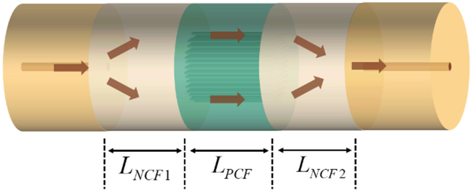
Figure 1. Schematic diagram of the MZI sensor based on the SNPNS composite structure.
Photonics 11 00941 g001
Figure 2. Cross-section of PCF under the electron microscope.
Photonics 11 00941 g002
Figure 3. Simulated field distributions of SNPNS composite structure with lengths of NCF1, PCF, and NCF2 equal to 3 cm.
Photonics 11 00941 g003
Figure 4. Diagram of the experimental setup.
Photonics 11 00941 g004
Figure 5. Transmission spectra of SNPNS composite structure sensors for PCF lengths of 3 cm, 4 cm, and 5 cm.
Photonics 11 00941 g005
Figure 6. Spatial spectra of SNPNS composite structure sensors for PCF lengths of 3 cm, 4 cm, and 5 cm.
Photonics 11 00941 g006
Figure 7. The SNPNS composite structure sensor with PCF length of 3 cm and (ac) transmission spectra responses for dips A, B, and C with different RI values; (d) relationships between interference dips and RI values.
Photonics 11 00941 g007
Figure 8. The SNPNS composite structure sensor with PCF length of 4 cm and (a) transmission spectra responses with different RI values and (b) relationships between interference dips and RI values.
Photonics 11 00941 g008
Figure 9. The SNPNS composite structure sensor with PCF length of 5 cm and (a) transmission spectra response with different RI values and (b) relationships between interference dips and RI values.
Photonics 11 00941 g009
Figure 10. (a) Transmission spectrum of the sensor with PCF length of 3 cm in water; (b) stability lines of different dips at the water temperature of 23 °C; (c) relationships between interference dips and water temperature.
Photonics 11 00941 g010
Table 1. Characteristic parameters of PCF.
ParameterValues
Cladding diameter (μm)125
Air hole diameter (μm)2.576
Air hole pitch (μm)5.6
Core diameter (μm)8.3
Table 2. RI sensing comparison with different MZI structures based on PCF.
StructuresSensitivity (nm/RIU)RI RangeReference
Tapered PCF51.9021.3411–1.3737[20]
MMF–PCF MZI1081.333–1.374[24]
TCF–PCF MZI119.291.3333–1.3735[25]
HTCR–PCF–HTCR MZI181.961.3333–1.3574[23]
Up-tapered MZI2521.333–1.379[26]
Tapered SMF–PCF–SMF260.8/243.41.3333–1.3737[27]
SNPNS MZI176.901.3365–1.3767This work
Table 3. RI sensing comparison with other different MZI structures.
StructuresSensitivity (nm/RIU)RI RangeReference
Tapered LPFG178.871.3333–1.3624[28]
etched, DRLPFGs2577/46811.333–1.343[29]
etched, DRLPFGs25001.333–1.353[30]
CC–LPFG>6001.42[31]
Bent SMF-peanut shape- SMF- core-off section-SMF−167.271.333–1.373[14]
SMF–NCF-offset NCF–NCF–SMF−11,078.81.3320–1.3355[32]
SMF–MMF-offset SSHF–SMF–MMF–SMF−101,6221.3311–1.3335[33]
Two cascaded SMF tapers15701.315–1.3618[34]
Two cascaded SMF tapers1548.41.3333–1.3792[35]
V-shape1437.611.3333–1.3459[17]
Rectangular-shape1489.101.3371–1.3407[17]
U-shape1584.471.31–1.335[36]
SMF-microfiber-SMF15371.44[37]
SMF-microfiber-SMF1376.031.4444–1.4462[38]
Disclaimer/Publisher’s Note: The statements, opinions and data contained in all publications are solely those of the individual author(s) and contributor(s) and not of MDPI and/or the editor(s). MDPI and/or the editor(s) disclaim responsibility for any injury to people or property resulting from any ideas, methods, instructions or products referred to in the content.