Review

Biological Photonic Devices Designed for the Purpose of Bio-Imaging with Bio-Diagnosis

1
Cell Therapy and Regeneration Medicine, Taipei Medical University, Taipei 11031, Taiwan
2
Department of Chemical Engineering and Biotechnology, National Taipei University of Technology, Taipei 10608, Taiwan
3
Hsuan Chen Technology Co., Ltd., New Taipei City 22055, Taiwan
4
Department of Orthopedics, College of Medicine, Taipei Medical University Hospital, Taipei 11031, Taiwan
5
Department of Orthopedics and Trauma, Faculty of Medicine, University of Medicine and Pharmacy, Ho Chi Minh City 700000, Vietnam
6
Graduate Institute of Biomedical Materials and Tissue Engineering, College of Biomedical Engineering, Taipei Medical University, Taipei 11031, Taiwan
7
International Ph.D. Program in Biomedical Engineering, College of Biomedical Engineering, Taipei Medical University, Taipei 11031, Taiwan
8
Cell Physiology and Molecular Image Research Center, Taipei Medical University-Wan Fang Hospital, Taipei 11696, Taiwan
*
Author to whom correspondence should be addressed.
These authors contributed equally to this work.
Photonics 2023, 10(10), 1124; https://doi.org/10.3390/photonics10101124
Submission received: 30 August 2023 / Revised: 26 September 2023 / Accepted: 1 October 2023 / Published: 6 October 2023

Abstract

:
The rapid progress in the fields of biomedical and biological photonic sciences has given rise to a substantial demand for biological photonic structures capable of interacting with living systems. These structures are expected to facilitate precise manipulation of incident light at small scales, enabling the detection of sensitive biological signals and the achievement of highly accurate cell structural imaging. The concept of designing biological photonic devices using innate biomaterials, particularly natural entities such as cells, viruses, and organs, has gained prominence. These innovative devices offer the capability of multimodal light manipulation at specific sites, enhancing biological compatibility while minimizing disruptions to the delicate biological microenvironment. This article delves into recent advancements within the realm of biological photonic devices, with a dedicated focus on their applications in bio-imaging and -diagnosis. The central theme revolves around devices derived from biological entities possessing the requisite optical properties, biocompatibility, biofunctionality, and the ability to induce biological effects. These devices encompass a diverse range of optical functionalities, including light generation, transportation, and modulation, all of which play pivotal roles in bio-detection and imaging, thereby contributing notably to the advancement of these fields. The potential future directions and opportunities for the enhancement of biological photonic devices were outlined.

1. Introduction

The precise monitoring of pathological alterations as well as the accurate identification of biological signals are crucial in facilitating the timely diagnosis and effective management of health issues such as infectious diseases, cancer, and other medical conditions. Detecting the specific targets of interest presents a significant challenge due to the limited presence of biological and chemical signals and the intricate environment within living systems. Fortunately, the rapid advancement of photonic as well as optical tools in up-to-date times has introduced a multitude of options for optical detection and imaging. This development brings about the substantial potential for biological signals’ real-time visualization within intricate bio-structural and dynamic processes [1,2]. Optical detection harnesses optical reactions, including phenomena like the absorption of light, fluorescence, reflectance, and scattering. These responses are triggered by biophysical and biochemical alterations, serving the purpose of identifying biological entities and diagnosing diseases [3]. Because of its inherent lack of reliance on labelling, optical detection presents a potent substitute for traditional detection methods, such as mass or electrochemical techniques [4,5,6,7]. By utilizing optical detection methods, it becomes possible to capture real-time signals from various biological test specimens, spanning from biomolecular markers to cells, pathogens, and even tissues and organs, in a non-invasive way that has both elevated resolution and elevated sensitivity [8,9,10,11,12]. Until now, optical imaging, as well as detection, have proven themselves as remarkably potent technologies for identifying biological signals and diagnosing an array of conditions, encompassing infectious diseases, cancer, and other disorders [13,14,15,16,17,18,19].
To achieve accurate and adaptable optical detection within a biological setting, the utilization of photonic devices featuring micro/nanostructures is consistently sought after. To fulfil this objective, the careful choice of suitable optical materials holds significant importance, as the effectiveness of probing is greatly influenced by their chemical and mechanical attributes, optical capabilities, and biological characteristics [20]. Up to now, the prevailing materials employed for creating versatile photonic components and devices predominantly consist of inorganic substances like glass, silica, and so on [21,22,23,24]. Alternatively, polymeric organic materials like polymeric nanowires have also been utilized as an option for constructing photonic components and devices [25,26].
Due to their exceptional optical characteristics, including high transparency and adequate mechanical strength, these materials have found application in a variety of fields for the development of integrated nanophotonic devices [27]. For instance, optical waveguides utilizing optical fibres have undergone extensive investigation and even found utility in animal implants, including fibre-optical bioimplants within the deep tissue. Nevertheless, a notable limitation of these optical components, which are constructed from conventional materials, pertains to their insufficient biocompatibility and biodegradability, thereby considerably constraining their potential in the realm of biomedical applications. A paramount criterion for materials for in vivo applications is high biocompatibility, which necessitates the nonexistence of biotoxicity and minimal risk to biological systems [28]. Furthermore, elevated biological compatibility extends to the material’s capability to fulfil its intended functions within the living environment. Equally essential is biodegradability, allowing the material to naturally degrade and be metabolized by the body without necessitating additional removal procedures.
The natural abundance of biomaterials and biological entities has long served as a source of inspiration for designing tracers/devices as well as photonic structures that are capable of manipulating incident light [29]. Biological cells, microorganisms, and their derivatives, encompassing DNA, proteins, silk, biomaterials, and polysaccharides, exhibit diverse interactions with light, facilitating their use as photonics components like waveguides, micro lenses, lasers, and even gratings [30,31,32,33,34,35,36]. These innate biological materials and biological beings possess immense potential for innovating novel photonics tracers/devices tailored for biological imaging, detection, and restorative uses [20]. They intrinsically manifest exceptional living properties, comprising high biological compatibility, mini-invasiveness, restorability, and biological degradability. Notably, a fascinating characteristic of natural beings such as cells, organs, and viruses is their dual role as both optical devices and diagnostic specimens, facilitating real-time imaging as well as detection within biologically compatible cellular environments. Consequently, the focus of biological photonic devices discussed in the study is predominantly on substantial biological entities like bacteria, viruses, algae, fungi, organs, mammalian cells, and biologically derived materials or biomolecules like proteins and nucleic acids. By translating biological principles into engineered designs, these biological photonic devices seamlessly bridge the gap between the optical, biological, and biomedical realms [37,38].
Despite existing comprehensive examinations of functional biological photonic structures derived from bio-inspired, naturally derived, and synthetic biomaterials, a notable gap remains in reviews dedicated to biological photonic devices centred around large natural beings, including cells, viruses, and organs, and the phototherapeutic and diagnostic aspects of translational medicines. This emerging area of natural-being-based photonics tracers/devices has considerably augmented the field of biological photonics, holding immense possibilities for applications in healthcare as well as biomedicine. In this article, the focus is directed toward recent advancements in biological photonics tracers/devices predicated on natural beings, with a substantial emphasis on cells, viruses, and organs and their uses in biological imaging as well as detection. We scientifically present biological laser systems, photonic waveguides, and photonic-based theragnostic applications in translational medicine (Scheme 1). These devices encompass a diverse array of optical functionalities ranging from incident light generation to light modulation as well as transportation. Moreover, we proposed the challenges that remain and offered insights into the potential future directions of these biological photonics tracers/devices for medical applications.
Achieving the promising medical uses of photonics tracers/devices demands effective modulation as well as control of the generation of light, especially in intricate biological/chemical microenvironments such as in vivo deep organs as well as cells. The distinct attributes of laser-emitted light, encompassing monochromatic emission, high intensity, and directionality, have established lasers as invaluable means in biological medical uses [39]. The previous study employed biophysical and biomaterial concepts for applications in the biomedical interface [40]. Subsequently, the exploration and advancement of lasers have paved the way for novel avenues in biomedical applications, resulting in the widespread incorporation of laser-based biomedical devices within clinical practices. The prior investigation focused on the exploration of selectively manipulating upconversion emission channels using tuneable biological photonic devices (Figure 1, [41]).
The biological laser [42] circumvents the biological hazards associated with traditional devices of the laser. By embedding or introducing biological lasers into organs or cells, they function as devices of photonics for the imaging as well as detection of diverse signals at the biomolecular [43,44,45], cell [46,47], and organ levels [48,49,50]. Biological lasers founded on cellular and tissue structures offer heightened sensitivity for bio-imaging as well as detection, as their optic output closely correlates with the biostructures as well as bioactivities intrinsic to the biosystems.
A defining feature of biological lasers is the integration of natural beings, including organs, cells, and viruses, as the gain medium—either wholly or partially. Given the intimate link between the gain medium and lasing output, biological changes within the gain medium are swiftly detectable via biological lasers, underscoring their remarkable sensitivity. Selecting suitable gain materials necessitates careful consideration to ensure biological compatibility as well as biological degradability. Illustrative instances of biologically compatible biomaterials encompass biological dyes as well as fluorescent proteins. Fluorescent proteins, harvested from diverse organisms, boast excellent biocompatibility, and robust photostability [51]. Successful emission of the laser via proteins of fluorescence like monomeric Cherry (mCherry), Venus, indocyanine green (ICG), and green fluorescent protein (GFP) is being attained by applying varied cavity configurations [42,48,52,53,54,55]. Alternatively, natural biological dyes present in the tissues of humans and other species, for example, riboflavin (vitamin B2) as well as luciferin, have been harnessed as gain materials for biological laser systems [56,57]. By integrating these biologically compatible fluorophores in viruses, tissues, and cells, biological laser systems are enabled to be fabricated, capitalizing on these natural beings to serve as the biological gain medium. The preceding study centered on enhancing photosensitivity in reassembled one-dimensional VB2 submicron rods, aiming to achieve efficient antibacterial therapy (Figure 2, [58]).
Typically comprised of three primary components—an optic cavity for light confinement, a gain medium to amplify incident light within the cavity, and a source of energy of pump—the laser system’s photons undergo iterative relations with the initiated gain medium, resulting in photon amplification. The phenomenon of emission of laser materializes only once the pool of accessible excited gain molecules exceeds the cumulative losses within the cavity, thereby surpassing the lasing threshold [42]. This threshold can be mathematically described [59]. The Q-factor (quality factor) gauges the effects of damping of the modes of the resonator and is expressed as the fraction of stored energy to energy dissipation per radian of the oscillation [60].
Contrasted with conventional fluorescence emission, the laser-based devices/probes exhibit significantly narrower line widths, heightened light intensity, greater spatial resolution, superior sensitivity, and spectral resolution. This is attributed to the distinctive optical feedback mechanism and threshold characteristics of the lasers [61].
An optical waveguide is a physical structure designed to direct electromagnetic waves within the optical spectrum. Electromagnetic wave-based analogue computing is being become an attractive computing paradigm indicating the promise for low power, high-throughput, and parallel functions [62]. Common types of optical waveguides encompass optical fibre waveguides, transparent dielectric waveguides crafted from materials such as plastic and glass, liquid light guides, and liquid waveguides. Optical waveguides serve various purposes, acting as integral components within integrated optical circuits or functioning as the transmission medium in optical communication systems. These waveguides can be categorized based on several factors, including their geometry (such as planar, strip, or fibre waveguides), mode structure (including single-mode or multi-mode), refractive index distribution (featuring step or gradient index profiles), and the materials employed (which range from glass and polymers to semiconductors).
Cellular-based lasers are enabled to be classified into distinct categories based on the position of the resonator: extracellular lasers (where the resonator structure exists external to the cell) and intracellular lasers (where the resonator structure is inside the cell). Previous research has demonstrated the remarkable phenomenon of self-sustained lasing occurring within living cells equipped with intracellular optical resonators. The cellular laser system is depicted in [63]. Under typical culture conditions, cells undergo natural endocytosis to internalize whispering gallery-mode microsphere resonators. This ingenious process eliminates the necessity for an external resonator structure. Extracellular lasers are commonly established using a Fabry–Pérot (FP) cavity configuration, involving the placement of fluorophore-stained cells between two reflective mirrors, thus creating an optical cavity for laser emission [61,64,65]. The earliest instances of extracellular lasers involved GFP-expressing mammalian cells enclosed within an FP resonator [65]. Furthermore, the spectra emission from the cell laser resided in multiple narrow-band peaks, facilitating the identification of distinct spectral components for cell phenotyping. Similarly, E. coli bacteria that stably expressed GFP were integrated into biomaterials [55] and biological systems [66] to act as a biological gain medium. By supplying adequate nutrients for E. coli’s steady-state growth, functional GFP expression was maintained, ensuring the self-replenishment of the gain medium. However, while endogenous fluorescence proteins present an appealing option for generating optical gain within cells, the practicality is hindered by the protracted transfection process required for fluorescent protein expression. In the previous work, single cell was used to induce optical confinement in biological lasers [67].
In contrast to the use of intracellular fluorescence proteins, a more diverse array of synthetic fluorescence biomolecules, including fluorescein sodium salt [64], 5-chloromethyl fluorescein diacetate (CMFDA) [68,69], Calcein-AM [70], molecular beacon [71], and fluorescein isothiocyanate (FITC)/Rhodamine 6G [72,73], are being explored as convenient gain media for generating cellular-based lasers across various cellular types. Of particular note, the rapid dye staining processes can be executed in a given period, drastically reducing the time needed for laser preparation compared to the time-intensive fluorescence protein transfection methods. Additionally, an extensive range of lasing wavelengths, spanning from green to red, are capable of being achieved as per the chosen dyes and mirrors.
Once the entire resonator structure, accompanied by the dye gain medium, is uptaken with the cell, an independent intracellular laser, often referred to as a “whispering gallery mode” (WGM) laser, can be realized. WGMs [74] are specific modes accessible in optical resonators, renowned for their application in precision physical determinations as well as biologically chemical sensing at the single-molecule level [75,76,77]. Typically compact and characterized by relatively low thresholds due to high Q-factors [78], micro- as well as nano-sized optical resonators can be readily uptaken by cells, presenting an ideal platform for cellular lasers. The polymeric/organic optical device/dyes could possess less toxicity and biocompatible properties and have been indicated to be used in a laser system based on a real cell [79]. The previous investigation revolved around the phenomenon of cavity-enhanced fluorescence observed in colliding droplets of aqueous solutions containing Rhodamine 6G (Figure 3, [80]).
Viruses, the most prevalent microorganisms on Earth, are characterized by their fundamental structure: genetic biomaterial (DNA or RNA) enclosed within the shell of a protein known as a capsid [81]. Within the realm of viruses, bacteriophages (phages) have garnered considerable attention; these viruses constitute a diverse group that exclusively infects bacteria [82]. Utilizing phage display techniques, an array of proteins as well as polypeptides boasting diverse bio-functionalities can be systematically and meticulously showcased on the phage capsids’ surface [83]. Driven by their stability, biosafety, and availability attributes, phages have currently developed as multipurpose means with a plethora of biological medical uses [84,85,86,87,88]. A groundbreaking phage-derived biolaser tailored for bio-detection has emerged, featuring the utilization of the M13 phage—a filamentous phage characterized by its rod-like architecture, having a length of 900 nm as well as a diameter of 7 nm [43]. The host cell for the M13 phage is a strain of E. coli bacteria expressing F pilus, ensuring its safety for human use. Through the chemical conjugation of fluorescent dyes onto the M13 surface, a virus-based laser system was crafted, offering adaptable laser output because of the repetitive arrangement of M13. Leveraging the capabilities of phage display technology, M13 can be engineered with various target binding specificities, thereby detecting an extensive array of biomolecules of interest. The resulting laser emissions from M13 demonstrate remarkable sensitivity of detection. These virus-driven lasing probes/devices exhibit substantial possibility to supplant conventional antibody-construed ways of bio-detecting, offering heightened precision and accelerated detection rates. The preceding study was centred on the distinguishing of small molecules within a microcavity using molecular laser polarization (Figure 4, [89]).
Beyond cell and virus-based lasers, the application of biological lasers within tissues holds greater significance for clinical utilization, given that tissues, composed of cells and the surrounding extracellular matrix, offer a more accurate representation of the complex physiological conditions in vivo. In pursuit of this objective, considerable efforts have been directed toward the development of tissue lasers over recent years. Leveraging the scattering properties inherent in tissues, tissue-based lasing can be conveniently achieved via random lasing, accomplished via the administration of fluorescence dyes [90,91]. This phenomenon of random lasing has been successfully demonstrated in a variety of tissue types, including bone and heart [50,92], brain and muscle [93,94], kidney, cancerous, and colon tissue [91,95,96]. Given that the output features of random lasing intimately correspond with tissue microenvironment as well as structure, the resulting lasing spectra can serve as a valuable tool for distinguishing between healthy and malignant tissues [97], investigating subtle structural changes [50], and detecting biological variations [98]. The advantage of random lasing lies in its relatively straightforward methodology for generating tissue-based lasers without the necessity of introducing external cavities. Nevertheless, the inherent uncertainty in lasing thresholds and spectral peaks can potentially compromise sensitivity as well as detection accuracy [31].
Besides random lasers, the creation of bio-implantable and standalone whispering gallery mode (WGM) lasers within tissues has also been successfully realized through the incorporation of solid microbeads or liquid microdroplets into structures such as tissues. Nevertheless, current tissue lasing techniques still lack the capability for identifying as well as detecting specific biological markers in organs, a capability that holds substantial significance in rendering biological lasers more applicable for clinical settings.
Addressing these limitations, the research group led by the scientist has devised a multipurpose organ-lasing platform known as the laser-emission-based microscope (LEM). This novel approach integrates a high-quality Fabry–Pérot (FP) cavity, formed by two highly reflective mirrors, with tissues stained by biocompatible fluorescent dyes, effectively sandwiching the tissues in between [72]. In this setup, distinct lasing emissions were observed from human tissues stained with dyes, like muscle or adipose, with remarkably low threshold intensities. For the generation of lasing within specific cells amidst the tissue, biomarkers like proteins or nucleic acids within the cells were labelled using fluorescently labelled nucleic acids or antibody dyes [61]. Integration with a two-dimensional raster-scanning stage facilitated the acquisition of maps or images of tumour tissues extracted from patients, with the laser emissions stemming from the labelled biomarkers enabling high-resolution discrimination between cancerous and normal tissues. Notably, the technique even allowed the early-stage diagnosis of lung cancer. The LEM approach stands poised to significantly contribute to precision medicine, harnessing its attributes of potent background suppression, high intensity, and impressive spatial/spectral resolution.
Beyond organs as well as solid tumours, the realm of lasing extends to blood as well. A notable example involves the use of the human-safe near-infrared dye indocyanine green (ICG), enabling lasing within whole human blood flowing through an optofluidic ring resonator (OFRR) capillary [48]. ICG, present at a biocompatible concentration below 0.04 mM, demonstrated an affinity for lipoproteins (LDL) within the blood, inducing ICG-based lasing. Furthermore, lasing within human blood was also accomplished by directly dispersing dye-doped polymeric beads into the tissue of the blood [99]. Such blood-based lasing holds immense potential for diverse biomedical as well as clinical applications, particularly in the domain of blood diagnosis.
As discussed earlier, the employment of cell- and tissue-based biological lasers facilitates the localized delivery of optic energy toward biosystems, achieving both temporal as well as spectral optical localization. These biological lasers stand as promising devices of photonics with broad applications in the biomedical realm, spanning from cell tracking as well as tagging to diagnostics, cellular sensing, and innovative imaging.
Leveraging the unique spectral signatures of cellular lasers, it becomes achievable to distinguish and track individual cells amidst large populations of cells. For instance, previous works, including [70,100] integrated WGM microdisk-based lasers into various cell types. The utilization of microdisks with slightly varying diameters caused discernibly distinct lasing output spectra, enabling the tracking as well as tagging of individual cells. Given the minute size of microdisks, cells could accommodate multiple ones, facilitating wavelength-multiplexed spectrum signals and enabling the simultaneous unique tagging of large cell populations. Within a 3D tumour organ model in vitro, numerous tumour cells displaying differing motility patterns were effectively tagged and tracked. These microdisk-based cellular lasers demonstrated robust stability and high biofunctionality within cellular and tissue environments. However, a limitation of these disc-shaped laser particles lies in their inherent directional emission, primarily occurring within the cavity resonance plane [101]. For tracking cells, this inherent property could lead to random fluctuations in intensity and obstruct the optical reading of lasers, given the arbitrary and erratic change in orientation as cells move. Addressing this issue, Tang et al. introduced modifications to microdisk lasers [102].
The potential of cell-based biological lasers extends further through their integration with other imaging techniques, enhancing the precision of tracking the trajectories of individual cells. For instance, Li et al. [103] devised a double-modality bioimaging system by merging nanowire-based lasers with optical coherence tomography (OCT), a well-established bioimaging technique.
Notably, the spectral information of lasers directly correlates with biological alterations within tissues or cells, particularly at the individual tissue or cell level. This attribute can be harnessed for biological medical diagnoses, particularly in the context of cancer detection. For instance, an array of cell lasers was engineered through the integration of micro-well arrays with high-quality Fabry–Pérot (FP) cavities [47]. Actual monitoring of shifts in spectral peak wavelength and lasing thresholds allowed the recognition of anomalous cells amidst a larger population of healthy cells. A recent advancement, the laser-emission-based microscope (LEM), has exhibited heightened sensitivity and specificity in screening pathological cancer tissues [61]. Through the labelling of cells having fluorochrome-engineered tumour biomarkers, substantial differences in lasing thresholds in normal as well as tumour cells facilitated the detection of tumours with impressive sensitivity. Up to the present time, this versatile diagnostic platform has been successfully applied across various cancer types with samples prepared through diverse methods [72,104,105,106].
The utility of cellular-based biological lasers extends to their capacity for real-time measurement of subtle physiological bioactivities within living cells—a capability not easily attainable through conventional techniques. For example, the LEM technique enabled the detection of minute changes in cellular ion concentrations at the nanomolar level in single neurons and neural networks [107]. This “neuron laser” exhibited an enhancement in detection sensitivity compared to traditional fluorescence detection methods. Additionally, cell lasers showcased potential as sensing probes/devices for monitoring alterations in intracellular osmotic pressure [64]. In a recent development, the research team introduced a cellular laser-constructed contraction biosensor, incorporating WGM lasers into heart cells [108]. This novel biosensor facilitated the all-optical measurement of contractile transient bioactivities within heart cells, having exceptional sensitivity as well as subcellular precision. This technique carries the substantial potential for diverse biological medical uses, for example, the recording of contraction in vivo profiles, the extended tracking of individual heart cells, and at the tissue level. The utilization of emerging cellular lasers as noninvasive, biologically integrated sensing tracers/devices represents a potent avenue for observing various physiological bioactivities within living cells, achieving subcellular resolution. The previous investigation focused on the application of controlled outcoupling methods for whispering-gallery-mode lasers, making usage of self-assembled organic single-crystalline microrings (Figure 5, [109]).

2. Limitations as Well as Possible Improvements

Despite the promising prospects of laser-based photonic devices, their current stage of development is relatively nascent. However, these bio-photonic devices have immense potential across a wide array of biomedical applications. As a fledgling technology, it also confronts certain challenges that necessitate further investigation and innovative strategies for designing and cultivating diverse biolayer forms. Presented below are a few of the limitations encountered, along with potential avenues for improvement.
To initiate efficient lasing emissions, surpassing the lasing threshold is imperative. Consequently, the external pump’s intensity or the concentration of gain molecules must be sufficiently high, especially when the cavity’s Q-factor is relatively low. Nonetheless, this requirement introduces a dilemma, as intense external pumping and concentrated fluorophores within the cytoplasm or tissues might trigger notable cellular and tissue damage and toxicity [110]. The development of enhanced cavities boasting higher Q-factors and lower thresholds stands as a pressing objective, as it would mitigate the biological harm stemming from pumping light and fluorophores into tissues as well as living cells.
The preceding study focused on the phenomenon of sublethal exposure to crude oil enhancing positive phototaxis in the calanoid copepod Calanus finmarchicus (Figure 6, [111]).
The primary choice for conducting in vivo fluorescence imaging techniques is to utilize visible light (ranging from 400 nm to 700 nm) and near-infrared I (NIR-I, spanning from 700 nm to 900 nm) wavelengths. This preference is largely driven by the ready availability of light sources and detectors that operate effectively within this wavelength range. Even though NIR-I light possesses reduced scattering as well as absorption within biological tissues, enabling deeper penetration compared to UV–visible light, its reach is limited to approximately 10 mm into subcutaneous tissues [112]. Consequently, the utilization of biological lasers for bio-detection and imaging within deeper body tissues remains a considerable challenge. Exploring gain materials that can be stimulated within the NIR-II region (ranging from 1000 nm to 1700 nm) could prove beneficial for deep tissue applications. Alternately, coupling biological lasers with biofunctional optical waveguides presents a potential solution, effectively bypassing the tissue penetration constraints by guiding light into deeper tissues. Employing a waveguide or fibre optic system to transmit light from within the human body might enable circumvention of the limitations posed by tissue penetration. Two-photon bioimaging techniques entail reduced optical harm to tissues as well as cells, with the absorption of NIR light limited to the focal plane, thereby enhancing tissue optical sectioning. NIR-based phototherapies and diagnosis hold promising and innovative applications in the field of biomedicine [113]. A prior study investigated the improved anticancer impact of photothermal therapy amplified by reactive oxygen species (ROS), utilizing polypyrrole nanoparticles coated with fucoidan (Figure 7, [114]). A prior research undertaking investigated the application of photothermal-irradiated polyethyleneimine–polypyrrole nanopigment film-coated polyethylene fabrics for infrared-inspired purposes, along with an assessment of their efficacy against pathogens. (Figure 8, [115]). A previous study centred on the development of dual-targeting polypyrrole nanoparticles decorated with glycol chitosan and heparin, aiming to enhance the efficacy of photothermal-based thrombolytic therapy (Figure 9, [116]).

2.1. Cell-Constructed Biological Photonic Waveguides

Enabling controlled light guidance and transport within biological systems holds paramount importance across various biomedical as well as biological uses, encompassing biomedical diagnosis as well as biomolecular sensing. However, the reach of near-infrared as well as visible-wavelength light within tissues as well as biological media is significantly restricted due to light scattering, thereby limiting the efficacy of light penetration. As foundational constituents for the integration of photonics, optic waveguides play a pivotal role in facilitating the propagation of light through well-defined structures. Within the context of biomedical applications, optical waveguides possess the potential to surmount light penetration constraints, efficiently guiding light into deeper tissues.
Traditional materials such as silica glass and rigid plastics are commonly employed to generate optical waveguides due to their practical limitations [117]. Furthermore, the inherent fragility of silica glass poses risks when interacting with living organisms. Hence, the quest for biological photonic waveguides characterized by elasticity, biocompatibility/biofunctionality, and biodegradability has gained prominence, particularly in interfacing with biological systems for advanced biomedical applications.
Among the diversity of innate biomedical materials, living cells emerge as prime candidates for creating in-situ biological photonic waveguides that fulfil the aforementioned criteria for the propagation of light. This part explores the waveguides of biological photonics constructed using various cells through an array of techniques of fabrication. These living cell-constructed biologically photonic waveguides demonstrate wanted optical features, such as effective light guiding, while crucially remaining biofunctional and adaptable compared to conventional synthetic materials. Additionally, we delve into the possibility of living cellular-based biologically photonic waveguides within the realms of bio-detection and imaging.

2.2. Biologically Inspired Light Guidance

Within nature, a myriad of optic waveguide structures biologically designed for light harvesting and guidance can be found in both plant and animal species. For example, jellyfish utilize the fibre-like structures present in their tentacles to guide bioluminescent light, effectively luring prey [118,119]. Plant vascular systems, including living leaves as well as stems, function as natural optical waveguides, facilitating the capture and transmission of sunlight toward their roots [120]. Deep-sea sponge E. aspergillum’s spicules exhibit notable fibre-optic features akin to glass fibres available commercially [121]. Within vertebrate eye retinas, light traverses’ multiple cellular layers before attaining photoreceptor cells. Intriguingly, Müller glial cells, primarily responsible for intercellular connections within the retina, act as optical waveguides, ushering light from the retinal interface toward the photoreceptor cell midsection via tubular cell bodies [122,123].
Beyond natural waveguiding structures in biological systems, innately originated biomedical materials like silks, cellulose, and/or intracellular proteins present themselves as robust contenders for generating optical waveguides or utilizing them in optical applications [124,125,126]. These biomedical materials boast exceptional optical traits, including low transmission loss as well as high transparency [127,128,129,130,131]. For instance, even native spider silk filaments, though experiencing comparatively high light propagation loss, display light-guiding potentials [132]. Silkworm silks have been harnessed for crafting microscale optical waveguides characterized by adjustable structure as well as composition [129]. Furthermore, cellularly endogenous proteins have been manipulated into all-protein nanowire-constructed optical waveguides, showcasing diverse complex micro/nano architectures and proficient propagation of light along waveguide structures [133].

2.3. Cell-Constructed Biologically Photonic Waveguides

The revelation of individual cell capabilities for light guidance has prompted the design of biological photonic waveguides using living cells. In principle, the refractive index (RI) of the optical material should exceed that of the surrounding medium to facilitate the entire internal reflection [134]. Bigger RI discrepancies between the material and its surroundings serve to minimize waveguide losses. Although RI inherently varies at the molecular as well as atomic level, it is pragmatically described as an averaged value across the optical wavelength’s dimension. The RI parameters of a living system were mentioned [135].
The capacity of individual cells to guide light paves the way for assembling biological photonic waveguides using multiple living cells. Optical trapping, offering a non-invasive methodology for manipulating various targets, facilitates the assembly of multiple cells. For instance, E. coli bacteria have been successfully assembled into cell chains using optical trapping, achieved through an extended optical gradient force or a collaboration between optical scattering and gradient force [37]. These assemblies, formed within aqueous solutions using an abrupt tapered optical fibre (ATF), generate biological photonic waveguides. E. coli cells, with an RI slightly different from water’s IR, allow for total internal reflection, permitting light to propagate across cell chains. The length of these biological photonic waveguides is modifiable based on the number of cells assembled and/or the related parameters, dictated by the laser’s intensity [37]. This optical trapping approach extends beyond E. coli cells, accommodating various cell types for creating diverse biological photonic waveguides or optical features [136]. The technique is biocompatible/biofunctional and flexible, assembling optical devices from living cells with the added benefit of direct sensing and detection of biological signals, thus eliminating the necessity for invasive synthetic optical materials.
The challenge of light propagation over extended distances within a biological environment, hindered by strong scattering and absorption losses, finds a solution in nonlinear optical effects. Nonlinear optical methodologies have been employed to overcome such obstacles, enabling enhanced light propagation through scattering media like colloids and nanoparticle suspensions [137,138,139].
Intriguingly, nonlinear optical effects can also augment light propagation within otherwise lossy biological suspensions composed of living cells, such as algae [140] and erythrocytes [141,142]. Research demonstrates that nonlinear optical response and heightened light transmission occur within such biological suspensions. Cyanobacteria chains and red blood cell suspensions have been subjected to experiments showcasing waveguide features [140,141,142]. These studies underscore the potential of nonlinear optical effects to pave the way for extended-range waveguides in suspensions. Such extended-distance transmission of light carries substantial possibilities for biological medical applications necessitating deep light penetration, such as deep tissue diagnosis as well as imaging. Other functional photonic/optical-based biomaterials were developed and applied in the theragnostic vascular diseases [143,144,145,146,147,148,149], urinary system [150], anticancer/translational medicine/characterizations [114,151,152,153,154], and regenerative medicine applications [145,155,156,157,158].

2.4. Biologically Medical Uses

Biological photonic waveguides composed of biological cells offer distinct compensations for facilitating the transportation of light within bio-systems due to their natural biocompatibility/biofunctionality and flexibility, which mitigate potential risks to living organisms. Similar to innate structural properties found in living biological organisms, individual biological cells exhibit the ability to guide incident light. This incident light-navigating potential within single cells represents a substantial advancement toward constructing biological photonic traces/devices for various biologically medical uses, particularly for improved non-invasiveness in biosensing as well as bioimaging in biological microenvironments.
Beyond individual cells, biological photonic waveguide-constructed devices generated by cellular chains have also found applications in bioimaging as well as detection within bio-systems. It was developed as a cellular-based biological photonic waveguide [159]. This biological photonic waveguide serves as a probe/device for cell imaging and fluorescence signal detection. In a separate study, it was made for a cellular-based biological photonic waveguide created from a chain of E. faecalis cells optically trapped in a chamber of microfluidic compartments [160].
It was investigated for its capacity as a biological photonic tracer/device by sensing backscattered signals from red blood cells (RBCs), indicating its potential for single-cell analysis as well as biologically medical sensing.
Living cells not only serve as the constructing blocks for biological photonic waveguides but also function as the assessing samples themselves. Given that the refractive index, as well as the shape of living red blood cells (RBCs), are intricately linked with the physical and chemical features of the membrane and surrounding environment, biological photonic waveguides constructed from RBCs offer a promising technique of detection for diagnosing blood-associated disorders. The scientist devised biological photonic features that relied on RBC waveguides for sensing the pH of blood [161].

2.5. Further Limitations and Potential Improvements

Undoubtedly, the application of biological cell-constructed biological photonic waveguides/optical devices marks a novel direction for biological medical detection as well as sensing in bio-systems because of their high biological functionality. However, the photonic system also faces considerable challenges that restrict their broader clinical applications. The issues, limitations, and possible improvements are as follows (Table 1).
Despite these limitations, living cell-based biological photonic waveguides hold immense potential for advancing various biomedical applications, from cellular analysis to disease diagnosis. Further research and technological innovations are necessary to address the existing challenges and fully realize the capabilities of these unique biological photonic devices.
The use of sophisticated techniques for biosensing, such as multiphonic effects and the assistance of machine learning, represents a potential and novel development in the field of biotechnology and bioengineering [162]. The ability to automatically interpret information through biosensors seems like a fundamental cornerstone of modern technology. One crucial application of this capability is in biological sensors, which could evaluate biological data in living organisms, detect potential threats, and predict urgent situations. These biological sensors rely on physical as well as chemical phenomena to provide valuable analytical insights. Machine learning techniques play a vital role in approximating functions that could uncover patterns in the detection of dynamic entities within the living body. They could possibly identify pathological viruses, unsafe substances, or rare cellular kinetics, augmenting the capability to respond to emerging threats and defend public health.

3. Conclusions

This review aims to highlight the notable advancements in developing biological photonic tracers/devices constructed from natural beings, including bacteria, viruses, tissues, and cells, for imaging as well as bio-detection. Notably, they inherently facilitate biocompatibility/biofunctionality and biodegradability, distinguishing them from synthetic materials. Furthermore, the utilization of biological cells and tissues as both optical components and test subjects enables real-time sensing, preclinical imaging, and detection.
In the realm of biological photonic waveguides/probes/devices, intensified efforts are needed to develop robust structures capable of long-range light delivery for noninvasive biomedical diagnoses within deep organ regions. Integrating biological photonic probes/devices with optical readouts boasting high sensitivity and selectivity is pivotal for real-time diagnostics. Notably, the recent strides in smartphone technology have paved the way for portable, home-care smartphone-based molecular diagnosis platforms. Consequently, the integration of bio-optical devices into such a platform holds tremendous promise for on-the-go optic bioimaging as well as the analysis of blood from clinical healthcare test samples, especially in resource-constrained zones.

Author Contributions

S.-C.C., S.-A.Y., P.-C.H. and H.-T.L. contributed to the writing of various sections of the manuscript. H.-T.N. was responsible for data collection for the study. E.-Y.C. played a crucial role in the finalization of the manuscript. All authors have read and agreed to the published version of the manuscript.

Funding

This study was financially supported by grants from the National Science and Technology Council (NSTC), Taiwan [grant numbers 112-2221-E-038-002-MY3, and 112-2622-E-038-006-]; Taipei Medical University/National Taiwan University of Science and Technology cross-university collaboration project (TMU-NTUST-109-09).

Institutional Review Board Statement

The published figures’ copyright have been obtained reasonably.

Informed Consent Statement

All of the authors involved in this study have agreed to the publication of this manuscript.

Data Availability Statement

The datasets used in this study are available from the corresponding author upon reasonable request.

Conflicts of Interest

The authors declare no conflict of interest.

References

  1. Baker, M.J.; Hughes, C.S.; Hollywood, K.A. Biophotonics: Vibrational Spectroscopic Diagnostics; Morgan & Claypool Publishers: San Rafael, CA, USA, 2016. [Google Scholar]
  2. Xin, H.; Namgung, B.; Lee, L. Nanoplasmonic optical antennas for life sciences and medicine. Nat. Rev. Mater. 2018, 3, 228–243. [Google Scholar]
  3. Fan, X.; White, I.M.; Shopova, S.I.; Zhu, H.; Suter, J.D.; Sun, Y. Sensitive optical biosensors for unlabeled targets: A review. Anal. Chim. Acta 2008, 620, 8–26. [Google Scholar]
  4. Bai, W.; Yang, H.; Ma, Y.; Chen, H.; Shin, J.; Liu, Y.; Yang, Q.; Kandela, I.; Liu, Z.; Kang, S.-K.; et al. Flexible Transient Optical Waveguides and Surface-Wave Biosensors Constructed from Monocrystalline Silicon. Adv. Mater. 2018, 30, 1801584. [Google Scholar] [CrossRef]
  5. Andreu, N.; Zelmer, A.; Wiles, S. Noninvasive biophotonic imaging for studies of infectious disease. FEMS Microbiol. Rev. 2011, 35, 360–394. [Google Scholar]
  6. Wu, L.; Sedgwick, A.C.; Sun, X.; Bull, S.D.; He, X.-P.; James, T.D. Reaction-Based Fluorescent Probes for the Detection and Imaging of Reactive Oxygen, Nitrogen, and Sulfur Species. Acc. Chem. Res. 2019, 52, 2582–2597. [Google Scholar] [CrossRef] [PubMed]
  7. Liao, J.; Yang, L. Optical whispering-gallery mode barcodes for high-precision and wide-range temperature measurements. Light Sci. Appl. 2021, 10, 32. [Google Scholar]
  8. Yang, S.-T.; Cao, L.; Luo, P.G.; Lu, F.; Wang, X.; Wang, H.; Meziani, M.J.; Liu, Y.; Qi, G.; Sun, Y.-P. Carbon Dots for Optical Imaging in Vivo. J. Am. Chem. Soc. 2009, 131, 11308–11309. [Google Scholar] [CrossRef] [PubMed]
  9. Zhu, H.; Isikman, S.O.; Mudanyali, O.; Greenbaum, A.; Ozcan, A. Optical imaging techniques for point-of-care diagnostics. Lab. Chip 2013, 13, 51–67. [Google Scholar]
  10. Zhang, K.Y.; Yu, Q.; Wei, H.; Liu, S.; Zhao, Q.; Huang, W. Long-lived emissive probes for time-resolved photoluminescence bioimaging and biosensing. Chem. Rev. 2018, 118, 1770–1839. [Google Scholar] [PubMed]
  11. Stender, A.S.; Marchuk, K.; Liu, C.; Sander, S.; Meyer, M.W.; Smith, E.A.; Neupane, B.; Wang, G.; Li, J.; Cheng, J.-X. Single cell optical imaging and spectroscopy. Chem. Rev. 2013, 113, 2469–2527. [Google Scholar] [PubMed]
  12. Lu, H.; Li, W.; Dong, H.; Wei, M. Graphene quantum dots for optical bioimaging. Small 2019, 15, 1902136. [Google Scholar] [CrossRef]
  13. Doong, R.-A.; Lee, P.-S.; Anitha, K. Simultaneous determination of biomarkers for Alzheimer’s disease using sol–gel-derived optical array biosensor. Biosens. Bioelectron. 2010, 25, 2464–2469. [Google Scholar] [CrossRef]
  14. Law, W.-C.; Yong, K.-T.; Baev, A.; Prasad, P.N. Sensitivity improved surface plasmon resonance biosensor for cancer biomarker detection based on plasmonic enhancement. ACS Nano 2011, 5, 4858–4864. [Google Scholar] [CrossRef] [PubMed]
  15. Budz, H.; Ali, M.; Li, Y.; LaPierre, R. Photoluminescence model for a hybrid aptamer-GaAs optical biosensor. J. Appl. Phys. 2010, 107, 104702. [Google Scholar] [CrossRef]
  16. Yao, J.; Yang, M.; Duan, Y. Chemistry, biology, and medicine of fluorescent nanomaterials and related systems: New insights into biosensing, bioimaging, genomics, diagnostics, and therapy. Chem. Rev. 2014, 114, 6130–6178. [Google Scholar] [PubMed]
  17. Cennamo, N.; Varriale, A.; Pennacchio, A.; Staiano, M.; Massarotti, D.; Zeni, L.; D’Auria, S. An innovative plastic optical fiber-based biosensor for new bio/applications. The case of celiac disease. Sens. Actuators B Chem. 2013, 176, 1008–1014. [Google Scholar] [CrossRef]
  18. Feng, K.; Qiu, L.-P.; Yang, Y.; Wu, Z.-S.; Shen, G.-L.; Yu, R.-Q. Label-free optical bifunctional oligonucleotide probe for homogeneous amplification detection of disease markers. Biosens. Bioelectron. 2011, 29, 66–75. [Google Scholar] [CrossRef]
  19. Pan, T.; Lu, D.; Xin, H.; Li, B. Biophotonic probes for bio-detection and imaging. Light Sci. Appl. 2021, 10, 124. [Google Scholar] [CrossRef]
  20. Xiong, R.; Luan, J.; Kang, S.; Ye, C.; Singamaneni, S.; Tsukruk, V.V. Biopolymeric photonic structures: Design, fabrication, and emerging applications. Chem. Soc. Rev. 2020, 49, 983–1031. [Google Scholar] [CrossRef]
  21. Ballato, J.; Ebendorff-Heidepriem, H.; Zhao, J.; Petit, L.; Troles, J. Glass and process development for the next generation of optical fibers: A review. Fibers 2017, 5, 11. [Google Scholar] [CrossRef]
  22. Qiu, J.; Miura, K.; Hirao, K. Femtosecond laser-induced microfeatures in glasses and their applications. J. Non-Cryst. Solids 2008, 354, 1100–1111. [Google Scholar] [CrossRef]
  23. Bradley, D.; Hugtenburg, R.; Nisbet, A.; Rahman, A.T.A.; Issa, F.; Noor, N.M.; Alalawi, A. Review of doped silica glass optical fibre: Their TL properties and potential applications in radiation therapy dosimetry. Appl. Radiat. Isot. 2012, 71, 2–11. [Google Scholar] [CrossRef]
  24. Zhang, X.; Cao, Q.-T.; Wang, Z.; Liu, Y.-X.; Qiu, C.-W.; Yang, L.; Gong, Q.; Xiao, Y.-F. Symmetry-breaking-induced nonlinear optics at a microcavity surface. Nat. Photonics 2019, 13, 21–24. [Google Scholar] [CrossRef]
  25. Xing, X.; Zhu, H.; Wang, Y.; Li, B. Ultracompact photonic coupling splitters twisted by PTT nanowires. Nano Lett. 2008, 8, 2839–2843. [Google Scholar] [CrossRef] [PubMed]
  26. Tang, S.J.; Liu, Z.; Qian, Y.J.; Shi, K.; Sun, Y.; Wu, C.; Gong, Q.; Xiao, Y.F. A tunable optofluidic microlaser in a photostable conjugated polymer. Adv. Mater. 2018, 30, 1804556. [Google Scholar] [CrossRef] [PubMed]
  27. Tong, L.; Gattass, R.R.; Ashcom, J.B.; He, S.; Lou, J.; Shen, M.; Maxwell, I.; Mazur, E. Subwavelength-diameter silica wires for low-loss optical wave guiding. Nature 2003, 426, 816–819. [Google Scholar] [CrossRef]
  28. Williams, D.F. On the mechanisms of biocompatibility. Biomaterials 2008, 29, 2941–2953. [Google Scholar] [CrossRef] [PubMed]
  29. Tadepalli, S.; Slocik, J.M.; Gupta, M.K.; Naik, R.R.; Singamaneni, S. Bio-Optics and Bio-Inspired Optical Materials. Chem. Rev. 2017, 117, 12705–12763. [Google Scholar] [CrossRef]
  30. Humar, M.; Kwok, S.J.J.; Choi, M.; Yetisen, A.K.; Cho, S.; Yun, S.-H. Toward biomaterial-based implantable photonic devices. Nanophotonics 2017, 6, 414–434. [Google Scholar] [CrossRef]
  31. Chen, Y.-C.; Fan, X. Biological Lasers for Biomedical Applications. Adv. Opt. Mater. 2019, 7, 1900377. [Google Scholar] [CrossRef]
  32. Apter, B.; Lapshina, N.; Handelman, A.; Rosenman, G. Light waveguiding in bioinspired peptide nanostructures. J. Pept. Sci. 2019, 25, e3164. [Google Scholar] [CrossRef] [PubMed]
  33. Zhang, Y.; Gong, X.; Yuan, Z.; Wang, W.; Chen, Y.-C. DNA Self-Switchable Microlaser. ACS Nano 2020, 14, 16122–16130. [Google Scholar] [CrossRef]
  34. Mysliwiec, J.; Cyprych, K.; Sznitko, L.; Miniewicz, A. Biomaterials in light amplification. J. Opt. 2017, 19, 033003. [Google Scholar] [CrossRef]
  35. Xin, H.; Li, Y.; Liu, Y.-C.; Zhang, Y.; Xiao, Y.-F.; Li, B. Optical Forces: From Fundamental to Biological Applications. Adv. Mater. 2020, 32, 2001994. [Google Scholar] [CrossRef]
  36. Puchkova, A.; Vietz, C.; Pibiri, E.; Wünsch, B.; Sanz Paz, M.; Acuna, G.P.; Tinnefeld, P. DNA Origami Nanoantennas with over 5000-fold Fluorescence Enhancement and Single-Molecule Detection at 25 μM. Nano Lett. 2015, 15, 8354–8359. [Google Scholar] [CrossRef] [PubMed]
  37. Xin, H.; Li, Y.; Liu, X.; Li, B. Escherichia coli-Based Biophotonic Waveguides. Nano Lett. 2013, 13, 3408–3413. [Google Scholar] [CrossRef] [PubMed]
  38. Lee, G.-H.; Moon, H.; Kim, H.; Lee, G.H.; Kwon, W.; Yoo, S.; Myung, D.; Yun, S.H.; Bao, Z.; Hahn, S.K. Multifunctional materials for implantable and wearable photonic healthcare devices. Nat. Rev. Mater. 2020, 5, 149–165. [Google Scholar] [CrossRef] [PubMed]
  39. Yun, S.H.; Kwok, S.J. Light in diagnosis, therapy and surgery. Nat. Biomed. Eng. 2017, 1, 0008. [Google Scholar] [CrossRef] [PubMed]
  40. Turco, A.; Corvaglia, S.; Pompa, P.P.; Malitesta, C. An innovative and simple all electrochemical approach to functionalize electrodes with a carbon nanotubes/polypyrrole molecularly imprinted nanocomposite and its application for sulfamethoxazole analysis. J. Colloid Interface Sci. 2021, 599, 676–685. [Google Scholar] [CrossRef] [PubMed]
  41. Gao, T.; Zhu, X.; Wu, X.J.; Zhang, B.; Liu, H.L. Selectively Manipulating Upconversion Emission Channels with Tunable Biological Photonic Crystals. J. Phys. Chem. C 2021, 125, 732–739. [Google Scholar] [CrossRef]
  42. Fan, X.; Yun, S.-H. The potential of optofluidic biolasers. Nat. Methods 2014, 11, 141–147. [Google Scholar] [CrossRef]
  43. Hales, J.E.; Matmon, G.; Dalby, P.A.; Ward, J.M.; Aeppli, G. Virus lasers for biological detection. Nat. Commun. 2019, 10, 3594. [Google Scholar] [CrossRef] [PubMed]
  44. Cho, S.; Humar, M.; Martino, N.; Yun, S.H. Laser particle stimulated emission microscopy. Phys. Rev. Lett. 2016, 117, 193902. [Google Scholar] [CrossRef] [PubMed]
  45. Chen, Q.; Liu, H.; Lee, W.; Sun, Y.; Zhu, D.; Pei, H.; Fan, C.; Fan, X. Self-assembled DNA tetrahedral optofluidic lasers with precise and tunable gain control. Lab A Chip 2013, 13, 3351–3354. [Google Scholar] [CrossRef] [PubMed]
  46. Schubert, M.; Volckaert, K.; Karl, M.; Morton, A.; Liehm, P.; Miles, G.B.; Powis, S.J.; Gather, M.C. Lasing in live mitotic and non-phagocytic cells by efficient delivery of microresonators. Sci. Rep. 2017, 7, 40877. [Google Scholar] [CrossRef] [PubMed]
  47. Chen, Q.; Chen, Y.-C.; Zhang, Z.; Wu, B.; Coleman, R.; Fan, X. An integrated microwell array platform for cell lasing analysis. Lab A Chip 2017, 17, 2814–2820. [Google Scholar] [CrossRef]
  48. Chen, Y.-C.; Chen, Q.; Fan, X. Lasing in blood. Optica 2016, 3, 809–815. [Google Scholar] [CrossRef]
  49. Gongora, J.S.T.; Fratalocchi, A. Optical force on diseased blood cells: Towards the optical sorting of biological matter. Opt. Lasers Eng. 2016, 76, 40–44. [Google Scholar] [CrossRef]
  50. Song, Q.; Xiao, S.; Xu, Z.; Liu, J.; Sun, X.; Drachev, V.; Shalaev, V.M.; Akkus, O.; Kim, Y.L. Random lasing in bone tissue. Opt. Lett. 2010, 35, 1425–1427. [Google Scholar] [CrossRef]
  51. Chudakov, D.M.; Matz, M.V.; Lukyanov, S.; Lukyanov, K.A. Fluorescent proteins and their applications in imaging living cells and tissues. Physiol. Rev. 2010, 90, 1103–1163. [Google Scholar] [CrossRef] [PubMed]
  52. Kuehne, A.J.; Gather, M.C. Organic lasers: Recent developments on materials, device geometries, and fabrication techniques. Chem. Rev. 2016, 116, 12823–12864. [Google Scholar] [CrossRef] [PubMed]
  53. Gather, M.C.; Yun, S.H. Bio-optimized energy transfer in densely packed fluorescent protein enables near-maximal luminescence and solid-state lasers. Nat. Commun. 2014, 5, 5722. [Google Scholar] [CrossRef]
  54. Oh, H.J.; Gather, M.C.; Song, J.-J.; Yun, S.H. Lasing from fluorescent protein crystals. Opt. Express 2014, 22, 31411–31416. [Google Scholar] [CrossRef] [PubMed]
  55. Jonáš, A.; Aas, M.; Karadag, Y.; Manioğlu, S.; Anand, S.; McGloin, D.; Bayraktar, H.; Kiraz, A. In vitro and in vivo biolasing of fluorescent proteins suspended in liquid microdroplet cavities. Lab A Chip 2014, 14, 3093–3100. [Google Scholar] [CrossRef]
  56. Wu, X.; Chen, Q.; Sun, Y.; Fan, X. Bio-inspired optofluidic lasers with luciferin. Appl. Phys. Lett. 2013, 102, 203706. [Google Scholar] [CrossRef]
  57. Nizamoglu, S.; Gather, M.C.; Yun, S.H. All-Biomaterial Laser Using Vitamin and Biopolymers. Adv. Mater. 2013, 25, 5943–5947. [Google Scholar] [CrossRef]
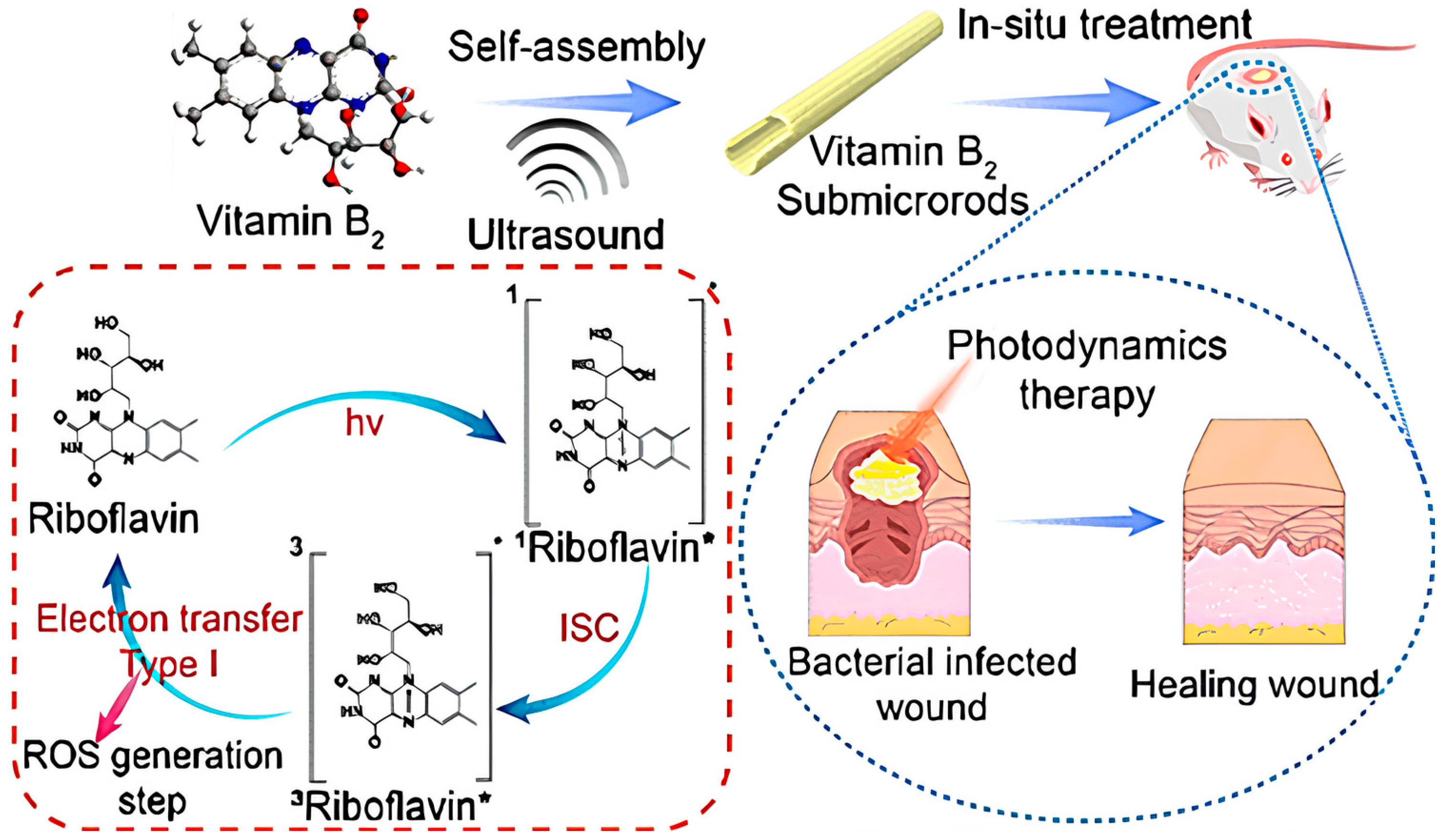
  58. Feng, J.; Yang, X.; Wang, T.; Du, T.; Zhuo, J.; Zhang, P.; Zhang, L.; Sun, H.; Han, Y.; Xia, Y.; et al. Reassembled One-Dimensional VB2 Submicrorods with Enhanced Photosensitivity and H2O2 Supply for Efficient Antibacterial Therapy. ACS Sustain. Chem. Eng. 2023, 11, 13081–13095. [Google Scholar] [CrossRef]
  59. Siegman, A.E. Lasers; University Science Books: Herndon, VA, USA, 1986. [Google Scholar]
  60. Toropov, N.; Cabello, G.; Serrano, M.P.; Gutha, R.R.; Rafti, M.; Vollmer, F. Review of biosensing with whispering-gallery mode lasers. Light Sci. Appl. 2021, 10, 42. [Google Scholar] [CrossRef]
  61. Chen, Y.-C.; Tan, X.; Sun, Q.; Chen, Q.; Wang, W.; Fan, X. Laser-emission imaging of nuclear biomarkers for high-contrast cancer screening and immunodiagnosis. Nat. Biomed. Eng. 2017, 1, 724–735. [Google Scholar] [CrossRef] [PubMed]
  62. Glyn MacDonald, R.; Yakovlev, A.; Pacheco-Peña, V. Time derivatives via interconnected waveguides. Sci. Rep. 2023, 13, 13126. [Google Scholar] [CrossRef] [PubMed]
  63. Schubert, M.; Steude, A.; Liehm, P.; Kronenberg, N.M.; Karl, M.; Campbell, E.C.; Powis, S.J.; Gather, M.C. Lasing within Live Cells Containing Intracellular Optical Microresonators for Barcode-Type Cell Tagging and Tracking. Nano Lett. 2015, 15, 5647–5652. [Google Scholar] [CrossRef] [PubMed]
  64. Humar, M.; Gather, M.C.; Yun, S.-H. Cellular dye lasers: Lasing thresholds and sensing in a planar resonator. Opt. Express 2015, 23, 27865–27879. [Google Scholar] [CrossRef] [PubMed]
  65. Gather, M.C.; Yun, S.H. Single-cell biological lasers. Nat. Photonics 2011, 5, 406–410. [Google Scholar] [CrossRef]
  66. Gather, M.C.; Yun, S.H. Lasing from Escherichia coli bacteria genetically programmed to express green fluorescent protein. Opt. Lett. 2011, 36, 3299–3301. [Google Scholar] [CrossRef]
  67. Abbadessa, A.; Blokzijl, M.M.; Mouser, V.H.; Marica, P.; Malda, J.; Hennink, W.E.; Vermonden, T. A thermo-responsive and photo-polymerizable chondroitin sulfate-based hydrogel for 3D printing applications. Carbohydr. Polym. 2016, 149, 163–174. [Google Scholar] [CrossRef] [PubMed]
  68. Karl, M.; Dietrich, C.; Schubert, M.; Samuel, I.; Turnbull, G.; Gather, M. Single cell induced optical confinement in biological lasers. J. Phys. D Appl. Phys. 2017, 50, 084005. [Google Scholar] [CrossRef]
  69. Nizamoglu, S.; Lee, K.B.; Gather, M.C.; Kim, K.S.; Jeon, M.; Kim, S.; Humar, M.; Yun, S.H. A simple approach to biological single-cell lasers via intracellular dyes. Adv. Opt. Mater. 2015, 3, 1197–1200. [Google Scholar] [CrossRef]
  70. Fikouras, A.H.; Schubert, M.; Karl, M.; Kumar, J.D.; Powis, S.J.; Di Falco, A.; Gather, M.C. Non-obstructive intracellular nanolasers. Nat. Commun. 2018, 9, 4817. [Google Scholar] [CrossRef]
  71. Sun, Y.; Fan, X. Distinguishing DNA by Analog-to-Digital-like Conversion by Using Optofluidic Lasers. Angew. Chem. Int. Ed. 2012, 51, 1236–1239. [Google Scholar] [CrossRef]
  72. Chen, Y.-C.; Chen, Q.; Zhang, T.; Wang, W.; Fan, X. Versatile tissue lasers based on high-Q Fabry–Pérot microcavities. Lab A Chip 2017, 17, 538–548. [Google Scholar] [CrossRef]
  73. Septiadi, D.; Barna, V.; Saxena, D.; Sapienza, R.; Genovese, D.; De Cola, L. Biolasing from Individual cells in a low-Q resonator enables spectral fingerprinting. Adv. Opt. Mater. 2020, 8, 1901573. [Google Scholar] [CrossRef]
  74. Wang, J.; Wang, L.; Su, X.; Gao, D.; Yu, H. CdTe quantum dot-based self-supporting films with enhanced stability for flexible light-emitting devices. Soft Matter. 2022, 18, 7324–7332. [Google Scholar] [CrossRef] [PubMed]
  75. Matsko, A.B.; Ilchenko, V.S. Optical resonators with whispering-gallery modes-part I: Basics. IEEE J. Sel. Top. Quantum Electron. 2006, 12, 3–14. [Google Scholar] [CrossRef]
  76. Jiang, X.; Qavi, A.J.; Huang, S.H.; Yang, L. Whispering-gallery sensors. Matter 2020, 3, 371–392. [Google Scholar] [CrossRef]
  77. Zhi, Y.; Yu, X.C.; Gong, Q.; Yang, L.; Xiao, Y.F. Single nanoparticle detection using optical microcavities. Adv. Mater. 2017, 29, 1604920. [Google Scholar] [CrossRef] [PubMed]
  78. He, L.; Özdemir, Ş.K.; Yang, L. Whispering gallery microcavity lasers. Laser Photonics Rev. 2013, 7, 60–82. [Google Scholar] [CrossRef]
  79. Guo, C.; Hu, J.; Kao, L.; Pan, D.; Luo, K.; Li, N.; Gu, Z. Pepetide Dendron-Functionalized Mesoporous Silica Nanoparticle-Based Nanohybrid: Biocompatibility and Its Potential as Imaging Probe. ACS Biomater. Sci. Eng. 2016, 2, 860–870. [Google Scholar] [CrossRef]
  80. Kamoshita, A.; Kohno, J.-Y. Cavity-Enhanced Fluorescence in Colliding Droplets of Rhodamine 6G Aqueous Solutions. J. Phys. Chem. A 2023, 127, 7605–7611. [Google Scholar] [CrossRef]
  81. Ma, Y.; Nolte, R.J.; Cornelissen, J.J. Virus-based nanocarriers for drug delivery. Adv. Drug Deliv. Rev. 2012, 64, 811–825. [Google Scholar] [CrossRef]
  82. Labrie, S.J.; Samson, J.E.; Moineau, S. Bacteriophage resistance mechanisms. Nat. Rev. Microbiol. 2010, 8, 317–327. [Google Scholar] [CrossRef]
  83. Kutter, E.; De Vos, D.; Gvasalia, G.; Alavidze, Z.; Gogokhia, L.; Kuhl, S.; Abedon, S.T. Phage therapy in clinical practice: Treatment of human infections. Curr. Pharm. Biotechnol. 2010, 11, 69–86. [Google Scholar] [CrossRef] [PubMed]
  84. Morales, N.M.; McClean, M.N. Engineered bacteria self-organize to sense pressure. Nat. Biotechnol. 2017, 35, 1045–1047. [Google Scholar] [CrossRef] [PubMed]
  85. Peivandi, A.; Tian, L.; Mahabir, R.; Hosseinidoust, Z. Hierarchically structured, self-healing, fluorescent, bioactive hydrogels with self-organizing bundles of phage nanofilaments. Chem. Mater. 2019, 31, 5442–5449. [Google Scholar] [CrossRef]
  86. Lee, J.H.; Warner, C.M.; Jin, H.-E.; Barnes, E.; Poda, A.R.; Perkins, E.J.; Lee, S.-W. Production of tunable nanomaterials using hierarchically assembled bacteriophages. Nat. Protoc. 2017, 12, 1999–2013. [Google Scholar] [CrossRef] [PubMed]
  87. Arter, J.A.; Taggart, D.K.; McIntire, T.M.; Penner, R.M.; Weiss, G.A. Virus-PEDOT nanowires for biosensing. Nano Lett. 2010, 10, 4858–4862. [Google Scholar] [CrossRef] [PubMed]
  88. Li, L.; Zhang, H.; Song, D.; Xu, K.; Zheng, Y.; Xiao, H.; Liu, Y.; Li, J.; Song, X. Simultaneous detection of three zoonotic pathogens based on phage display peptide and multicolor quantum dots. Anal. Biochem. 2020, 608, 113854. [Google Scholar] [CrossRef]
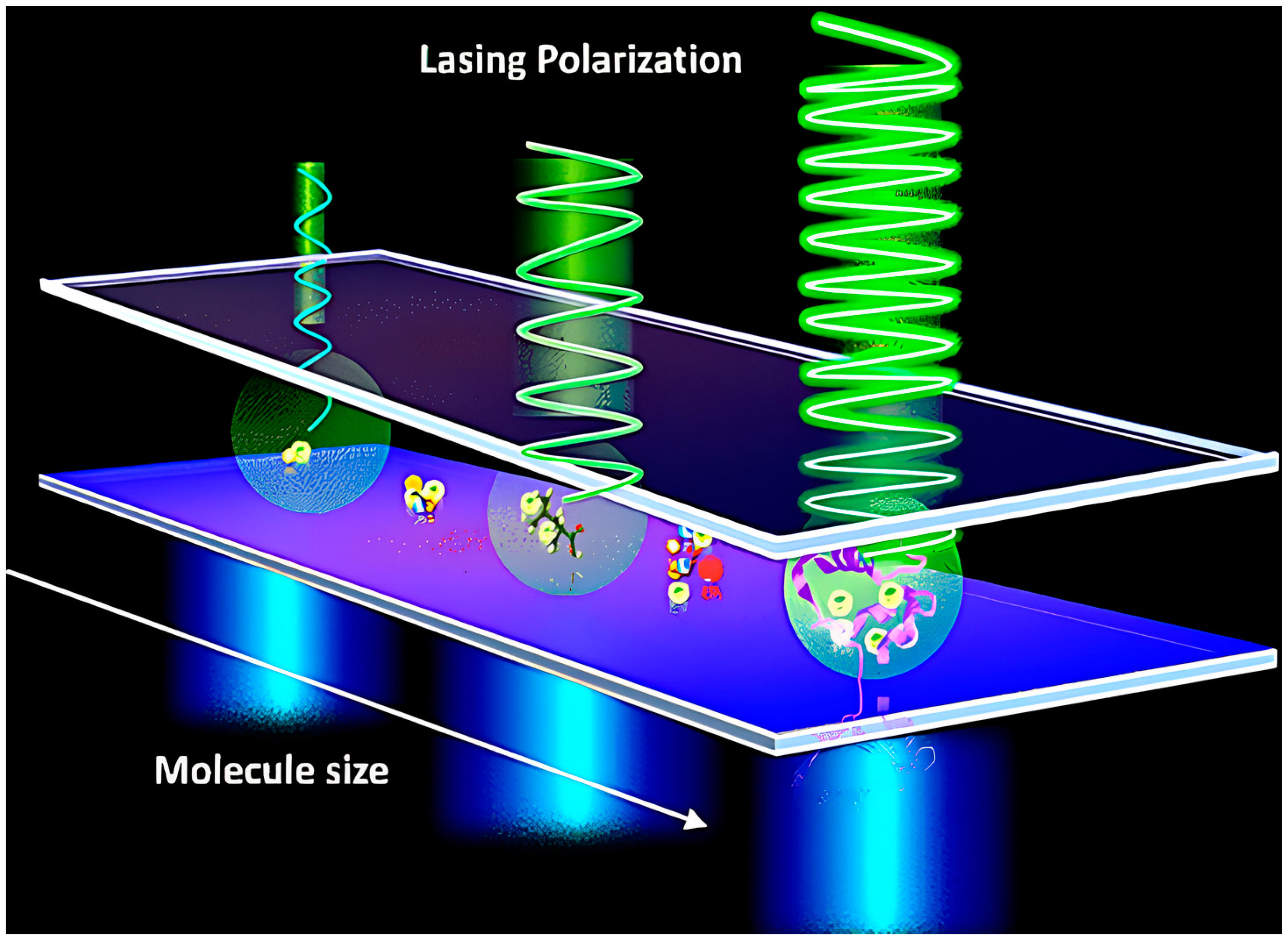
  89. Yuan, Z.; Cheng, X.; Zhou, Y.; Tan, X.; Gong, X.; Rivy, H.; Gong, C.; Fan, X.; Wang, W.-J.; Chen, Y.-C. Distinguishing Small Molecules in Microcavity with Molecular Laser Polarization. ACS Photonics 2020, 7, 1908–1914. [Google Scholar] [CrossRef]
  90. Ignesti, E.; Tommasi, F.; Fini, L.; Martelli, F.; Azzali, N.; Cavalieri, S. A new class of optical sensors: A random laser based device. Sci. Rep. 2016, 6, 35225. [Google Scholar] [CrossRef]
  91. Polson, R.C.; Vardeny, Z.V. Random lasing in human tissues. Appl. Phys. Lett. 2004, 85, 1289–1291. [Google Scholar] [CrossRef]
  92. Briones-Herrera, J.; Cuando-Espitia, N.; Sánchez-Arévalo, F.; Hernández-Cordero, J. Evaluation of mechanical behavior of soft tissue by means of random laser emission. Rev. Sci. Instrum. 2013, 84, 104301. [Google Scholar] [CrossRef]
  93. Wang, L.; Liu, D.; He, N.; Jacques, S.L.; Thomsen, S.L. Biological laser action. Appl. Opt. 1996, 35, 1775–1779. [Google Scholar] [CrossRef] [PubMed]
  94. Lahoz, F.; Acebes, A.; González-Hernández, T.; de Armas-Rillo, S.; Soler-Carracedo, K.; Cuesto, G.; Mesa-Infante, V. Random lasing in brain tissues. Org. Electron. 2019, 75, 105389. [Google Scholar] [CrossRef]
  95. Lahoz, F.; Martín, I.R.; Urgellés, M.; Marrero-Alonso, J.; Marín, R.; Saavedra, C.J.; Boto, A.; Díaz, M. Random laser in biological tissues impregnated with a fluorescent anticancer drug. Laser Phys. Lett. 2015, 12, 045805. [Google Scholar] [CrossRef]
  96. Wang, Y.; Duan, Z.; Qiu, Z.; Zhang, P.; Wu, J.; Zhang, D.; Xiang, T. Random lasing in human tissues embedded with organic dyes for cancer diagnosis. Sci. Rep. 2017, 7, 8385. [Google Scholar] [CrossRef] [PubMed]
  97. Zhang, D.; Wang, Y.; Tang, J.; Mu, H. Random laser marked PLCD1 gene therapy effect on human breast cancer. J. Appl. Phys. 2019, 125, 203102. [Google Scholar] [CrossRef]
  98. Hohmann, M.; Dörner, D.; Mehari, F.; Chen, C.; Späth, M.; Müller, S.; Albrecht, H.; Klämpfl, F.; Schmidt, M. Investigation of random lasing as a feedback mechanism for tissue differentiation during laser surgery. Biomed. Opt. Express 2019, 10, 807–816. [Google Scholar] [CrossRef] [PubMed]
  99. Humar, M.; Dobravec, A.; Zhao, X.; Yun, S.H. Biomaterial microlasers implantable in the cornea, skin, and blood. Optica 2017, 4, 1080–1085. [Google Scholar] [CrossRef]
  100. Martino, N.; Kwok, S.J.J.; Liapis, A.C.; Forward, S.; Jang, H.; Kim, H.-M.; Wu, S.J.; Wu, J.; Dannenberg, P.H.; Jang, S.-J.; et al. Wavelength-encoded laser particles for massively multiplexed cell tagging. Nat. Photonics 2019, 13, 720–727. [Google Scholar] [CrossRef]
  101. Jiang, X.F.; Zou, C.L.; Wang, L.; Gong, Q.; Xiao, Y.F. Whispering-gallery microcavities with unidirectional laser emission. Laser Photonics Rev. 2016, 10, 40–61. [Google Scholar] [CrossRef]
  102. Tang, S.-J.; Dannenberg, P.H.; Liapis, A.C.; Martino, N.; Zhuo, Y.; Xiao, Y.-F.; Yun, S.-H. Laser particles with omnidirectional emission for cell tracking. Light Sci. Appl. 2021, 10, 23. [Google Scholar] [CrossRef]
  103. Li, X.; Zhang, W.; Wang, W.Y.; Wu, X.; Li, Y.; Tan, X.; Matera, D.L.; Baker, B.M.; Paulus, Y.M.; Fan, X. Optical coherence tomography and fluorescence microscopy dual-modality imaging for in vivo single-cell tracking with nanowire lasers. Biomed. Opt. Express 2020, 11, 3659–3672. [Google Scholar] [CrossRef] [PubMed]
  104. Chen, Y.-C.; Chen, Q.; Wu, X.; Tan, X.; Wang, J.; Fan, X. A robust tissue laser platform for analysis of formalin-fixed paraffin-embedded biopsies. Lab A Chip 2018, 18, 1057–1065. [Google Scholar] [CrossRef] [PubMed]
  105. Chen, Y.-C.; Chen, Q.; Tan, X.; Chen, G.; Bergin, I.; Aslam, M.N.; Fan, X. Chromatin laser imaging reveals abnormal nuclear changes for early cancer detection. Biomed. Opt. Express 2019, 10, 838–854. [Google Scholar] [CrossRef]
  106. Tan, X.; Chen, Q.; Zhu, H.; Zhu, S.; Gong, Y.; Wu, X.; Chen, Y.-C.; Li, X.; Li, M.W.-H.; Liu, W. Fast and reproducible ELISA laser platform for ultrasensitive protein quantification. ACS Sens. 2019, 5, 110–117. [Google Scholar] [CrossRef] [PubMed]
  107. Chen, Y.-C.; Li, X.; Zhu, H.; Weng, W.-H.; Tan, X.; Chen, Q.; Sun, Y.; Wang, X.; Fan, X. Monitoring neuron activities and interactions with laser emissions. ACS Photonics 2020, 7, 2182–2189. [Google Scholar] [CrossRef]
  108. Schubert, M.; Woolfson, L.; Barnard, I.R.; Dorward, A.M.; Casement, B.; Morton, A.; Robertson, G.B.; Appleton, P.L.; Miles, G.B.; Tucker, C.S. Monitoring contractility in cardiac tissue with cellular resolution using biointegrated microlasers. Nat. Photonics 2020, 14, 452–458. [Google Scholar] [CrossRef]
  109. Lv, Y.; Xiong, X.; Liu, Y.; Yao, J.; Li, Y.J.; Zhao, Y.S. Controlled Outcoupling of Whispering-Gallery-Mode Lasers Based on Self-Assembled Organic Single-Crystalline Microrings. Nano Lett. 2019, 19, 1098–1103. [Google Scholar] [CrossRef] [PubMed]
  110. Ettinger, A.; Wittmann, T. Fluorescence live cell imaging. Methods Cell Biol. 2014, 123, 77–94. [Google Scholar]
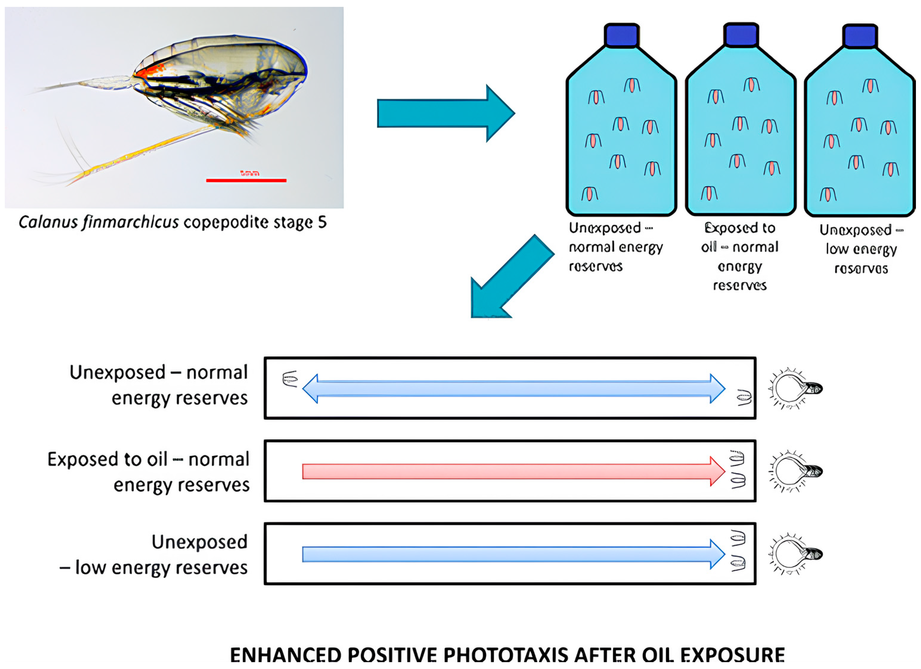
  111. Miljeteig, C.; Olsen, A.J.; Nordtug, T.; Altin, D.; Jenssen, B.M. Sublethal Exposure to Crude Oil Enhances Positive Phototaxis in the Calanoid Copepod Calanus finmarchicus. Environ. Sci. Technol. 2013, 47, 14426–14433. [Google Scholar] [CrossRef]
  112. Wang, C.; Tao, H.; Cheng, L.; Liu, Z. Near-infrared light induced in vivo photodynamic therapy of cancer based on upconversion nanoparticles. Biomaterials 2011, 32, 6145–6154. [Google Scholar] [CrossRef] [PubMed]
  113. Chen, H.; Yao, Y.; Zhao, X.; Tan, N. Programmable site-specific delivery of an alkaline phosphatase-activatable prodrug and a mitochondria-targeted cyclopeptide for combination therapy in colon cancer. Biomater. Sci. 2023. [Google Scholar] [CrossRef] [PubMed]
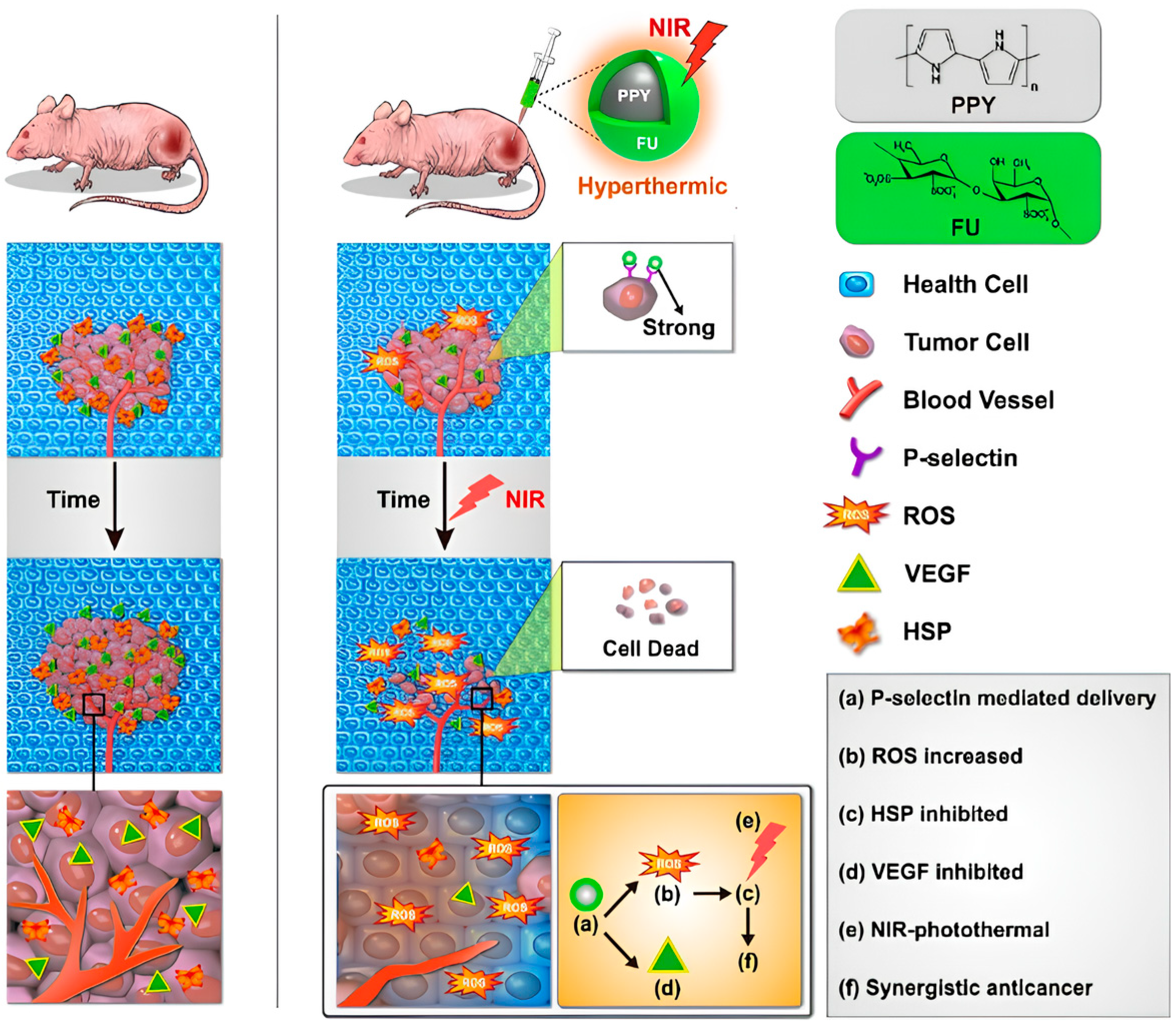
  114. Lu, K.-Y.; Jheng, P.-R.; Lu, L.-S.; Rethi, L.; Mi, F.-L.; Chuang, E.-Y. Enhanced anticancer effect of ROS-boosted photothermal therapy by using fucoidan-coated polypyrrole nanoparticles. Int. J. Biol. Macromol. 2021, 166, 98–107. [Google Scholar] [CrossRef]
  115. Hsiao, Y.-C.; Jheng, P.-R.; Nguyen, H.T.; Chen, Y.-H.; Manga, Y.B.; Lu, L.-S.; Rethi, L.; Chen, C.-H.; Huang, T.-W.; Lin, J.-D.; et al. Photothermal-Irradiated Polyethyleneimine–Polypyrrole Nanopigment Film-Coated Polyethylene Fabrics for Infrared-Inspired with Pathogenic Evaluation. ACS Appl. Mater. Interfaces 2021, 13, 2483–2495. [Google Scholar] [CrossRef] [PubMed]
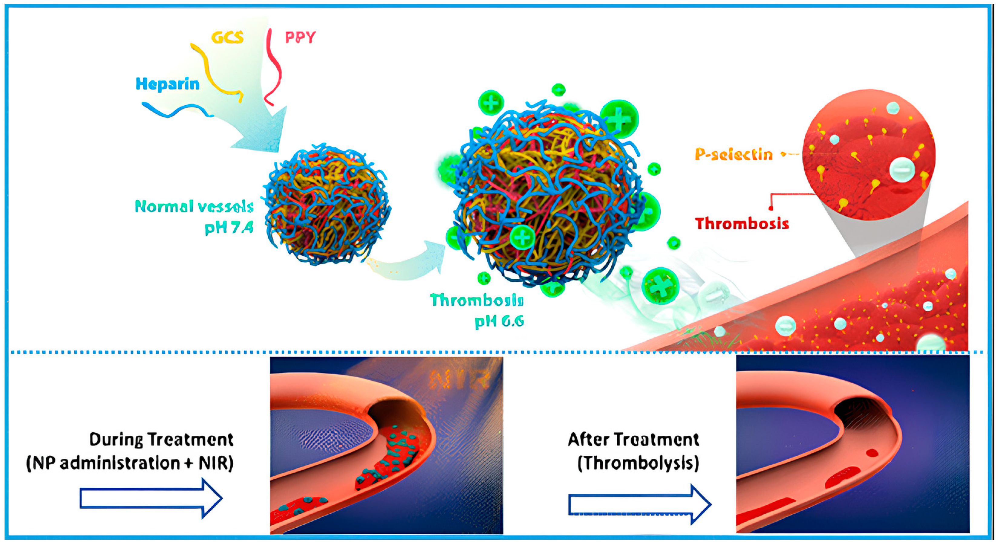
  116. Lu, T.-Y.; Chiang, C.-Y.; Fan, Y.-J.; Jheng, P.-R.; Quiñones, E.D.; Liu, K.-T.; Kuo, S.-H.; Hsieh, H.Y.; Tseng, C.-L.; Yu, J.; et al. Dual-Targeting Glycol Chitosan/Heparin-Decorated Polypyrrole Nanoparticle for Augmented Photothermal Thrombolytic Therapy. ACS Appl. Mater. Interfaces 2021, 13, 10287–10300. [Google Scholar] [CrossRef]
  117. Dianov, E.M. Bismuth-doped optical fibers: A challenging active medium for near-IR lasers and optical amplifiers. Light Sci. Appl. 2012, 1, e12. [Google Scholar] [CrossRef]
  118. Haddock, S.H.; Moline, M.A.; Case, J.F. Bioluminescence in the sea. Annu. Rev. Mar. Sci. 2010, 2, 443–493. [Google Scholar] [CrossRef] [PubMed]
  119. Oertner, T.G. The Bright Side of Life; Nature Publishing Group: London, UK, 2006. [Google Scholar]
  120. Lee, H.-J.; Ha, J.-H.; Kim, S.-G.; Choi, H.-K.; Kim, Z.H.; Han, Y.-J.; Kim, J.-I.; Oh, Y.; Fragoso, V.; Shin, K. Stem-piped light activates phytochrome B to trigger light responses in Arabidopsis thaliana roots. Sci. Signal. 2016, 9, ra106. [Google Scholar] [CrossRef] [PubMed]
  121. Sundar, V.C.; Yablon, A.D.; Grazul, J.L.; Ilan, M.; Aizenberg, J. Fibre-optical features of a glass sponge. Nature 2003, 424, 899–900. [Google Scholar] [CrossRef]
  122. Franze, K.; Grosche, J.; Skatchkov, S.N.; Schinkinger, S.; Foja, C.; Schild, D.; Uckermann, O.; Travis, K.; Reichenbach, A.; Guck, J. Müller cells are living optical fibers in the vertebrate retina. Proc. Natl. Acad. Sci. USA 2007, 104, 8287–8292. [Google Scholar] [CrossRef]
  123. Agte, S.; Junek, S.; Matthias, S.; Ulbricht, E.; Erdmann, I.; Wurm, A.; Schild, D.; Käs, J.A.; Reichenbach, A. Müller glial cell-provided cellular light guidance through the vital guinea-pig retina. Biophys. J. 2011, 101, 2611–2619. [Google Scholar] [CrossRef] [PubMed]
  124. Pena-Francesch, A.; Florez, S.; Jung, H.; Sebastian, A.; Albert, I.; Curtis, W.; Demirel, M.C. Materials fabrication from native and recombinant thermoplastic squid proteins. Adv. Funct. Mater. 2014, 24, 7401–7409. [Google Scholar] [CrossRef]
  125. Shan, D.; Gerhard, E.; Zhang, C.; Tierney, J.W.; Xie, D.; Liu, Z.; Yang, J. Polymeric biomaterials for biophotonic applications. Bioact. Mater. 2018, 3, 434–445. [Google Scholar] [CrossRef]
  126. Jeevarathinam, A.S.; Pai, N.; Huang, K.; Hariri, A.; Wang, J.; Bai, Y.; Wang, L.; Hancock, T.; Keys, S.; Penny, W. A cellulose-based photoacoustic sensor to measure heparin concentration and activity in human blood samples. Biosens. Bioelectron. 2019, 126, 831–837. [Google Scholar] [CrossRef] [PubMed]
  127. Nazempour, R.; Zhang, Q.; Fu, R.; Sheng, X. Biocompatible and implantable optical fibers and waveguides for biomedicine. Materials 2018, 11, 1283. [Google Scholar] [CrossRef] [PubMed]
  128. Monks, J.N.; Yan, B.; Hawkins, N.; Vollrath, F.; Wang, Z. Spider silk: Mother nature’s bio-superlens. Nano Lett. 2016, 16, 5842–5845. [Google Scholar] [CrossRef] [PubMed]
  129. Parker, S.T.; Domachuk, P.; Amsden, J.; Bressner, J.; Lewis, J.A.; Kaplan, D.L.; Omenetto, F.G. Biocompatible silk printed optical waveguides. Adv. Mater. 2009, 21, 2411–2415. [Google Scholar] [CrossRef]
  130. Li, D.; Wang, L. Cellulose acetate polymer film modified microstructured polymer optical fiber towards a nitrite optical probe. Opt. Commun. 2010, 283, 2841–2844. [Google Scholar] [CrossRef]
  131. Roxby, D.N.; Yuan, Z.; Krishnamoorthy, S.; Wu, P.; Tu, W.C.; Chang, G.E.; Lau, R.; Chen, Y.C. Enhanced biophotocurrent generation in living photosynthetic optical resonator. Adv. Sci. 2020, 7, 1903707. [Google Scholar] [CrossRef]
  132. Huby, N.; Vié, V.; Renault, A.; Beaufils, S.; Lefevre, T.; Paquet-Mercier, F.; Pézolet, M.; Bêche, B. Native spider silk as a biological optical fiber. Appl. Phys. Lett. 2013, 102, 123702. [Google Scholar] [CrossRef]
  133. Sun, Y.-L.; Sun, S.-M.; Wang, P.; Dong, W.-F.; Zhang, L.; Xu, B.-B.; Chen, Q.-D.; Tong, L.-M.; Sun, H.-B. Customization of Protein Single Nanowires for Optical Biosensing. Small 2015, 11, 2869–2876. [Google Scholar] [CrossRef]
  134. Shabahang, S.; Kim, S.; Yun, S.H. Light-guiding biomaterials for biomedical applications. Adv. Funct. Mater. 2018, 28, 1706635. [Google Scholar] [CrossRef]
  135. Jacques, S.L. Optical properties of biological tissues: A review. Phys. Med. Biol. 2013, 58, R37. [Google Scholar] [CrossRef]
  136. Xin, H.; Li, Y.; Li, B. Bacteria-based branched structures for bionanophotonics. Laser Photonics Rev. 2015, 9, 554–563. [Google Scholar] [CrossRef]
  137. Fardad, S.; Salandrino, A.; Heinrich, M.; Zhang, P.; Chen, Z.; Christodoulides, D.N. Plasmonic resonant solitons in metallic nanosuspensions. Nano Lett. 2014, 14, 2498–2504. [Google Scholar] [CrossRef]
  138. Kelly, T.S.; Ren, Y.-X.; Samadi, A.; Bezryadina, A.; Christodoulides, D.; Chen, Z. Guiding and nonlinear coupling of light in plasmonic nanosuspensions. Opt. Lett. 2016, 41, 3817–3820. [Google Scholar] [CrossRef]
  139. Yashin, V.; Chizhov, S.; Sabirov, R.; Starchikova, T.; Vysotina, N.; Rozanov, N.; Semenov, V.; Smirnov, V.; Fedorov, S. Formation of soliton-like light beams in an aqueous suspension of polystyrene particles. Opt. Spectrosc. 2005, 98, 466–469. [Google Scholar] [CrossRef]
  140. Bezryadina, A.; Hansson, T.; Gautam, R.; Wetzel, B.; Siggins, G.; Kalmbach, A.; Lamstein, J.; Gallardo, D.; Carpenter, E.J.; Ichimura, A. Nonlinear self-action of light through biological suspensions. Phys. Rev. Lett. 2017, 119, 058101. [Google Scholar] [CrossRef] [PubMed]
  141. Gautam, R.; Xiang, Y.; Lamstein, J.; Liang, Y.; Bezryadina, A.; Liang, G.; Hansson, T.; Wetzel, B.; Preece, D.; White, A. Optical force-induced nonlinearity and self-guiding of light in human red blood cell suspensions. Light Sci. Appl. 2019, 8, 31. [Google Scholar] [CrossRef] [PubMed]
  142. Perez, N.; Chambers, J.; Chen, Z.; Bezryadina, A. Nonlinear self-trapping and guiding of light at different wavelengths with sheep blood. Opt. Lett. 2021, 46, 629–632. [Google Scholar] [CrossRef] [PubMed]
  143. Burnouf, T.; Chen, C.H.; Tan, S.J.; Tseng, C.L.; Lu, K.Y.; Chang, L.H.; Nyambat, B.; Huang, S.C.; Jheng, P.R.; Aditya, R.N.; et al. A bioinspired hyperthermic macrophage-based polypyrrole-polyethylenimine (Ppy-PEI) nanocomplex carrier to prevent and disrupt thrombotic fibrin clots. Acta Biomater. 2019, 96, 468–479. [Google Scholar] [CrossRef] [PubMed]
  144. Xie, S.; Li, S.; Cao, W.; Mo, C.; Zhang, Z.; Huang, K.; Li, X. Bacteria-Templated and Dual Enzyme-Powered Microcapsule Motors To Promote Thrombus Penetration and Thrombolytic Efficacy. ACS Appl. Mater. Interfaces 2022, 14, 37553–37565. [Google Scholar] [CrossRef]
  145. Chang, L.H.; Chuang, E.Y.; Cheng, T.M.; Lin, C.; Shih, C.M.; Wu, A.T.; Jheng, P.R.; Lu, H.Y.; Shih, C.C.; Mi, F.L. Thrombus-specific theranostic nanocomposite for codelivery of thrombolytic drug, algae-derived anticoagulant and NIR fluorescent contrast agent. Acta Biomater. 2021, 134, 686–701. [Google Scholar] [CrossRef] [PubMed]
  146. Chen, Y.-T.; Liu, C.-H.; Pan, W.-Y.; Jheng, P.-R.; Hsieh, Y.S.; Burnouf, T.; Fan, Y.-J.; Chiang, C.-C.; Chen, T.-Y.; Chuang, E.-Y. Biomimetic Platelet Nanomotors for Site-Specific Thrombolysis and Ischemic Injury Alleviation. ACS Appl. Mater. Interfaces 2023, 15, 32967–32983. [Google Scholar] [CrossRef] [PubMed]
  147. Liu, K.T.; Quiñones, E.D.; Liu, M.H.; Lin, C.W.; Chen, Y.T.; Chiang, C.C.; Wu, K.C.W.; Fan, Y.J.; Chuang, E.Y.; Yu, J. A Biomimicking and Multiarm Self-Indicating Nanoassembly for Site-Specific Photothermal-Potentiated Thrombolysis Assessed in Microfluidic and In Vivo Models. Adv. Healthc. Mater. 2023, 12, 2300682. [Google Scholar] [CrossRef]
  148. Liu, C.H.; Liu, M.C.; Jheng, P.R.; Yu, J.; Fan, Y.J.; Liang, J.W.; Hsiao, Y.C.; Chiang, C.W.; Bolouki, N.; Lee, J.W. Plasma-Derived Nanoclusters for Site-Specific Multimodality Photo/Magnetic Thrombus Theranostics. Adv. Healthc. Mater. 2023, 2301504. [Google Scholar] [CrossRef] [PubMed]
  149. Liu, C.-H.; Jheng, P.-R.; Rethi, L.; Godugu, C.; Lee, C.Y.; Chen, Y.-T.; Nguyen, H.T.; Chuang, E.-Y. P-Selectin mediates targeting of a self-assembling phototherapeutic nanovehicle enclosing dipyridamole for managing thromboses. J. Nanobiotechnol. 2023, 21, 260. [Google Scholar] [CrossRef]
  150. Liang, J.-W.; Liu, C.-H.; Wu, K.C.-W.; Jheng, P.-R.; Chuang, E.-Y. Plasma-treated nano-enabled multimodal coatings made of phototherapeutic molybdenum disulfide and fucoidan prevent catheter-associated urinary tract issues. Chem. Eng. J. 2023, 468, 143749. [Google Scholar] [CrossRef]
  151. Burnouf, T.; Jheng, P.-R.; Chen, Y.-H.; Rethi, L.; Rethi, L.; Lu, L.-S.; Ho, Y.-C.; Chuang, E.-Y. Near-infrared-driven photoablation of lung cancer tumors utilizing biomimetic platelet-polyethyleneimine-polypyrrole drug-free nanoparticles. Mater. Des. 2022, 215, 110481. [Google Scholar] [CrossRef]
  152. Lu, Z.; Zhou, H.; Ye, C.; Chen, S.; Ning, J.; Halim, M.A.; Donaev, S.B.; Wang, S. Fabrication of Iron Pyrite Thin Films and Photovoltaic Devices by Sulfurization in Electrodeposition Method. Nanomaterials 2021, 11, 2844. [Google Scholar] [CrossRef] [PubMed]
  153. Chiang, C.-W.; Chuang, E.-Y. Biofunctional core-shell polypyrrole–polyethylenimine nanocomplex for a locally sustained photothermal with reactive oxygen species enhanced therapeutic effect against lung cancer. Int. J. Nanomed. 2019, 14, 1575–1585. [Google Scholar] [CrossRef]
  154. Mi, F.L.; Burnouf, T.; Lu, S.Y.; Lu, Y.J.; Lu, K.Y.; Ho, Y.C.; Kuo, C.Y.; Chuang, E.Y. Self-Targeting, Immune Transparent Plasma Protein Coated Nanocomplex for Noninvasive Photothermal Anticancer Therapy. Adv. Healthc. Mater. 2017, 6, 1700181. [Google Scholar] [CrossRef]
  155. Satapathy, M.K.; Nyambat, B.; Chiang, C.-W.; Chen, C.-H.; Wong, P.-C.; Ho, P.-H.; Jheng, P.-R.; Burnouf, T.; Tseng, C.-L.; Chuang, E.-Y. A gelatin hydrogel-containing nano-organic PEI–Ppy with a photothermal responsive effect for tissue engineering applications. Molecules 2018, 23, 1256. [Google Scholar] [CrossRef] [PubMed]
  156. Chiang, C.-W.; Hsiao, Y.-C.; Jheng, P.-R.; Chen, C.-H.; Manga, Y.B.; Lekha, R.; Chao, K.-M.; Ho, Y.-C.; Chuang, E.-Y. Strontium ranelate-laden near-infrared photothermal-inspired methylcellulose hydrogel for arthritis treatment. Mater. Sci. Eng. C 2021, 123, 111980. [Google Scholar] [CrossRef] [PubMed]
  157. Bolouki, N.; Hsu, Y.-N.; Hsiao, Y.-C.; Jheng, P.-R.; Hsieh, J.-H.; Chen, H.-L.; Mansel, B.W.; Yeh, Y.-Y.; Chen, Y.-H.; Lu, C.-X. Cold atmospheric plasma physically reinforced substances of platelets-laden photothermal-responsive methylcellulose complex restores burn wounds. Int. J. Biol. Macromol. 2021, 192, 506–515. [Google Scholar] [CrossRef] [PubMed]
  158. Chuang, E.-Y.; Huang, W.-H.; Ho, T.-L.; Wang, P.-C.; Hsiao, Y.-C. IR-inspired visual display/response device fabricated using photothermal liquid crystals for medical and display applications. Chem. Eng. J. 2022, 429, 132213. [Google Scholar] [CrossRef]
  159. Li, Y.; Xin, H.; Zhang, Y.; Lei, H.; Zhang, T.; Ye, H.; Saenz, J.J.; Qiu, C.-W.; Li, B. Living Nanospear for Near-Field Optical Probing. ACS Nano 2018, 12, 10703–10711. [Google Scholar] [CrossRef]
  160. Wu, T.; Chen, X.; Gong, Z.; Li, Y.; Zhang, Y. Waveguiding and focusing in a bio-medium with an optofluidic cell chain. Acta Biomater. 2020, 103, 165–171. [Google Scholar] [CrossRef]
  161. Li, Y.; Liu, X.; Xu, X.; Xin, H.; Zhang, Y.; Li, B. Red-Blood-Cell Waveguide as a Living Biosensor and Micromotor. Adv. Funct. Mater. 2019, 29, 1905568. [Google Scholar] [CrossRef]
  162. Arano-Martinez, J.A.; Martínez-González, C.L.; Salazar, M.I.; Torres-Torres, C. A Framework for Biosensors Assisted by Multiphoton Effects and Machine Learning. Biosensors 2022, 12, 710. [Google Scholar] [CrossRef]
Scheme 1. The illustration portrays the biological laser system, photonic waveguides, and photonic-based theragnostic applications in translational medicine.
Photonics 10 01124 sch001
Figure 1. Illustration of the previous study’s emphasis on manipulating upconversion emission channels through the utilization of adaptable biological photonic devices, which was reproduced from [41]. Copyright 2021 American Chemical Society.
Photonics 10 01124 g001
Figure 2. Illustration depicting the primary objective of the prior research: augmenting photosensitivity within reassembled one-dimensional VB2 submicron rods, to facilitate effective antibacterial therapy, which was reproduced from [58]. Copyright 2023 American Chemical Society.
Photonics 10 01124 g002
Figure 3. Illustration depicting the central focus of the earlier study: the observation of cavity-enhanced fluorescence within colliding droplets of aqueous solutions containing Rhodamine 6G, which was reproduced from [80]. Copyright 2023 American Chemical Society.
Photonics 10 01124 g003
Figure 4. Illustration depicting the primary focus of the prior study: distinguishing of small molecules within a microcavity achieved through the utilization of molecular laser polarization, which was reproduced from [89]. Copyright 2020 American Chemical Society.
Photonics 10 01124 g004
Figure 5. Illustration indicates that the earlier research centred on implementing controlled outcoupling techniques for whispering-gallery-mode lasers, employing self-assembled organic single-crystalline microrings [109]. Copyright 2019 American Chemical Society.
Photonics 10 01124 g005
Figure 6. Illustration depicting the primary subject of the previous study: the enhancement of positive phototaxis in the calanoid copepod Calanus finmarchicus due to sublethal exposure to crude oil, which was reproduced from [111]. Copyright 2013 American Chemical Society.
Photonics 10 01124 g006
Figure 7. Illustration depicting the augmented anticancer effect of photothermal therapy amplified by reactive oxygen species (ROS) utilizing fucoidan-coated polypyrrole nanoparticles, which was reproduced from [114]. Copyright 2021 Elsevier.
Photonics 10 01124 g007
Figure 8. Illustration depicting the application of photothermal-irradiated polyethyleneimine–polypyrrole nanopigment film-coated polyethylene fabrics for infrared-inspired purposes, along with the assessment of their pathogen-fighting capabilities, which was reproduced from [115]. Copyright 2021 American Chemical Society.
Photonics 10 01124 g008
Figure 9. Illustration depicting the concept of dual-targeting glycol chitosan/heparin-decorated polypyrrole nanoparticles for augmented photothermal thrombolytic therapy, which was reproduced from [116]. Copyright 2021 American Chemical Society.
Photonics 10 01124 g009
Table 1. Issues, limitations, and possible improvements on the photonic-system uses.
IssueLimitationsPossible Improvements
Fabrication challengesGenerating long-length cellular-based biological photonic waveguides/devices remains a challenge because of the geometric properties of cells. Moreover, maintaining the biological functionality as well as the viability of living cells in fabrication is crucial, limiting the ways to those that can create these waveguides without harming living cells. Further research is vital for exploring novel ways of producing long-living cellular-based biological photonic waveguides/devices under biologically compatible situations.
Limited penetration depth of lightThe depth to which light can be delivered along formed biological cellular-based waveguides/devices remains constrained, posing a hurdle for applications in vivo. Developing methods to overcome scattering losses and enhance light transmission efficiency is essential to achieve deeper tissue penetration in the body.
Cellular morphology dependenceThe optical properties of cell-based bio-micro lenses are greatly influenced by various factors, including cell morphology and the surrounding medium. Precisely controlling these factors for consistent and reliable performance is needed and it is required for further investigation.
Disclaimer/Publisher’s Note: The statements, opinions and data contained in all publications are solely those of the individual author(s) and contributor(s) and not of MDPI and/or the editor(s). MDPI and/or the editor(s) disclaim responsibility for any injury to people or property resulting from any ideas, methods, instructions or products referred to in the content.

