Systematic Review

Photobiomodulation and Growth Factors in Dentistry: A Systematic Review

by
Gianna Dipalma
1,†,
Angelo Michele Inchingolo
1,†,
Assunta Patano
1,†,
Irene Palumbo
1,
Mariafrancesca Guglielmo
1,
Irma Trilli
1,
Anna Netti
1,
Irene Ferrara
1,
Fabio Viapiano
1,
Alessio Danilo Inchingolo
1,*,
Gianfranco Favia
1,
Leonardo Dongiovanni
1,
Andrea Palermo
2,
Francesco Inchingolo
1,*,‡ and
Luisa Limongelli
1,‡
1
Department of Interdisciplinary Medicine, School of Medicine, University of Bari “Aldo Moro”, 70124 Bari, Italy
2
College of Medicine and Dentistry, Birmingham B4 6BN, UK
*
Authors to whom correspondence should be addressed.
These authors contributed equally to this work.
These authors also contributed equally to this work.
Photonics 2023, 10(10), 1095; https://doi.org/10.3390/photonics10101095
Submission received: 20 July 2023 / Revised: 18 September 2023 / Accepted: 27 September 2023 / Published: 29 September 2023

Abstract

:
Photobiomodulation, or Low-Level Laser Therapy, is a therapeutic technique that can be applied in tissue regenerative surgery. By stimulating the cellular compartment, photobiomodulation promotes cell proliferation, enabling tissue restoration after dental extractions, periodontal surgical treatments, or the management of traumatic oral injuries. On the other hand, Platelet-Rich Plasma (PRP) or Platelet-Rich Fibrin (PRF) therapy is particularly effective in providing a source of growth factors that enhance tissue healing. Authors carried out a Systematic Review following the PRISMA (Preferred Reporting Items for Systematic Reviews and Meta-Analyses) guidelines by searching PubMed, Web of Science (WoS), Scopus, and Cochrane Library databases, using the combination of the following keywords: (“low laser therapy”) AND (“growth factors”) AND (dent* OR oral) and (“photobiomodulation”) AND (“growth factors”) AND (dent* OR oral). A total of 10 publications were deemed eligible for qualitative analysis. The evidence analyzed confirms that the use of photobiomodulation and PRP/PRF (alone or in combination) can stimulate tissue regeneration, allowing for a reduction in postoperative inflammation, wound healing, and new tissue formation. Moreover, these effects are strictly dependent on the intrinsic characteristics of the individual patient, the needs, and the clinical assessment of the practitioner who will appropriately direct the treatment. Furthermore, it is necessary to investigate with evidence-based methodologies (Evidence-based Medicine, EBM) which types of patients and/or lesions are more susceptible to treatment with these tools, as well as to understand the best ways of using (alone or in combination) these important and cutting-edge therapeutic options.

1. Introduction

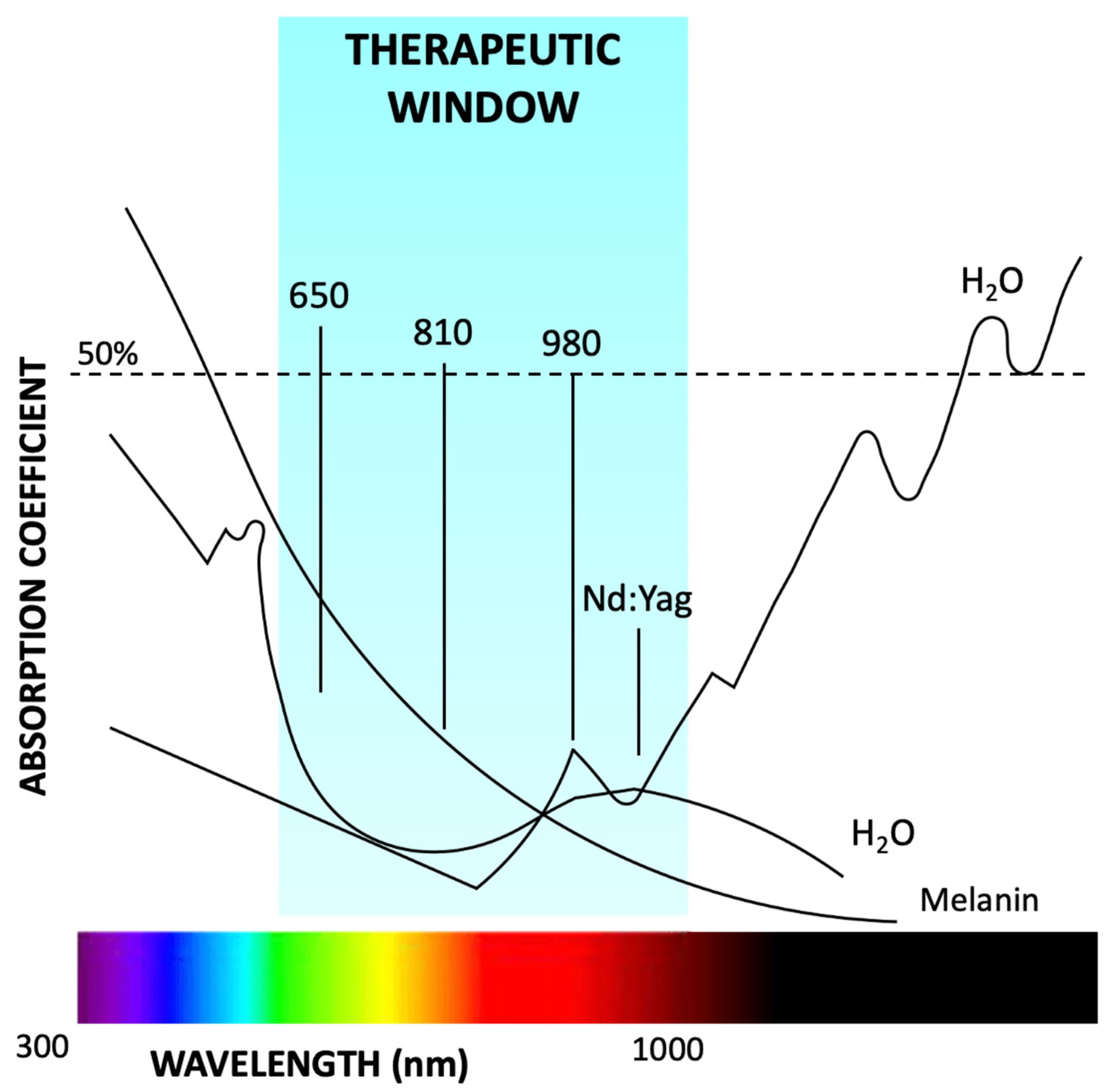
1.1. Photobiomodulation

Photobiomodulation (PBM), also known as low-level laser therapy (LLLT), represents a promising therapeutic approach to managing a wide range of medical conditions. This technique relies on the use of low-level laser radiation or LED lights with specific wavelengths to stimulate cellular and tissue responses [1].
Low Level Laser Therapy (LLLT) is low-power laser therapy, which is a different form of using the same surgical laser, but with a lower, sub-ablative power setting at a low average irradiance that produces minimal and non-destructive thermal effects on tissues. Rather, it produces stimulation and/or inhibition of biological processes and allows tissues to generate an intracellular biological response [1].
The scientific interest in PBM likely stems from the growing interest in laser technology for medical applications, particularly in stimulating healing processes, analgesics, and other treatments [2].
Results show high compliance with PBM, with no invasiveness, atraumatic, and no collateral effects. This, along with the rapid operative recovery time, has contributed to the technology’s diffusion in various fields and different patient categories, such as special needs and pediatric patients [3].

1.2. PBM Mechanisms of Action

PBM transmits energy to intracellular mitochondrial chromophores, i.e., light-absorbing molecules such as endogenous porphyrins and elements of the mitochondrial electron transport chain such as cytochrome-C oxidase. Others replaced the theory of adsorption to unit 4 of cytochrome c oxidase with that of viscosity changes in pore water [4]. At this level, laser energy is converted into metabolic energy via the respiratory chain with the resulting redox transfer of net photons leading to the production of adenosine triphosphate (ATP), and the by-product release of nitric oxide (NO) and reactive oxygen species (monatomic O). Intracellular ROS contributes to gene transcription and NO may act as a potent extracellular vasodilator [5,6].
The mitochondrial respiratory complexes serve as the major photoacceptors for PBM produced at wavelengths in the visible spectrum, whereas light-sensitive ion channels located at the level of the cell membrane serve as the key photoacceptors for PBM produced at wavelengths in the infrared spectrum [7,8,9].
Two methods are used by PBM to activate cells: a direct mechanism that involves photobiological activity (activation of the respiratory chain) and intracellular redox chains (increase in ATP); and an indirect mechanism that involves secondary messengers produced by directly stimulated cells [10]. Arany investigates the mechanism underlying the selective activation of TGF-β1 (endogenous latent transforming growth factor) through photobiomodulation; it is a redox-mediated pathway involved in the pathogenesis of malignant tumours, as well as in embryonic development and the immune and tissue healing response [11]. LPL can be utilized as a minimally invasive method to activate an endogenous latent growth factor complex, transforming growth factor-1 (TGF-1), which then differentiates host stem cells to facilitate tissue regeneration. LPL treatment caused a dose-dependent increase in reactive oxygen species (ROS), which activated latent TGF-1 (LTGF-1) via a particular methionine residue [12].
Because the absorption and scattering of light in the tissue depends on the wavelength and chromophores of the tissue, wavelengths shorter than 600 nm would be too absorbed by proteins including flavin, cytochromes, cryptochromes, porphyrins, inclusion bodies, and nuclear chromatin, and those longer than 1150 nm would be too absorbed by water in the tissue, the “optical window” in the field of PBM is defined as the range of wavelengths useful and usable for this type of application. This window is between 600 and 1150 nm [13,14] (Figure 1).
However, research has broadened the concepts of bioactivation with respect to shorter and longer wavelengths [15,16].
Cell cycle stage and irradiation duration have also been linked to the biological effects of PBM [7,17]. Furthermore, the prevalence of experiments utilizing helium-neon or He-Ne lasers in the literature supports the notion that wavelengths in the visible light spectrum (380–780 nm) are mostly “stimulating” wavelengths (632.8 in most cases) and diode lasers (wavelengths varying from about 630 to 940 nm) [17].
Due to its low energy density and suitability for preventing collateral effects, PBM can be regarded as relatively safe. However, the need to comply with international safety standards in order to use the device safely is emphasized. Class III lasers have been linked to PBM by the US Food and Drug Administration, with possible side effects from direct laser beam exposure to the eyes [18,19,20,21].
Here are some fields of use for laser-induced biostimulation [22]:
-
Endodontics
-
Maxillofacial
-
Oral pathology
-
Oral surgery
-
Orthodontics
-
Pediatric dentistry
-
Periodontics
-
Prosthodontics

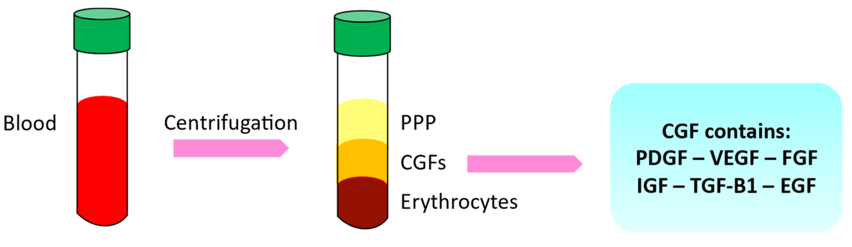
1.3. Growth Factors and Autologous Platelet Concentrates

Growth factors are polypeptide ligands that are crucial for tissue repair, fibrosis, angiogenesis, proliferation, differentiation, and survival [23,24].
Research has focused on the use of autologous growth factors as the best strategy to stimulate tissue regeneration in order to overcome problems and make dental procedures very predictable. There are methods for using blood’s capacity to repair in dentistry as well [25].
Autologous platelet concentrates (APCs) contain high levels of growth factors (GFs). The main generations of APCs are platelet-rich plasma (PRP), platelet-rich fibrin (PRF), and concentrated growth factor (CGF) techniques [26,27].
Platelet-derived growth factor (PDGF), transforming growth factor-1 (TGF-1) and 2 (TGF-2), growth factor fibroblasts (FGF), vascular endothelial growth factor (VEGF), and insulin-like growth factor (IGF) are among the many growth factors found in blood platelets. These factors promote cell proliferation, extracellular matrix remodeling, and angiogenesis [28].
Many methods have recently been discovered that can separate the various growth factors from the patient’s blood. In reality, it is well known that vital growth factors like TGF-1 are present in not just platelets but also other blood cells including erythrocytes and leukocytes [29].
The development of platelet-rich plasma (PRP), growth factor-rich plasma (PRGF), and fibrin-rich plasma (PRF) marked the beginning of growth factor utilization in dentistry. Their efficacy has not always been clearly demonstrated, though [30].
Recombinant human platelet-derived growth factor (rh PDGF-BB) and bone morphogenetic proteins (BMPs) have both been studied and used in dentistry more recently.
A cytokine found in the alpha granules of blood platelets and the bone matrix, platelet-derived growth factor (or PDGF, platelet-derived growth factor) is produced in cases of damage to soft tissue and bone. The physiologically active protein is a dimer made up of the related polypeptides A and B [14,31,32].
CGF is an autologous biological matrix involved in osteoblast differentiation and in vivo bone repair [30,33].
CGF is produced by centrifuging venous blood at varying speeds. It is rich in growth factors (Figure 2). This platelet-rich product is of the third generation [34].
PRP is an autologous concentration of growth factors that are created by centrifuging the patient’s whole blood [35,36].
A platelet concentration generated from whole blood without the use of coagulants was shown to be able to further used to promote wound healing after extensive research using PRP [30,37,38,39].
PRF has demonstrated boosting effects on stem cell proliferation, differentiation, migration, and mineralization during bone formation [20,40,41,42].
Transforming growth factor (TGF-), VEGF, PDGF, Bone Morphogenetic Protein 2 (BMP2), and Insulin-like Growth Factor (IGF-1) are all released by CGF, and these factors promote proliferation and extracellular matrix mineralization through the BMP2/SMAD5/RUNX2 signaling pathway [43,44,45,46].
For angiogenesis and tissue remodeling, CGF also comprises CD34-positive cells, fibroblasts, leukocytes, and endothelial cells, and it acts as a matrix for cell migration [47].
TGF-, platelet-derived growth factors (PDGF-AB and PDGF-BB), IGF, VEGFs, epidermal growth factors (EGFs), and fibroblast growth factor (FGF)-2 are among the substances that make up PRP [8,48,49].
Mesenchymal cell proliferation is promoted by TGF- and PDGF. Additionally, collagen synthesis in the extracellular matrix is stimulated by TGF-1 [50].
To facilitate the earliest stages of tissue healing, these factors primarily stabilize the injured tissue [36].
They also instruct the local mesenchymal and epithelial cells to migrate, divide, and produce more collagen and matrix, which ultimately results in the creation of fibrous connective tissue and scars. VEGF and FGF-2 play a crucial role in promoting the growth of new blood vessels that provide nutrients and progenitor cells to the site of injury [46].
By over-expressing IGF-1, PRP is hypothesized to enhance tendon defect early repair. IGF-binding proteins transport this 70 amino acid polypeptide hormone, which is a typical part of plasma [36].
Given the potential of both PBM and growth factors, the following systematic review aims to investigate the various uses and effects of PBM in combination with growth factors and/or its direct effect on endogenous growth factors in dentistry.

2. Materials and Methods

2.1. Protocol and Registration

This systematic review was conducted according to Preferred Reporting Items for Systematic Reviews and Meta-Analyses (PRISMA), and the protocol was registered at PROSPERO under the ID: 441458.

2.2. Search Processing

The PRISMA protocol was adhered to in conducting the present review. A search was performed on PubMed, Web of Science, and Scopus to identify articles relevant to the study topic, limited to those published in English from January 2013 to 30 May 2023. The search strategy utilized a combination of relevant terms for the study purpose, with the following Boolean keywords applied: (“low laser therapy”) AND (“growth factors”) AND (dent* OR oral) and (“photobiomodulation”) AND (“growth factors”) AND (dent* OR oral).

2.3. Inclusion Criteria

The relevant studies were assessed by the reviewers in pairs, based on the following inclusion criteria: (1) randomized controlled clinical trial design; (2) studies involvement of human participants of any age; (3) open-access publications; (4) studies written in the English language; and (5) studies investigating the effect of LLT in combination with growth factors or its effect on endogenous ones. Studies that failed to meet these criteria were excluded from consideration.
The review was conducted using the PICOS criteria:
Participants: adult patients, both male and female;
Interventions: application of LLLT in combination with growth factors or its effect on endogenous ones;
Comparisons: no LLLT applications;
Outcomes: comparison between the application of the LLLT and its non-use;
Study: randomized clinical trials, retrospective and observational studies.

2.4. Exclusion Criteria

The exclusion criteria were as follows: (1) animal studies; (2) in vitro studies; (3) off-topic; (4) reviews, case reports, case series, letters, or comments; (5) not written in English.

2.5. Data Processing

Four reviewers (M.G., I.F., A.N., and F.V.) independently consulted the databases to collect the studies and rated their quality, based on the selection criteria. The selected articles were downloaded into Zotero (version 6.0.15). Any divergence between the four authors was settled by a discussion with a senior reviewer (F.I.).

2.6. Quality Assessment

The quality of the included papers was assessed by two reviewers, RF and EI, using the reputable Cochrane risk-of-bias assessment for randomized trials (RoB 2). The following six areas of possible bias are evaluated by this tool: random sequence generation, allocation concealment, participant and staff blinding, outcome assessment blinding, inadequate outcome data, and selective reporting. A third reviewer (FI) was consulted in the event of a disagreement until an agreement was reached

3. Results

A search across four databases, namely Pubmed (184), Web of Science (247), and Scopus (209), yielded a total of 640 publications. After the removal of duplicates (206), 434 records were screened based on the title and abstract, leading to the exclusion of 372 articles. From the remaining 62 records, 52 papers were excluded after full-text evaluation, either for being off-topic or failing to meet the inclusion criteria. Ultimately, a total of 10 publications were deemed eligible for qualitative analysis (Table 1). The selection process is outlined in Figure 3.

Quality Assessment and Risk of Bias

The risk of bias in the included studies is reported in Figure 4. Regarding the randomization process, 50% of studies present a high risk of bias and allocation concealment. All other studies ensure a low risk of bias. Seventy-five percent of studies exclude a performance; half of the studies confirm an increased risk of detection bias (self-reported outcome), and 75% of the included studies present a low detection bias (objective measures) (Figure 4). Seventy-five percent of studies ensure a low risk regarding attrition and reporting bias.

4. Discussion

4.1. LLLT, GF, and Oral Cancer

LLLT is a promising therapeutic approach for treating various conditions in the oral cavity, including inflammatory lesions and healing disorders. An intriguing aspect of LLLT is its ability to interact with growth factors, which play a crucial role in tissue healing and regeneration in the oral cavity.
Growth factors are bioactive proteins involved in the regulation of cell proliferation, differentiation, and survival. In the oral cavity, growth factors play a crucial role in wound healing, tissue regeneration, and modulation of the inflammatory response. Some of the key growth factors found in the oral cavity include EGF, TGF-β, VEGF, and basic fibroblast growth factor (bFGF).
Oton-Leite et al. provided some insights into the interaction between laser therapy and growth factors in the oral cavity [51]. The study discussed the potential effects of LLLT on the expression of various growth factors, including EGF, VEGF, bFGF, and PDGF. The findings indicated that LLLT has the potential to modulate the expression of these growth factors, which play crucial roles in tissue repair and regeneration. The results suggested that LLLT may contribute to a less exacerbated inflammatory response and a reduction in tissue degradation. The study also highlighted the importance of further investigations to understand the exact mechanisms of action and the optimal parameters for laser irradiation in promoting the therapeutic effects of growth factors in the oral mucosa [51].
Lauria et al. discussed the use of LLLT in the prevention and treatment of oral mucositis in patients undergoing hematopoietic stem cell transplantation (HSCT) [52]. The study conducted clinical trials and randomized trials to evaluate the efficacy of LLLT in reducing the severity of oral mucositis in HSCT patients. The document presents data on the levels of inflammatory mediators, such as cytokines (IL-6, IL-10), and matrix metalloproteinases (MMP-2, MMP-9), in saliva and blood plasma before and after LLLT treatment. The results showed that LLLT had a positive effect in preventing grades 3–4 oral mucositis in HSCT patients as shown by Cronshaw et al. in a recent systematic review [15]. However, the positive effect did not seem to be completely linked to the modulation of inflammatory mediators.
Lauria’s paper highlights the efficacy of laser light in enhancing the effectiveness of growth factors in promoting tissue regeneration and wound healing. The combination of laser therapy and growth factors appears to have a synergistic effect, leading to improved outcomes in various medical and cosmetic applications. Laser therapy, through its photobiomodulation properties, can stimulate cellular activity and enhance the absorption and utilization of growth factors. This interaction between laser therapy and growth factors holds great potential for advancing therapeutic approaches and improving patient outcomes in the field of regenerative medicine [52].

4.2. LLLT, GF, and Surgery

Ali Arakeeb et al. (2019) aimed to evaluate bone osseointegration around dental implants using innovative techniques. The study involved 40 male patients with missing lower posterior teeth who received dental implants. The patients were divided into four groups: control, LLLT, leukocyte and platelet-rich fibrin (L-PRF), and L-PRF plus LLLT. Cone-beam computed tomography (CBCT) was used to evaluate bone density around the implants at different time points. The results showed significant differences between the groups, with the L-PRF group demonstrating the best results, followed by the L-PRF plus LLLT group. LLLT alone had less improvement compared to the other groups. The study concluded that L-PRF performed better in promoting osseointegration compared to LLLT [56].
The effects of photobiomodulation on implant stability were looked at in the study by Gokmenoglu et al. In more detail, 15 partially edentulous patients were involved and randomly assigned to one group (8 patients, 10 implants) that obtained post-implant LED irradiation and the other (7 patients, 12 implants) that did not. For 20 min, laser stimulation (Osseopulse) at a wavelength of 626 nm and an intensity of 38.5 mW/cm2 was applied to the LED group. In the three weeks following surgery, LED treatment was conducted three times each week. For what concerns the biochemical markers, it was found the increase in IL-1b levels in the control group and, conversely, the decrease in IL-1b and PGE2 in the group that received LEDs served as evidence of the photobiomodulation’s anti-inflammatory effects. TGF-b levels increased in both groups. The NO levels in the LED group, however, slightly increased but not significantly. Therefore, all of the alterations in the biochemical markers appeared to represent the action of LED therapy alone because there were no inflammatory conditions near the implant. The findings demonstrated that LED therapy could have a good impact on bone and peri-implant tissue healing while also helping to maintain implant stability over the first three months following surgery [57].
Kamal et al. (2020) have studied the management of dry sockets, a complication following tooth extraction. The study compared the efficacy of CGF and LLLT to conventional treatment. Sixty patients with dry sockets were divided into three groups: conventional treatment, CGF, and LLLT. The outcomes were evaluated based on pain score, inflammation, tenderness, and granulation tissue formation. The results showed that CGF treatment demonstrated superior outcomes compared to LLLT in terms of granulation tissue formation and pain control by day 7. The conventional treatment group took longer to achieve similar healing. The study emphasized the need for regenerative approaches in managing dry sockets and mentioned other techniques such as platelet-rich plasma (PRP) and low-intensity pulsed ultrasound therapy in dentistry [58].

4.3. LLLT, GF, and Orthodontics

LLLT has been of interest in the field of orthodontics due to its potential effect on orthodontic movement by stimulating the cellular metabolism of bone cells and surrounding tissues. Low-level laser irradiation can increase the activity of osteoblastic cells, which are involved in bone formation, promoting the production of extracellular matrix and osteogenesis.
The study by Sevin Erol Üretürk et Al. aims to evaluate the effect of LLLT irradiation on the speed and efficiency of orthodontic movement by 3D measurement of intraoral scans and assessment of IL-1β (interleukin-1β) and TGF-β1 (transforming growth factor-beta) levels in the gingival crevicular fluid. Fifteen patients with Angle class II division I malocclusion requiring maxillary first premolar extraction and canine distalization were analyzed. The split-mouth study compared the right quadrant, treated with traditional technique, with the left quadrant, on which 5 irradiations of gallium-aluminium-arsenide diode laser were applied at regular intervals. The laser unit generated a constant wave with an average power of 20 mW in continuous mode, having an irradiation area of 4 mm2. An output of 20 mW with an energy density of 5 J/cm2 was used, and each point was irradiated for 10 s, corresponding to an energy of 0.2 J per point.
The study group showed a 40% higher displacement velocity and increased TGF-β1 in the crevicular fluid. In the field of orthodontics, TGF-β1 has been the subject of interest due to its ability to influence the speed and direction of tooth movement. It has been observed that TGF-β1 is involved in regulating the mechanical forces acting on teeth during their orthodontic movement. It may influence the response of bone cells to orthodontic forces by modulating cell proliferation and differentiation as well as extracellular matrix synthesis in the process of bone remodeling. The application of orthodontic forces can cause an inflammatory response in the gums and surrounding bone tissue, and TGF-β1 may be involved in the regulation of this inflammatory response by influencing tumor necrosis factor (TNF) receptor and pro-inflammatory cytokines. Understanding these mechanisms may contribute to the development of more effective and personalized orthodontic therapies. In conclusion, the use of lasers, through the involvement of these mechanisms and growth factors, accelerated tooth movement and could reduce the overall duration of treatment [59].
Cytokines, TGF, and tumor necrosis factors are also responsible for regulating the inflammatory process in the oral cavity, and the use of LLLT to reduce inflammation has shown promising results. This is due to the possible effects of the diode laser acting as a biostimulator in the tissue repair process. Irradiation with LLLT can stimulate blood circulation in the affected area, reduce inflammatory mediators, and improve cell proliferation and collagen production. In this regard, the study by Yassaei et al. investigated the effects of 980 nm diode laser irradiation on the stability of an MS and the concentration of IL-1β and TGF-β1 in the peri-miniscrew crevicular fluid (PMCF) at four-time points after MS placement. In this double-blind randomized controlled clinical trial, the effect of diode laser irradiation on MS stability and the concentration of IL-1β and TGF-β1 in the PMCF was examined. 18 patients with Class II Division I malocclusion requiring maxillary first premolar extraction and anterior retraction by MS were recruited. A diode laser with a wavelength of 980 nm was used in the laser quadrant, while the control quadrant underwent pseudo-laser application. The 980 nm diode laser was chosen for its effectiveness against bacteria and its compatibility with implants. Inflammation around MS is a negative factor affecting implant stability. In this study, cytokines IL-1β and TGF-β1 in PMCF were assessed and biochemical evaluations were performed. The results showed a significant reduction in IL-1β levels in the laser group compared to the control group. As for TGF-β1, the levels were lower in the laser group, but the difference was not statistically significant. Furthermore, laser irradiation was shown to increase the stability of MS over time. These results support the efficacy of diode laser therapy in reducing inflammation and increasing MS stability. However, further research with a larger number of participants and more in-depth evaluations are needed to fully understand the effects of LLLT in the context of MS [60].

4.4. LLLT, GF and Periodontology

A centrifugal machine operating at a certain speed can extract growth factors from blood [25]. Hematopoietic stem cells that are CD34+ can differentiate into endothelial stem cells [61]. The platelets, white blood cells, and growth factors that make up the fibrin network primarily are capable of independently causing vascular regeneration and acting as a matrix for cell migration [62,63]. Growth factors promote angiogenesis and cell division. Low Lever Laser Therapy (LLLT) is a type of light therapy that uses non-thermal processes with endogenous chromophores to trigger photophysical and photochemical events at various biological scales [64]. Non-thermal processes with endogenous chromophores include lasers, light-emitting semiconductors (LEDs), and broadband light in the visible and infrared spectrum. Immunomodulation, pain or inflammation relief, wound healing promotion, and tissue regeneration are just a few of the positive therapeutic consequences of this treatment [65,66].
Chen et al. used LLLT in their investigation to encourage stem cells to proliferate, differentiate, and growth factor activity [67].
Nd: YAG by PBMT effect is irradiated on the local periodontal area with a semiconductor laser (810 nm) to stimulate local microcirculation, activate the growth factors in CGF, and promote CD34 + stem cells to grow and differentiate [68].
Additionally, local gingival papilla tissue exposed to radiation aids in boosting gingival fibroblast growth and type I collagen molecule production [69,70].
There was no discernible increase in gingival papilla height following the therapies. Additionally, the gingival papilla’s volume only slightly increased. LPCGF was gradually absorbed by tissues 14 to 21 days after each injection [71].
The findings demonstrated that the regeneration of the damaged gingival papilla in natural teeth is significantly impacted by the injection of LPCGF. This may suggest that the three sources of soft tissue regeneration in LPCGF caused gingival fibroblast proliferation and angiogenesis, which led to the gradual repair of the inadequate gingival papilla after therapy [67].
In a split-mouth trial, Pamuk et al. used 30 people with chronic periodontitis (15 smokers and 15 nonsmokers) and 30 healthy people who were gender, age, and smoking paired as controls [72].
On day 30 and before beginning non-surgical periodontal therapy, clinical indicators were noted. Samples of crevicular fluid from the gingival region were taken both before and after periodontal therapy on days 7, 14, and 30. The enzyme-linked immunosorbent assay was used to determine the concentrations of tPA, transforming growth factor (TGF)-1, and plasminogen activator inhibitor 1 (PAI-1).
Between the baseline and day 30 following SRP therapy in both groups, all clinical markers significantly decreased. Between the LLLT and sham groups of smokers or never-smokers, no appreciable changes were found. In both the laser and SRP treatment groups of smokers, gingival crevicular fluid PAI-1 levels reduced as expected. In all treatment groups, the amount of TGF-1 in the gingival crevicular fluid was reduced.
The results of this study demonstrate that the adjunctive use of LLLT promotes clinical improvements. Furthermore, LLLT could promote modulation of cytokine levels [72].
In opposition, Gündoğar et al. in their study showed no statistically significant change between groups in these biochemical parameters at any time point. They believe that these inconsistencies are related to the nature of lasers (such as penetration depth), study design, and laser duration. However, the biochemical parameters showed no statistical differences, and this could be related to minor clinical improvement [73].
In this study, no statistically significant difference was found in the intergroup assessments of CGF levels of IL-1β found at any time point [73].

4.5. Energy Dose and Photobiomodulation

Studies by Cronshaw et al. and Arany et al. demonstrate that the dose of laser energy plays a pivotal role in the efficacy of Photobiomodulation Therapy (PBM) [74,75]. The first article underscores the significance of laser spot size, demonstrating that larger optical spot sizes are associated with optimal clinical outcomes for deeper targets, while smaller spot sizes prove ineffective [74]. To ensure an effective dose reaches a depth of 1 cm, an increase in surface-applied dose is recommended. The second article delves into the complexity of PBM device parameters, emphasizing the nonlinear nature of PBM biological responses. It highlights the importance of understanding factors beyond just wavelength, such as coherence, collimation, polarization, and pulsing, in determining PBM therapy outcomes. Pulsing, in particular, is noted for its efficacy in various PBM treatments [75] In essence, these articles underscore the critical role of laser dose and the need to consider multifaceted device parameters to optimize clinical outcomes in PBM therapy [74,75].

5. Limitations

This review explores the potential effects of LLLT on the interaction between growth factors and various oral conditions, but it has several limitations:
  • Limited sample size: Some of the studies mentioned in the text had a small sample size, which can limit the generalizability of the findings. Small sample sizes may not accurately represent the larger population and can increase the risk of bias.
  • Lack of control groups: In some studies, there was a lack of control groups or inadequate comparison groups. Without proper control groups, it becomes challenging to determine the specific effects of LLLT and growth factors independently.
  • Varied study designs: The studies mentioned in the text had different designs, including clinical trials, randomized trials, and observational studies. While each study design has its merits, the lack of consistency in study designs makes it difficult to draw definitive conclusions and compare the results across studies.
  • Limited understanding of mechanisms: Although the studies suggest potential interactions between LLLT, growth factors, and oral conditions, the exact underlying mechanisms of action are not well understood. Further research is needed to elucidate the precise mechanisms involved and to determine the optimal parameters for LLLT application.
  • Need for more rigorous research: While the studies discussed provide preliminary evidence, additional well-designed and rigorous research is necessary to establish the efficacy, safety, and long-term effects of LLLT in conjunction with growth factors. Further investigations should include larger sample sizes, standardized protocols, and control groups to strengthen the scientific validity of the findings.
In summary, while the mentioned studies provide valuable insights into the potential effects of LLLT on the interaction between growth factors and oral conditions, it is important to consider their limitations. Further research with larger sample sizes, rigorous study designs, and long-term follow-up is needed to validate and expand upon these preliminary findings.

6. Conclusions

In conclusion, the studies reviewed in this article highlight the potential interaction between LLLT, GF, and various oral health conditions. LLLT has shown promising results in modulating the expression of growth factors involved in tissue healing and regeneration, such as EGF, (TGF-β), VEGF, and bFGF. The combination of LLLT and growth factors has demonstrated synergistic effects in promoting tissue repair and wound healing in conditions such as oral mucositis, oral submucous fibrosis, and orthodontic movement.
Furthermore, LLLT has shown potential in reducing inflammation, controlling pain, and improving oral health-related quality of life in various surgical procedures, including sinus lift surgery and dental implant therapy. The use of LLLT in conjunction with growth factors like CGF has shown promising outcomes in managing complications such as dry sockets and promoting bone osseointegration around dental implants.
In the field of orthodontics, LLLT has been found to accelerate orthodontic tooth movement by stimulating osteoblastic cell activity and increasing the levels of growth factors like TGF-β1. The application of LLLT in orthodontic treatment may lead to shorter treatment durations and improved outcomes. However, further research is needed to optimize LLLT parameters and protocols for specific orthodontic applications.
Additionally, LLLT has shown potential in periodontal treatment by promoting angiogenesis, tissue regeneration, and growth factor activity. It has been observed to positively influence CD34+ stem cells, gingival fibroblast growth, and collagen production. The combination of LLLT with growth factors derived from blood components, such as PRF, has shown promising results in promoting tissue repair and regeneration in periodontal tissues.
Overall, the findings presented in these studies suggest that LLLT, in combination with growth factors, holds great potential for advancing therapeutic approaches in oral health and improving patient outcomes in various clinical scenarios. However, all this must not disregard the fact that the effect of each therapy is a direct function of the type of patient treated and/or the type of lesion, so it is necessary to investigate with evidence-based methodologies (Evidence-based Medicine, EBM) which types of patients and/or lesions are more susceptible to treatment with these tools, as well as to understand the best ways of using (alone or in combination) these important and cutting-edge therapeutic options. However, further research is needed to fully understand the underlying mechanisms of the interaction between LLLT and growth factors and to establish optimal treatment protocols for different oral health conditions.

Author Contributions

Conceptualization, G.D., F.I., A.M.I., A.P. (Assunta Patano), I.F., I.P., M.G. and A.D.I.; methodology, A.P. (Assunta Patano), I.F., I.P., L.L., G.F., F.V., G.D. and I.T.; software, A.N., F.V., I.T., A.D.I., F.I. and A.M.I.; validation, G.D., A.P. (Assunta Patano), A.N., F.I. and I.P.; formal analysis, F.V., I.P., I.T., G.D., A.N., L.L., G.F. and A.M.I.; investigation, A.P. (Assunta Patano), F.V., I.T., M.G., I.F., G.F., A.D.I. and F.I.; resources, I.T., M.G., A.D.I., G.D., I.P., G.F., L.L. and A.M.I.; data curation, I.F., A.N., I.T., A.D.I., I.F. and A.N.; writing—original draft preparation, I.P., M.G., I.T., A.P., F.I, A.N., F.V., I.F. and G.D.; writing—review and editing, G.D., A.N., F.I., I.P. and A.D.I.; visualization, I.F., F.V., A.D.I. and A.M.I.; supervision, G.D., A.P. (Assunta Patano), A.N., I.P., I.F., L.D. and F.V.; project administration, I.F., I.P., L.L., G.F., G.D., A.P. (Andrea Palermo) and F.I. All authors have read and agreed to the published version of the manuscript.

Funding

This research received no external funding.

Institutional Review Board Statement

Not applicable.

Informed Consent Statement

Not applicable.

Data Availability Statement

Not applicable.

Conflicts of Interest

The authors declare no conflict of interest.

Abbreviations

APCsAutologous platelet concentrates
ATPAdenosine triphosphate
bFGFbasic fibroblast growth factor
BMP2Bone Morphogenetic Protein 2
BTA black triangle area
BTH black triangle height
CBCTCone-beam computed tomography
CGFConcentrated growth factor
EGFEpidermal growth factor
FGFFibroblast Growth Factor
GFsGrowth factors
HSCThematopoietic stem cell transplantation
IGF-1Insulin-like Growth Factor
IL-1βinterleukin-1β
IL-6interleukin-6
L-PRFLeukocyte and platelet-rich fibrin
LEDs light emitting semiconductors
LLLTLow-level laser therapy
LPCGF Liquid Phase Concentrated Growth Factors
MMPMatrix Metalloproteinase
MSminiscrew
NCINational Cancer Institute
Nd:YAGNeodymium-doped yttrium aluminum garnet
OHRQoLOral health-related quality of life
OMOral Mucositis
OSMFOral submucous fibrosis
PAI-1plasminogen activator inhibitor 1
PBMphotobiomodulation
PBMTPhotobiomodulation therapy
PDGFPlatelet-Derived Growth Factor
PICF peri-implant crevicular fluid
PMCFperi-miniscrew crevicular fluid
PRFPlatelet-Rich Fibrin
PRPPlatelet-rich plasma
RBDRelative bone density
SRPscaling and root planning
TGFtransforming growth factor
tPAtissue plasminogen activator
VEGFVascular Endothelial Growth Factor
WHOWord Health Organization

References

  1. Almasri, M.; Mandal, N.; Kulkarni, P.; Raj, A.; Zeya, A.; Mann, N.; Tiwari, R.C. Low-Level Laser Therapy Role in Surgical Extractions: An Original Research. J. Pharm. Bioall. Sci. 2022, 14, 245. [Google Scholar] [CrossRef]
  2. John, S.S.; Mohanty, S.; Chaudhary, Z.; Sharma, P.; Kumari, S.; Verma, A. Comparative Evaluation of Low Level Laser Therapy and Cryotherapy in Pain Control and Wound Healing Following Orthodontic Tooth Extraction: A Double Blind Study. J. Cranio-Maxillofac. Surg. 2020, 48, 251–260. [Google Scholar] [CrossRef] [PubMed]
  3. Da Silva, J.P.; Da Silva, M.A.; Almeida, A.P.F.; Junior, I.L.; Matos, A.P. Laser Therapy in the Tissue Repair Process: A Literature Review. Photomed. Laser Surg. 2010, 28, 17–21. [Google Scholar] [CrossRef]
  4. Vermesan, D.; Inchingolo, F.; Patrascu, J.M.; Trocan, I.; Prejbeanu, R.; Florescu, S.; Damian, G.; Benagiano, V.; Abbinante, A.; Caprio, M.; et al. Anterior Cruciate Ligament Reconstruction and Determination of Tunnel Size and Graft Obliquity. Eur. Rev. Med. Pharmacol. Sci. 2015, 19, 357–364. [Google Scholar] [PubMed]
  5. Mester, E.; Mester, A.F.; Mester, A. The Biomedical Effects of Laser Application. Lasers Surg. Med. 1985, 5, 31–39. [Google Scholar] [CrossRef] [PubMed]
  6. Inchingolo, A.M.; Malcangi, G.; Ferrara, I.; Viapiano, F.; Netti, A.; Buongiorno, S.; Latini, G.; Azzollini, D.; De Leonardis, N.; De Ruvo, E.; et al. Laser Surgical Approach of Upper Labial Frenulum: A Systematic Review. Int. J. Environ. Res. Public Health 2023, 20, 1302. [Google Scholar] [CrossRef] [PubMed]
  7. Rapone, B.; Ferrara, E.; Santacroce, L.; Topi, S.; Gnoni, A.; Dipalma, G.; Mancini, A.; Di Domenico, M.; Tartaglia, G.M.; Scarano, A.; et al. The Gaseous Ozone Therapy as a Promising Antiseptic Adjuvant of Periodontal Treatment: A Randomized Controlled Clinical Trial. Int. J. Environ. Res. Public Health 2022, 19, 985. [Google Scholar] [CrossRef]
  8. Inchingolo, F.; Tatullo, M.; Abenavoli, F.M.; Marrelli, M.; Inchingolo, A.D.; Inchingolo, A.M.; Dipalma, G. Comparison between Traditional Surgery, CO2 and Nd:Yag Laser Treatment for Generalized Gingival Hyperplasia in Sturge-Weber Syndrome: A Retrospective Study. J. Investig. Clin. Dent. 2010, 1, 85–89. [Google Scholar] [CrossRef]
  9. Hamblin, M.R.; Liebert, A. Photobiomodulation Therapy Mechanisms Beyond Cytochrome c Oxidase. Photobiomodul. Photomed. Laser Surg. 2022, 40, 75–77. [Google Scholar] [CrossRef]
  10. Sun, G.; Tunér, J. Low-Level Laser Therapy in Dentistry. Dent. Clin. N. Am. 2004, 48, 1061–1076. [Google Scholar] [CrossRef]
  11. Arany, P.R. Photobiomodulation-Activated Latent Transforming Growth Factor-Β1: A Critical Clinical Therapeutic Pathway and an Endogenous Optogenetic Tool for Discovery. Photobiomodul. Photomed. Laser Surg. 2022, 40, 136–147. [Google Scholar] [CrossRef] [PubMed]
  12. Arany, P.R.; Cho, A.; Hunt, T.D.; Sidhu, G.; Shin, K.; Hahm, E.; Huang, G.X.; Weaver, J.; Chen, A.C.-H.; Padwa, B.L.; et al. Photoactivation of Endogenous Latent Transforming Growth Factor–Β1 Directs Dental Stem Cell Differentiation for Regeneration. Sci. Transl. Med. 2014, 6, 238ra69. [Google Scholar] [CrossRef] [PubMed]
  13. Huang, Y.-Y.; Sharma, S.K.; Carroll, J.; Hamblin, M.R. Biphasic Dose Response in Low Level Light Therapy—An Update. Dose Response 2011, 9, 602–618. [Google Scholar] [CrossRef] [PubMed]
  14. De Benedittis, M.; Petruzzi, M.; Pastore, L.; Inchingolo, F.; Serpico, R. Nd:YAG Laser for Gingivectomy in Sturge-Weber Syndrome. J. Oral Maxillofac. Surg. 2007, 65, 314–316. [Google Scholar] [CrossRef]
  15. Cronshaw, M.; Parker, S.; Anagnostaki, E.; Mylona, V.; Lynch, E.; Grootveld, M. Photobiomodulation and Oral Mucositis: A Systematic Review. Dent. J. 2020, 8, 87. [Google Scholar] [CrossRef]
  16. Serrage, H.; Heiskanen, V.; Palin, W.M.; Cooper, P.R.; Milward, M.R.; Hadis, M.; Hamblin, M.R. Under the Spotlight: Mechanisms of Photobiomodulation Concentrating on Blue and Green Light. Photochem. Photobiol. Sci. 2019, 18, 1877–1909. [Google Scholar] [CrossRef]
  17. Karu, T. Primary and Secondary Mechanisms of Action of Visible to Near-IR Radiation on Cells. J. Photochem. Photobiol. B Biol. 1999, 49, 1–17. [Google Scholar] [CrossRef]
  18. Navratil, L.; Kymplova, J. Contraindications in Noninvasive Laser Therapy: Truth and Fiction. J. Clin. Laser Med. Surg. 2002, 20, 341–343. [Google Scholar] [CrossRef]
  19. Qamar, Z.; Alghamdi, A.M.S.; Haydarah, N.K.B.; Balateef, A.A.; Alamoudi, A.A.; Abumismar, M.A.; Shivakumar, S.; Cicciù, M.; Minervini, G. Impact of Temporomandibular Disorders on Oral Health-related Quality of Life: A Systematic Review and Meta-analysis. J. Oral Rehabil. 2023. [Google Scholar] [CrossRef]
  20. Grassi, F.R.; Ciccolella, F.; D’Apolito, G.; Papa, F.; Iuso, A.; Salzo, A.E.; Trentadue, R.; Nardi, G.M.; Scivetti, M.; De Matteo, M.; et al. Effect of Low-Level Laser Irradiation on Osteoblast Proliferation and Bone Formation. J. Biol. Regul. Homeost. Agents 2011, 25, 603–614. [Google Scholar]
  21. Tarullo, A.; Laino, L.; Tarullo, A.; Inchingolo, F.; Flace, P.; Inchingolo, A.M.; Inchingolo, A.D.; Dipalma, G.; Podo Brunetti, S.; Cagiano, R. Use of a Diode Laser in an Excisional Biopsy of Two Spoonlike Neoformations on the Tongue Tip. Acta Biomed. 2011, 82, 63–68. [Google Scholar] [PubMed]
  22. Carroll, J.D.; Milward, M.R.; Cooper, P.R.; Hadis, M.; Palin, W.M. Developments in Low Level Light Therapy (LLLT) for Dentistry. Dent. Mater. 2014, 30, 465–475. [Google Scholar] [CrossRef] [PubMed]
  23. Stone, W.L.; Leavitt, L.; Varacallo, M. Physiology, Growth Factor; StatPearls Publishing: Treasure Island, FL, USA, 2023. [Google Scholar]
  24. Minervini, G.; Franco, R.; Marrapodi, M.M.; Ronsivalle, V.; Shapira, I.; Cicciù, M. Prevalence of Temporomandibular Disorders in Subjects Affected by Parkinson Disease: A Systematic Review and Metanalysis. J. Oral Rehabil. 2023. [Google Scholar] [CrossRef] [PubMed]
  25. Intini, G. The Use of Platelet-Rich Plasma in Bone Reconstruction Therapy. Biomaterials 2009, 30, 4956–4966. [Google Scholar] [CrossRef]
  26. Mijiritsky, E.; Assaf, H.D.; Peleg, O.; Shacham, M.; Cerroni, L.; Mangani, L. Use of PRP, PRF and CGF in Periodontal Regeneration and Facial Rejuvenation—A Narrative Review. Biology 2021, 10, 317. [Google Scholar] [CrossRef] [PubMed]
  27. Inchingolo, F.; Ballini, A.; Cagiano, R.; Inchingolo, A.D.; Serafini, M.; De Benedittis, M.; Cortelazzi, R.; Tatullo, M.; Marrelli, M.; Inchingolo, A.M.; et al. Immediately Loaded Dental Implants Bioactivated with Platelet-Rich Plasma (PRP) Placed in Maxillary and Mandibular Region. Clin. Ter. 2015, 166, e146–e152. [Google Scholar] [CrossRef]
  28. Inchingolo, A.D.; Ceci, S.; Patano, A.; Inchingolo, A.M.; Montenegro, V.; Di Pede, C.; Malcangi, G.; Marinelli, G.; Coloccia, G.; Garibaldi, M.; et al. Elastodontic Therapy of Hyperdivergent Class II Patients Using AMCOP® Devices: A Retrospective Study. Appl. Sci. 2022, 12, 3259. [Google Scholar] [CrossRef]
  29. Choukroun, J.; Diss, A.; Simonpieri, A.; Girard, M.-O.; Schoeffler, C.; Dohan, S.L.; Dohan, A.J.J.; Mouhyi, J.; Dohan, D.M. Platelet-Rich Fibrin (PRF): A Second-Generation Platelet Concentrate. Part IV: Clinical Effects on Tissue Healing. Oral Surg. Oral Med. Oral Pathol. Oral Radiol. Endodontol. 2006, 101, e56–e60. [Google Scholar] [CrossRef]
  30. Dohan Ehrenfest, D.M.; Del Corso, M.; Inchingolo, F.; Sammartino, G.; Charrier, J.-B. Platelet-Rich Plasma (PRP) and Platelet-Rich Fibrin (PRF) in Human Cell Cultures: Growth Factor Release and Contradictory Results. Oral Surg. Oral Med. Oral Pathol. Oral Radiol. Endodontol. 2010, 110, 418–421. [Google Scholar] [CrossRef]
  31. Islam, M.S.; Greco, S.; Janjusevic, M.; Ciavattini, A.; Giannubilo, S.R.; D’Adderio, A.; Biagini, A.; Fiorini, R.; Castellucci, M.; Ciarmela, P. Growth Factors and Pathogenesis. Best Pract. Res. Clin. Obstet. Gynaecol. 2016, 34, 25–36. [Google Scholar] [CrossRef]
  32. Mummolo, S.; Mancini, L.; Quinzi, V.; D’Aquino, R.; Marzo, G.; Marchetti, E. Rigenera® Autologous Micrografts in Oral Regeneration: Clinical, Histological, and Radiographical Evaluations. Appl. Sci. 2020, 10, 5084. [Google Scholar] [CrossRef]
  33. Rochira, A.; Siculella, L.; Damiano, F.; Palermo, A.; Ferrante, F.; Carluccio, M.A.; Calabriso, N.; Giannotti, L.; Stanca, E. Concentrated Growth Factors (CGF) Induce Osteogenic Differentiation in Human Bone Marrow Stem Cells. Biology 2020, 9, 370. [Google Scholar] [CrossRef] [PubMed]
  34. Shirbhate, U.; Bajaj, P. Third-Generation Platelet Concentrates in Periodontal Regeneration: Gaining Ground in the Field of Regeneration. Cureus 2022, 14, e28072. [Google Scholar] [CrossRef] [PubMed]
  35. Cole, B.J.; Seroyer, S.T.; Filardo, G.; Bajaj, S.; Fortier, L.A. Platelet-Rich Plasma: Where Are We Now and Where Are We Going? Sports Health 2010, 2, 203–210. [Google Scholar] [CrossRef] [PubMed]
  36. Boccellino, M.; Di Stasio, D.; Dipalma, G.; Cantore, S.; Ambrosio, P.; Coppola, M.; Quagliuolo, L.; Scarano, A.; Malcangi, G.; Borsani, E.; et al. Steroids and Growth Factors in Oral Squamous Cell Carcinoma: Useful Source of Dental-Derived Stem Cells to Develop a Steroidogenic Model in New Clinical Strategies. Eur. Rev. Med. Pharmacol. Sci. 2019, 23, 8730–8740. [Google Scholar] [CrossRef]
  37. Everts, P.A.M.; Knape, J.T.A.; Weibrich, G.; Schönberger, J.P.A.M.; Hoffmann, J.; Overdevest, E.P.; Box, H.A.M.; van Zundert, A. Platelet-Rich Plasma and Platelet Gel: A Review. J. Extra Corpor. Technol. 2006, 38, 174–187. [Google Scholar] [CrossRef]
  38. Borsani, E.; Bonazza, V.; Buffoli, B.; Nocini, P.F.; Albanese, M.; Zotti, F.; Inchingolo, F.; Rezzani, R.; Rodella, L.F. Beneficial Effects of Concentrated Growth Factors and Resveratrol on Human Osteoblasts In Vitro Treated with Bisphosphonates. Biomed. Res. Int. 2018, 2018, 4597321. [Google Scholar] [CrossRef]
  39. Naik, B.; Karunakar, P.; Jayadev, M.; Marshal, V.R. Role of Platelet Rich Fibrin in Wound Healing: A Critical Review. J. Conserv. Dent. 2013, 16, 284–293. [Google Scholar] [CrossRef]
  40. Kim, T.-H.; Kim, S.-H.; Sándor, G.K.; Kim, Y.-D. Comparison of Platelet-Rich Plasma (PRP), Platelet-Rich Fibrin (PRF), and Concentrated Growth Factor (CGF) in Rabbit-Skull Defect Healing. Arch. Oral Biol. 2014, 59, 550–558. [Google Scholar] [CrossRef]
  41. Liu, Y.; Sun, X.; Yu, J.; Wang, J.; Zhai, P.; Chen, S.; Liu, M.; Zhou, Y. Platelet-Rich Fibrin as a Bone Graft Material in Oral and Maxillofacial Bone Regeneration: Classification and Summary for Better Application. BioMed Res. Int. 2019, 2019, 3295756. [Google Scholar] [CrossRef]
  42. Bonazza, V.; Borsani, E.; Buffoli, B.; Parolini, S.; Inchingolo, F.; Rezzani, R.; Rodella, L.F. In Vitro Treatment with Concentrated Growth Factors (CGF) and Sodium Orthosilicate Positively Affects Cell Renewal in Three Different Human Cell Lines. Cell Biol. Int. 2018, 42, 353–364. [Google Scholar] [CrossRef] [PubMed]
  43. Palermo, A.; Ferrante, F.; Stanca, E.; Damiano, F.; Gnoni, A.; Batani, T.; Carluccio, M.A.; Demitri, C.; Siculella, L. Release of VEGF from Dental Implant Surface (IML® Implant) Coated with Concentrated Growth Factors (CGF) and the Liquid Phase of CGF (LPCGF): In Vitro Results and Future Expectations. Appl. Sci. 2019, 9, 2114. [Google Scholar] [CrossRef]
  44. Wang, L.; Wan, M.; Li, Z.; Zhong, N.; Liang, D.; Ge, L. A Comparative Study of the Effects of Concentrated Growth Factors in Two Different Forms on Osteogenesis In vitro. Mol. Med. Rep. 2019, 20, 1039–1048. [Google Scholar] [CrossRef] [PubMed]
  45. Dohan Ehrenfest, D.M.; Bielecki, T.; Mishra, A.; Borzini, P.; Inchingolo, F.; Sammartino, G.; Rasmusson, L.; Everts, P.A. In Search of a Consensus Terminology in the Field of Platelet Concentrates for Surgical Use: Platelet-Rich Plasma (PRP), Platelet-Rich Fibrin (PRF), Fibrin Gel Polymerization and Leukocytes. Curr. Pharm. Biotechnol. 2012, 13, 1131–1137. [Google Scholar] [CrossRef]
  46. Dohan Ehrenfest, D.M.; Bielecki, T.; Jimbo, R.; Barbé, G.; Del Corso, M.; Inchingolo, F.; Sammartino, G. Do the Fibrin Architecture and Leukocyte Content Influence the Growth Factor Release of Platelet Concentrates? An Evidence-Based Answer Comparing a Pure Platelet-Rich Plasma (P-PRP) Gel and a Leukocyte- and Platelet-Rich Fibrin (L-PRF). Curr. Pharm. Biotechnol. 2012, 13, 1145–1152. [Google Scholar] [CrossRef]
  47. Stanca, E.; Calabriso, N.; Giannotti, L.; Nitti, P.; Damiano, F.; Stanca, B.D.C.; Carluccio, M.A.; De Benedetto, G.E.; Demitri, C.; Palermo, A.; et al. Analysis of CGF Biomolecules, Structure and Cell Population: Characterization of the Stemness Features of CGF Cells and Osteogenic Potential. Int. J. Mol. Sci. 2021, 22, 8867. [Google Scholar] [CrossRef]
  48. Nicosia, R.F.; Nicosia, S.V.; Smith, M. Vascular Endothelial Growth Factor, Platelet-Derived Growth Factor, and Insulin-like Growth Factor-1 Promote Rat Aortic Angiogenesis in Vitro. Am. J. Pathol. 1994, 145, 1023–1029. [Google Scholar]
  49. Dohan Ehrenfest, D.M.; Bielecki, T.; Del Corso, M.; Inchingolo, F.; Sammartino, G. Shedding Light in the Controversial Terminology for Platelet-Rich Products: Platelet-Rich Plasma (PRP), Platelet-Rich Fibrin (PRF), Platelet-Leukocyte Gel (PLG), Preparation Rich in Growth Factors (PRGF), Classification and Commercialism. J. Biomed. Mater. Res. A 2010, 95, 1280–1282. [Google Scholar] [CrossRef]
  50. Throckmorton, D.C.; Brogden, A.P.; Min, B.; Rasmussen, H.; Kashgarian, M. PDGF and TGF-β Mediate Collagen Production by Mesangial Cells Exposed to Advanced Glycosylation End Products. Kidney Int. 1995, 48, 111–117. [Google Scholar] [CrossRef]
  51. Oton-Leite, A.F.; Silva, G.B.L.; Morais, M.O.; Silva, T.A.; Leles, C.R.; Valadares, M.C.; Pinezi, J.C.D.; Batista, A.C.; Mendonça, E.F. Effect of Low-Level Laser Therapy on Chemoradiotherapy-Induced Oral Mucositis and Salivary Inflammatory Mediators in Head and Neck Cancer Patients: Effect of Low-Level Laser Therapy in Cancer Patients. Lasers Surg. Med. 2015, 47, 296–305. [Google Scholar] [CrossRef]
  52. Silva, G.B.L.; Sacono, N.T.; Othon-Leite, A.F.; Mendonça, E.F.; Arantes, A.M.; Bariani, C.; Duarte, L.G.L.; Abreu, M.H.N.; Queiroz-Júnior, C.M.; Silva, T.A.; et al. Effect of Low-Level Laser Therapy on Inflammatory Mediator Release during Chemotherapy-Induced Oral Mucositis: A Randomized Preliminary Study. Lasers Med. Sci. 2015, 30, 117–126. [Google Scholar] [CrossRef] [PubMed]
  53. Kuhn, A.; Porto, F.A.; Miraglia, P.; Brunetto, A.L. Low-Level Infrared Laser Therapy in Chemotherapy-Induced Oral Mucositis: A Randomized Placebo-Controlled Trial in Children. J. Pediatr. Hematol. Oncol. 2009, 31, 33–37. [Google Scholar] [CrossRef] [PubMed]
  54. Sukanya, D.; Upasana, L.; Deepak, T.; Abhinethra, M.; Choudary, S. Determination of Effectiveness of Photobiomodulation in the Treatment of Oral Submucous Fibrosis. J. Pharm. Bioall. Sci. 2022, 14, 475. [Google Scholar] [CrossRef] [PubMed]
  55. Ozturan, S.; Sirali, A.; Sur, H. Effects of Nd:YAG Laser Irradiation for Minimizing Edema and Pain After Sinus Lift Surgery: Randomized Controlled Clinical Trial. Photomed. Laser Surg. 2015, 33, 193–199. [Google Scholar] [CrossRef]
  56. Arakeeb, M.A.A.; Zaky, A.A.; Harhash, T.A.-H.; Salem, W.S.; El-Mofty, M. Effect of Combined Application of Growth Factors and Diode Laser Bio-Stimulation on the Osseo Integration of Dental Implants. Open Access Maced. J. Med. Sci. 2019, 7, 2520–2527. [Google Scholar] [CrossRef]
  57. Gokmenoglu, C.; Ozmeric, N.; Erguder, I.; Elgun, S. The Effect of Light-Emitting Diode Photobiomodulation on Implant Stability and Biochemical Markers in Peri-Implant Crevicular Fluid. Photomed. Laser Surg. 2014, 32, 138–145. [Google Scholar] [CrossRef]
  58. Kamal, A.; Salman, B.; Razak, N.H.A.; Samsudin, A.R. A Comparative Clinical Study between Concentrated Growth Factor and Low-Level Laser Therapy in the Management of Dry Socket. Eur. J. Dent. 2020, 14, 613–620. [Google Scholar] [CrossRef]
  59. Üretürk, S.E.; Saraç, M.; Fıratlı, S.; Can, Ş.B.; Güven, Y.; Fıratlı, E. The Effect of Low-Level Laser Therapy on Tooth Movement during Canine Distalization. Lasers Med. Sci. 2017, 32, 757–764. [Google Scholar] [CrossRef]
  60. Yassaei, S.; Kordi, S.; Aghili, H.; Zavar Reza, J.; Ebrahiminik, Z. Diode Laser Irradiation Effects on Miniscrew Stability and IL-1β and TGF-Β1 Levels: A Split-Mouth Randomized Controlled Clinical Trial. J. Lasers Med. Sci. 2023, 14, e3. [Google Scholar] [CrossRef]
  61. Marrelli, M.; Tatullo, M.; Dipalma, G.; Inchingolo, F. Oral Infection by Staphylococcus Aureus in Patients Affected by White Sponge Nevus: A Description of Two Cases Occurred in the Same Family. Int. J. Med. Sci. 2012, 9, 47–50. [Google Scholar] [CrossRef]
  62. Felmeden, D. Angiogenesis: Basic Pathophysiology and Implications for Disease. Eur. Heart J. 2003, 24, 586–603. [Google Scholar] [CrossRef] [PubMed]
  63. Adina, S.; Dipalma, G.; Bordea, I.R.; Lucaciu, O.; Feurdean, C.; Inchingolo, A.D.; Septimiu, R.; Malcangi, G.; Cantore, S.; Martin, D.; et al. Orthopedic Joint Stability Influences Growth and Maxillary Development: Clinical Aspects. J. Biol. Regul. Homeost. Agents 2020, 34, 747–756. [Google Scholar]
  64. Folkman, J.; Klagsbrun, M. Angiogenic Factors. Science 1987, 235, 442–447. [Google Scholar] [CrossRef] [PubMed]
  65. Li, J.; Zhang, Y.-P.; Kirsner, R.S. Angiogenesis in Wound Repair: Angiogenic Growth Factors and the Extracellular Matrix. Microsc. Res. Tech. 2003, 60, 107–114. [Google Scholar] [CrossRef] [PubMed]
  66. Liekens, S.; De Clercq, E.; Neyts, J. Angiogenesis: Regulators and Clinical Applications. Biochem. Pharmacol. 2001, 61, 253–270. [Google Scholar] [CrossRef] [PubMed]
  67. Chen, Z.; Miao, D.; Zhang, L.; Zhong, L.; Liu, N.; Chen, Y. Efficacy of Concentrated Growth Factor with Low-Level Laser for the Regeneration of Interdental Papilla Defects. Odontology 2022, 110, 795–804. [Google Scholar] [CrossRef]
  68. Xue, G.; Wang, S.; Liu, Q.; Zhang, K.; Xin, P. Analysis of the Effects of Concentrated Growth Factor and Low-Level Laser Therapy on the Bone Healing. Heliyon 2023, 9, e12800. [Google Scholar] [CrossRef]
  69. Minervini, G.; Franco, R.; Marrapodi, M.M.; Fiorillo, L.; Cervino, G.; Cicciù, M. Economic Inequalities and Temporomandibular Disorders: A Systematic Review with Meta-analysis. J. Oral Rehabil. 2023, 50, 715–723. [Google Scholar] [CrossRef]
  70. Gkogkos, A.S.; Karoussis, I.K.; Prevezanos, I.D.; Marcopoulou, K.E.; Kyriakidou, K.; Vrotsos, I.A. Effect of Nd:YAG Low Level Laser Therapy on Human Gingival Fibroblasts. Int. J. Dent. 2015, 2015, 258941. [Google Scholar] [CrossRef]
  71. Minervini, G.; Franco, R.; Marrapodi, M.M.; Crimi, S.; Badnjević, A.; Cervino, G.; Bianchi, A.; Cicciù, M. Correlation between Temporomandibular Disorders (TMD) and Posture Evaluated Trough the Diagnostic Criteria for Temporomandibular Disorders (DC/TMD): A Systematic Review with Meta-Analysis. J. Clin. Med. 2023, 12, 2652. [Google Scholar] [CrossRef]
  72. Pamuk, F.; Lütfioğlu, M.; Aydoğdu, A.; Koyuncuoglu, C.Z.; Cifcibasi, E.; Badur, O.S. The Effect of Low-Level Laser Therapy as an Adjunct to Non-Surgical Periodontal Treatment on Gingival Crevicular Fluid Levels of Transforming Growth Factor-Beta 1, Tissue Plasminogen Activator and Plasminogen Activator Inhibitor 1 in Smoking and Non-Smoki. J. Periodont. Res. 2017, 52, 872–882. [Google Scholar] [CrossRef] [PubMed]
  73. Gündoğar, H.; Şenyurt, S.Z.; Erciyas, K.; Yalım, M.; Üstün, K. The Effect of Low-Level Laser Therapy on Non-Surgical Periodontal Treatment: A Randomized Controlled, Single-Blind, Split-Mouth Clinical Trial. Lasers Med. Sci. 2016, 31, 1767–1773. [Google Scholar] [CrossRef] [PubMed]
  74. Minervini, G.; Franco, R.; Marrapodi, M.M.; Fiorillo, L.; Cervino, G.; Cicciù, M. Prevalence of Temporomandibular Disorders (TMD) in Pregnancy: A Systematic Review with Meta-analysis. J. Oral Rehabil. 2023, 50, 627–634. [Google Scholar] [CrossRef] [PubMed]
  75. Arany, P.R. Craniofacial Wound Healing with Photobiomodulation Therapy: New Insights and Current Challenges. J. Dent. Res. 2016, 95, 977–984. [Google Scholar] [CrossRef] [PubMed]
Figure 1. The therapeutic window for the wavelengths used in PBM.
Photonics 10 01095 g001
Figure 2. Preparation of CGF.
Photonics 10 01095 g002
Figure 3. The literature search Preferred Reporting Items for Systematic Reviews and Meta-Analyses (PRISMA) flow diagram and database search indicators.
Photonics 10 01095 g003
Figure 4. Risk of bias; red indicates high risk and green indicates low risk of bias [51,52,53,54,55,56,57,58,59,60].
Photonics 10 01095 g004
Table 1. Descriptive summary of item selection.
AuthorsStudy TypeNumber of PatientsAverage AgeMaterial and MethodsResults
Oton-Leite AF et al.
(2015) [51]
Double-blinded, placebo-controlled RCT3018 yearsTo evaluate the effect of LLLT on the severity of oral mucositis (OM) and the release of salivary molecules during chemoradiation treatment for head and neck cancer. The study aimed to investigate the potential benefits of LLLT in reducing the severity of OM, a common side effect of chemotherapy and radiotherapy, and to understand its impact on the release of inflammatory mediators and growth factors involved in the pathogenesis of OM.The severity of OM, as assessed by both the National Cancer Institute (NCI) and Word Health Organization (WHO) grading scales, was significantly lower in the laser group compared to the control group at the 7th, 21st, and 35th sessions of radiotherapy. Salivary concentrations of interleukin-6 (IL-6), FGF, EGF, and VEGF were lower in the laser group compared to the control group, although not all differences reached statistical significance. Matrix metalloproteinase (MMP) levels showed a slight decrease in the laser group compared to the control group, but the difference was not statistically significant. The study also mentions a reduction in IL-6 concentrations at the end of radiotherapy after 21 sessions of laser therapy.
Silva et al. (2015) [52]Randomized, controlled, and single-blinded clinical trial25 N.D.Patients were randomly assigned to either the laser group or the control group. Salivary and blood samples were collected at multiple time points. Laser therapy was administered using a diode laser with specific parameters. The severity of oral mucositis was assessed using the WHO mucositis scale. Saliva and plasma samples were analyzed for cytokine, growth factor, and enzyme concentrations.The study demonstrated a significant reduction in the severity of oral mucositis in the laser group compared to the control group. The laser group had a higher proportion of patients without ulcers and a lower proportion of patients with severe mucositis. The levels of certain cytokines, growth factors, and enzymes showed differences between the laser and control groups, indicating potential modulatory effects of low-level laser therapy.
Ali Arakeeb et al. (2019) [53]RCCT4040 Males
(average age: 35.2 years)
The patients were randomly allocated into four groups, each consisting of 10 patients: Group A: Control Group-Implant procedure without growth factors or LLLT. Group B: LLLT Group-Implant procedure with LLLT (Diode laser 808 nm). Group C: L-PRF Group-Implant procedure with the addition of L-PRF. Group D: Combined Group-Implant procedure with both L-PRF and LLLT.Cone-beam computed tomography (CBCT) assessed implant relative bone density (RBD) at 1, 6, and 12 weeks. Results showed the best outcomes at 12 weeks. Group A had decreased RBD at 6 weeks, while other treatment groups demonstrated increases. L-PRF exhibited the highest effect.
Kamal et al. (2020) [54]RCCT6038 Males
(average age: 34.5 years)
22 Females (average age: 27.1 years)
60 patients with individual dry sockets at University Dental Hospital Sharjah were assigned to three treatment groups based on their preferences. Group I (n = 30) received conventional treatment involving gentle socket curettage and saline irrigation. Group II (n = 15) received CGF treatment, while Group III (n = 15) underwent LLLT. Patients were assessed for pain score, perisocket inflammation, perisocket tenderness, and granulation tissue formation at day 0 and followed up at 4, 7, 14, and 21 days.CGF and LLLT speed up gum tissue formation and pain reduction. CGF outperforms LLLT by promoting faster gum tissue growth and eliminating pain within a week.
Üretürk et al. (2017) [55]RCT 158 Males-7 FemalesSplit-mouth study: distalization of the canine after extraction of the maxillary first premolar; LLLT irradiation (gallium-aluminum-arsenide diode laser with a power of 20 mW on days 0, 3, 7, 14, 21, 30, 33, 37, 60, 63 and 67); crevicular fluid collection and cytokine measurement IL-1β and TGF-β1; 3D scan to measure the extent of displacementIncreased TGF-β1 concentration and accelerated movement by 40% in the study group
Yassaei et al. (2023) [56]RCT18N.D.Split-mouth study: distalization of the canine after extraction of the maxillary first premolar and use of miniscrew (MS) (diode laser in continuous wave mode, the wavelength is 980 nm, and the output power is 100 mW at four-time points); crevicular fluid collection and cytokine measurement IL-1β and TGF-β1TGF-β1 lower in the laser group but the difference was not statistically significant
Chen et al., 2022 [57]RCT87 sites from 12 participants Female participantsLiquid Phase Concentrated Growth Factors (LPCGF) was injected at time 0 and 2, 4, 8, 16, and 24 weeks after the initial injection into the connective tissue layer measuring. Then, Nd:YAG or Semiconductor laser was irradiated to the surface of the gingival papilla at the labial and lingual surface. CBCT was used to measure the BTH (black triangle height) and BTA (black triangle area)Low-level laser treatment has been widely employed, but concentrated growth factor (CGF) was formerly thought to be the sole material capable of soft tissue regeneration.
Pamuk et al., 2017 [58] 60 N.D.On the day that SRP was applied, as well as on days 2 and 7, LLLT was also used. Clinical indicators were noted before and after day 30. On days 7, 14, and 30, samples of gingival crevicular fluid were taken before and during follow-up visits. ELISpot test was used to assess the levels of TGF-1, tissue plasminogen activator (tPA), and Plasminogen Activator Inhibitor (PAI)-1.Particularly in smokers with chronic periodontitis, LLLT may be thought to have a role in the regulation of periodontal tissue tPA and PAI-1 gingival crevicular fluid levels.
Gündoğar et al., 2016 [59] RCT25 adults with chronic periodontitis9 Males, 16 FemalesGingival index (GI), plaque index (PI), and clinical attachment level (CAL) are measured to determine the periodontal status. Gingival crevicular fluid samples are taken at baseline, 1 week, and 1 month after treatment. Strips of paper (Periopaper®) are installed inside the crack until slight resistance has occurred. Levels of cytokines, chemokines, and growth factors are determined using a MAGPIX system.They showed no statistical significance between-group changes in these biochemical parameters at any time point
Gokmenoglu et al., 2014 [60]RCT15 patientsUnspecifiedIn the trial, 15 patients (8 control, 7 LED) took part. Three times each week beginning on the day of the procedure, an LED device was placed for 20 min over the surgical region for three weeks. In postoperative weeks 4 and 12, peri-implant crevicular fluid (PICF) samples were taken, and the levels of IL-1b, TGF-b, PGE2, and NO were measured.Changes in biochemical parameters were found to be similar between groups over time.
Disclaimer/Publisher’s Note: The statements, opinions and data contained in all publications are solely those of the individual author(s) and contributor(s) and not of MDPI and/or the editor(s). MDPI and/or the editor(s) disclaim responsibility for any injury to people or property resulting from any ideas, methods, instructions or products referred to in the content.

