
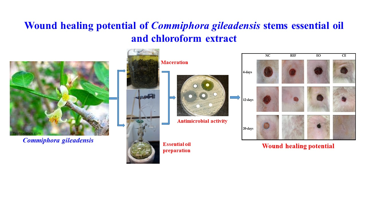
Wound Healing Potential of Commiphora gileadensis Stems Essential Oil and Chloroform Extract

 , , ,
, , ,  ,
, 
Abstract
:
1. Introduction
2. Materials and Methods
2.1. Plant Material
2.2. Chemicals
2.3. Preparation of the Oils
2.4. GC/MS Analysis
2.5. GC Analysis
2.6. Extraction
2.7. Antimicrobial Activity
2.7.1. Bacterial Strains
2.7.2. Antimicrobial Assay
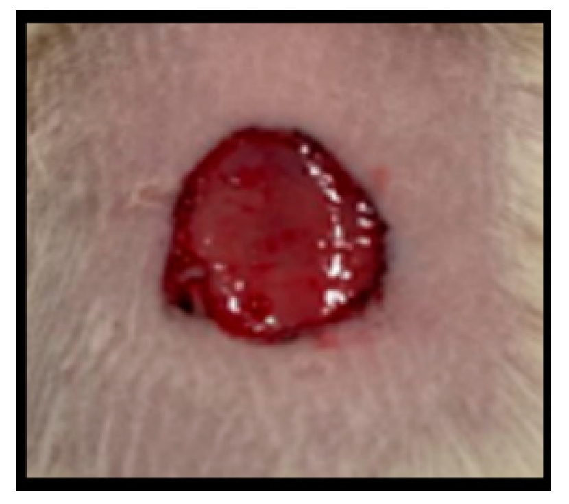
2.8. Evaluation of Wound Healing Activity
2.8.1. Experimental Animals
2.8.2. Preparation of Creams
2.8.3. Experimental Design
- Group 1: Negative control group (NC); treated with the plain cream base topically.
- Group 2: Reference group (REF); topically with 2% Fucidin cream.
- Group 3 and 4 were treated with either 1% EO or with 3% CE creams, respectively.
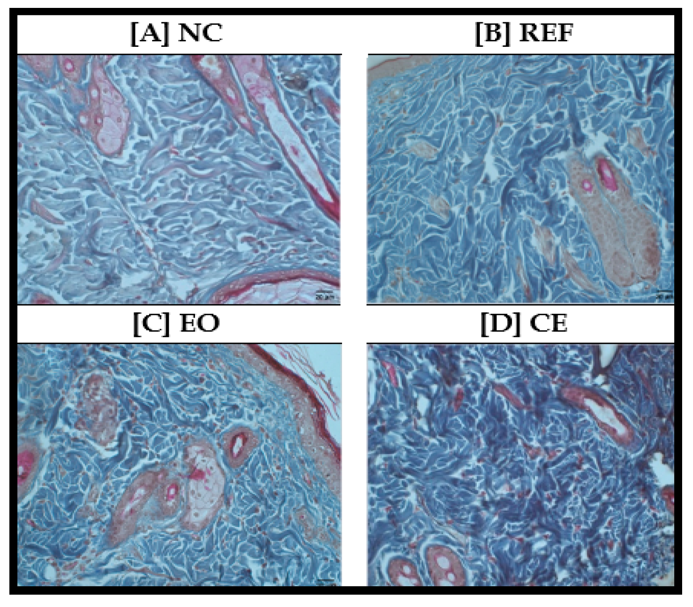
2.9. Histopathological Examination
2.10. Data Analysis
3. Results and Discussion
3.1. Preparation of the Oil and GC-MS Study
3.2. Antimicrobial Activity
3.3. Wound Healing Activity
3.4. Histopathological Study
4. Conclusions
Author Contributions
Funding
Institutional Review Board Statement
Informed Consent Statement
Data Availability Statement
Acknowledgments
Conflicts of Interest
References
- Marcotullio, M.C.; Rosati, O.; Lanari, D. Phytochemistry of Commiphora erythraea: A review. Nat. Prod. Comm. 2018, 13, 1209–1212. [Google Scholar] [CrossRef]
- El Rabey, H.A.; Al-sieni, A.I.; Al-seeni, M.N.; Alsieni, M.A.; Alalawy, A.I.; Almutairi, F.M. The antioxidant and antidiabetic activity of the Arabian balsam tree “Commiphora gileadensis” in hyperlipidaemic male rats. J. Taibah Univ. Sci. 2020, 14, 831–841. [Google Scholar] [CrossRef]
- Miller, A.G.; Morris, M.; Stuart-Smith, S. Plants of Dhofar, the Southern Region of Oman: Traditional, Economic, and Medicinal Uses; Office of the Adviser for Conservation of the Environment, Diwan of Royal Court: Muscat, Oman, 1988. [Google Scholar]
- Al-mahbashi, H.M.; El-shaibany, A.; Saad, F.A. Evaluation of acute toxicity and antimicrobial effects of the bark extract of Bisham (Commiphora gileadensis L.). J. Chem. Pharm. Res. 2015, 7, 810–814. [Google Scholar]
- Al Zoubi, O.M. Evaluation of anti-microbial activity of ex vitro and callus extracts from Commiphora gileadensis. Pak. J. Biol. Sci. 2019, 22, 73–82. [Google Scholar]
- Iluz, D.; Hoffman, M.; Gilboa-Garber, N.; Amar, Z. Medicinal properties of Commiphora gileadensis. Afr. J. Pharm. Pharmacol. 2010, 4, 516–520. [Google Scholar]
- Bouslama, L.; Kouidhi, B.; Alqurashi, Y.M.; Chaieb, K.; Papetti, A. Virucidal Effect of Guggulsterone Isolated from Commiphora gileadensis. Planta Med. 2019, 85, 1225–1232. [Google Scholar] [CrossRef]
- Abdel-Kader, M.S.; Ibnouf, E.O.; Alqarni, M.H.; AlQutaym, A.S.; Salkini, M.A.; Foudah, A.I. Terpenes from the Fresh Stems of Commiphora gileadensis with Antimicrobial Activity. Rec. Nat. Prod. 2022, 16, 605–613. [Google Scholar] [CrossRef]
- Alkahtani, J.; Elshikh, S.M.; Almaary, K.S.; Ali, S.; Imtiyaz, Z.; Ahmad, B.S. Anti-bacterial, anti-scavenging and cytotoxic activity of garden cress polysaccharides. Saudi J. Biol. Sci. 2020, 27, 2929–2935. [Google Scholar] [CrossRef]
- Gonelimali, F.D.; Lin, J.; Miao, W.; Xuan, J.; Charles, F.; Chen, M.; Hatab, S.R. Antimicrobial Properties and Mechanism of Action of Some Plant Extracts Against Food Pathogens and Spoilage Microorganisms. Front. Microbiol. 2018, 9, 1639. [Google Scholar] [CrossRef]
- Chauhan, L.; Gupta, S. Creams: A Review on Classification, Preparation Methods, Evaluation and its Applications. J. Drug Deliv. Ther. 2020, 10, 281–289. [Google Scholar] [CrossRef]
- Mukherjee, P.K.; Verpoorte, R.; Suresh, B. Evaluation of in-vivo wound healing activity of Hypericum patulum (Family: Hypericaceae) leaf extract on different wound model in rats. J. Ethnopharmacol. 2000, 70, 315–321. [Google Scholar] [CrossRef]
- Ponrasu, T.; Suguna, L. Efficacy of Annona squamosa on wound healing in streptozotocin-induced diabetic rats. Int. Wound J. 2012, 9, 613–623. [Google Scholar] [CrossRef] [PubMed]
- Sadaf, F.; Saleem, R.; Ahmed, M.; Ahmad, S.I.; Navaid-ul-Zafar. Healing potential of cream containing extract of Sphaeranthus indicus on dermal wounds in Guinea pigs. J. Ethnopharmacol. 2006, 107, 161–163. [Google Scholar] [CrossRef] [PubMed]
- Manjunatha, B.K.; Vidya, S.M.; Rashmi, K.V.; Mankani, K.L.; Shilpa, H.J.; Singh, S.D. Evaluation of wound-healing potency of Vernonia arborea Hk. Ind. J. Pharmacol. 2005, 37, 223–226. [Google Scholar] [CrossRef]
- Hamad, A.M.; Ahmed, H.G. Association of some carbohydrates with estrogen expression in breast lesions among Sudanese females. J. Histotechnol. 2018, 41, 2–9. [Google Scholar] [CrossRef]
- Hamad, A.M.; Ahmed, H.G. Association of connective tissue fibers with estrogen expression in breast lesions among Sudanese females. Int. Clin. Pathol. J. 2016, 2, 97–102. [Google Scholar] [CrossRef]
- Suvarna, S.K.; Christopher, L.; Bancroft, J.D. The Hematoxyline and Eosin. Bancroft’s Theory and Practice of Histological Techniques, 8th ed.; Elsevier: Amsterdam, The Netherlands, 2018. [Google Scholar]
- Alqarni, M.H.; Salkini, M.A.; Abujheisha, K.Y.; Daghar, M.F.; Al-khuraif, F.A.; Abdel-Kader, M.S. Qualitative, Quantitative and Antimicrobial Activity Variations of the Essential Oils Isolated from Thymus Vulgaris and Micromeria Fruticosa Samples Subjected to Different Drying Conditions. Arab. J. Sci. Eng. 2022, 47, 6861–6867. [Google Scholar] [CrossRef]
- Bassolé, I.H.; Juliani, H.R. Essential oils in combination and their antimicrobial properties. Molecules 2012, 17, 3989–4006. [Google Scholar] [CrossRef]
- Gebrehiwot, M.; Asres, K.; Bisrat, D.; Mazumder, A.; Lindemann, P.; Bucar, F. Evaluation of the wound healing property of Commiphora guidottii Chiov. ex. Guid. BMC Complement. Altern. Med. 2015, 15, 282. [Google Scholar] [CrossRef]
- Bisrat, D.; Mazumder, A.; Lindemann, P. Effects of Resin and Essential Oil from Commiphora myrrha Engl. on Wound Healing. Ethiop. Pharm. J. 2016, 32, 85–100. [Google Scholar]
- Krishna, M. Role of special stains in diagnostic liver pathology. Clin. Liver Dis. 2013, 2, 8–10. [Google Scholar] [CrossRef] [PubMed]


| Condition | Weight (g) | Weight of Oil (g) | % w/w |
|---|---|---|---|
| Fresh | 300.00 | 6.69 | 2.23 |
| Dried | 200.00 | 3.55 | 1.77 |
| Components | RT | RRI | Area % | |
|---|---|---|---|---|
| Fresh | Dry | |||
| β-Pinene (1) | 9.7833 | 978 | 62.974 | 4.462 |
| Eugenol | 26.7184 | 1365 | 5.834 | 35.366 |
| Caryophyllene | 28.7625 | 1444 | 4.543 | 34.469 |
| Humulene (2) | 29.8557 | 1460 | - | 1.718 |
| cis-Calamenene | 32.0615 | 1533 | 12.29 | 0.687 |
| Isoeugenol acetate | 32.2167 | 1618 | 1.926 | 0.483 |
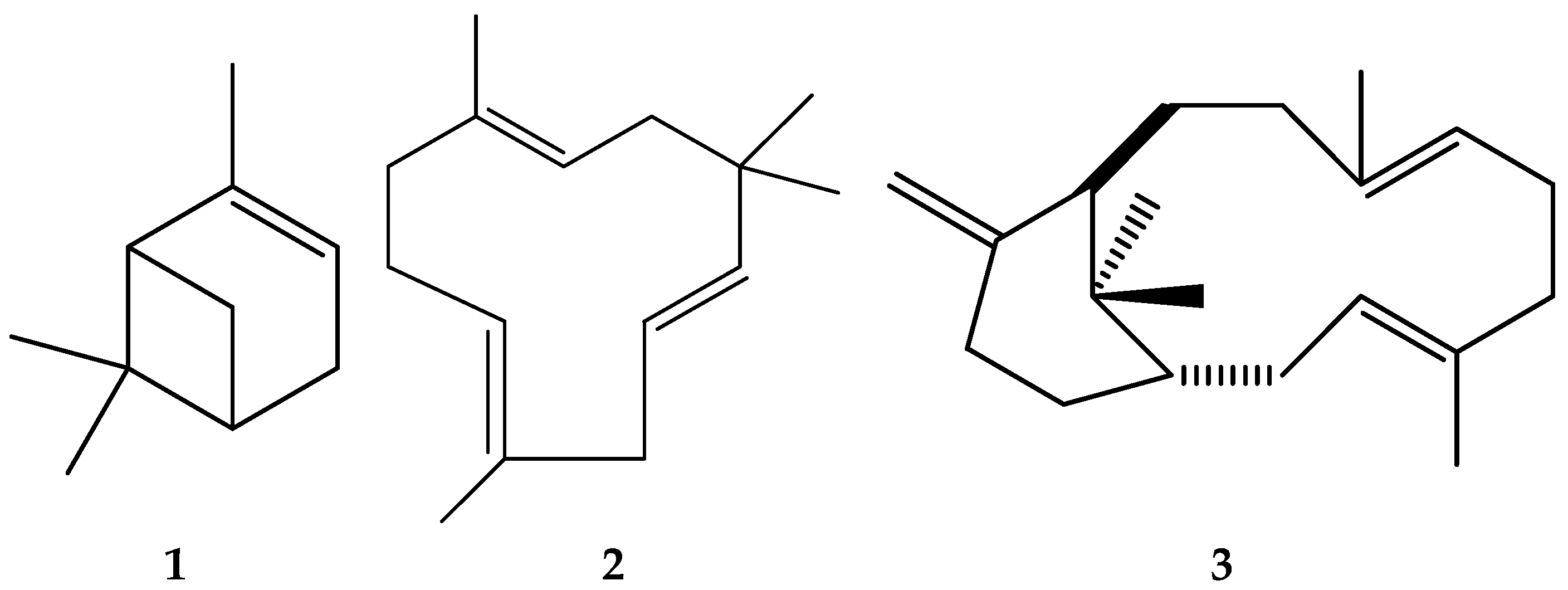
| 1S,3E,7E,11R)-(+)-verticilla-3,7,12(18)-triene (3) | 63.1821 | 2040 | 11.374 | 20.638 |
| Total | 98.941 | 97.823 | ||
| Staph. aureus | B. subtilis | E. coli | k. pneumonia | C. albicans | |
|---|---|---|---|---|---|
| Fresh Stem Oil | 0.75 | 0.75 | 0.5 | 0.5 | 0.25 |
| Dried stem Oil | 0.75 | 0.75 | 0.5 | 0.5 | 0.25 |
| Fresh stem CHCl3 Ext. | 0.75 | 0.75 | 0.75 | 0.25 | 0.125 |
| Fresh stem MeOH Ext. | 75 | 25 | 75 | 50 | 25 |
| Fresh Leaves CHCl3 Ext. | 0.75 | 0.5 | 0.75 | 0.25 | 0.125 |
| Fresh Leaves MeOH Ext. | 75 | 25 | 75 | 25 | 75 |
Publisher’s Note: MDPI stays neutral with regard to jurisdictional claims in published maps and institutional affiliations. |
© 2022 by the authors. Licensee MDPI, Basel, Switzerland. This article is an open access article distributed under the terms and conditions of the Creative Commons Attribution (CC BY) license (https://creativecommons.org/licenses/by/4.0/).
Share and Cite
Althurwi, H.N.; Salkini, M.A.A.; Soliman, G.A.; Ansari, M.N.; Ibnouf, E.O.; Abdel-Kader, M.S. Wound Healing Potential of Commiphora gileadensis Stems Essential Oil and Chloroform Extract. Separations 2022, 9, 254. https://doi.org/10.3390/separations9090254
Althurwi HN, Salkini MAA, Soliman GA, Ansari MN, Ibnouf EO, Abdel-Kader MS. Wound Healing Potential of Commiphora gileadensis Stems Essential Oil and Chloroform Extract. Separations. 2022; 9(9):254. https://doi.org/10.3390/separations9090254
Chicago/Turabian StyleAlthurwi, Hassan N., Mohammad Ayman A. Salkini, Gamal A. Soliman, Mohd Nazam Ansari, Elmutasim O. Ibnouf, and Maged S. Abdel-Kader. 2022. "Wound Healing Potential of Commiphora gileadensis Stems Essential Oil and Chloroform Extract" Separations 9, no. 9: 254. https://doi.org/10.3390/separations9090254
APA StyleAlthurwi, H. N., Salkini, M. A. A., Soliman, G. A., Ansari, M. N., Ibnouf, E. O., & Abdel-Kader, M. S. (2022). Wound Healing Potential of Commiphora gileadensis Stems Essential Oil and Chloroform Extract. Separations, 9(9), 254. https://doi.org/10.3390/separations9090254