Article

Antiallergic Properties of Biflavonoids Isolated from the Flowers of Mesua ferrea Linn.

1
Pharmaceutical Research and Technology Institute, Kindai University, 3-4-1 Kowakae, Higashi-Osaka 577-8502, Osaka, Japan
2
Faculty of Agro-Industry, Rajamangala University of Technology Srivijaya, Thungyai, Nakhon Si Thammarat 80240, Thailand
3
Faculty of Science and Technology, Rajamangala University of Technology Srivijaya, Thungyai, Nakhon Si Thammarat 80240, Thailand
*
Author to whom correspondence should be addressed.
Separations 2022, 9(5), 127; https://doi.org/10.3390/separations9050127
Submission received: 27 April 2022 / Revised: 17 May 2022 / Accepted: 17 May 2022 / Published: 18 May 2022
(This article belongs to the Section Purification Technology)

Abstract

:
The methanolic extract from the flowers of Mesua ferrea Linn. (Calophyllaceae) showed significant hyaluronidase inhibitory activity. Following a bioassay-guided separation of the extract, two biflavonoids, viz., mesuaferrone-A (1) and mesuaferrone-B (2), were isolated, along with ten flavonoids (312), two xanthones (13 and 14), three triterpenes (1517), a phenylpropanoid (18), and five aromatics (1924). Among the isolates, 1 and 2 (IC50 = 51.1 µM and 54.7 µM, respectively) exhibited hyaluronidase inhibitory activity equivalent to that of the commercially available antiallergic agents disodium cromoglycate (64.8 μM) and ketotifen fumarate (76.5 μM). These biflavonoids (1 and 2) are 8-8″ linked dimers that are composed of naringenin (1a) or apigenin (3), with their corresponding monomers lacking inhibitory activity (IC50 > 300 μM). In addition, 1 and 2 (IC50 = 49.4 µM and 49.2 µM, respectively) inhibited the release of β-hexosaminidase, which is a marker of antigen-IgE-mediated degranulation, in rat basophilic leukemia (RBL-2H3) cells. These inhibitory activities were more potent than those of the antiallergic agents tranilast and ketotifen fumarate (IC50 = 282 μM and 158 μM, respectively), as well as one of the corresponding monomers (1a; IC50 > 100 μM). Nonetheless, these effects were weaker than those of the other monomer (3; IC50 = 6.1 μM).

1. Introduction

Mesua ferrea Linn. (Ceylon ironwood in English and locally known as “bunnak” in Thai), of the Calophyllaceae family, is a tropical tree that is widely distributed across Southeast Asia, Thailand, India, and Sri Lanka [1,2,3,4,5]. In traditional Indian medicine, different aerial parts of the plant and their extracts are used to manage a wide range of bodily disorders, such as the use of essential oils to treat skin diseases and rheumatism. In addition, powders from the flowers and fruit of the plant, when mixed with butter, are applied locally for the management of piles, while the seeds are used for treating pain and inflammatory conditions such as arthritis [2]. Previous studies of the chemical constituents from the rhizomes of this plant have led to the isolation and characterization of numerous compounds, including biflavonoids [4,5,6], xanthones [2,4,5], coumarins [4,5,7], flavanone glycosides [8], cyclohexanedione derivatives [9], triterpenes [10], and essential oil [11]. Furthermore, the biological effects of the extract and constituents have been reported, such as the antioxidant [4], antibacterial [5], anti-inflammatory [5], and antitumor [12] properties. During our characterization of the bioactive constituents of plants in Thailand [13,14,15,16,17,18,19,20,21,22,23,24,25,26], a methanolic extract of the M. ferrea flower was found to inhibit hyaluronidase activity. Following the use of a bioassay-guided separation technique, two 8-8″ linked biflavonoids, mesuaferrone-A (1) and mesuaferrone-B (2), were isolated, along with ten flavonoids (312), two xanthones (13 and 14), three triterpenes (1517), a phenylpropanoid (18), and five aromatics (1924). Herein, we report on the isolation, structure elucidation, and antiallergic activities, such as the hyaluronidase and degranulation inhibitory properties, of the isolates.

2. Materials and Methods

2.1. General

The following instruments were used to obtain spectroscopic data: specific rotation, JASCO P-2200 polarimeter (JASCO Corporation, Tokyo, Japan, l  =  5 cm); UV spectra, Shimadzu UV-1600 spectrometer; IR spectra, IRAffinity-1 spectrophotometer (Shimadzu Co., Kyoto, Japan); 1H NMR spectra, JNM-ECA800 (800 MHz), JNM-LA500 (500 MHz), JNM-ECS400 (400 MHz), and JNM-AL400 (400 MHz) spectrometers; 13C NMR spectra, JNM-ECA800 (200 MHz), JNM-LA500 (125 MHz), JNM-ECA400 (100 MHz), and JNM-AL400 (100 MHz) spectrometers (JEOL Ltd., Tokyo, Japan) by using tetramethylsilane as the internal standard; ESIMS and HRESIMS, Exactive Plus mass spectrometer (Thermo Fisher Scientific Inc., Waltham, MA, USA).
Instruments and tools for analytical determinations included the following: HPLC detectors, the Shimadzu RID-6A refractive index (RI) and SPD-10A UV-VIS detectors, and a Shodex OR-2 optical rotation detector; HPLC columns, Cosmosil 5C18-MS-II (Nacalai Tesque, Inc., Kyoto, Japan), Cosmosil Πnap (Nacalai Tesque, Inc., Kyoto, Japan), and Wakopak Navi C30-5 (FUJIFILM Wako Pure Chemical Co., Osaka, Japan). Columns with 4.6 mm i.d. × 250 mm and 20 mm i.d. × 250 mm were used for analytical and preparative purposes, respectively.
The following experimental chromatographic materials were used for column chromatography (CC): highly porous synthetic resin, Diaion HP-20 (Mitsubishi Chemical Co., Tokyo, Japan); normal-phase silica gel CC, silica gel 60 N (Kanto Chemical Co., Ltd., Tokyo, Japan; 63–210 mesh, spherical, neutral); reversed-phase ODS CC, Chromatorex ODS DM1020T (Fuji Silysia Chemical, Ltd., Aichi, Japan; 100–200 mesh); TLC, precoated TLC plates with silica gel 60F254 (Merck, Darmstadt, Germany, 0.25 mm) (normal-phase) and silica gel RP-18 WF254S (Merck, 0.25 mm) (reversed-phase); reversed-phase HPTLC, precoated TLC plates with silica gel RP-18 WF254S (Merck, 0.25 mm). Detection was performed by spraying with 1% Ce(SO4)2–10% aqueous H2SO4, followed by heating.

2.2. Plant Material

The flowers of M. ferrea (loss on drying: 4.35% at 105 °C for 6 h) were collected from the Nakhon Si Thammarat Province of Thailand in September 2006. The plant material was identified by one of the authors (Y.P.). A voucher specimen (2006.09. Raj-07) of the plant is on file in our laboratory.

2.3. Extraction and Isolation

Dried flowers (986.2 g) of M. ferrea were extracted three times following reflux with methanol for 3 h. Solvent evaporation from the combined extracts under reduced pressure yielded an aqueous acetone extract (156.8 g, 15.9%). An aliquot (127.0 g) was partitioned in an EtOAc–H2O (1:1, v/v) mixture to furnish an EtOAc-soluble fraction (75.68 g, 9.47%) and an aqueous phase. The aqueous phase was subjected to Diaion HP-20 CC (2.0 kg, H2O→MeOH) to yield H2O-eluted (40.89 g, 5.12%) and MeOH-eluted (10.43 g, 1.31%) fractions. An aliquot (60.20 g) of the EtOAc-soluble fraction was subjected to normal-phase silica gel CC [3.00 kg, n-hexane–EtOAc (20:1→5:1→1:1→1:2, v/v)→EtOAc→MeOH] to yield seven fractions [Fr. E1 (0.49 g), Fr. E2 (2.02 g), Fr. E3 (19.70 g), Fr. E4 (11.10 g), Fr. E5 (4.67 g), Fr. E6 (1.17 g), and Fr. E7 (16.10 g)]. Fraction E2 (2.02 g) was subjected to reversed-phase silica gel CC [70.0 g, MeOH–H2O (80:20→95:5, v/v)→MeOH] to yield ten fractions [Fr. E2-1 (77.0 mg), Fr. E2-2 (163.7 mg), Fr. E2-3 (62.4 mg), Fr. E2-4 (87.1 mg), Fr. E2-5 (46.0 mg), Fr. E2-6 (91.9 mg), Fr. E2-7 (192.0 mg), Fr. E2-8 [=lupeol (15, 430.1 mg, 0.068%)], Fr. E2-9 (133.7 mg), and Fr. E2-10 (430.8 mg)]. Fraction E2-7 (436.8 mg) was analyzed using HPLC [detection: RI, MeOH-1% aqueous AcOH (95:5, v/v)] to yield 15 (7.1 mg, 0.0011%). Fraction E3 (19.7 g) was subjected to reversed-phase silica gel CC [600.0 g, MeOH–H2O (80:20→90:10, v/v)→MeOH] to yield eight fractions [Fr. E3-1 (78.0 mg), Fr. E3-2 (181.3 mg), Fr. E3-3 (127.2 mg), Fr. E3-4 (583.1 mg), Fr. E3-5 (193.1 mg), Fr. E3-6 (12.6 g), Fr. E3-7 (348.5 mg), and Fr. E3-8 (1.78 mg)]. Fraction E3-2 (181.3 mg) was analyzed using HPLC [detection: RI, MeOH-1% aqueous AcOH (60:40, v/v)] to yield 1,7-dihydroxyxanthone (13, 12.1 mg, 0.019%). Fraction E3-7 (348.5 mg) was purified by HPLC [detection: RI, MeOH-1% aqueous AcOH (95:5, v/v)] to yield betulinaldehyde (16, 15.5 mg, 0.0024%). Fraction E4 (11.05 g) was subjected to reversed-phase silica gel CC [330.0 g, MeOH–H2O (80:20→90:10, v/v)→MeOH] to yield 11 fractions [Fr. E4-1 (58.1 mg), Fr. E4-2 (473.6 mg), Fr. E4-3 (189.4 mg), Fr. E4-4 (799.1 mg), Fr. E4-5 (2301.3 mg), Fr. E4-6 (72.5 mg), Fr. E4-7 (920.0 mg), E4-8 (723.5 mg), E4-9 (254.6 mg), E4-10 (1050.0 mg), and Fr. E4-11 (734.2 mg)]. Fraction E4-1 (58.1 mg) was analyzed using HPLC [detection: UV (254 nm), MeOH-1% aqueous AcOH (20:80, v/v)] to yield p-hydroxybenzoic acid (19, 20.1 mg, 0.00080%), protocatechuic acid (20, 18.2 mg, 0.0018%), protocatechuic aldehyde (22, 5.6 mg, 0.0019%), and vanillic acid (21, 7.8 mg, 0.00074%). Fraction E4-2 (473.6 mg) was analyzed using HPLC [detection: UV (254 nm), MeOH-1% aqueous AcOH (50:50, v/v)] to yield quercetin (10, 27.2 mg, 0.00428%) and trans-cinnamic acid (18, 8.1 mg, 0.00128%). Fraction E4-3 (189.4 mg) was analyzed by HPLC [detection: UV (254 nm), MeOH-1% aqueous AcOH (50:50, v/v)] to yield 1,3,7-trihydroxyxanthone (14, 3.2 mg, 0.0020%). Fraction E4-4 (624.1 mg) was analyzed using HPLC [detection: UV (254 nm), MeOH-1% aqueous AcOH (60:40, v/v)] to yield apigenin (3, 8.6 mg, 0.0020%). Fraction E4-10 (300.0 mg) was analyzed using HPLC [detection: UV (254 nm), MeOH-1% aqueous AcOH (90:10, v/v)] to yield ursolic acid (17, 20.1 mg, 0.0048%). Fraction E5 (4.67 g) was subjected to reversed-phase silica gel CC [150 g, MeOH–H2O (40:60→60:40→70:30→80:20, v/v)→MeOH] to yield eight fractions [Fr. E5-1 (480.0 mg), Fr. E5-2 (250.0 mg), Fr. E5-3 (60.0 mg), Fr. E5-4 (110.0 mg), Fr. E5-5 (870.0 mg), Fr. E5-6 (420.0 mg), Fr. E5-7 (0.51 mg), and Fr. E5-8 (1.18 g)]. Fraction E5-1 (230.0 mg) was analyzed using HPLC [detection: UV (254 nm), MeOH-1% aqueous AcOH (5:95, v/v)] to yield 20 (71.0 mg, 0.0113%) and gallic acid (23, 37.6 mg, 0.0149%). Fraction E5-5 (300.0 mg) was characterized using HPLC [detection: UV (254 nm), MeOH-1% aqueous AcOH (60:40, v/v)] to yield mesuaferrone-A (1, 85.3 mg, 0.0387%), mesuaferrone-B (2, 99.8 mg, 0.0452%), and luteolin (4, 7.0 mg, 0.0032%). Fraction E5-6 (420 mg) was analyzed using HPLC [detection: UV (254 nm), CH3CN-1% aqueous AcOH (50:50 v/v)] to yield 2 (13.9 mg, 0.0022%). Fraction E7 (16.1 g) was subjected to reversed-phase silica gel CC [60.0 g, MeOH–H2O (40:60→60:40→70:30→80:20, v/v)→MeOH] to yield ten fractions [Fr. E7-1 (0.097 g), Fr. E7-2 (1.22 g), Fr. E7-3 (0.48 g), Fr. E7-4 (0.74 g), Fr. E7-5 (1.04 g), Fr. E7-6 (3.55 g), Fr. E7-7 (2.33 g), E7-8 (2.05 g), E7-9 (1.70 g), and Fr. E7-10 (1.50 g)]. Fraction E7-4 (300.0 mg) was analyzed using HPLC [detection: UV (254 nm), MeOH-1% aqueous AcOH (30:70, v/v)] to yield orientin (6, 13.9 mg, 0.0032%). Fraction E7-5 (500.0 mg) was characterized using HPLC [detection: UV (254 nm), MeOH-1% aqueous AcOH (30:70, v/v)] to yield vitexin (5, 14.6 mg, 0.0126%). Fraction E7-6 (500.0 mg) was analyzed using HPLC [detection: UV (254 nm), MeOH-1% aqueous AcOH (50:50, v/v)] to yield 5 (12.7 mg, 0.0140%), saponaretin (7, 89.5 mg, 0.100%), and quercetin-3-O-α-l-rhamnopyranoside (12, 135.4 mg, 0.151%). Fraction E7-7 (500.0 mg) was analyzed using HPLC [detection: UV (254 nm), MeOH-1% aqueous AcOH (40:60, v/v)] to yield 12 (17.0 mg, 0.011%) and kaempferol-3-O-α-l-rhamnopyranoside (11, 48.5 mg, 0.033%). An aliquot (8.20 g) of the MeOH-eluted fraction was subjected to reversed-phase silica gel CC [500 g, MeOH–H2O (35:65→50:50→70:30→90:10, v/v)→EtOAc→MeOH] to yield seven fractions [Fr. M1 (0.80 g), Fr. M2 (0.78 g), Fr. M3 (1.13 g), Fr. M4 (1.72 g), Fr. M5 (1.46 g), Fr. M6 (0.39 g), and Fr. M7 (1.48 g)]. Fraction M4 (500.0 mg) was subjected to HPLC [detection: UV (254 nm), MeOH-1% aqueous AcOH (50:50, v/v)] to yield five fractions {Fr. M4-1 (13.4 mg), Fr. M4-2 (3.4 mg), Fr. M4-3 (31.2 mg), Fr. E4-4 [=5 (51.7 mg, 0.028%)], and Fr. E4-5 [=7 (11.1 g, 0.0061%)]}. Fraction M4-3-1 (31.2 mg) was analyzed using HPLC [detection: UV (254 nm), MeOH-1% aqueous AcOH (35:65, v/v)] to yield homoorientin (8, 3.7 mg, 0.0020%). Fraction M5 (500.0 mg) was analyzed using HPLC [detection: UV (254 nm), MeOH-1% aqueous AcOH (50:50, v/v)] to yield 5 (21.5 mg, 0.040%), 6 (10.7 mg, 0.020%), 7 (31.1 mg, 0.0582%), and apigenin-7-O-rutinoside (9, 23.7 mg, 0.044%) (Figure 1).

2.4. Hyaluronidase Inhibitory Activity

Hyaluronidase inhibitory activity was determined in accordance with a previously reported method [27], with slight modifications. Briefly, the assay was performed in 96-well microplates. Preincubation of 10 μL hyaluronidase enzyme (Type IV-S from bovine testes; 340 NF unit/mL, Sigma-Aldrich Co. LLC, St. Louis, MO, USA) or a blank buffer (0.1 M acetate buffer, pH 3.5) with 20 μL of sample or control was performed at 37 °C for 20 min. Calcium dichloride (20 μL, final concentration: 2.0 mM) was added to the buffer, and the mixture was incubated at 37 °C for 40 min. Next, 50 μL of hyaluronic acid potassium salt (final concentration: 0.6 mg/mL, Sigma-Aldrich Co. LLC, St. Louis, MO, USA) was added, and the mixture was incubated at 37 °C for 40 min. The reaction was stopped by the addition of 0.4 M NaOH (10 μL) and 0.08 M borate solution (pH 9.1, 10 μL), and was immediately heated using boiling water for 3 min. The reaction solution (20 μL) was transferred to another 96-well microplate. p-Dimethylaminobenzaldehyde (80 μL, final concentration: 8.0 mg/mL, Wako Pure Chemical Industries Ltd., Osaka, Japan) acetate solution was added to the reaction mixture and incubated at 37 °C for 20 min. The optical density (OD) of the reaction mixture was measured using a microplate reader (SH-9000, CORONA ELECTRIC Co., Ltd., Ibaraki, Japan) at a wavelength of 585 nm (reference 670 nm). The final concentration of dimethyl sulfoxide (DMSO) in the test solution was 1.0%, and no influence of DMSO on the inhibitory activity was detected. All experiments were performed in quadruplicate, and IC50 values were determined graphically. Disodium cromoglycate (DSCG), ketotifen fumarate, and tranilast, which are clinically prescribed antiallergic medicines, were used as the reference compounds. Equation (1) below was used to calculate the percentage inhibition.
Inhibition (%) = [(OD (C) − OD (N)) − (OD (T) − OD (B))]/(OD (C) − OD (N)) × 100
Control (C): enzyme (+), test sample (−); Test (T): enzyme (+), test sample (+); Blank (B): Missouri, enzyme (−), test sample (+); Normal (N): enzyme (−), test sample (−); OD, optical density.

2.5. Inhibitory Effects on the Release of β-Hexosaminidase from RBL-2H3 Cells

Inhibitory effects on the release of β-hexosaminidase in RBL-2H3 cells [Cell No. JCRB0023, obtained from the Health Science Research Resources Bank (Osaka, Japan)] were evaluated by using a previously reported method [28,29]. Briefly, RBL-2H3 cells in 24-well plates (2 × 10⁵ cells/well in minimal essential medium (MEM) containing 10% fetal bovine serum (FBS), penicillin (100 units/mL), and streptomycin (100 µg/mL)) were sensitized with anti-dinitrophenyl immunoglobulin E (anti-DNP IgE, 0.45 µg/mL). The cells were washed with Siraganian buffer (119 mM NaCl, 5 mM KCl, 0.4 mM MgCl2, 25 mM piperazine-N,N′-bis(2-ethanesulfonic acid) (PIPES), and 40 mM NaOH, pH 7.2) supplemented with 5.6 mM glucose, 1 mM CaCl2, and 0.1% bovine serum albumin (BSA) (incubation buffer), and then incubated in 160 μL of the incubation buffer for 10 min at 37 °C. Next, 20 μL of the test sample solution was added to each well and was incubated for 10 min, followed by an addition of 20 μL of antigen (DNP-BSA, final concentration: 10 μg/mL) and incubation at 37 °C for 10 min to stimulate cell degranulation. Subsequently, the reaction was stopped by cooling in an ice bath for 10 min. The supernatant (50 μL) was then transferred into a 96-well plate and incubated with 50 µL of substrate (1 mM p-nitrophenyl-N-acetyl-β-d-glucosaminide) in 0.1 M citrate buffer (pH 4.5) at 37 °C for 1 h. The reaction was stopped by adding 200 μL of stop solution (0.1 M Na2CO3/NaHCO3, pH 10.0). The absorbance was measured by using a microplate reader set at a wavelength of 405 nm. The test sample was dissolved in DMSO, and the solution added to incubation buffer (final DMSO concentration: 0.1%). The inhibition (%) of the release of β-hexosaminidase by the test samples was calculated using Equation (2), and IC50 values were determined graphically:
Inhibition (%) = [1 − (T − B − N)/(C − N)] × 100
Control (C): DNP-BSA (+), test sample (−); Test (T): DNP-BSA (+), test sample (+); Blank (B): DNP-BSA (−), test sample (+); Normal (N): DNP-BSA (−), test sample (−).
To ascertain whether the antiallergic effects of the samples were due to the inhibition of β-hexosaminidase release, and not due to a false positive from the inhibition of β-hexosaminidase activity, we performed the following test. The cell suspension (5 × 10⁷ cells) was placed in 6 mL of phosphate-buffered saline and sonicated. The solution was then centrifuged, and the supernatant was diluted with the incubation buffer and adjusted to equal the enzyme activity of the above-tested degranulation. The enzyme solution (45 μL) and test sample solution (5 μL) were transferred into a 96-well microplate, and the enzyme activity was examined as described above (Equation (2)). Under these conditions, the total β-hexosaminidase activity of the cell suspension after sonication was calculated from the cells in the control groups. Tranilast and ketotifen fumarate, which are clinically prescribed antiallergic medicines, were used as the reference compounds.

2.6. Statistics

All data are expressed as means ± standard error of the mean. One-way analysis of variance, followed by Dunnett’s test, were used for statistical analysis. Probability (p) values < 0.05 were considered statistically significant.

3. Results and Discussion

3.1. Inhibitory Effects of the M. ferrea Flower Methanolic Extract and Its Fractions on Hyaluronidase

Hyaluronidases are enzymes that break down hyaluronic acid, which is a mucopolysaccharide that is related to inflammation, through the release of histamine from mast cells. Hyaluronidase inhibitors are effective therapies for the suppression of allergies and inflammation [30,31,32]. It is known that DSCG, which is a commercially available antiallergic agent, exhibits a strong inhibitory effect against hyaluronidase [31]. Therefore, a close relationship has been defined between allergic reactions and hyaluronidase inhibitory activity [31,32].
The dried flowers of M. ferrea were used to obtain a methanol extract (15.9% from the dried material). The methanol was partitioned by using EtOAc–H2O (1:1, v/v) to yield an EtOAc-soluble fraction (9.47%) and an aqueous phase. The aqueous phase was subjected to Diaion HP-20 CC (H2O→MeOH) to yield MeOH- and H2O-eluted fractions (5.12% and 1.31%, respectively). As shown in Table 1, the methanolic extract was found to have hyaluronidase inhibitory activity [inhibition (%): 52.1 ± 4.6 at 1000 μg/mL]. Following the use of a bioassay-guided separation procedure, the EtOAc-soluble and the MeOH-eluted fractions were found to be the active fractions (IC50 = 430 and 360 μg/mL, respectively), while the H2O-eluted fraction showed no notable activity.

3.2. Chemical Constituents of the M. ferrea Flower

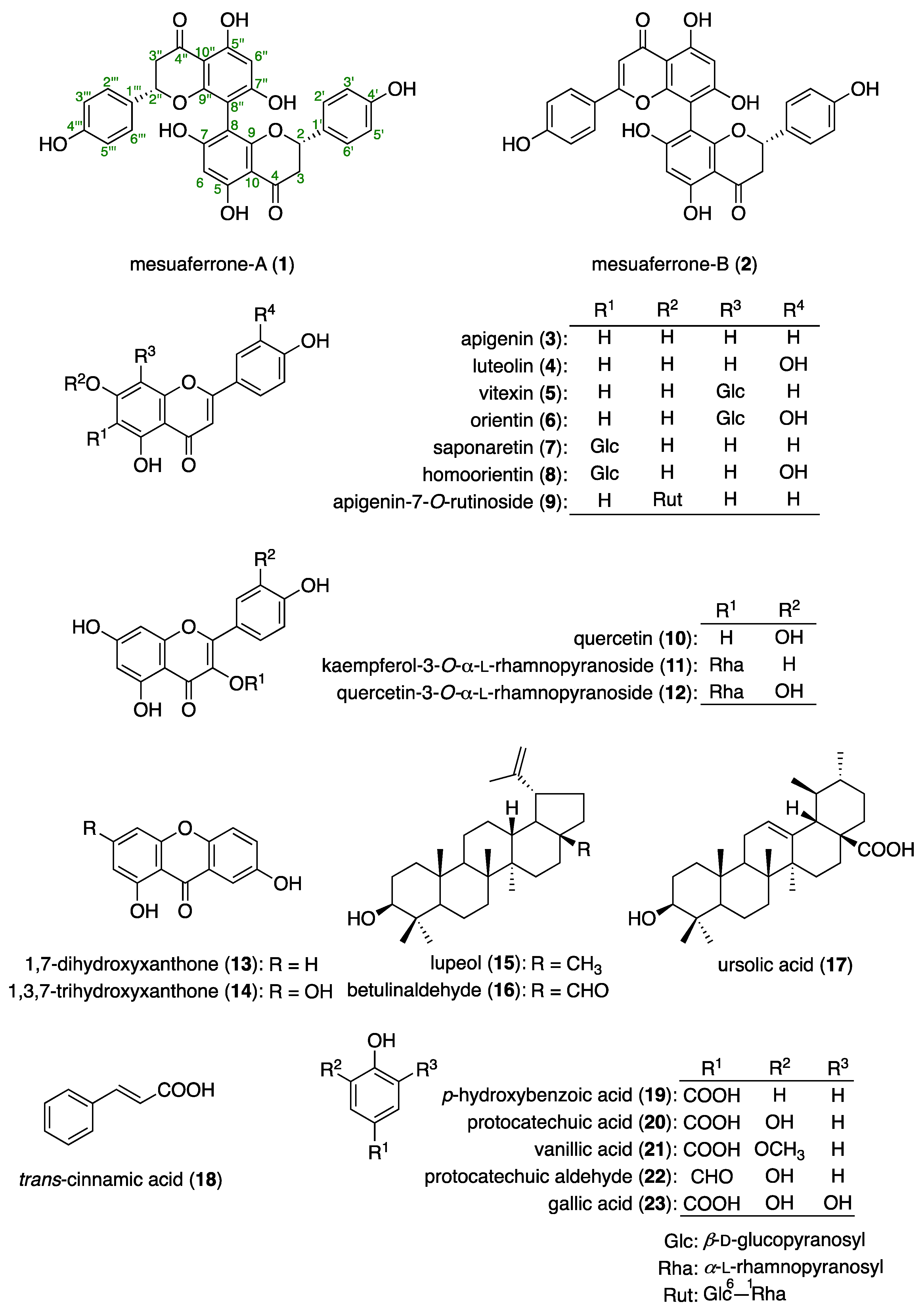
The EtOAc-soluble fraction was subjected to normal-phase silica gel and reversed-phase ODS column CC, and finally to HPLC to obtain the following: two 8-8″ linked biflavonoids, mesuaferrone-A (1, 0.039%) [33] and mesuaferrone-B (2, 0.0047%) [34]; eight flavonoids, apigenin (3, 0.0020%), luteolin (4, 0.0032%), vitexin (5, 0.032%) [35], orientin (6, 0.015%) [36], saponaretin (7, 0.16%) [37], quercetin (10, 0.0043%), kaempferol 3-O-α-l-rhamnopyranoside (11, 0.033%) [38], and quercetin 3-O-α-l-rhamnopyranoside (12, 0.16%) [39]; two xanthones, 1,7-dihydroxyxanthone (13, 0.0024%) [40] and 1,3,7-trihydroxyxanthone (14, 0.0019%) [41]; three triterpenes, lupeol (15, 0.069%) [42], betulinaldehyde (16, 0.0048%) [43], and ursolic acid (17, 0.048%) [44]; a phenylpropanoid, trans-cinnamic acid (18, 0.0013%); and five aromatics, p-hydroxybenzoic acid (19, 0.0008%), protocatechuic acid (20, 0.013%), vanillic acid (21, 0.00074%), protocatechuic aldehyde (22, 0.0019%) [45], and gallic acid (23, 0.19%). From the MeOH-eluted fraction, five flavonoids, 5 (0.068%), 6 (0.020%), 7 (0.064%), homoorientin (8, 0.0020%) [46], and apigenin 7-O-rutinoside (9, 0.044%) [47], were isolated (Figure 2 and Table S1). The isolates were identified by a comparison of their physical and spectral data with those of commercially available samples (3, 4, 10, 1821, and 23), or with reported values [33,34,35,36,37,38,39,40,41,42,43,44,45,46,47].

3.3. Inhibitory Effects of M. ferrea Flower Isolates (123) on Hyaluronidase

In previous studies of compounds from natural medicines that possess hyaluronidase inhibitory activity, it was reported that a phenylethanoid glycoside that was isolated from the flowers of Mimusops elengi L. (Sapotaceae) [20], aporphine- and benzylisoquinoline-type alkaloids from the flowers of Nelumbo nucifera Gaertn. (Nelumbonaceae) [27], and iridoids from the rhizomes of Picrorhiza kurroa Royle ex Benth. (Plantaginaceae) [48] possess this property. To add on to these findings, the hyaluronidase inhibitory activity of the isolates from the flowers of M. ferrea were examined. Among the isolates, the two 8-8″ linked biflavonoids mesuaferrone-A (1, IC50 = 51.1 μM) and B (2, IC50 = 54.7 μM) exhibited hyaluronidase inhibitory activity that was equivalent to that of the commercial antiallergic agents DSCG (64.8 μM) and ketotifen fumarate (76.5 μM), which are therapeutically effective owing to their inhibitory activity on degranulation [49] (Table 2). These biflavonoids (1 and 2) are 8-8″ linked dimers that are composed of naringenin (1a) or apigenin (3). However, their corresponding monomers did not show similarly potent inhibitory activity (IC50 > 300 μM).

3.4. Inhibitory Effects of 1 and 2 on the Release of β-Hexosaminidase in RBL-2H3 Cells

Basophils and mast cells play important roles in both the immediate- and late-phase reactions of type I allergies. The aggregation of high-affinity Fcε receptor I (FcεRI) by antigens results in tyrosine phosphorylation, Ca2+ release from intracellular Ca2+ stores, and Ca2+ influx via release-activated Ca2+ channels. The levels of intracellular free Ca2+ ([Ca2+]i) play an essential role in the degranulation process [49,50,51,52]. Histamine, which is a chemical mediator that is released from mast cells and basophils when they are stimulated by an immunoglobulin E (IgE)-antigen complex or a degranulation inducer, is usually considered to be a degranulation marker of immediate allergic reactions in in vitro experiments. β-Hexosaminidase is also stored in the secretory granules of cells and is released concomitantly with histamine when the cells are immunologically activated. Therefore, the enzymatic activity of β-hexosaminidase can be used as a marker for the degranulation of the cells [53]. Previously, our studies on compounds from natural medicines that possess degranulation inhibitory activity have shown that phenylpropanoids [54], neolignans [49], flavonoids [52,55], stilbenoids [28,56,57], diarylheptanoids [29,58,59], terpenoids [55,60,61,62,63], and alkaloids [64,65] have this property. In addition, it has been found that there is a close relationship between allergic reactions and hyaluronidase inhibitory activity [31,32]. Therefore, the degranulation inhibitory activities of mesuaferrone-A (1) and mesuaferrone-B (2), which have hyaluronidase activity, were examined. Our findings show that 1 (IC50 = 49.4 µM) and 2 (C50 = 49.2 µM) inhibited the release of β-hexosaminidase, which is a marker of antigen-IgE-mediated degranulation in RBL-2H3 cells (Table 3). These inhibitory activities were more potent than those of tranilast (282 μM), ketotifen fumarate (158 μM), and one of the corresponding monomers (1a; >100 μM), but they were weaker than that of the other monomer (3; 6.1 μM). Next, to confirm that these observations were definitely the result of inhibiting the release of β-hexosaminidase, and not a false positive from inhibiting the enzymatic activity of β-hexosaminidase, the effects of the abovementioned active degranulation inhibitors on the enzyme activity were examined. The results show that none of the investigated molecules significantly inhibited β-hexosaminidase activity at a concentration of 100 μM (data not shown).

4. Conclusions

In conclusion, we found that the methanolic extract of the flowers of M. ferrea inhibits the enzymatic activity of hyaluronidase. Through a bioassay-guided separation of the extract, two biflavonoids, mesuaferrone-A (1) and mesuaferrone-B (2), were isolated, along with ten flavonoids (312), two xanthones (13 and 14), three triterpenes (1517), a phenylpropanoid (18), and five aromatics (1924). Among the isolates, the biflavonoids mesuaferrone-A (1, IC50 = 51.1 µM) and B (2, IC50 = 54.7 µM) were identified as the active constituents. Their inhibitory activities were equivalent to those of the antiallergic medicines DSCG (64.8 μM) and ketotifen fumarate (76.5 μM). As for the corresponding monomer flavonoids, naringenin (1a) or apigenin (3), they did not show similar inhibitory activity (IC50 > 300 μM). In addition, 1 (IC50 = 49.4 µM) and 2 (IC50 = 49.2 µM) were found to possess degranulation inhibitory activities. These inhibitory activities were more potent than those of the antiallergic medicines tranilast (IC50 = 282 μM) and ketotifen fumarate (158 μM), and one of the corresponding monomers (1a; >100 μM), but they were weaker than that of the other monomer (3; 6.1 μM). These results suggest that the presence of a biflavonoid skeleton may be important for antiallergic properties through both the hyaluronidase and degranulation inhibitory pathways. Further studies on the mechanisms of action of these constituents, as well as the associated structural requirements, are in progress.

Supplementary Materials

The following supporting information can be downloaded at: https://www.mdpi.com/article/10.3390/separations9050127/s1. Figures S1–S10: NMR spectra of mesuaferrone-A (1) and mesuaferrone-B (2); and Table S1: List of the isolates (123) from the flowers of M. ferrea.

Author Contributions

Conceptualization, Y.M.; data curation, Y.M., Y.S., T.M. (Taiki Miyachi), M.N., Y.H. and T.M. (Toshio Morikawa); formal analysis, Y.M., Y.S., T.M. (Taiki Miyachi) and M.N.; funding acquisition, T.M. (Toshio Morikawa); investigation, Y.M. and T.M. (Toshio Morikawa); resources, S.C. and Y.P.; methodology, Y.M. and T.M. (Toshio Morikawa); project administration, T.M. (Toshio Morikawa); supervision, T.M. (Toshio Morikawa); visualization, Y.M. and T.M. (Toshio Morikawa); writing—original draft, Y.M.; writing—review and editing, T.M. (Toshio Morikawa). All authors have read and agreed to the published version of the manuscript.

Funding

This work was supported in part by JSPS KAKENHI, Japan [Grant Numbers 18K06726 and 22K06688 (T.M. (Toshio Morikawa))].

Institutional Review Board Statement

Not applicable.

Informed Consent Statement

Not applicable.

Data Availability Statement

The data that support the findings of this study are available from the corresponding author upon reasonable request.

Acknowledgments

The authors thank the Division of Joint Research Center of Kindai University for the NMR and MS measurements.

Conflicts of Interest

The authors declare no conflict of interest.

References

  1. Patil, A.B.; Shinde, S.S.; Raghavendra, S.; Satish, B.N.; Kushalappa, C.G.; Vijay, N. The genome sequence of Mesua ferrea and comparative demographic histories of forest trees. Gene 2021, 15, 145214. [Google Scholar] [CrossRef] [PubMed]
  2. Chukaew, A.; Saithong, S.; Chusri, S.; Limsuwan, S.; Watanapokasin, R.; Voravuthikunchai, S.P.; Chakthong, S. Cytotoxic xanthones from the roots of Mesua ferrea L. Phytochemistry 2019, 157, 64–70. [Google Scholar] [CrossRef] [PubMed]
  3. Shirsat, P.; Ziyaurrahman, A.R.; Kashikar, R.; Athavale, M.; Athavale, T.; Taware, P.; Saldanha, T.; Kolhe, S.; Tembhurne, S. Subacute toxicity study of the ethanolic extract of Mesua ferrea (L.) flowers in rats. Drug Chem. Toxicol. 2020, 18, 1–8. [Google Scholar] [CrossRef] [PubMed]
  4. Manoj, K.C.; Sanjaya, K.D.S.; Geetha, L.; Lokesh, T.; Manohara, K.P. Mesua ferrea L.: A review of the medical evidence for its phytochemistry and pharmacological actions. Afr. J. Pharm. Pharmacol. 2013, 7, 211–219. [Google Scholar] [CrossRef] [Green Version]
  5. Sruthikrishna, P.K. Review on Ethnobotany and Phytopharmacology on Mesua ferrea Linn. Res. J. Pharmacogn. Phytochem. 2021, 13, 195–199. [Google Scholar] [CrossRef]
  6. Zhang, X.; Gao, R.; Liu, Y.; Cong, Y.; Zhang, D.; Zhang, Y.; Yang, X.; Lu, C.; Shen, Y. Anti-virulence activities of biflavonoids from Mesua ferrea L. flower. Drug. Discov. Ther. 2019, 13, 222–227. [Google Scholar] [CrossRef] [Green Version]
  7. Wang, S.; Guo, Y.; Yao, D.; Liu, L.; Duan, H.; Meng, L.; Yang, H.; Zhang, K.; Huang, J.; Li, Q.; et al. 4-Alkyl-5,7-dihydroxycoumarins from the flowering buds of Mesua ferrea. Fitoterapia 2019, 138, 104192. [Google Scholar] [CrossRef]
  8. Alam, M.S.; Jain, N.; Kamil, M. Mesuein: A novel flavanone glycoside from Mesua ferrea. Chem. Ind. Lond. 1987, 16, 565–566. [Google Scholar]
  9. Dennis, T.J.; Akshay, K.K.; Srimannarayana, G. A new cyclo hexadione from Mesua ferrea. Phytochemistry 1988, 27, 2325–2327. [Google Scholar] [CrossRef]
  10. Kamarusalihin, N.; Faujan, B.H.A.; Taufiq-Yap, Y.H.; Ali, A.M. Volatile components of methanol extract from the flower of Malaysian Mesua ferrea Linn. Orient. J. Chem. 2004, 20, 69–72. [Google Scholar]
  11. Chahar, M.K.; Sanjaya-Kumar, D.S.; Lokesh, T.; Manohara, K.P. In-vivo antioxidant and immunomodulatory activity of mesuol isolated from Mesua ferrea L. seed oil. Int. Immunopharmacol. 2012, 13, 386–391. [Google Scholar] [CrossRef] [PubMed]
  12. Asif, M.; Shafaei, A.; Jafari, S.F.; Mohamed, S.K.; Ezzat, M.O.; Abdul-Majid, A.S.; Oon, C.E.; Petersen, S.H.; Kono, K.; Abdul-Majid, A.M. Isoledene from Mesua ferrea oleo-gum resin induces apoptosis in HCT 116 cells through ROS-mediated modulation of multiple proteins in the apoptotic pathways: A mechanistic study. Toxicol. Lett. 2016, 22, 84–96. [Google Scholar] [CrossRef] [PubMed]
  13. Morikawa, T.; Akaki, J.; Ninomiya, K.; Kinouchi, E.; Tanabe, G.; Pongpiriyadacha, Y.; Yoshikawa, M.; Muraoka, O. Salacinol and related analogs: New leads for type 2 diabetes therapeutic candidates from the Thai traditional natural medicine Salacia chinensis. Nutrients 2015, 7, 1480–1493. [Google Scholar] [CrossRef] [PubMed] [Green Version]
  14. Ninomiya, K.; Matsumoto, T.; Chaipech, S.; Miyake, S.; Katsuyama, Y.; Tsuboyama, A.; Pongpiriyadacha, Y.; Hayakawa, T.; Muraoka, O.; Morikawa, T. Simultaneous quantitative analysis of 12 methoxyflavones with melanogenesis inhibitory activity from the rhizomes of Kaempferia parviflora. J. Nat. Med. 2016, 70, 179–189. [Google Scholar] [CrossRef] [PubMed]
  15. Ninomiya, K.; Shibatani, K.; Sueyoshi, M.; Chaipech, S.; Pongpiriyadacha, Y.; Hayakawa, T.; Muraoka, O.; Morikawa, T. Aromatase inhibitory activity of geranylated coumarins, mammeasins C and D, isolated from the flowers of Mammea siamensis. Chem. Pharm. Bull. 2016, 64, 880–885. [Google Scholar] [CrossRef] [PubMed] [Green Version]
  16. Manse, Y.; Ninomiya, K.; Nishi, R.; Kamei, I.; Katsuyama, Y.; Imagawa, T.; Chaipech, S.; Muraoka, O.; Morikawa, T. Melanogenesis inhibitory activity of a 7-O-9′-linked neolignan from Alpinia galanga fruit. Bioorg. Med. Chem. 2016, 24, 6215–6224. [Google Scholar] [CrossRef] [PubMed]
  17. Ninomiya, K.; Chaipech, S.; Kunikata, Y.; Yagi, R.; Pongpiriyadacha, Y.; Muraoka, O.; Morikawa, T. Quantitative determination of stilbenoids and dihydroisocoumarins in Shorea rexburghii and evaluation of their hepatoprotective activity. Int. J. Mol. Sci. 2017, 18, 451. [Google Scholar] [CrossRef] [Green Version]
  18. Tanabe, G.; Tsutsui, N.; Shibatani, K.; Marumoto, S.; Ishikawa, F.; Ninomiya, K.; Morikawa, T. Total syntheses of the aromatase inhiitors, mammeasins C and D, from Thai medicinal plant Mammea siamensis. Tetrahedron 2017, 73, 4481–4486. [Google Scholar] [CrossRef]
  19. Manse, Y.; Ninomiya, K.; Nishi, R.; Hashimoto, Y.; Chaipech, S.; Muraoka, O.; Morikawa, T. Labdane-type diterpenes, galangalditerpenes A-C, with melanogenesis inhibitory activity from the fruit of Alpinia galanga. Molecules 2017, 22, 2279. [Google Scholar] [CrossRef] [Green Version]
  20. Morikawa, T.; Manse, Y.; Koda, M.; Chaipech, S.; Pongpiriyadacha, Y.; Muraoka, O.; Ninomiya, K. Two new aromatic glycosides, elengiosides A and B, from the flowers of Mimusops elengi. J. Nat. Med. 2018, 72, 542–550. [Google Scholar] [CrossRef]
  21. Tanabe, G.; Manse, Y.; Ogawa, T.; Sonoda, N.; Marumoto, S.; Ishikawa, F.; Ninomiya, K.; Chaipech, S.; Pongpiriyadacha, Y.; Muraoka, O.; et al. Total synthesis of γ-alkylidenebutenolides, potent melanogenesis inhibitors from Thai medicinal plant Melodorum fruticosum. J. Org. Chem. 2018, 83, 8250–8264. [Google Scholar] [CrossRef] [PubMed]
  22. Morikawa, T.; Akaki, J.; Pongpiriyadacha, Y.; Yoshikawa, M.; Ninomiya, K.; Muraoka, O. Simultaneous quantitative determination of polyphenol constituents in Salacia species from different regions by LC-MS. Jpn. J. Food Chem. Saf. 2018, 25, 130–138. [Google Scholar] [CrossRef]
  23. Kobayashi, M.; Akaki, J.; Yamaguchi, Y.; Yamasaki, H.; Ninomiya, K.; Pongpiriyadacha, Y.; Yoshikawa, M.; Muraoka, O.; Morikawa, T. Salacia chinensis stem extract and its thiosugar sulfonium constituent, neokotalanol, improves HbA1c in ob/ob mice. J. Nat. Med. 2019, 73, 584–588. [Google Scholar] [CrossRef] [PubMed]
  24. Morikawa, T.; Luo, F.; Manse, Y.; Sugita, H.; Saeki, S.; Chaipech, S.; Pongpiriyadacha, Y.; Muraoka, O.; Ninomiya, K. Geranylated coumarins from Thai medicinal plant Mammea siamensis with testosterone 5α-reductase inhibitory activity. Front. Chem. 2020, 20, 199. [Google Scholar] [CrossRef] [PubMed] [Green Version]
  25. Luo, F.; Sugita, H.; Muraki, K.; Saeki, S.; Chaipech, S.; Pongpiriyadacha, Y.; Muraoka, O.; Morikawa, T. Anti-proliferative activities of coumarins from the Thai medicinal plant Mammea siamensis (Miq.) T. Anders. against human digestive tract carcinoma cell lines. Fitoterapia 2021, 148, 104780. [Google Scholar] [CrossRef]
  26. Morikawa, T.; Ninomiya, K.; Tanabe, G.; Matsuda, H.; Yoshikawa, M.; Muraoka, O. A review of antidiabetic active thiosugar sulfoniums, salacinol and neokotalanol, from plants of the genus Salacia. J. Nat. Med. 2021, 75, 449–466. [Google Scholar] [CrossRef]
  27. Morikawa, T.; Okugawa, S.; Manse, Y.; Muraoka, O.; Yoshikawa, M.; Ninomiya, K. Quantitative determination of principal aporphine and benzylisoquinoline alkaloids due to blooming state in lotus flower (flower buds of Nelumbo nucifera) and their hyaluronidase inhibitory activity. Nat. Prod. Commun. 2019, 14, 1934578X19857834. [Google Scholar] [CrossRef] [Green Version]
  28. Morikawa, T.; Xu, F.; Matsuda, H.; Yoshikawa, M. Structures of novel Norstilbene dimer, Longusone A, and three new stilbene dimers, longusols A, B, and C, with antiallergic and radical scavenging activities from Egyptian natural medicine Cyperus longus. Chem. Pharm. Bull. 2010, 58, 1379–1385. [Google Scholar] [CrossRef] [Green Version]
  29. Matsuda, H.; Tewtrakul, S.; Morikawa, T.; Nakamura, A.; Yoshikawa, M. Anti-allergic principles from Thai zedoary: Structural requirements of curcuminoids for inhibition of degranulation and effect on the release of TNF-α and IL-4 in RBL-2H3 cells. Bioorg. Med. Chem. 2004, 12, 5891–5898. [Google Scholar] [CrossRef]
  30. Girish, K.S.; Kemparaju, K. The magic glue hyaluronan and its eraser hyaluronidase: A biological overview. Life Sci. 2007, 80, 1921–1943. [Google Scholar] [CrossRef]
  31. Kakegawa, H.; Matsumoto, H.; Satoh, T. Inhibitory effects of some natural products on the activation of hyaluronidase and their anti-allergic actions. Chem. Pharm. Bull. 1992, 40, 1439–1442. [Google Scholar] [CrossRef] [PubMed] [Green Version]
  32. Murata, T.; Watahiki, M.; Tanaka, Y.; Miyase, T.; Yoshizaki, F. Hyaluronidase inhibitors from Takuran, Lycopus lucidus. Chem. Pharm. Bull. 2010, 58, 394–397. [Google Scholar] [CrossRef] [PubMed] [Green Version]
  33. Subramanyam, R.M.; Rao, G.; Subba, N.V. Structure of mesuaferrone-A, a new biflavanone from the stamens of Mesua ferrea Linn. Indian J. Chem. Sect. B 1978, 16, 167–168. [Google Scholar]
  34. Subramanyam, R.M.; Srimannarayana, G.; Subba-Rao, N.V.; Bala, K.R.; Seshadri, T.R. Structure of mesuaferrone-B a new biflavanone from the stamens of Mesua ferrea Linn. Tetrahedron Lett. 1976, 17, 4509–4512. [Google Scholar] [CrossRef]
  35. Jayasinghe, U.L.B.; Balasooriya, B.A.I.S.; Bandara, A.G.D.; Fujimoto, Y. Glycosides from Grewia damine and Filicium decipiens. Nat. Prod. Res. 2004, 18, 499–502. [Google Scholar] [CrossRef]
  36. Kato, T.; Morita, Y. C-Glycosylflavones with acetyl substitution from Rumex acetosa L. Chem. Pharm. Bull. 1990, 38, 2277–2280. [Google Scholar] [CrossRef] [Green Version]
  37. Lin, C.N.; Kuo, S.H.; Chung, M.I.; Ko, F.N.; Teng, C.T. A new flavone C-glycoside and antiplatelet and vasorelaxing flavones from Gentiana arisanensis. J. Nat. Prod. 1997, 60, 851–853. [Google Scholar] [CrossRef]
  38. Zhang, Z.; ElSohly, H.N.; Li, X.C.; Khan, S.I.; Sheldon, E.; Broedel, S.E.J.; Raulli, R.E.; Cihlar, R.L.; Burandt, C.; Walker, L.A. Phenolic compounds from Nymphaea odorata. J. Nat. Prod. 2003, 66, 548–550. [Google Scholar] [CrossRef]
  39. Fukunaga, T.; Nishiya, K.; Kajikawa, I.; Watanabe, Y.; Suzuki, N.; Takeya, K.; Itokawa, H. Chemical studies on the constituents of Hyphear tanakae Hosokawa from different host trees. Chem. Pharm. Bull. 1988, 36, 1180–1184. [Google Scholar] [CrossRef]
  40. Yang, X.D.; Xu, L.Z.; Yang, S.L. Xanthones from the stems of Securidaca inappendiculata. Phytochemistry 2001, 58, 1245–1249. [Google Scholar] [CrossRef]
  41. Mohammad, A.; Mumtaz, A.; Habib, A.; Itrat, A.; Ajmal, K.; Iqbal, C.; Muhammad, R.S. Urease inhibitors from Hypericum oblongifolium WALL. J. Enzym. Inhib. Med. Chem. 2010, 25, 296–299. [Google Scholar] [CrossRef] [Green Version]
  42. Wenkert, E.; Vernon-Baddeley, G.; Burfitt, I.R.; Moreno, L.N. Carbon-13 nuclear magnetic resonance spectroscopy of naturally-occurring substances-LVII triterpenes related to lupane and hopane. Magn. Reson. Chem. 1978, 11, 337–343. [Google Scholar] [CrossRef]
  43. Pohjala, L.; Alakurtti, S.; Ahola, T.; Yli-Kauhaluoma, J.; Tammela, P. Betulin-derived compounds as inhibitors of alphavirus replication. J. Nat. Prod. 2009, 72, 1917–1926. [Google Scholar] [CrossRef] [PubMed]
  44. Acebey-Castellon, I.L.; Voutquenne-Nazabadioko, L.; Thi-Mai, H.D.; Roseau, N.; Bouthagane, N.; Muhammad, D.; Debar, E.L.M.; Gangloff, S.C.; Litaudon, M.; Sevenet, T.; et al. Triterpenoid saponins from Symplocos lancifolia. J. Nat. Prod. 2011, 74, 163–168. [Google Scholar] [CrossRef] [PubMed]
  45. Detsi, A.; Majdalani, M.; Kontogiorgis, C.A.; Hadjipavlou-Litina, D.; Kefalas, P. Natural and synthetic 2′-hydroxy-chalcones and aurones: Synthesis, characterization and evaluation of the antioxidant and soybean lipoxygenase inhibitory activity. Bioorg. Med. Chem. 2009, 17, 8073–8085. [Google Scholar] [CrossRef] [PubMed]
  46. Maatooq, G.T.; El-Sharkawy, S.H.; Afifi, M.S.; Rosazza, J.P. C-p-Hydroxybenzoylglycoflavones from Citrullus colocynthis. Phytochemistry 1997, 44, 187–190. [Google Scholar] [CrossRef]
  47. Wang, M.; Simon, J.E.; Aviles, I.F.; He, K.; Zheng, Q.; Tadmor, Y. Analysis of antioxidative phenolic compounds in artichoke (Cynara scolymus L.). J. Agric. Food Chem. 2003, 51, 601–608. [Google Scholar] [CrossRef]
  48. Morikawa, T.; Nakanishi, Y.; Inoue, N.; Manse, Y.; Matsuura, H.; Hamasaki, S.; Yoshikawa, M.; Muraoka, O.; Ninomiya, K. Acylated iridoid glycosides with hyaluronidase inhibitory activity from the rhizomes of Picrorhiza kurroa Royle ex Benth. Phytochemistry 2020, 169, 112185. [Google Scholar] [CrossRef]
  49. Morikawa, T.; Hachiman, I.; Ninomiya, K.; Hata, H.; Sugawara, K.; Muraoka, O.; Matsuda, H. Degranulation inhibitors from the arils of Myristica fragrans in antigen-stimulated rat basophilic leukemia cells. J. Nat. Med. 2018, 72, 464–473. [Google Scholar] [CrossRef]
  50. Platts-Mills, T.A.E. The role of immunoglobulin E in allergy and asthma. Am. J. Respir. Crit. Care Med. 2001, 164, S1–S5. [Google Scholar] [CrossRef]
  51. Matsubara, M.; Masaki, S.; Ohmori, K.; Karasawa, A.; Hasegawa, K. Differential regulation of IL-4 expression and degranulation by anti-allergic olopatadine in rat basophilic leukemia (RBL-2H3) cells. Biochem. Pharmacol. 2004, 67, 1315–1326. [Google Scholar] [CrossRef]
  52. Matsuda, H.; Morikawa, T.; Ueda, K.; Managi, H.; Yoshikawa, M. Structural requirements of flavonoids for inhibition of antigen-induced degranulation, TNF-α and IL-4 production. Bioorg. Med. Chem. 2002, 10, 3123–3128. [Google Scholar] [CrossRef]
  53. Tanaka, Y.; Takagaki, Y.; Nishimune, T. Effects of metal elements on β-hexosaminidase release from rat basophilic leukemia cells (RBL-2H3). Chem. Pharm. Bull. 1991, 39, 2072–2076. [Google Scholar] [CrossRef] [Green Version]
  54. Matsuda, H.; Morikawa, T.; Managi, H.; Yoshikawa, M. Antiallergic principles from Alpinia galanga: Structural requirements of phenylpropanoids for inhibition of degranulation and release of TNF-α and IL-4 in RBL-2H3 cells. Bioorg. Med. Chem. Lett. 2003, 13, 3197–3202. [Google Scholar] [CrossRef]
  55. Matsuda, H.; Sugimoto, S.; Morikawa, T.; Matsuhira, K.; Mizuguchi, E.; Nakamura, S.; Yoshikawa, M. Bioactive constituents from Chinese natural medicines. XX. Inhibition of antigen-induced degranulation in RBL-2H3 cells from the seeds of Psoralea corylifolia. Chem. Pharm. Bull. 2007, 55, 106–110. [Google Scholar] [CrossRef] [Green Version]
  56. Matsuda, H.; Morikawa, T.; Xie, H.; Yoshikawa, M. Antiallergic phenanthrenes and stilbenes from the tubers of Gymnadenia conopsea. Planta Med. 2004, 70, 847–855. [Google Scholar] [CrossRef]
  57. Matsuda, H.; Tewtrakul, S.; Morikawa, T.; Yoshikawa, M. Anti-allergic activity of stilbenes from Korean rhubarb (Rheum undulatum L.): Structure requirements for inhibition of antigen-induced degranulation and their effects on the release of TNF-α and IL-4 production from RBL-2H3 cells. Bioorg. Med. Chem. 2004, 12, 4871–4876. [Google Scholar] [CrossRef]
  58. Matsuda, H.; Morikawa, T.; Tao, J.; Ueda, K.; Yoshikawa, M. Bioactive constituents of Chinese natural medicines. VII. inhibitors of degranulation in RBL-2H3 cells and absolute stereostructures of three new diarylheptanoid glycosides from the bark of Myrica rubra. Chem. Pharm. Bull. 2002, 50, 208–215. [Google Scholar] [CrossRef] [Green Version]
  59. Morikawa, T.; Tao, J.; Ueda, K.; Matsuda, H.; Yoshikawa, M. Medicinal foodstuffs. XXXI. Structures of new aromatic constituents and inhibitors of degranulation in RBL-2H3 cells from a Japanese folk medicine, the stem of Acer nikoence. Chem. Pharm. Bull. 2003, 51, 62–67. [Google Scholar] [CrossRef] [Green Version]
  60. Morikawa, T.; Matsuda, H.; Sakamoto, Y.; Ueda, K.; Yoshikawa, M. New farnesane-type sesquiterpenes, hedychiols A and B 8,9-diacetate, and inhibitors of degranulation in RBL-2H3 cells from the rhizomes of Hedychium coronarium. Chem. Pharm. Bull. 2002, 50, 1045–1049. [Google Scholar] [CrossRef] [Green Version]
  61. Morikawa, T.; Matsuda, H.; Toguchida, I.; Ueda, K.; Yoshikawa, M. Absolute stereostructures of three new sesquiterpenes from the fruit of Alpinia oxyphylla with inhibitory effects on nitric oxide production and degranulation in RBL-2H3 cells. J. Nat. Prod. 2002, 65, 1468–1474. [Google Scholar] [CrossRef]
  62. Tao, J.; Morikawa, T.; Ando, S.; Matsuda, S.; Matsuda, H.; Yoshikawa, M. Bioactive constituents from Chinese natural medicines. XI. Inhibitors on NO production and degranulation in RBL-2H3 from Rubia yunnanensis: Structures of rubianosides II, III, and IV, rubianol-g, and rubianthraquinone. Chem. Pharm. Bull. 2003, 51, 654–662. [Google Scholar] [CrossRef] [Green Version]
  63. Morikawa, T.; Nakamura, S.; Kato, Y.; Muraoka, O.; Matsuda, H.; Yoshikawa, M. Bioactive saponins and glycosides. XXVIII. New triterpene saponins, foliatheasaponins I, II, III, IV, and V, from tencha (the leaves of Camellia sinensis). Chem. Pharm. Bull. 2007, 55, 293–298. [Google Scholar] [CrossRef] [Green Version]
  64. Sun, B.; Morikawa, T.; Matsuda, H.; Tewtrakul, S.; Wu, L.J.; Harima, S.; Yoshikawa, M. Structures of new β-carboline-type alkaloids with antiallergic effects from Stellaria dichotoma. J. Nat. Prod. 2004, 67, 1464–1469. [Google Scholar] [CrossRef]
  65. Morikawa, T.; Sun, B.; Matsuda, H.; Wu, L.J.; Harima, S.; Yoshikawa, M. Bioactive constituents from Chinese natural medicines. XIV. New glycosides of β-carboline-type alkaloid, neolignan, and phenylpropanoid from Stellaria dichotoma L. var. lanceolata and their antiallergic activities. Chem. Pharm. Bull. 2004, 52, 1194–1199. [Google Scholar] [CrossRef] [Green Version]
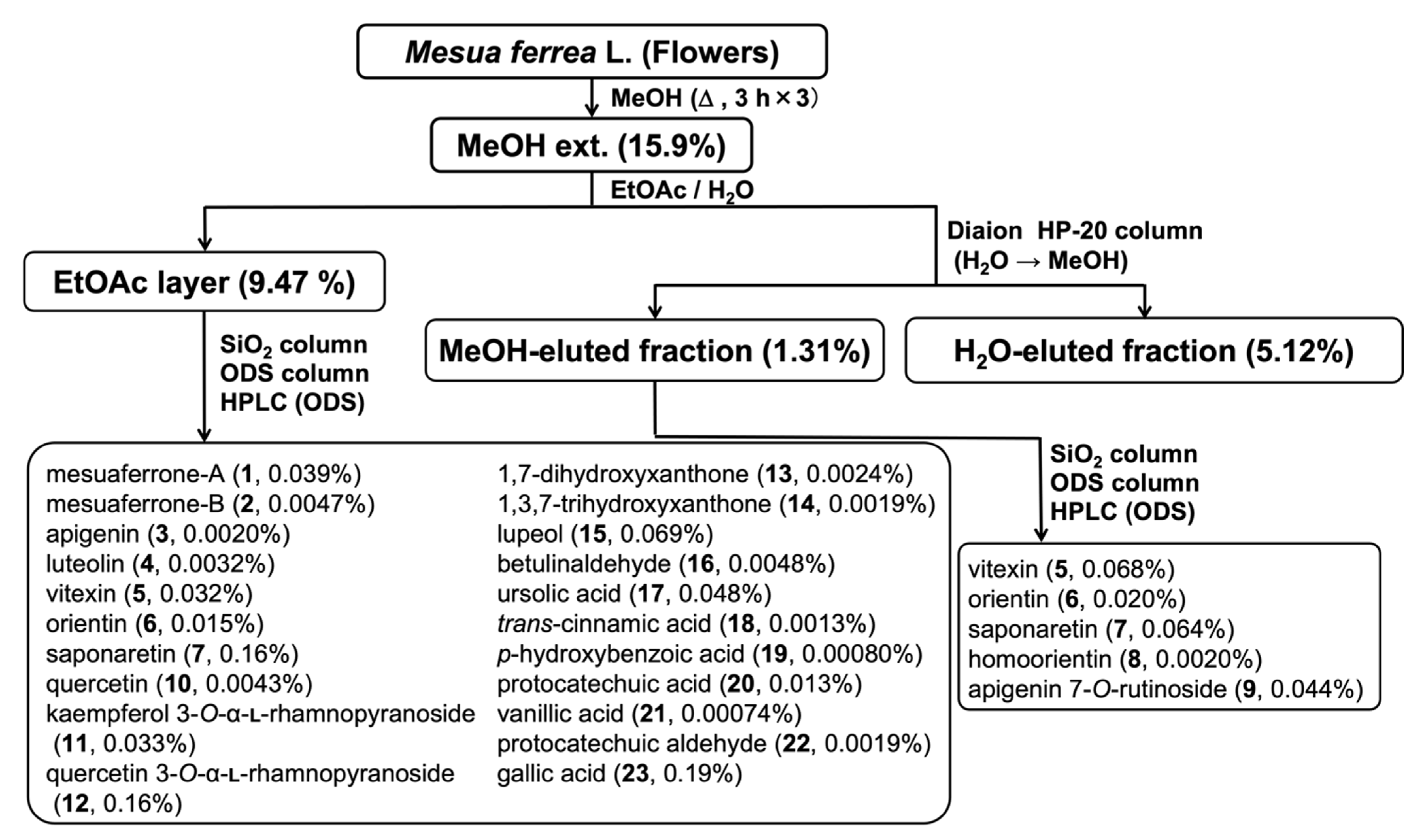
Figure 1. Isolation protocol of the chemical constituents (123) from the flowers of M. ferrea.
Separations 09 00127 g001
Figure 2. Isolates (123) from the flowers of M. ferrea.
Separations 09 00127 g002
Table 1. Hyaluronidase inhibitory activity of the MeOH extract and its fractions obtained from flowers of M. ferrea.
Inhibition (%)
0 μg/mL125 μg/mL250 μg/mL500 μg/mL1000 μg/mL
MeOH extract0.0 ± 8.15.1 ± 6.810.7 ± 5.624.2 ± 6.452.1 ± 4.5 b
EtOAc-soluble fraction0.0 ± 3.319.6 ± 7.7 a27.2 ± 5.1 b52.8 ± 3.8 b72.0 ± 3.7 b
MeOH-eluted fraction0.0 ± 8.416.6 ± 4.844.9 ± 5.1 b61.9 ± 4.7 b79.6 ± 1.4 b
H2O-eluted fraction0.0 ± 10.0−5.7 ± 8.411.7 ± 7.712.1 ± 7.06.9 ± 7.4
Each value represents the mean ± S.E.M. (N = 4). Significantly different from the control, a p < 0.05, b p < 0.01.
Table 2. Inhibitory effects of the isolates (123) from the flowers of M. ferrea on hyaluronidase.
Inhibition (%)IC50
0 μM12.5 μM25 μM50 μM100 μM(μM)
mesuaferrone-A (1)0.0 ± 8.110.2 ± 14.626.5 ± 7.348.8 ± 6.8 b71.1 ± 1.4 b51.1
mesuaferrone-B (2)0.0 ± 4.37.3 ± 1.923.6 ± 2.5 b46.6 ± 0.9 b54.5 ± 1.4 b54.7
Inhibition (%)IC50
0 μM32.5 μM75 μM150 μM300 μM(μM)
naringenin (1a)0.0 ± 8.8−0.4 ± 5.3−8.8 ± 2.00.4 ± 2.220.5 ± 1.7
apigenin (3)0.0 ± 6.215.8 ± 7.727.5 ± 8.132.9 ± 8.238.3 ± 6.4 b
luteolin (4) 0.0 ± 4.65.7 ± 2.88.9 ± 3.810.5 ± 3.512.3 ± 3.4
vitexin (5)0.0 ± 8.30.4 ± 8.2−7.0 ± 7.9−11.7 ± 3.8−10.9 ± 5.2
orientin (6)0.0 ± 9.34.9 ± 5.95.9 ± 3.29.0 ± 2.913.2 ± 2.6
saponaretin (7)0.0 ± 3.51.1 ± 2.04.8 ± 3.14.6 ± 3.02.2 ± 2.4
homoorientin (8)0.0 ± 7.9−7.1 ± 8.5−2.9 ± 7.6−5.0 ± 4.01.7 ± 5.9
apigenin 7-O-Rut (9)0.0 ± 7.64.4 ± 4.53.8 ± 2.12.5 ± 3.11.3 ± 2.4
quercetin (10)0.0 ± 3.7−1.0 ± 1.4−4.3 ± 5.2−4.3 ± 3.80.7 ± 4.5
kaempferol 3-O-Rha (11)0.0 ± 1.20.5 ± 2.5−4.7 ± 3.6−1.2 ± 3.76.3 ± 3.1
quercetin 3-O-Rha (12)0.0 ± 4.31.4 ± 4.24.0 ± 2.35.9 ± 3.313.2 ± 2.6
1,7-dihydroxyxthantone (13)0.0 ± 3.03.3 ± 3.00.4 ± 1.4−1.8 ± 3.65.4 ± 3.9
1,3,7-trihydroxyxthantone (14)0.0 ± 7.77.8 ± 2.74.7 ± 1.811.1 ± 2.718.4 ± 1.9
lupeol (15)0.0 ± 1.110.3 ± 3.29.4 ± 2.80.8 ± 2.2−0.4 ± 6.2
betulinaldehyde (16)0.0 ± 3.6−3.9 ± 2.10.2 ± 1.91.0 ± 1.95.3 ± 5.6
ursolic acid (17)0.0 ± 1.30.7 ± 1.50.5 ± 0.72.4 ± 1.5−5.7 ± 2.8
trans-cinnamic acid (18)0.0 ± 1.42.4 ± 0.9−1.7 ± 4.7−4.0 ± 5.1−1.2 ± 5.0
p-hydroxybenzoic acid (19)0.0 ± 5.0−2.8 ± 2.2−0.7 ± 1.42.6 ± 2.7−0.2 ± 4.3
protocatechuic acid (20)0.0 ± 2.51.0 ± 2.5−2.1 ± 1.54.0 ± 2.35.6 ± 4.2
vanillic acid (21)0.0 ± 3.84.0 ± 1.42.3 ± 3.1−0.7 ± 3.35.7 ± 3.2
protocatechuic aldehyde (22)0.0 ± 1.00.7 ± 1.84.1 ± 1.14.7 ± 3.01.5 ± 0.8
gallic acid (23)0.0 ± 1.4−0.6 ± 1.6−0.3 ± 3.13.6 ± 3.54.4 ± 4.2
disodium cromoglycate [48]0.0 ± 2.04.0 ± 2.414.4 ± 0.4 a39.0 ± 4.9 b69.1 ± 2.2 b64.8
ketotifen fumarate [48]0.0 ± 6.111.9 ± 1.926.7 ± 4.936.4 ± 2.9 b54.6 ± 2.5 b76.5
Each value represents the mean ± S.E.M. (N = 4). Significantly different from the control, a p < 0.05, b p < 0.01.
Table 3. Inhibitory effects of mesuaferrone-A (1) and B (2) on the release of β-hexosaminidase in RBL-2H3 cells.
Inhibition (%)IC50
0 μM3 μM10 μM30 μM100 μM(μM)
mesuaferrone-A (1)0.0 ± 7.124.9 ± 6.6 a24.8 ± 1.6 a37.8 ± 5.6 b86.0 ± 7.8 b49.4
mesuaferrone-B (2)0.0 ± 9.513.8 ± 5.34.2 ± 4.45.6 ± 6.2113.0 ± 10.1 b49.2
Inhibition (%)IC50
0 μM30 μM100 μM300 μM1000 μM(μM)
tranilast [49]0.0 ± 1.78.2 ± 1.822.4 ± 2.5 a56.9 ± 3.4 b75.0 ± 0.6 b282
ketotifen fumarate [49]0.0 ± 1.87.7 ± 1.527.6 ± 2.2 a80.7 ± 1.8 b100.7 ± 1.1 b158
Each value represents the mean ± S.E.M. (N = 4). Significantly different from the control, a p < 0.05, b p < 0.01.
Publisher’s Note: MDPI stays neutral with regard to jurisdictional claims in published maps and institutional affiliations.