Article

Tribulus terrestris Cytotoxicity against Breast Cancer MCF-7 and Lung Cancer A549 Cell Lines Is Mediated via Activation of Apoptosis, Caspase-3, DNA Degradation, and Suppressing Bcl-2 Activity

by
Ali Mohamed Alshabi
1,*,
Saad Ahmed Alkahtani
1,
Ibrahim Ahmed Shaikh
2,
Mohamed A. A. Orabi
3,
Basel A. Abdel-Wahab
2,
Ismail A. Walbi
1,
Mohammed Shafiuddin Habeeb
2,
Masood Medleri Khateeb
2,
Arun K. Shettar
4 and
Joy H. Hoskeri
5
1
Department of Clinical Pharmacy, College of Pharmacy, Najran University, Najran 64462, Saudi Arabia
2
Department of Pharmacology, College of Pharmacy, Najran University, Najran 64462, Saudi Arabia
3
Department of Pharmacognosy, College of Pharmacy, Najran University, Najran 64462, Saudi Arabia
4
Department of Pre-Clinical Research and Drug Development, Cytxon Biosolutions Pvt. Ltd., Hubballi 580031, Karnataka, India
5
Department of Bioinformatics and Biotechnology, Karnataka State Akkamahadevi Women’s University, Vijayapura 586102, Karnataka, India
*
Author to whom correspondence should be addressed.
Separations 2022, 9(11), 383; https://doi.org/10.3390/separations9110383
Submission received: 31 October 2022 / Revised: 15 November 2022 / Accepted: 16 November 2022 / Published: 21 November 2022
(This article belongs to the Special Issue Isolation, Elucidation and Synthesis of Bioactive Natural Products)

Abstract

:
The primary objective of this research was to use flow cytometry to gain mechanistic insights into the cytotoxic effects of Tribulus terrestris extracts on breast cancer (MCF7) and lung cancer (A549) cell lines. T. terrestris was extracted using a Soxhlet apparatus in a progressive process. GC–MS was used to establish the phytochemical constituents. The amounts of phenolic compounds and flavonoids in the plant extracts were calculated using spectrophotometric analysis. The cytotoxicity of plant extracts was initially evaluated in non-malignant L929 cells, then in carcinogenic MCF-7 and A549 cell lines. Then, we performed an Annexin V assay, an anti-Bcl-2 assay, a Caspase-3 assay, and a DNA fragmentation (TUNEL) assay, using flow cytometry to investigate the underlying molecular processes. Based on the data, the methanolic extract of T. terrestris contained the highest amounts of phenolic compounds and flavonoids, with values of 169.87 µg GAE/g dwt and 160.12 µg QE/g dwt, respectively. Analysis by GC–MS revealed the presence of bioactive phytochemicals with proven cytotoxicity. Based on the MTT experiment, we determined that the IC50 values for the methanol extract’s effect on the viability of the MCF-7 and A549 cell lines were 218.19 and 179.62 µg/mL, respectively. The aqueous and methanol extracts were less cytotoxic when tested against the cancer-free L929 cell line (IC50 = 224.35 µg/mL). In both breast and lung cancer cells, the methanolic extract was found to activate caspase-3 and inhibit the Bcl-2 protein, resulting in early and late apoptosis and cell death via DNA damage. These findings point to cytotoxic effects of T. terrestris methanol extract against breast and lung cancer cell lines. Due to its potential as a source of anti-cancer chemotherapeutic medicines, T. terrestris warrants further investigation.

1. Introduction

Cancer is one of the leading causes of death worldwide, and is an important barrier to increasing life expectancy. It is estimated that about 19.3 million new cases of cancer and about 10 million deaths were reported in the year 2020. Furthermore, this number may increase to 20 million cases by the year 2025 [1]. There is a remarkable increase in incidence and mortality that is associated with cancer seen in Saudi Arabia in recent years, when compared with the increase in population size. Changes in lifestyle of the people in Saudi Arabia may be one of the contributing factors to such an increase in cancer cases [2].
Breast cancer is the most common cancer seen in females worldwide and also in Saudi Arabia, and it is the second most cause of death due to cancer worldwide [3]. In Saudi Arabia, the prevalence rate of breast cancer was reported as 53% [4]. According to estimates from GLOBOCAN, breast cancer in females accounts for the highest incidence of about 11.7% of total new cancer cases diagnosed in the year 2020, followed by lung cancer with an incidence rate of 11.4%. While lung cancer has the highest mortality rate, 18% of all deaths due to cancer, breast cancer was reported to have a 6.9% death rate in the year 2020 [1]. Multiple factors, such as hormonal, environmental, and hormonal factors, play a role in breast cancer development. The incidence of breast cancer is increasing in the Arab population, and a significant number of cases are diagnosed at the advanced stage of the disease, contributing to a higher mortality rate [5]. According to a Saudi Cancer Registry data on incidence from 2017, breast cancer was the most common cancer among women, and was responsible for 18.1% of all cancers. It is also reported that the breast cancer incidence rate is common among young females [6]. According to Global Cancer Incidence, Mortality and Prevalence (GLOBOCAN) 2020 report, a total of 24,485 cases of cancer were reported in the year 2018, and 10,518 deaths were due to breast cancer. Furthermore, GLOBOCAN reported that breast cancer is foremost on the list of cancers in Saudi Arabia, with an incidence rate and mortality rate of 14.8% and 8.5% respectively; in comparison, the lung cancer incidence rate and mortality rate reported were 3.8% and 7.4%, respectively [7].
Natural plants have been used by mankind since the ancient era for medicinal purposes; it is estimated that more than 70 thousand plant species have been used for their medicinal benefits. Likewise, natural products from plants are taken as sources of food or as supplements and are well-known to possess anti-cancer activity [8]. It is estimated that more than 60% of anti-cancer drugs are obtained from natural resources, mainly plants. They act by inhibiting various steps that may promote carcinogenesis, and are capable of more selectively killing cancer cells with minimal effect on normal cells. Some natural compounds are even known to decrease the adverse effects of anti-cancer therapies. Hence, the use of natural products, along with conventional anti-cancer therapy, may have synergistic effects. The anti-cancer activities of natural products are due to their inhibition of cell proliferation activity, antioxidant activity, and anti-inflammatory activity [9].
Tribulus terrestris (common name: puncture vine, Gokharu) is an important member (among 20 species) of the genus Tribulus; it belongs to the family Zygophyllaceae [10]. It is commonly grown in dry and hot desert regions, and is also commonly grown in grazing land, farmland, and roadside. T. terrestris is native to the Mediterranean region, but is now widely grown in warm regions of Asia, Africa, America, and Australia [11]. It has been used for thousands of years in traditional Indian and Chinese systems of medicine. Furthermore, this plant is included in different official pharmacopeia [12]. Its fruit as well as roots, either alone or in combination, are used for the treatment of various ailments/disorders that are associated with genitourinary tracts, such as kidney and bladder stones, urinary tract infections, male infertility, sperm motility, and loss of libido both in men and women, as well as in ocular diseases [12]. Some studies also reported antimicrobial and cytotoxic effects of T. terrestris in the cardiovascular system [10].
It has been reported that T. terrestris is used by local tribal people in the southwest city of Saudi Arabia (Jazan Province) for pain and kidney stones, vitiligo, and other skin diseases [13]. A number of T. terrestris-containing pharmaceutical preparations and food supplements are available in the market, and are used for different therapeutic applications [11]. A literature survey showed that T. terrestris has antidiabetic, cardiotonic, hypolipidemic, hepatoprotective, antispasmodic, diuretic, antiurolithic, immunomodulatory, analgesic, anti-inflammatory, anti-cancer, anthelmintic, antibacterial, and larvicidal activities [14].
The main active principles of the T. terrestris include alkaloids, amides, flavonoids, glycosides, lignin, and saponins [15,16]. Kang et al. (2014) reported 14 different steroidal saponins, of which 7 are new compounds, called terrestrinins C-I [16]. Saponins are known to have an active therapeutic principle that exerts cytotoxic effects and other therapeutic responses. According to reports, the saponins content of T. terrestris is influenced by the plant’s geographic location, and accordingly the biological activities of the plant [17]. It was revealed that differences in sapogenin types, but also the number of sugars in saponins, were found in samples that were obtained from different geographical regions [11].
Natural products have many drawbacks as well; their improper use can lead to serious consequences. In contrast to this, there is a misconception that equates the term ‘natural’ with ‘safe’; consequently, their safety is either underestimated or overestimated [18]. Hence, for the rational use of natural compounds, proper selection, screening, isolation, characterization, and optimization, is an extreme need. The aim of the present study was to carry out qualitative and quantitative phytochemical screening of indigenously grown T. terrestris that was collected from the southern region of Saudi Arabia, and to investigate its cytotoxic effects using breast MCF7 and lung cancer A549 cell lines; moreover, its cytotoxic effects on the non-cancer fibroblast cell line L929 by MTT assay were investigated, followed by a mechanistic pathway analysis to elucidate its possible mechanism of action.

2. Materials and Methods

2.1. Plant Collection, Extraction, and Phytochemical Analysis

Whole plants of T. terrestris were collected from the Najran University Campus, Saudi Arabia, and identified by Prof. Mohammed A. A. Orabi, Department of Pharmacognosy, College of Pharmacy, Najran, Saudi Arabia. A herbarium specimen (TT-102023) was deposited in the Department of Pharmacognosy. Plant material was washed and dried after gathering. Using Soxhlet equipment, air-dried material was coarsely pulverized and extracted using solvents of increasing polarity, hexane, ethyl acetate, methanol, and distilled water. Each time, 100 g of powdered material and 1000 mL of solvent were used to create crude extract. Each cycle lasted 24 h, or until the siphon tube’s solution became colorless. Each cycle ended with air-dried plant material, and a new solvent extraction. End-of-cycle crude solvent extracts were concentrated, dried, and kept at −20 °C [19]. The qualitative phytochemical screening [20] was carried out to detect its phytocomponents.

2.2. Chemicals and Reagents

The cell lines L929, A549, and MCF7 were procured from the National Centre for Cell Science (NCCS), Pune, India. MTT (3-(4,5-dimethylthiazol-2-yl)-2,5-diphenyltetrazolium bromide) (purity 97.5%) was purchased from Sigma-Aldrich, St. Louis, MO, USA. All other chemical reagents were of analytical grade, and purchased from HiMedia, Mumbai, India.

2.3. Estimation of Total Phenolic Content

The Folin–Ciocalteu test was applied to the methanolic and aqueous extracts of T. terrestris to determine the total phenolic content that was present in the extract. An amount of 5 mL of a 1:10 v/v aqueous solution of Folin–Ciocalteu reagent, and 4 mL of a 75 g/L aqueous solution of sodium carbonate, were added to the extract [21,22]. The absorbance was then measured at a wavelength of 760 nm using a spectrophotometer [21], after the solution was vortexed and heated at 40 °C for approximately 30 min. Gallic acid (GA) concentrations between 20 and 100 µg/mL were used to generate a standard calibration curve, and the total phenolic content was reported in µg/g of GA. The following formula was used to determine the total phenolic content:
y = 0.001x + 0.113

2.4. Estimation of Total Flavonoid Content

A previously reported colorimetric technique was utilized to assess the total flavonoid concentration of the T. terrestris extracts [23,24]. Briefly stated, an aliquot of the extract was diluted in 5 mL of water, to which 0.3 mL of a 1:20 v/v sodium nitrite aqueous solution was added. After waiting 5 min, 3 mL of a 1:10 mL aluminum chloride solution was added, and after another 6 min, 2 mL of a 1 M sodium hydroxide solution was added. The resulting mixture was thoroughly blended, and 10 mL of distilled water was added to it. The mixture was left to stand at room temperature for 10 min, after which the absorbance at a wavelength of 510 nm was measured. The flavonoid content of the extract was quantified in terms of mg of quercetin/g of extract, using the equation y = 0.265x + 0.152, and a known concentration of quercetin was used to build a standard curve [25].

2.5. In Vitro Cytotoxicity of T. terrestris Extracts

An in vitro cell viability assay (MTT assay) [26] was used to test the cytotoxicity of various T. terrestris extracts. Cytotoxic activity in the non-cancerous cell line (L292), lung cancer (A549), and breast cancer (MCF-7) cell lines were determined by preparing various concentrations (50, 100, 150, 200, and 250 µg/mL) from different solvent extracts of T. terrestris. The formula for calculating the half-life (IC50) is as follows:
Inhibition   percentage = ( OD   of   test ÷ OD   of   control ) × 100

2.6. Determination of Apoptosis (Annexin-V Assay)

In the current investigation, MCF-7 breast cancer cell lines were exposed to T. terrestris extract for approximately twenty-four hours, prior to being analyzed by flow cytometry for the detection of early apoptosis and late apoptosis. In line with the instructions provided for the kit [27], the flow cytometric analysis was carried out using FlowJo X 10.0.7 software (10.0.7, Becton Dickinson, OR, USA).

2.7. Caspase Activation Assay

In the current study, breast cancer cell line MCF-7 was pretreated with caspase-3 inhibitor Z-DEVD-FMK (20 M, BD Biosciences, Franklin Lakes, NJ, USA) for 2 h. After that, the cells were incubated for 24 h with methanolic extract, and then subjected to flow cytometry to determine caspase-3 activation [19].

2.8. Anti-Bcl-2 Assay

According to the method outlined by Moraes et al. [21], we used flow cytometry to determine the amount of Bcl-2 protein present in cancer cell lines MCF-7 and A549 that had been treated with a methanolic extract of T. terrestris. Following treatment with the test specimen at its IC50 concentration, the cell preparations were examined flow cytometrically using FlowJo X 10.0.7 software, within thirty minutes of the end of the treatment.

2.9. DNA Degradation Analysis by TUNEL Assay

Using a TUNEL assay kit (The APO-DIRECTTM Kit, Waltham, MA, USA), we were able to assess whether the methanol extract of T. terrestris caused DNA to break as a result of apoptosis. Flow cytometric analysis, with the software Flowzo 10.1, was used to determine the proportion of TUNEL-positive cells. Acquisition equipment included a 7 Cytomics FC500 flow cytometer (Beckman Coulter, Brea, CA, USA).

2.10. GC–MS Analysis

The methanolic extract was analyzed by GC–MS with a ELITE-5MS column (30 m × 0.25 mm, i.d.), a GCMS-QP2010S model, helium as the carrier gas, a flow rate of 1 mL/min, and an injector volume of 1 µL. Injector and ion source temperatures were both set at 260 °C, while the column temperature was initialized at 80 °C and increased by 10 °C every minute. The scan mode used was 0.50 s, and the entire MS process took 7 min to complete. The scan rate was 1000 Hz, and the beginning and ending m/z values were 50 and 500, respectively. Components were identified by comparing their mass spectra to those in the NIST mass spectral collection [25].

2.11. Statistical Analysis

Three independent experiments’ data (n = 3) are summarized and presented in terms of mean ± standard deviation. The software ‘GraphPad Prism version 5’ was used to conduct one-way ANOVA and Dunnett’s t-test analyses, and a p-value of less than 0.05 was considered statistically significant.

3. Results

3.1. Phytochemical Analysis

Analysis was performed, based on the standard procedure of biochemical tests, to detect different secondary metabolites that were present in the plant extracts. The results revealed that among the tested extracts, methanol and aqueous extracts showed the presence of phenols and terpenoids as major phytoconstituents. Meanwhile, hexane extracts showed the presence of steroids and saponins. The overall results are shown in Table S1.

3.2. Estimation of Total Phenols Content

Based on the phytochemical results, the methanol and aqueous extracts of the T. terrestris were subjected to a quantification study, in order to evaluate total phenol and flavonoid content by following a standard procedure. The quantification results showed that between the methanol and aqueous extracts, the methanol extract exhibited the higher phenolic content, i.e., 169.87 µg GAE/g dwt, whereas the aqueous extract exhibited 114.06 µg GAE/g dwt. The total phenolic content was calculated from the standard calibration curve (y = 0.001x + 0.113) (Figure S1).

3.3. Estimation of Total Flavonoid Content

The methanol and aqueous extracts revealed total flavonoid contents to be 160.12 µg QE/g dwt and 113.01 µg QE/g dwt, respectively.

3.4. In Vitro Cytotoxicity of T. terrestris Extracts

In the present study, the hexane, ethyl acetate, methanol, and aqueous extracts were taken as treatments for the L929 cell line, along with standard drug cisplatin as a positive and untreated cells as a negative control. In all of the treated extracts, there was a dose-dependent activity; with an increase in the concentrations of extracts, the percentage of cell viability decreased. Almost all of the tested extracts showed a cytotoxic nature at higher concentrations. At higher concentrations, hexane, ethyl acetate, methanol, and aqueous extract showed cell viabilities as 47.05 ± 0.01, 32.76 ± 0.008, 32.76 ± 0.02, and 53.23 ± 0.01, respectively. The results are shown in Table 1 and Figure S2.
After the toxicity assay, the extracts were subjected to cytotoxic studies against breast cancer MCF-7 and lung cancer A549 cell lines, along with cisplatin as a standard drug for comparison. In both tested cell lines, the selected solvent extracts showed significant activity by suppressing the cell’s growth through a decrease in cell viability. At the initial concentrations of hexane, ethyl acetate, methanol, and aqueous extract, cell viability in lung cancer was observed to be 96.38 ± 0.008, 90.99 ± 0.001, 43.57 ± 0.003, and 49.39 ± 0.012 (Table 2 and Figure S3), respectively; in case of breast cancer, cell viability was found to be 97.43 ± 0.018, 91.14 ± 0.009, 98.30 ± 0.003, and 91.31 ± 0.008 (Table 3 and Figure S4), respectively. In comparison, the selected solvent extracts showed more effective cytotoxic activity against lung cancer than breast cancer. The cell viability and preliminary observations indicated that the methanol extract was the most effective of the extracts tested.
The untreated MCF-7 and A549 cells were compared to the treated cells, and the results showed that there were differences in the cellular morphology of the test samples. In a set of untreated cells, the shape of the cells could be seen clearly, and there were greater proportions of them without any intracellular voids. In contrast, in the treated test samples, the number of cells gradually dropped as the concentration grew, and intracellular voids were observed between the cells. In addition to this observation, it was found that apoptotic bodies, cell shrinkage, membrane blabbing, cell turgidity, and other abnormalities were present in the cells. All of these morphological features are characteristics of cells that are going through the process of apoptosis. The results of the MTT as well as the microscopic study indicated that the test samples had the potential to induce apoptosis in both breast cancer MCF-7 and lung cancer A549 cell lines.
A standardized calibration curve was used to determine the IC50 for the extracts. For lung cancer, the results were 277.14, 256.38, 179.62, and 189.70 g/mL for hexane, ethyl acetate, methanol, and aqueous extracts, respectively; for breast cancer, the results were 293.27, 238.11, 218.19, and 271.07 g/mL for the same solvents, respectively. The IC50 details are shown in Table 4.

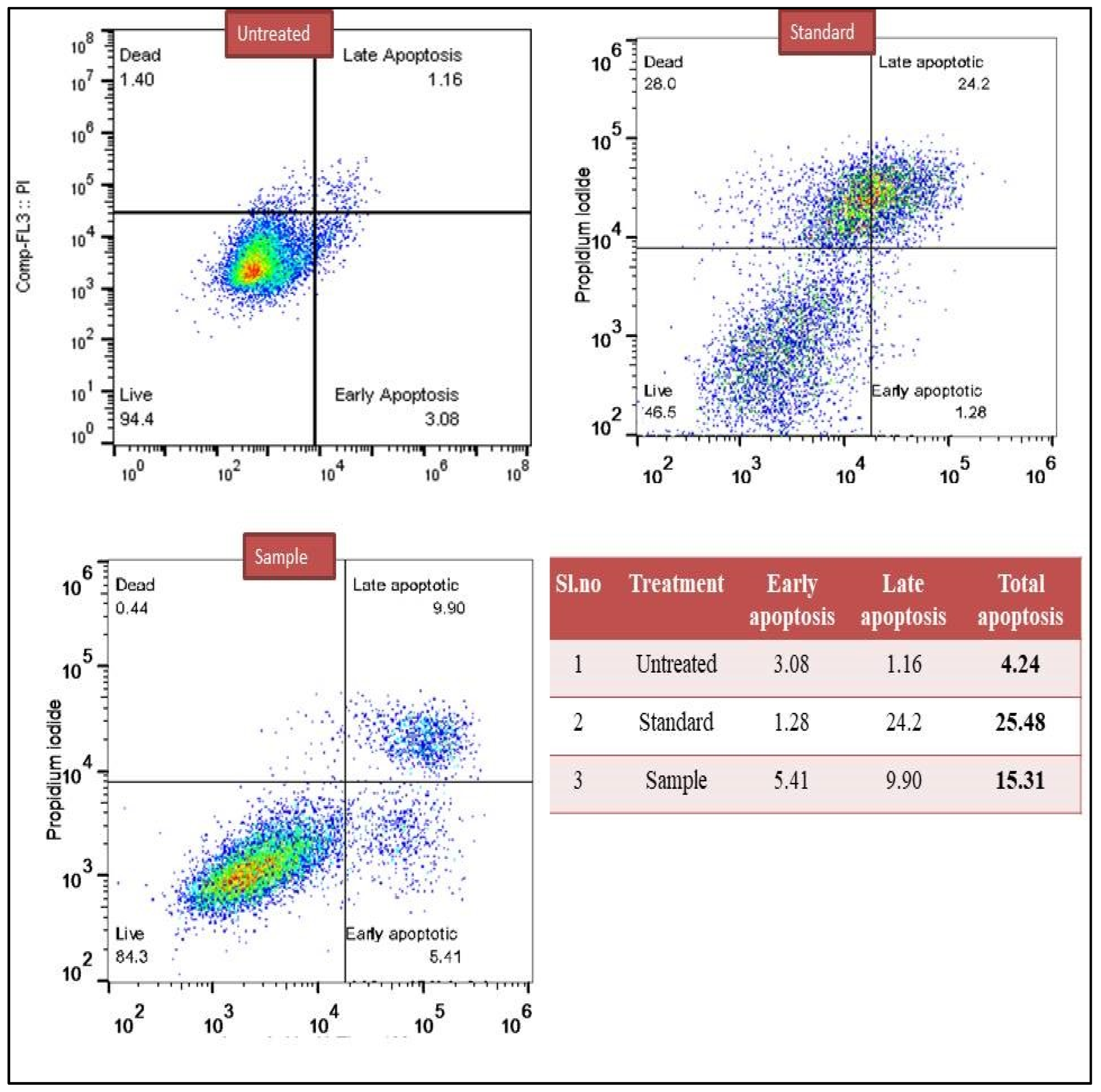
3.5. Determination of Early and Late Apoptosis (Annexin V Assay)

In the present study in treated breast cancer, MCF-7, cells subjected to a detection of early apoptosis and late apoptosis were studied on the basis of flow cytometry. The results showed that in the case of breast cancer untreated cells, around 4.24% of cell showed apoptosis, whereas the standard drug cisplatin showed 25.48% of apoptotic cells, and the methanol extract test sample showed 15.31% of apoptotic cells (Figure 1).

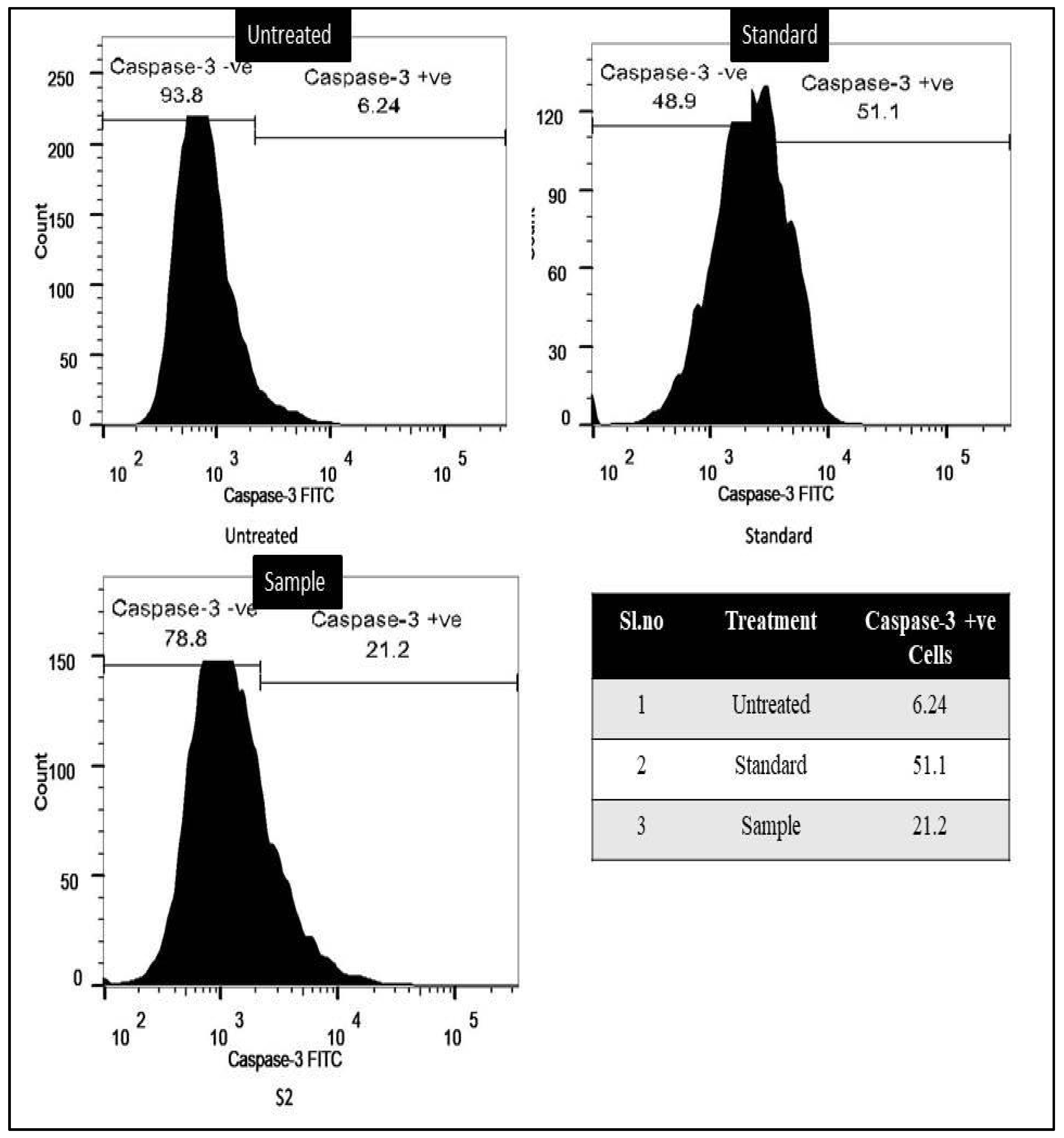
3.6. Caspase-3 Activation Assay

On breast cancer MCF-7 cells, the methanol extract of T. terrestris was examined alongside the control group and the standard drug, cisplatin. The results of the caspase-3 assay showed that breast cancer cells had clearly undergone apoptotic induction. The data revealed that 51.1% of MCF-7 breast cancer cells tested positive for caspase-3 when exposed to the standard drug cisplatin, while 21.2% tested positive for the methanol extract. Figure 2 displays the outcomes of caspase-3 flow cytometry.

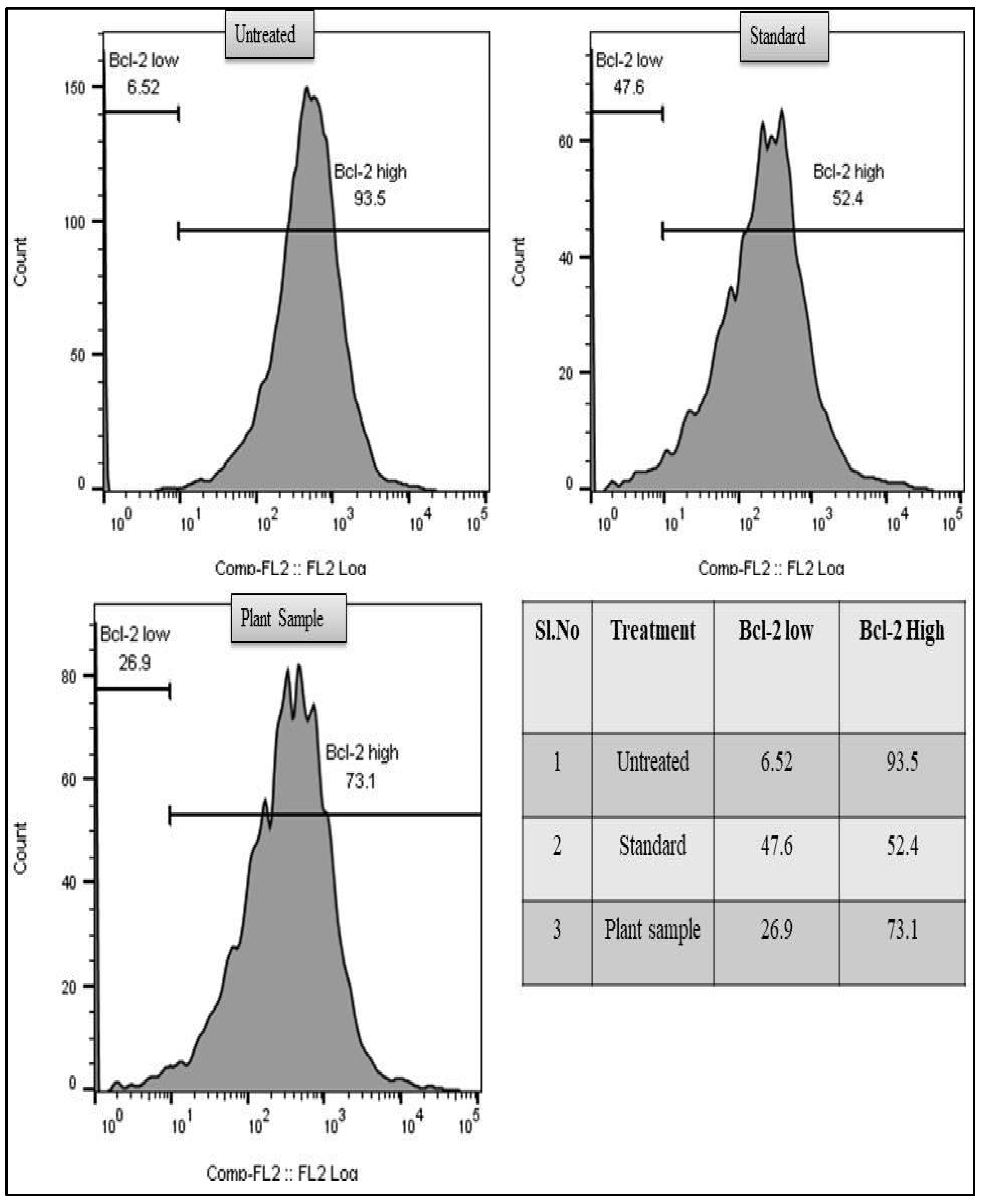
3.7. Anti-Bcl-2 Assay

In the current study, the Bcl-2 assay results demonstrated that untreated cells had a lower degree of apoptosis, with a high level of Bcl-2-positive cells (93.5%). In contrast, the test sample (73.1%) and cisplatin (52.4%) treated for MCF-7 revealed lower levels of Bcl-2-positive cells. Thus, compared to the untreated samples, a higher degree of apoptosis was observed in the test drug and standard treated groups (Figure 3).

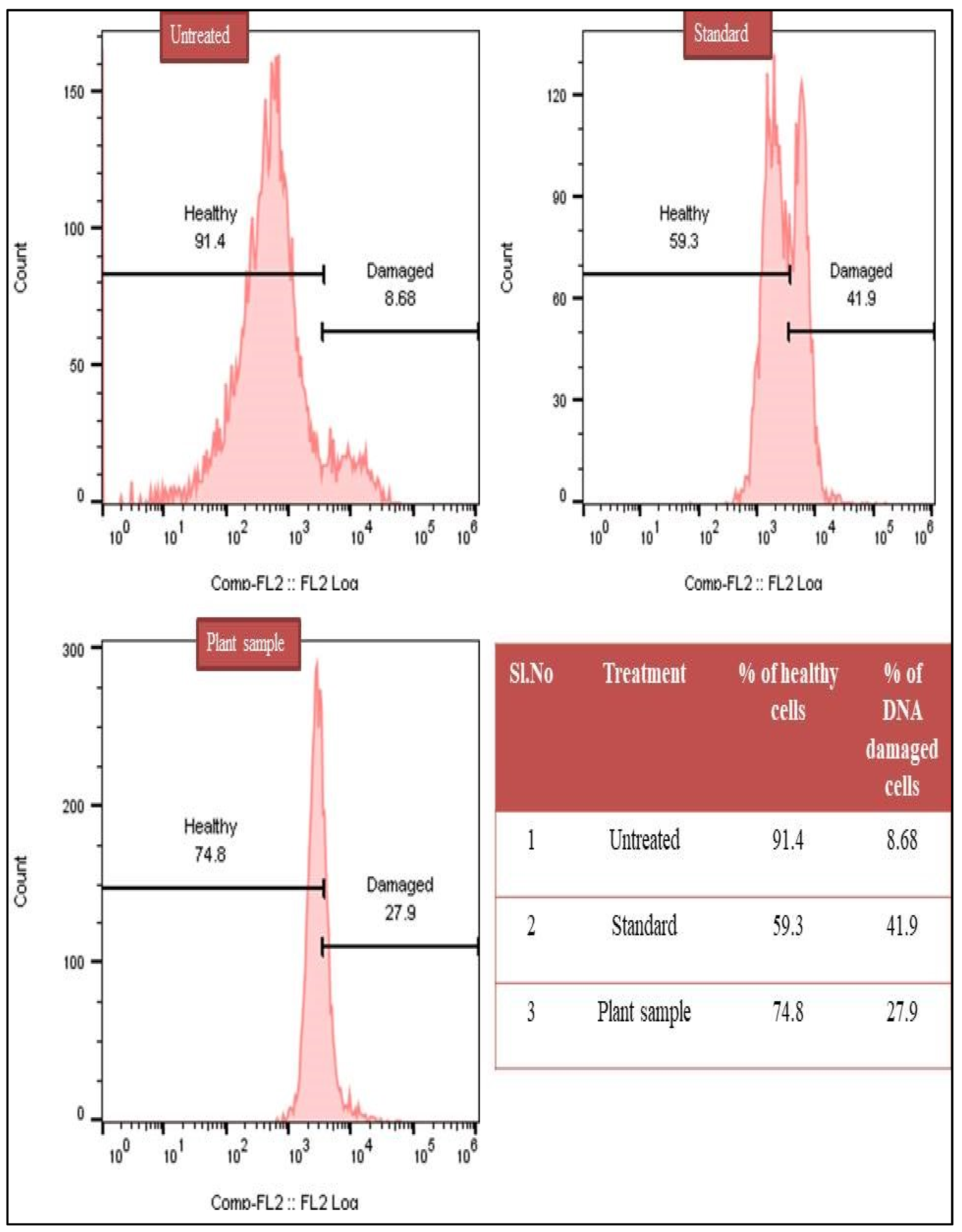
3.8. DNA Degradation Analysis by TUNEL Assay

The cell death that occurred in the apoptotic cells may have been due to DNA damage. Hence, in the present study, DNA damage induced by the methanol extract of T. terrestris and cisplatin was studied in breast cancer MCF-7 on the basis of a flow cytometry TUNEL assay. The results revealed that in the untreated group, around 8.68% of cells showed DNA damage. In the treated breast cancer MCF-7 cell test sample, around 27.9% of cells showed DNA damage, whereas in the case of the standard drug cisplatin, DNA damage was observed in 41.9% of cells (Figure 4).

3.9. GCMS Analysis

After the phytochemical analysis, the methanol extract was observed to be a potent extract; hence, the methanol extract was subjected to GC–MS analysis, in order to identify the phytochemical compounds. The GC–MS results showed the presence of seven sharp peaks in the gas chromatogram. Each peak was subjected to a similarity search using the NIST library database to identify the compounds. Mainly, the GC–MS analysis showed the presence of 9-Octadecene, (E), 1-Deoxy-d-mannitol, (E)-Phytol, 8-Pentadecanone, Methyl palmitate, Methyl 9,12-octadecadienoate, Methyl linolenate, Isophytol acetate, Methyl isostearate, and Squalene as major compounds. These compounds are known for several applications, such as industrial applications, organic synthesis, and biological activities (Table 5, and Figures S5 and S6).

4. Discussion

Cancer is one of the most significant non-communicable diseases. It is responsible for 71% of total deaths worldwide, and the number of cancer cases is estimated to increase by 80% in underdeveloped and developing countries [48]. It is anticipated that by 2030, the number of cancer cases may reach 21.7 million, with about 13 million deaths [49]. Saudi Arabia accounts for more than half of the cancer burden in GCC countries. The high prevalence of cases in Saudi Arabia is attributed to its population size, which accounts for about 60% of the total GCC population [50]. The rising incidence of cancer can be attributed to a variety of demographic shifts, including rising life expectancy, larger populations, and the prevalence of risk factors including fast food consumption and tobacco use. [49,51,52,53,54]. The main aim of cancer treatment is to kill cancer cells, prolong the life expectancy of the patient, reduce the relapse rate, and improve the quality of life of the patient [55]. However, there is currently no effective treatment available for cancer that has minimal adverse effects or no ill effects on the quality of patients’ life post-treatment. Hence, there is always a need to search for a more effective treatment that can be used either alone, or as an adjuvant to currently approved therapy.
Natural products are renowned for their effectiveness and tolerability in the treatment of various diseases, including cancer. It has also been proven that natural products possess synergistic effects with other drug therapies [56]. They serve as one of the sources of drugs, and/or may serve as potential lead compounds in drug development [57]. Between 1930 and 2012, 183 drugs were authorized for use in treating cancer; of these, 30% came from natural origins, 57% were semisynthetic in origin, and 34% had pharmacophores that mimicked those of naturally occurring products [58]. However, the isolation and characterization of constituents that are responsible for beneficial effects are challenging [56]. The present study focused on the cytotoxic effects on noncancerous as well as breast and lung cancer cell lines, followed by the identification, and characterization of active constituents present in T. terrestris.
In the present study, the major active constituents from plants were found to be flavonoids, phenolics, terpenoids, and steroids, which is consistent with previous reports [12,59,60]. Furthermore, results from quantification studies showed there was a higher percentage of phenolic and flavonoid content in the methanolic extract compared with the aqueous extract. These polyphenolics are very well-known for their antioxidant activity, and they have been proven to have a role in many different types of diseases/disorders, such as cardiovascular disease, hepatic disorder, diabetes, allergies, infection, inflammation, and cancer, to name a few [61,62,63]. The anti-cancer effect of flavonoids is due to the antioxidant effect, which, in turn, is dependent on a number of phenolic hydroxyl groups in their structure [64,65]. Similarly, many reports are available that show anti-cancer properties of terpenoids [66,67], as well as anti-inflammatory properties [68].
The MTT assay is a well-known assay technique that is based on the metabolic activity of cells, and is used for the detection of the viability, cytotoxicity, and proliferation of cells [69,70,71]. The MTT assay in the present study, with microscopic examination, found that both methanolic and aqueous extracts of T. terrestris showed significant cytotoxic activity in cancer cell lines. Moreover, higher cytotoxicity was recorded with the lung cancer cell line (A549) compared to the breast cancer cell line (MCF-7), and a good cytotoxic effect was recorded, even at a low dose of extract used (50 µg/mL). In comparing the cytotoxic effects of the methanol and aqueous extracts, the methanolic extract was found to have a higher cytotoxic effect. The IC50 values of 179.62 and 189.70 µg/mL were obtained with methanol and aqueous extract, respectively. Similarly, studies also reported the cytotoxicity activity of alcoholic, hydroalcoholic, and aqueous extracts of T. terrestris [72,73,74]. It was observed that T. terrestris extract does not have much cytotoxic effect on normal cell lines. The hexane and ethyl acetate extracts showed higher numbers of viable cells in all treated cell lines compared to a methanolic extract. A previous report on T. terrestris extract showed cytotoxic effects on fibroblast cell lines [75]. Moreover, Pourali M, et al., found higher cytotoxicity in colon and prostate cancer cell line assays than in normal fibroblast cell lines [74].
It has been shown that natural products are shown to exert anti-cancer and antitumor activities by the following mechanism: anti-angiogenesis, cell cycle inhibition, induction of apoptosis, suppression of proliferation of cells, cell migration, and inhibition of metastasis [58]. To further elucidate the possible anti-cancer mechanism of T. terrestris, the methanolic extract was subjected to flowcytometry analysis using evaluation of early and late apoptosis, followed by its confirmation with a Caspase-3 assay. Breast cancer cell lines were employed for the mechanistic study using flow cytometry. It was found that the extract showed considerable apoptosis action when compared to untreated MCF-7 cell lines. However, the apoptotic effect of the extract was less than that of the standard drug, cisplatin. The result of caspase-3 activity showed that the T. terrestris extract-treated MCF-7 cell lines showed a high degree of caspase-3 activity when compared with untreated cell lines. This indicated that one of the possible mechanisms of cytotoxicity of T. terrestris extract may be due to the induction of apoptosis by activation of proteolytic enzyme caspase-3. A similar activity in apoptosis induction and enhanced caspase-3 activity was reported in previous studies [76,77]. However, another possible mechanism of apoptosis may also be involved, such as by decreased expression of caspase-8 [78], down-regulating the expression of Bax and p53 [77,78], DNA fragmentation [76,77], Bcl-2 [78,79], and down-regulation of NF-kB [73].
GC–MS analysis in a previous report found the presence of α-amyrin as a major constituent, and seven minor constituents in a methanolic extract of T. terrestris [80]. Similarly, in the present study, we found ten major compounds through GC–MS analysis. Some of these compounds were reported to have anti-cancer and antioxidant activities, and may be responsible for the cytotoxic effects seen in the present study of normal non-cancerous cell lines, as well as in cancer cells (Table 5).
It is well known that phenolic compounds present in medicinal plants are known to exhibit potent antioxidant effects, and are quite effective in the treatment of different diseases due to oxidative stress, including cancer [61,81,82,83]. T. terrestris was also reported to contain saponins as major active compound with promising cytotoxicity [11]. Saponins present in T. terrestris were previously reported to have anti-cancer activity on various cell lines [75,78,79,84,85]. A steroidal saponin, nuatigenin, from seeds of T. terrestris, was reported to have anti-cancer activity against breast cell line MCF-7 [78].
Patel et al., 2019, demonstrated that T. terrestris induced an intrinsic apoptotic pathway in an MCF-7 cell line, as evidenced by increased expressions of the Bax and p53 genes, DNA degradation, and decreased expression of the Bcl-2 gene [77,78]. The protein Bcl-2 inhibits cellular suicide by acting as an anti-apoptotic agent. Overexpression of Bcl-2 was shown to prevent cell death and improve cell survival.
This study compared the effects of a T. terrestris methanolic extract with those of a standard drug, and those of a control group of cells that had not been treated. There was an increase in apoptosis as compared to untreated cells, in both the test sample’s and the standard drug’s abilities to inhibit the Bcl-2 protein. To further elucidate the mechanism of apoptosis, a TUNEL assay was employed to determine the extent of DNA degradation. The results revealed that the methanolic extract and cisplatin exhibited significant DNA damage compared to untreated cells. Thus, T. terrestris can induce apoptosis by decreased Bcl-2 protein levels, increased caspase-3 activity, and DNA degradation in the breast cancer cell line MCF-7. In light of this, the possibility exists that naturopathic remedies isolated from T. terrestris could undergo further clinical development as potential therapeutic medications.

5. Conclusions

In conclusion, the cytotoxic action of the T. terrestris methanol extract was demonstrated by its ability to significantly suppress the growth of both the breast cancer MCF-7 and lung cancer A549 cell lines. The test medication caused DNA damage and apoptosis in breast cancer MCF-7, as shown by flow cytometry. Furthermore, T. terrestris showed powerful cytotoxic activity, comparable to that of the gold standard medication cisplatin. Therefore, T. terrestris should be further investigated to identify lead compounds that have cancer chemotherapeutic potential. Additional research is necessary before the results of this study can be applied to clinical trials.

Supplementary Materials

The following supporting information can be downloaded at: https://www.mdpi.com/article/10.3390/separations9110383/s1, Figure S1: Calibration curve for the standard gallic acid; Figure S2. Images of cytotoxicity of methanol extract of T. terrestris on non-cancerous L929 cell line; Figure S3. Images of cytotoxicity of methanol extract of T. terrestris on lung cancer (A549) cell line; Figure S4. Images of cytotoxicity of methanol extract of T. terrestris on breast cancer (MCF-7) cell line; Figure S5. GC spectrum of methanol extract of T. terrestris; Figure S6. MS spectra of methanol extract of T. terrestris; Table S1: Qualitative phytochemical analysis of different solvent extracts of T. terrestris.

Author Contributions

Conceptualization, M.A.A.O., S.A.A., I.A.S. and A.M.A.; methodology, M.A.A.O., A.K.S., B.A.A.-W. and J.H.H.; software, I.A.W., M.S.H. and M.M.K.; validation, B.A.A.-W. and J.H.H.; formal analysis, J.H.H.; investigation, S.A.A., I.A.S., J.H.H. and A.M.A.; resources, M.A.A.O., A.K.S., B.A.A.-W. and J.H.H.; data curation, A.K.S.; writing—original draft preparation, I.A.S., A.K.S., A.M.A. and M.M.K.; writing—review and editing, I.A.S., A.K.S., M.A.A.O. and A.M.A.; supervision, M.A.A.O. and A.K.S.; project administration, I.A.S. and A.K.S.; funding acquisition, A.M.A. All authors have read and agreed to the published version of the manuscript.

Funding

The authors would like to acknowledge the support of the Deputy for Research and Innovation—Ministry of Education, Kingdom of Saudi Arabia, for a grant (NU/IFC/ENT/01/006) under the Institutional Funding Committee at Najran University, Kingdom of Saudi Arabia.

Institutional Review Board Statement

Not applicable.

Informed Consent Statement

Not applicable.

Data Availability Statement

All data have been included in the manuscript.

Acknowledgments

The authors would like to acknowledge the support of the Deputy for Research and Innovation—Ministry of Education, Kingdom of Saudi Arabia, for a grant (NU/IFC/ENT/01/006) under the Institutional Funding Committee at Najran University, Kingdom of Saudi Arabia.

Conflicts of Interest

The authors declare no conflict of interest.

References

  1. Sung, H.; Ferlay, J.; Siegel, R.L.; Laversanne, M.; Soerjomataram, I.; Jemal, A.; Bray, F. Global Cancer Statistics 2020: GLOBOCAN Estimates of Incidence and Mortality Worldwide for 36 Cancers in 185 Countries. CA Cancer. J. Clin. 2021, 71, 209–249. [Google Scholar] [CrossRef] [PubMed]
  2. Althubiti, M.A.; Nour Eldein, M.M. Trends in the incidence and mortality of cancer in Saudi Arabia. Saudi Med. J. 2018, 39, 1259–1262. [Google Scholar] [CrossRef] [PubMed]
  3. Algehyne, E.A.; Jibril, M.L.; Algehainy, N.A.; Alamri, O.A.; Alzahrani, A.K. Fuzzy Neural Network Expert System with an Improved Gini Index Random Forest-Based Feature Importance Measure Algorithm for Early Diagnosis of Breast Cancer in Saudi Arabia. Big Data Cogn. Comput. 2022, 6, 13. [Google Scholar] [CrossRef]
  4. Alqahtani, W.S.; Almufareh, N.A.; Domiaty, D.M.; Albasher, G.; Alduwish, M.A.; Alkhalaf, H.; Almuzzaini, B.; Al-Marshidy, S.S.; Alfraihi, R.; Elasbali, A.M.; et al. Epidemiology of cancer in Saudi Arabia thru 2010-2019: A systematic review with constrained meta-analysis. AIMS Public Health 2020, 7, 679–696. [Google Scholar] [CrossRef] [PubMed]
  5. Messaoudi, S.A.; Al Sharhan, N.A.; Alharthi, B.; Babu, S.R.; Alsaleh, A.B.; Alasiri, A.M.; Assidi, M.; Buhmeida, A.; Almawi, W.Y. Detection of genetic mutations in patients with breast cancer from Saudi Arabia using Ion AmpliSeq™ Cancer Hotspot Panel v. 2.0. Biomed. Rep. 2022, 16, 26. [Google Scholar] [CrossRef]
  6. Alghamdi, I.G.; Hussain, I.I.; Alghamdi, M.S.; El-Sheemy, M.A. The incidence rate of female breast cancer in Saudi Arabia: An observational descriptive epidemiological analysis of data from Saudi Cancer Registry 2001–2008. Breast Cancer (Dove Med. Press) 2013, 5, 103–109. [Google Scholar] [CrossRef] [Green Version]
  7. WHO. International Agency for Research in Cancer (IARC) Saudi Arabia. Source: Globocan. 2018. Available online: https://gco.iarc.fr/today/data/factsheets/populations/682-saudi-arabia-fact-sheets.pdf (accessed on 30 August 2022).
  8. Kuruppu, A.I.; Paranagama, P.; Goonasekara, C.L. Medicinal plants commonly used against cancer in traditional medicine formulae in Sri Lanka. Saudi Pharm. J. 2019, 27, 565–573. [Google Scholar] [CrossRef]
  9. Maiuolo, J.; Gliozzi, M.; Carresi, C.; Musolino, V.; Oppedisano, F.; Scarano, F.; Nucera, S.; Scicchitano, M.; Bosco, F.; Macri, R.; et al. Nutraceuticals and Cancer: Potential for Natural Polyphenols. Nutrients 2021, 13, 3834. [Google Scholar] [CrossRef]
  10. Chhatre, S.; Nesari, T.; Somani, G.; Kanchan, D.; Sathaye, S. Phytopharmacological overview of Tribulus terrestris. Pharmacogn. Rev. 2014, 8, 45–51. [Google Scholar] [CrossRef] [PubMed] [Green Version]
  11. Kostova, I.; Dinchev, D. Saponins in Tribulus terrestris—Chemistry and Bioactivity. Phytochem. Rev. 2005, 4, 111–137. [Google Scholar] [CrossRef]
  12. Ștefănescu, R.; Tero-Vescan, A.; Negroiu, A.; Aurică, E.; Vari, C.E. A Comprehensive Review of the Phytochemical, Pharmacological, and Toxicological Properties of Tribulus terrestris L. Biomolecules 2020, 10, 752. [Google Scholar] [CrossRef]
  13. Tounekti, T.; Mahdhi, M.; Khemira, H. Ethnobotanical Study of Indigenous Medicinal Plants of Jazan Region, Saudi Arabia. Evid. Based Complement. Alternat. Med. 2019, 2019, 3190670. [Google Scholar] [CrossRef] [PubMed] [Green Version]
  14. Sirotkin, A.V.; Kolesárová, A. Puncture vine (Tribulus terrestris L.) in control of health and reproduction. Physiol. Res. 2021, 70 (Suppl. S4), S657–S667. [Google Scholar] [CrossRef] [PubMed]
  15. Shahid, M.; Riaz, M.; Talpur, M.M.; Pirzada, T. Phytopharmacology of Tribulus terrestris. J. Biol. Regul. Homeost. Agents 2016, 30, 785–788. [Google Scholar] [PubMed]
  16. Kang, L.P.; Wu, K.L.; Yu, H.S.; Pang, X.; Liu, J.; Han, L.F.; Zhang, J.; Zhao, Y.; Xiong, C.Q.; Song, X.B.; et al. Steroidal saponins from Tribulus terrestris. Phytochemistry 2014, 107, 182–189. [Google Scholar] [CrossRef] [PubMed]
  17. Dinchev, D.; Janda, B.; Evstatieva, L.; Oleszek, W.; Aslani, M.R.; Kostova, I. Distribution of steroidal saponins in Tribulus terrestris from different geographical regions. Phytochemistry 2008, 69, 176–186. [Google Scholar] [CrossRef] [PubMed]
  18. Gaston, T.E.; Mendrick, D.L.; Paine, M.F.; Roe, A.L.; Yeung, C.K. “Natural” is not synonymous with “Safe”: Toxicity of natural products alone and in combination with pharmaceutical agents. Regul. Toxicol. Pharmacol. 2020, 113, 104642. [Google Scholar] [CrossRef] [PubMed]
  19. Alkahtani, S.A.; Alshabi, A.M.; Shaikh, I.A.; Orabi, M.A.A.; Abdel-Wahab, B.A.; Walbi, I.A.; Habeeb, M.S.; Khateeb, M.M.; Shettar, A.K.; Hoskeri, J.H. In Vitro Cytotoxicity and Spectral Analysis-Based Phytochemical Profiling of Methanol Extract of Barleria hochstetteri, and Molecular Mechanisms Underlying Its Apoptosis-Inducing Effect on Breast and Lung Cancer Cell Lines. Separations 2022, 9, 298. [Google Scholar] [CrossRef]
  20. Islam, S.; Koly, S.; Zaman, S.; Sukorno, F.; Ahammed, S.; Munira, S.; Hridoy, R. Estimation of phytochemical, antioxidant screening profile and thrombolytic activities of methanolic extract of Antidesma bunius L. leaf. Int. J. Hortic. Sci. Technol. 2018, 6, 358–363. [Google Scholar] [CrossRef]
  21. Erdenechimeg, C.; Guiqide, A.; Dejidmaa, B.; Chimedragchaa, C.; Purevsuren, S. Total phenolic, flavonoid, alkaloid and iridoid content and preventive effect of Lider-7-tang on lipopolysaccharide-induced acute lung injury in rats. Braz. J. Med. Biol. Res. 2017, 50. [Google Scholar] [CrossRef] [PubMed]
  22. Adedapo, A.A.; Jimoh, F.O.; Koduru, S.; Masika, P.J.; Afolayan, A.J. Assessment of the medicinal potentials of the methanol extracts of the leaves and stems of Buddleja saligna. BMC Complement. Altern. Med. 2009, 9, 21. [Google Scholar] [CrossRef] [PubMed] [Green Version]
  23. Pękal, A.; Pyrzynska, K. Evaluation of Aluminium Complexation Reaction for Flavonoid Content Assay. Food Anal. Methods 2014, 7, 1776–1782. [Google Scholar] [CrossRef] [Green Version]
  24. Zhishen, J.; Mengcheng, T.; Jianming, W. The Determination of Flavonoid Contents in Mulberry and Their Scavenging Effects on Superoxide Radicals. Food Chem. 1999, 64, 555–559. [Google Scholar] [CrossRef]
  25. Alshabi, A.M.; Alkahtani, S.A.; Shaikh, I.A.; Orabi, M.A.; Abdel-Wahab, B.A.; Walbi, I.A.; Habeeb, M.S.; Khateeb, M.M.; Hoskeri, J.H.; Shettar, A.K.; et al. Phytochemicals from Corchorus olitorius methanolic extract induce apoptotic cell death via activation of caspase 3, anti-Bcl-2 activity, and DNA degradation in breast and lung cancer cell lines. J. King Saud Univ. Sci. 2022, 34, 102238. [Google Scholar] [CrossRef]
  26. Bahuguna, A.; Khan, I.; Bajpai, V.K.; Kang, S.C. MTT assay to evaluate the cytotoxic potential of a drug. Bangladesh J. Pharmacol. 2017, 12, 115–118. [Google Scholar] [CrossRef]
  27. Van Engeland, M.; Nieland, L.J.; Ramaekers, F.C.; Schutte, B.; Reutelingsperger, C.P. Annexin V-affinity assay: A review on an apoptosis detection system based on phosphatidylserine exposure. Cytom. J. Int. Soc. Anal. Cytol. 1998, 31, 1–9. [Google Scholar] [CrossRef]
  28. Faridha Begum, I.; Mohankumar, R.; Jeevan, M.; Ramani, K. GC–MS analysis of bio-active molecules derived from Paracoccus pantotrophus FMR19 and the antimicrobial activity against bacterial pathogens and MDROs. Indian J. Microbiol. 2016, 56, 426–432. [Google Scholar] [CrossRef] [Green Version]
  29. Thekkangil, A.; Suchithra, T.V. Antidermatophytic lead compounds from Streptomycetes albidoflavus STV1572a against Tinea infections by Tricophyton mentagrophytes. Microb. Pathog. 2020, 142, 104037. [Google Scholar] [CrossRef]
  30. Dills, W.L., Jr.; Meyer, W.L. 1976. Studies on 1-deoxy-D-fructose, 1-deoxy-D-glucitol, and 1-deoxy-D-mannitol as antimetabolites. Biochemistry 1976, 15, 4506–4512. [Google Scholar] [CrossRef]
  31. Roopa, M.S.; Shubharani, R.; Rhetso, T.; Sivaram, V. Comparative analysis of phytochemical constituents, free radical scavenging activity and GC-MS analysis of leaf and flower extract of Tithonia diversifolia (Hemsl.) A. Gray. Int. J. Pharm. Sci. Res. 2020, 11, 5081–5090. [Google Scholar]
  32. Gomathi, S.; Velayutham, P.; Karthi, C.; Santhoshkumar, S. In vitro callus induction and phytochemical screening of Corbichonia decumbens (Forssk.) Exell through GC MS analysis. J. Pharmacogn. Phytochem. 2019, 8, 566–571. [Google Scholar]
  33. Mohammed, D.Y.; Dwaish, A.S.; Jawad, A.L.M. Anti-phytopathogenic fungi activities of Cladophora glomerata extract. World J. Pharm. Res. 2013, 2, 1868–1877. [Google Scholar]
  34. Arora, S.; Meena, S. Pharmacological Studies on Flowers of Ceropegia bulbosa Roxb. var. bulbosa and lushii (Grah.) Hook. F from Thar Desert of Rajasthan, India. Res. J. Pharmacogn. Phytochem. 2018, 10, 226–232. [Google Scholar] [CrossRef]
  35. Pinto, M.; Araújo, S.G.; Morais, M.I.; Sá, N.P.; Lima, C.M.; Rosa, C.A.; Siqueira, E.P.; Johann, S.; Lima, L. Antifungal and antioxidant activity of fatty acid methyl esters from vegetable oils. An. Acad. Bras. Cienc. 2017, 89, 1671–1681. [Google Scholar] [CrossRef] [Green Version]
  36. El-Demerdash, E. Anti-inflammatory and antifibrotic effects of methyl palmitate. Toxicol. Appl. Pharmacol. 2011, 254, 238–244. [Google Scholar] [CrossRef]
  37. Saeed, N.M.; El-Demerdash, E.; Abdel-Rahman, H.M.; Algandaby, M.M.; Al-Abbasi, F.A.; Abdel-Naim, A.B. Anti-inflammatory activity of methyl palmitate and ethyl palmitate in different experimental rat models. Toxicol. Appl. Pharmacol. 2012, 264, 84–93. [Google Scholar] [CrossRef]
  38. Breeta, R.D.I.E.; Grace, V.M.B.; Wilson, D.D. Methyl Palmitate—A suitable adjuvant for Sorafenib therapy to reduce in vivo toxicity and to enhance anti-cancer effects on hepatocellular carcinoma cells. Basic Clin. Pharmacol. Toxicol. 2021, 128, 366–378. [Google Scholar] [CrossRef]
  39. Olowofolahan, A.O.; Oyebode, O.T.; Olorunsogo, O.O. Methyl palmitate reversed estradiol benzoate-induced endometrial hyperplasia in female rats. Toxicol. Mech. Methods 2021, 31, 43–52. [Google Scholar] [CrossRef]
  40. Ouf, S.A.; Ali, M.I.; Haggag, M.G.; Elsafty, D.O.; Faraag, A.H. Enhancement of antidermatophytic activities of Citrullus colocynthis Schrad collected from different ecological habitats in Egypt using fluconazole. Phytomedicine Plus 2022, 2, 100178. [Google Scholar] [CrossRef]
  41. Mu’nisa, A.; Pagarra, H.; Maulana, Z. Active compounds extraction of cocoa pod husk (Thebroma cacao L.) and potential as fungicides. J. Phys. Conf. Ser. 2018, 1028, 012013. [Google Scholar]
  42. Hentati, F.; El-Euch, G.; Bouhlal, Y.; Amouri, R. Ataxia with vitamin E deficiency and abetalipoproteinemia. Handb. Clin. Neurol. 2011, 103, 295–305. [Google Scholar]
  43. Joshi, S.; Mishra, D.; Bisht, G.; Khetwal, K.S. Essential oil composition and antimicrobial activity of Lobelia pyramidalis Wall. EXCLI J. 2011, 10, 274–279. [Google Scholar] [PubMed]
  44. Adekoyeni, O.O.; Ajayi, F.; Adegoke, A. GC-MS analysis and identification of pharmacological Components of doum palm nuts. Niger. J. Sci. Res. 2019, 18, 571–578. [Google Scholar]
  45. To’bungan, N.; Jati, W.N.; Zahida, F. Acute toxicity and anticancer potential of knob weed (Hyptis capitata) ethanolic leaf extract and fraction. Plant Sci. Today 2022, 9, 955–962. [Google Scholar]
  46. Ibrahim, N.; Fairus, S.; Zulfarina, M.S.; Naina Mohamed, I. The Efficacy of Squalene in Cardiovascular Disease Risk-A Systematic Review. Nutrients 2020, 12, 414. [Google Scholar] [CrossRef] [Green Version]
  47. Lozano-Grande, M.A.; Gorinstein, S.; Espitia-Rangel, E.; Dávila-Ortiz, G.; Martínez-Ayala, A.L. Plant sources, extraction methods, and uses of squalene. Int. J. Agron. 2018, 2018, 1829160. [Google Scholar] [CrossRef] [Green Version]
  48. WHO. Non-Communicable Diseases. 2019. Available online: https://www.who.int/en/news-room/fact-sheets/detail/noncommunicable-diseases (accessed on 21 August 2022).
  49. Torre, L.A.; Bray, F.; Siegel, R.L.; Ferlay, J.; Lortet-Tieulent, J.; Jemal, A. Global cancer statistics, 2012. CA. Cancer J. Clin. 2015, 65, 87–108. [Google Scholar] [CrossRef] [PubMed]
  50. Cheema, S.; Maisonneuve, P.; Lowenfels, A.B.; Abraham, A.; Doraiswamy, S.; Mamtani, R. Influence of Age on 2040 Cancer Burden in the Older Population of the Gulf Cooperation Council (GCC) Countries: Public Health Implications. Cancer Control 2021, 28, 10732748211027158. [Google Scholar] [CrossRef] [PubMed]
  51. Rady, I.; Mohamed, H.; Rady, M.; Siddiqui, I.A.; Mukhtar, H. Cancer preventive and therapeutic effects of EGCG, the major polyphenol in green tea. Egypt. J. Basic Appl. Sci. 2018, 5, 1–23. [Google Scholar] [CrossRef] [Green Version]
  52. Srivastava, A.; Jalink, M.; de Moraes, F.Y.; Booth, C.M.; Berry, S.R.; Rubagumya, F.; Roitberg, F.; Sengar, M.; Hammad, N. Tracking the workforce 2020–2030: Making the case for a cancer workforce registry. JCO Glob. Oncol. 2021, 7, 925–933. [Google Scholar] [CrossRef]
  53. United Nations. Transforming Our World: The 2030 Agenda for Sustainable Development; United Nations: New York, NY, USA, 2015.
  54. Tran, K.B.; Lang, J.J.; Compton, K.; Xu, R.; Acheson, A.R.; Henrikson, H.J.; Kocarnik, J.M.; Penberthy, L.; Aali, A.; Abbas, Q.; et al. The global burden of cancer attributable to risk factors, 2010–2019: A systematic analysis for the Global Burden of Disease Study 2019. Lancet 2022, 400, 563–591. [Google Scholar] [CrossRef]
  55. World-Health-Organization. Cancer: Fact Sheet No. 297. 2015. Available online: http://www.who.int (accessed on 2 October 2016).
  56. Atanasov, A.G.; Zotchev, S.B.; Dirsch, V.M.; International Natural Product Sciences Taskforce; Supuran, C.T. Natural products in drug discovery: Advances and opportunities. Nature reviews. Drug Discov. 2021, 20, 200–216. [Google Scholar] [CrossRef] [PubMed]
  57. Ahmadi, F.; Derakhshandeh, K.; Jalalizadeh, A.; Mostafaie, A.; Hosseinzadeh, L. Encapsulation in PLGA-PEG enhances 9-nitro-camptothecin cytotoxicity to human ovarian carcinoma cell line through apoptosis pathway. Res. Pharm. Sci. 2015, 10, 161–168. [Google Scholar] [PubMed]
  58. Shokoohinia, Y.; Jafari, F.; Mohammadi, Z.; Bazvandi, L.; Hosseinzadeh, L.; Chow, N.; Bhattacharyya, P.; Farzaei, M.H.; Farooqi, A.A.; Nabavi, S.M.; et al. Potential Anticancer Properties of Osthol: A Comprehensive Mechanistic Review. Nutrients 2018, 10, 36. [Google Scholar] [CrossRef] [Green Version]
  59. Burda, N.Y.; Zhuravel, I.O.; Dababneh, M.F.; Fedchenkova, Y.A. Analysis of diosgenin and phenol compounds in Tribulus terrestris L. Pharmacia 2019, 66, 41. [Google Scholar] [CrossRef]
  60. Mathur, M.; Sundaramoorthy, S. Ethnopharmacological studies of Tribulus terrestris (Linn). in relation to its aphrodisiac properties. Afr. J. Tradit. Complement. Altern. Med. 2012, 10, 83–94. [Google Scholar] [CrossRef] [Green Version]
  61. Batiha, G.E.-S.; Beshbishy, A.M.; Ikram, M.; Mulla, Z.S.; El-Hack, M.E.A.; Taha, A.E.; Algammal, A.M.; Elewa, Y.H.A. The Pharmacological Activity, Biochemical Properties, and Pharmacokinetics of the Major Natural Polyphenolic Flavonoid: Quercetin. Foods 2020, 9, 374. [Google Scholar] [CrossRef]
  62. Ekalu, A.; Habila, J.D. Flavonoids: Isolation, characterization, and health benefits. Beni-Suef Univ. J. Basic Appl. Sci. 2020, 9, 45. [Google Scholar] [CrossRef]
  63. Gul, S.; Maqbool, M.F.; Zheng, D.; Li, Y.; Khan, M.; Ma, T. Alpinetin: A Dietary Flavonoid with Diverse Anticancer Effects. Appl. Biochem. Biotechnol. 2022, 14, 1–24. [Google Scholar] [CrossRef]
  64. Kumar, R.; Vijayalakshmi, S.; Nadanasabapathi, S. Health Benefits of Quercetin. Def. Life Sci. J. 2017, 2, 142. [Google Scholar] [CrossRef] [Green Version]
  65. Glevitzky, I.; Dumitrel, G.A.; Glevitzky, M.; Pasca, B.; Otrisal, P.; Bungau, S.; Cioca, G.; Pantis, C.; Popa, M. Statistical analysis of the relationship between antioxidant activity and the structure of flavonoid compounds. Rev. Chim. 2019, 70, 3103–3107. [Google Scholar] [CrossRef]
  66. Kuete, V.; Omosa, L.K.; Midiwo, J.O.; Karaosmanoğlu, O.; Sivas, H. Cytotoxicity of naturally occurring phenolics and terpenoids from Kenyan flora towards human carcinoma cells. J. Ayurveda Integr. Med. 2019, 10, 178–184. [Google Scholar] [CrossRef] [PubMed]
  67. Kamran, S.; Sinniah, A.; Abdulghani, M.A.M.; Alshawsh, M.A. Therapeutic Potential of Certain Terpenoids as Anticancer Agents: A Scoping Review. Cancers 2022, 14, 1100. [Google Scholar] [CrossRef] [PubMed]
  68. Ge, J.; Liu, Z.; Zhong, Z.; Wang, L.; Zhuo, X.; Li, J.; Jiang, X.; Ye, X.Y.; Xie, T.; Bai, R. Natural terpenoids with anti-inflammatory activities: Potential leads for anti-inflammatory drug discovery. Bioorganic Chem. 2022, 124, 105817. [Google Scholar] [CrossRef]
  69. Tolosa, L.; Donato, M.T.; Gómez-Lechón, M.J. General cytotoxicity assessment by means of the MTT assay. In Protocols In Vitro Hepatocyte Research; Humana Press: New York, NY, USA, 2015; pp. 333–348. [Google Scholar]
  70. Tan, A.S.; Berridge, M.V. Superoxide produced by activated neutrophils efficiently reduces the tetrazolium salt, WST-1 to produce a soluble formazan: A simple colorimetric assay for measuring respiratory burst activation and for screening anti-inflammatory agents. J. Immunol. Methods 2000, 238, 59–68. [Google Scholar] [CrossRef]
  71. Pintor, A.V.B.; Queiroz, L.D.; Barcelos, R.; Primo, L.S.G.; Maia, L.C.; Alves, G.G. MTT versus other cell viability assays to evaluate the biocompatibility of root canal filling materials: A systematic review. Int. Endod. J. 2020, 53, 1348–1373. [Google Scholar] [CrossRef]
  72. Maqbul, M.S.; Alshabi, A.M.; Khan, A.A.; Iqubal, S.M.S.; Mohammed, T.; Shaikh, I.A.; Dawoud, A.; Muddapur, U.M.; Hussain, M.S.; Singh, S.K. Comparison of E-Test Values for Standard Antibiotics and Conventional Antimicrobial Assay Values for Ethanoic Acids against Nosocomial Multidrug-Resistant Pseudomonas Aeruginosa. J. Pure Appl. Microbiol. 2020, 14, 255–260. [Google Scholar] [CrossRef]
  73. Kim, H.J.; Kim, J.C.; Min, J.S.; Kim, M.J.; Kim, J.A.; Kor, M.H.; Yoo, H.S.; Ahn, J.K. Aqueous extract of Tribulus terrestris Linn induces cell growth arrest and apoptosis by down-regulating NF-κB signaling in liver cancer cells. J. Ethnopharmacol. 2011, 136, 197–203. [Google Scholar] [CrossRef]
  74. Pourali, M.; Yaghoobi, M.M.; Salehi Sormaghi, M.H. Cytotoxic, Anti-Proliferative and Apoptotic Effects of Tribulus terrestris L. Fruit Extract on Human Prostate Cancer Lncap and Colon Cancer HT-29 Cell Lines. Jundishapur J. Nat. Pharm. Prod. 2017, 12, e33561. [Google Scholar] [CrossRef]
  75. Neychev, V.K.; Nikolova, E.; Zhelev, N.; Mitev, V.I. Saponins from Tribulus terrestris L. are less toxic for normal human fibroblasts than for many cancer lines: Influence on apoptosis and proliferation. Exp. Biol. Med. (Maywood, N.J.) 2007, 232, 126–133. [Google Scholar]
  76. Basaiyye, S.S.; Naoghare, P.K.; Kanojiya, S.; Bafana, A.; Arrigo, P.; Krishnamurthi, K.; Sivanesan, S. Molecular mechanism of apoptosis induction in Jurkat E6-1 cells by Tribulus terrestris alkaloids extract. J. Tradit. Complement. Med. 2018, 8, 410–419. [Google Scholar] [CrossRef] [PubMed]
  77. Patel, A.; Soni, A.; Siddiqi, N.J.; Sharma, P. An insight into the anticancer mechanism of Tribulus terrestris extracts on human breast cancer cells. 3 Biotech 2019, 9, 58. [Google Scholar] [CrossRef]
  78. Patel, A.; Bhatt, M.; Soni, A.; Sharma, P. Identification of steroidal saponins from Tribulus terrestris and their in silico docking studies. J. Cell. Biochem. 2021, 122, 665–1685. [Google Scholar] [CrossRef] [PubMed]
  79. Sun, B.; Qu, W.J.; Zhang, X.L.; Yang, H.J.; Zhuang, X.Y.; Zhang, P. Investigation on inhibitory and apoptosis-inducing effects of saponins from Tribulus terrestris on hepatoma cell line BEL-7402. China J. Chin. Mater. Med. 2004, 29, 681–684. [Google Scholar]
  80. Abirami, P.; Rajendran, A. GC-MS analysis of Tribulus terrestris. l. Asian J. Plant Sci. Res. 2011, 1, 13–16. [Google Scholar]
  81. Cai, Y.; Luo, Q.; Sun, M.; Corke, H. Antioxidant activity and phenolic compounds of 112 traditional Chinese medicinal plants associated with anticancer. Life Sci. 2004, 74, 2157–2184. [Google Scholar] [CrossRef]
  82. Dai, J.; Mumper, R.J. Plant Phenolics: Extraction, Analysis and Their Antioxidant and Anticancer Properties. Molecules 2010, 15, 7313–7352. [Google Scholar] [CrossRef]
  83. Huda-Faujan, N.; Noriham, A.; Norrakiah, A.S.; Babji, A.S. Antioxidant activity of plants methanolic extracts containing phenolic compounds. Afr. J. Biotechnol. 2009, 8. [Google Scholar]
  84. Sun, B.; Qu, W.; Bai, Z. The inhibitory effect of saponins from Tribulus terrestris on Bcap-37 breast cancer cell line in vitro. J. Chin. Med. Mater. 2003, 26, 104–106. [Google Scholar]
  85. Su, L.; Chen, G.; Feng, S.G.; Wang, W.; Li, Z.F.; Chen, H.; Liu, Y.X.; Pei, Y.H. Steroidal saponins from Tribulus terrestris. Steroids 2009, 74, 399–403. [Google Scholar] [CrossRef]
Figure 1. Detection of early and late apoptosis induced by methanol extract of T. terrestris in the MCF-7 cell line. Green and orange red colour is due to cells reaction with dyes used in the flowcytometry, whereas the blue colour indicates the cells going through apoptosis death which are positively stained with Annexin V.
Separations 09 00383 g001
Figure 2. Confirmation of apoptosis induced by methanol extract of T. terrestris in the MCF-7 cell line through the caspase 3 assay.
Separations 09 00383 g002
Figure 3. Flow cytometry-based study of inhibition of anti-apoptotic protein Bcl-2 through methanol extract of T. terrestris in the MCF-7 cell line.
Separations 09 00383 g003
Figure 4. Flow cytometry-based study of DNA damage induced by methanol extract of the Tribulus plant in the breast cancer MCF-7 cell line.
Separations 09 00383 g004
Table 1. Growth inhibitory activity of different solvent extracts of T. terrestris against (L929) cell line.
Concentration (µg/mL)Percentage of Cell Viability
Hexane Ethyl Acetate Methanol Aqueous
5091.84 ± 0.017 *98.35 ± 0.01788.62 ± 0.002 *95.86 ± 0.008
10087.56 ± 0.008 *80.51 ± 0.021 *78.31 ± 0.002 *80.40 ± 0.011 *
15070.82 ± 0.007 *66.80 ± 0.008 *66.43 ± 0.005 *70.49 ± 0.001 *
20061.75 ± 0.018 *45.44 ± 0.001 *52.87 ± 0.022 *60.91 ± 0.018 *
25047.05 ± 0.010 *32.76 ± 0.008 *32.76 ± 0.022 *53.23 ± 0.012 *
Cell viability (%) is used to express the data. The data are presented as mean SEM (n = 3), with significance determined at p < 0.05. After that, Dunnett’s test was used to compare means using a one-way ANOVA. * p < 0.001 in comparison to the control group (100% cell viability).
Table 2. Growth inhibitory activity of different solvent extracts of T. terrestris against Lung Cancer (A549) cell line.
Concentration (µg/mL)Percentage of Cell Viability
Hexane Ethyl Acetate Methanol Aqueous
5096.38 ± 0.00890.99 ± 0.001 *43.57 ± 0.003 *49.39 ± 0.012 *
10088.35 ± 0.001 *79.95 ± 0.021 *27.13 ± 0.002 *44.71 ± 0.005 *
15078.45 ± 0.013 *71.96 ± 0.012 *22.80 ± 0.015 *36.01 ± 0.010 *
20067.94 ± 0.014 *63.38 ± 0.023 *17.52 ± 0.009 *29.71 ± 0.014 *
25053.60 ± 0.018 *49.51 ± 0.011 *9.90 ± 0.006 *18.72 ± 0.026 *
Cell viability (%) is used to express the data. The data are presented as mean SEM (n = 3), with significance determined at p < 0.05. After that, Dunnett’s test was used to compare means using a one-way ANOVA. * p < 0.001 in comparison to the control group (100% cell viability).
Table 3. Growth inhibitory activity of different solvent extracts of T. terrestris against Breast cancer MCF-7 cell line.
Concentration (µg/mL)Percentage of Cell Viability
Hexane Ethyl AcetateMethanolAqueous
5097.43 ± 0.01891.14 ± 0.009 *98.30 ± 0.00391.31 ± 0.008 *
10090.21 ± 0.006 *83.98 ± 0.006 *89.94 ± 0.014 *82.34 ± 0.009 *
15082.13 ± 0.008 *70.98 ± 0.011 *72.45 ± 0.014 *71.31 ± 0.027 *
20068.85 ± 0.009 *61.47 ± 0.018 *51.25 ± 0.007 *60.87 ± 0.014 *
25057.21 ± 0.017 *44.48 ± 0.006 *41.63 ± 0.005 *55.90 ± 0.019 *
Cell viability (%) is used to express the data. The data are presented as mean SEM (n = 3), with significance determined at p < 0.05. After that, Dunnett’s test was used to compare means using a one-way ANOVA. * p < 0.001 in comparison to the control group (100% cell viability).
Table 4. Cytotoxicity of different solvent extracts of T. terrestris against different cell lines.
IC50 (µg/mL)
Test SampleL929 Cell LineMCF-7 Cell LineA549 Cell Line
Hexane extract244.16293.27277.14
Ethyl acetate extract194.43238.19256.38
Methanol extract224.35218.19179.62
Aqueous extract255.89271.07189.70
Cisplatin9.872.5111.47
Table 5. GC–MS-identified compounds of methanol extract of T. terrestris.
Peak NoCompound NameRtBase m/zNatureUses
19-Octadecene, (E)21.19455.05Long-chain hydrocarbonAntimicrobial and antioxidant activity [28,29]
21-Deoxy-d-mannitol23.92373.05Sugar alcoholAntimetabolite [30]
3(E)-Phytol26.61168.10Acyclic hydrogenated diterpene alcoholCancer protective, antimicrobial, anti-inflammatory, diuretic action [31]
48-Pentadecanone27.353149.05Aliphatic ketone Anti-cancer [32], Anti-phytopathogenic fungi activities [33], Hepatotoxic, Demyelination [34]
5Methyl palmitate28.37974.05Fatty acid methyl esterPotent antioxidant property [35]
Anti-inflammatory effect, anti-fibrotic effect [36,37]
Hepatoprotective, reducing chemotherapy-induced toxicity in vivo, neuroprotective effects, and potent cardio-protective agent [38]
Anti-proliferative [39]
6Methyl 9,12-octadecadienoate31.60367.10Fatty acid methyl esterHypocholesterolemic hepatoprotective, Antieczemic, anti-histaminic, [34]
Antidermatophytic activities [40]
Antimicrobial, antioxidant [41]
7Methyl linolenate31.72979.10Polyunsaturated fatty acid methyl estercancer-preventive, anti-inflammatory,
antieczemic, antihistaminic, hepatoprotective, hypocholesterolemic, nematicide, insectifuge, antiacne, 5-alpha reductase
inhibitor- antiandrogenic, anti-arthritic [31], antioxidant and antifungal activity [35]
8Isophytol acetate31.95171.10Monoterpene acetate derivativesPrecursor of vitamin E [42], antimicrobial activity [43]
9Methyl isostearate32.15974.10Fatty acid methyl esterRecently patented for use in ulcers, antipruritic, keloids, alopecia and anti-inflammatory agents [44] Antioxidant [35], cancer [45]
10Squalene43.20169.05Acyclic polyunsaturated triterpeneAntioxidant, anti-cancer, cardiovascular protective effects [46]
Detoxifying agent, emollient and moisturizer [47]
Publisher’s Note: MDPI stays neutral with regard to jurisdictional claims in published maps and institutional affiliations.

