Abstract
Steroid hormones, such as 17β-testosterone, 11-ketotestorenone and 17β-estradiol, play an essential role not only in reproductive function but also are potential biomarkers of numerous additional functions in teleost fish. The presence of endocrine disruptor compounds in aquatic ecosystems has raised concern about their effect on hormone levels in fish target organs. Since hormones are present in very low concentrations in biological material, their determination still remains a challenge. A new analytical procedure has been developed to determine 17β-testosterone, 11-ketotestosterone and 17β-estradiol in the sea trout female and male gonads by liquid chromatography-tandem mass spectrometry (LC-MS/MS) system equipped with an ESI source operating in both positive and negative mode. Chromatographic separation of analytes was accomplished in Poroshell 120 EC-C18 (150 mm × 2.1 mm, 2.7 µm) column under isocratic elution conditions. The mobile phase consisted of acetonitrile, methanol and water (20:50:30/v/v/v) at a flow rate of 0.2 mL/min. Analytes were extracted from the gonad matrix with ethyl acetate, and co-extractives impurities were successfully removed by QuEChERS (quick, easy, cheap, effective, rugged and safe) method. The procedure was validated with good sensitivity, linearity, accuracy, and precision. Limits of quantifications were from 0.15 to 0.75 ng/g, linearity was obtained with correlation coefficient R > 0.99, accuracy was from 94.0 to 109.5%, precision expressed as RSD ranged from 1.7 to 27.2% (repeatability) and from 2.2 to 37.1% (reproducibility). Finally, the method was applied to determining 17β-testosterone, 11-ketotestosterone and 17β-estradiol in real samples of the female and male sea trout gonads, 8 and 22 samples, respectively.
1. Introduction
Gonadal androgenic and estrogenic steroid hormones, 17β-testosterone (17β-T), 11-ketotestosterone (11-KT) and 17β-estradiol (17β-E2) can modulate the normal sexual behaviour in the early developed or adult fish and also regulate many other critical physiological processes. Among others, 11-KT promotes both epidermal and dermal thickening, and markedly reduces the number of superficial goblet cells, whereas testosterone stimulates only epidermal thickening. Whereas 17β-E2 accelerates skin wound healing, which is associated with increased production of collagen, actin and myosin components in regenerating skin.
However, many androgen and anty-androgen active compounds, endocrine disrupters, enter the aquatic environment and, even at low concentrations, affect fish, causing a modification of behaviour and sexual or immune function, diseases, chemical exposure and other disturbances [1,2,3,4,5,6].
The sea trout (Salmo trutta L.) is an anadromous biological form of the brown trout species [7]. The stress related to migration movements from the sea to river and contamination of the aquatic environment cause changes in the concentration of hormones in target organs, and thus contribute to the development of lesions. This phenomenon is observed, among others, on the skin of male sea trout. Though 17β-T, 11-KT and 17β-E2 possess crucial physiological roles, their concentrations are very low at the levels of ng/g in the blood or other tissues and organs; additionally, periodical fluctuations of sex hormone concentrations are observed in fish. Therefore, precise determination of 17β-T, 11-KT and 17β-E2 levels in biological samples is critical, especially for impact assessment of endocrine disrupting compounds on the proper functioning of the fish organism.
Traditionally, radioimmunoassay (RIA) and enzyme-linked immunosorbent assays (ELISA) were used for hormone measurement in biological samples. However, such techniques are enough sensitive and selective but are designed for the explicit assay of one hormone, thus limiting the ability to determine multiple hormones per sample.
In contrast, chromatography-coupled with mass spectrometry, such as gas chromatography/mass spectrometry (GC-MS/MS) or liquid chromatography/mass spectrometry (LC-MS/MS), are easily capable of analysing multiple hormones per sample. GC-MS/MS offers multiple hormone assays, but this technique requires additional step, such as derivatization, limiting the analysis’s applicability [8,9,10,11]. Therefore, the use of liquid chromatography (LC) to separation of hormones remains more advantageous to GC technique.
Liquid chromatography-tandem mass spectrometry (LC-MS/MS) is an alternative mass spectrometry-based approach that has been successfully used to detect hormones in plasma and other biological samples collected from fish [12,13,14,15,16]. LC-MS/MS enables the simultaneous analysis of low concentrations of hormones belonging to different chemical groups in one sample, and this is especially important when samples are taken from small animals such as fish [17,18,19,20,21]. However, there is a significant limitation in chromatographic techniques. It is necessary to use various analytical approaches to eliminate endogenous proteins, lipoproteins, lipids and other substances present in the biological matrices [22,23,24,25,26]. Effective elimination of endogenous substances is a critical step in any sample preparation in LC-MS/MS analysis, because it can limit the matrix effect, lowering the detection sensitivity and poor analytical accuracy. This study aimed to develop a sensitive and accurate LC-MS/MS analytical procedure for determining 17β-T, 11-KT and 17β-E2 in the sea trout gonads. The developed procedure applies the effective QuEChERS technique for clean-up. The effectiveness of the previously validated method has been tested in studies of gonad samples collected from the healthy, female and male individuals.
2. Materials and Methods
2.1. Reagents
Acetonitrile (99.5% purity), ethyl acetate (99.6% purity), n-hexane (99.5% purity), methanol (99.8% purity)—all residue grade, and methanol (99.8%) HPLC–MS grade were obtained from Mall Baker (Devender, The Netherlands). Purified water was achieved with a Milli-Q apparatus (Millipore, Bedford, MA, USA).
For extraction, the QuEChERS Extract kit consisting centrifugal vial (vol. 50 mL) and a pouch consisting of 5 g magnesium sulphate (MgSO4) and 1.5 g sodium acetate (CH3COONa) was purchased from Agilent Technologies (Lake Forest, CA, USA). The dispersive SPE materials, 0.4 g of PSA (primary secondary amine), 0.4 g octadecyl silyl (C18EC) and 1.2 g of MgSO4 in 15 mL conical vials were obtained from Agilent Technologies (Lake Forest, CA, USA).
A standard of 17β-estradiol (CAS: 50-28-2), 17β-testosterone (CAS: 52-22-0), internal standard of 17β-estradiol-D3 and 17β-testosterone-D2 were purchased from the European Reference Laboratory for residues of growth promoting compounds—Wageningen Food Safety Research, WFSR (Wageningen, The Netherlands), 11-ketotestosterone (CAS: 564-35-2) was purchased from Sigma Aldrich (Darmstadt, Germany) (Figure 1).
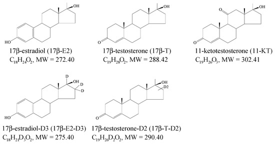
Figure 1.
Chemical structures, molecular formulas and weights of the compounds tested.
2.2. Preparation of Working Solutions
Primary standard stock solutions were prepared in methanol at concentrations of 1 mg/mL. Working solutions were obtained by a 10-fold dilution of primary standards to a concentration of µg/mL. Working solutions were stored in a freezer at 2–8 °C for six months.
2.3. Fish and Sampling
Microbiologically healthy, 12 female and 12 male sea trout (Salmo trutta L.) were obtained from the Department of Salmonidae Fish Breeding (Rudki, Poland), for the development and validation of the analytical procedure. In addition, 30 fish (8 female and 22 male) were obtained from the same place to determine the real concentrations of the tested hormones. (Consent to perform study from the Local Ethical Commission at the University of Life Sciences in Lublin, Poland, Resolution No. 52/2020 of 28 September 2020). Before sampling, fish were weighted, and the female or male gonads were dissected, weighted and stored at −20 °C until sample preparation. The percentage gonad somatic index (%GCI) was calculated using the formulae:
%GSI = (gonad weight/bodyweight) × 100
2.4. Sample Preparation
Sample (female or male gonad) at an amount of 5.0 ± 0.05 g was directly weighted in a 50 mL propylene centrifuge tube, IS was added, and after that, 5 mL of water was also added. The content of the tube was homogenized by vortex for 1 min.
Afterwards, 10 mL of ethyl acetate was added, and the content of the tube was shaken for 10 min. Next, the QuEChERS Extract Pouch (5 g MgSO4 and 1.5 g CH3COONa) was added, and the tube content was mechanically shaken for 1 min, centrifuged for 10 min at 3500 rpm and a temperature of 4 °C, for 10 min. The organic upper phase was transferred to the conical tube with dispersive SPE materials (PSA, C18EC and MgSO4).
The content of the tube was mechanically shaken for 1 min and centrifuged for 10 min at 10,000 rpm and at a temperature of 4 °C. The organic layer was transferred to the next tube and evaporated under a nitrogen current at a temperature of 60 °C. The dry residue was dissolved in 3 mL of methanol, and 0.5 mL of water was added and mixed for 1 min. To the tube, 5 mL of hexane was added and mechanically shaken for 10 min; after phase separation, hexane was removed. The procedure with hexane was repeated.
The content of the tube was evaporated under nitrogen current at a temperature of 60 °C. The dry residue was reconstituted in 300 µL of mobile phase, filtered by centrifugation with micro spin at 5000 rpm and transferred to an autosampler vial for analysis. The individual steps for handling the gonad samples are summarized briefly in the diagram below (Figure 2).
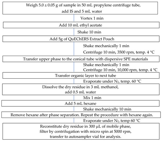
Figure 2.
Diagram of the analytical procedure with the sample.
2.5. LC-MS/MS Conditions
Chromatographic separation was performed on a Shimadzu Nexera X2 (Shimadzu, Japan) system equipped with Poroshell 120 EC-C18 (150 mm × 2.1 mm, 2.7 µm) (Agilent Technologies) column that was coupled with a C18 (4 mm × 2 mm) (Phenomenex, CA, USA) pre-column. The mobile phase consisted of a mixture of acetonitrile (eluent A), methanol (eluent B) and water (eluent C). The separation of analytes and their ISs was performed under isocratic elution condition (A:B:C = 20:50:30, v/v/v) at a flow rate of 0.2 mL/min. The column temperature was kept at 40 °C, and an injection volume of 25 μL was used.
Mass spectrometry analysis was carried out with the AB SCIEX 5500 QTRAP instrument (Applied Biosystems, Foster City, CA, USA) equipped with an ESI source operating in positive and negative modes. Data acquisition and quantitation were performed using Analyst® Software 1.6.3.
2.6. Method Validation
The reliability of the developed method was established by an in-house validation procedure, in accordance with the European Union Decision 2002/675/EC [27], using spiked female and male gonad samples of sea salmon trout. The analytical parameters such as selectivity, linearity, repeatability, within-lab reproducibility, relative recovery, detection and quantitation limits were evaluated. Additionally, uncertainty was calculated.
2.6.1. Selectivity
The analysis of 6 female and male gonad samples investigated selectivity to test for potential interference at the retention times of analytes with endogenous substances.
2.6.2. Linearity
Matrix calibration curves were obtained after analysing extracts of target gonad samples spiked with a known amount of each analyte and IS. Linear regression analysis was applied to determine the slope of the calibration curve, standard error of intercept and correlation coefficient. The calibration curve was used for estimation precision, relative recovery, and quantitation limit and was included in analysing all batches.
2.6.3. Precision
Repeatability (intra-day precision), within-laboratory reproducibility (inter-day precision) and recovery (accuracy) were estimated after analysis of the gonad samples spiked at 3 concentration levels of analytes corresponding to 1.0, 2.0, and 5.0 ng/g. Fixed levels of 17β-E2-D3 (1 ng/g) and 17β-T-D2 (1 ng/g) were added to each sample as internal standard (IS). Repeatability was estimated after analysis of spiked samples on the same day with the same instrument and one operator, and within-laboratory reproducibility was calculated after an analysis of spiked another two sets of samples on two different days with the same instrument and two other operators. The assay precision was evaluated by the measured concentration’s relative standard deviation (RSD, %).
2.6.4. Recovery
Relative recovery was calculated in the same experiment as repeatability by comparing the mean measured concentration with the spiked concentration.
2.6.5. Measurement Uncertainty
The uncertainty of the measurement of hormones in the female and male gonads was calculated by ResVal software at a 2 ng/g validation level. The expanded uncertainty (U) was calculated by the coverage factor of 2.
2.6.6. Carry-Over
Carry-over was evaluated by injection of the blank sample extracts directly following matrix calibration curves. The occurrence of carry-over was assessed by visual inspection of the chromatograms obtained for the blank samples.
3. Results and Discussion
This study aimed to develop a sensitive sample preparation and LC-MS/MS to quantify 17β-T, 11-KT and 17β-E2 in fish gonads and assess hormone levels in real fish samples. We found that this method is suitable for determining the interested hormones in samples obtained from fish.
3.1. LC-MS/MS Method Development
Positive ESI modes were selected to optimise the mass responses of 17β-T and 11-KT, and negative ones were chosen for 17β-E2, respectively. Detection parameters were obtained by diffusing the methanol solution of the analyte directly into the MS. The optimized ionisation source parameters are shown in Table 1.

Table 1.
The optimized ionisation source parameters.
The product ion mass spectrum of 17β-T, 11-KT and 17β-E2 and corresponding IS are presented in Table 2. The schemes of proposed fragmentation of 17β-E2, 17β-T and 11-KT are presented in Figure 3.
Table 2.
Monitored ion of analytes and MRM parameters.
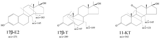
Figure 3.
Proposed schemes of fragmentation of 17β-E2, 17β-T and 11-KT molecules.
The optimum chromatographic condition was achieved using a mobile phase of acetonitrile-methanol-water (20:50:30, v/v/v) eluted isocratically at a flow rate of 0.2 mL/min and the total run time for sample analysis was 14 min. The chromatographic conditions allowed the resolution of 17β-T, 11-KT, 17β-E2 and ISs.
Using Poroshell 120 EC-C18 with acetonitrile-methanol-water mobile phase yielded satisfactory separation of both target analytes, and corresponding IS standards without formic acid or any other modifiers. Additionally, the addition of acetonitrile to the mobile phase improved the high of separated peaks.
3.2. Sample Preparation
The QuEChERS method has been evaluated in this study as a simple and not time-consuming sample preparation that was described by some authors [26]. Ethyl acetate was chosen as extracting solvent versus acetonitrile and methanol. It was found that ethyl acetate provided the best results in peak areas for tested hormones. Although acetonitrile and methanol were used to extract steroids from serum or other biota samples, 17β-T, 11-KT and 17β-E2 were isolated poorly from the tested fish gonads. Before vortexing with ethyl acetate, water was added to improve sample homogeneity and maximise extraction efficiency. The optimum water and ethyl acetate were adjusted at 5 and 10 mL, respectively, facilitating the extract collection.
The classical QuEChERS method contains two steps, a salting-out extraction and a dispersive SPE clean-up. In the preliminary study, different dehydrating reagents were compared for the efficiency of the salting-out extraction. Still, using 5 g magnesium sulphate and 1.5 g sodium acetate provided better phase separation and a more accessible collection of ethyl acetate.
The first, typical clean-up sorbents mixed at different proportions, were tested. The comparative studies found that a mixture of PSA, octadecylsilyl (C18) and MgSO4 gave good results. However, the highest recoveries and the lowest matrix effects were obtained when the commercially available mixture of 0.4 g PSA, 0.4 g C18EC and 1.2 g MgSO4 was used.
Additionally, the clean-up by double partitioning with hexane was applied to purify the ethyl acetate extract after the QuEChERS procedure. This additional step of sample cleaning allowed the elimination of fatty matrix extractives, which negatively affected the chromatographic analysis results.
3.3. Validation
Using the deuterated internal standard of target analytes was ideal for analysing any compounds in biological matrices because this can compensate extraction efficiency, matrix effect, instrumental, personal and/or other analytical variabilities that influence method performance. Due to the unavailability of a suitable 17β-T, 11-KT and 17β-E2-free gonads matrix, in the studies, deuterated internal standards (17β-estradiol-D3, 17β-testosterone-D2) were used to validate the developed procedure.
3.3.1. Selectivity
As shown in Figure 4 and Figure 5 17β-T, 11-KT, 17β-E2, and ISs were separated successfully, and no endogenous or exogenous interferences were detected at the retention times of target analytes.
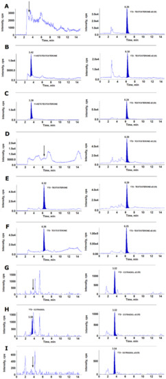
Figure 4.
Ion chromatogram from the analysis of a male gonad sample: (A)—blank sample for 11-KT; (B)—sample spiked with 11-KT to the concentration of 5 ng/g; (C)—sample with 11-KT at a concentration of 23.67 ng/g; (D)—blank sample for 17β-T; (E)—sample spiked with 17β-T to a concentration of 1 ng/g; (F)—sample with 17β-T at a concentration of 0.57 ng/g; (G)—blank sample for 17β-E2; (H)—sample spiked with 17β-E2 to a concentration of 1 ng/g; (I)—sample tested with no 17β-E2.
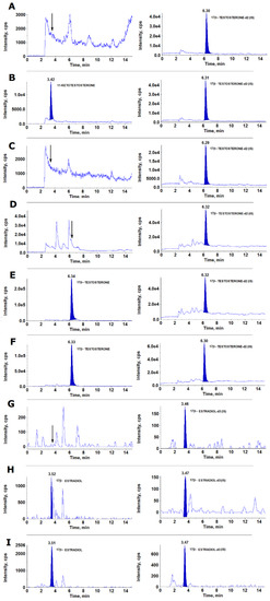
Figure 5.
Ion chromatogram from the analysis of a female gonad sample: (A)—blank sample for 11-KT; (B)—sample spiked with 11-KT to the concentration of 5 ng/g; (C)—sample tested with no residues of 11-KT; (D)—blank sample for 17β-T; (E)—sample spiked with 17β-T to a concentration of 1 ng/g; (F)—sample with 17β-T at a concentration of 3.32 ng/g; (G)—blank sample for 17β-E2; (H)—sample spiked with 17β-E2 to a concentration of 2 ng/g; (I)—sample with 17β-E2 at a concentration of 2.50 ng/g.
3.3.2. Linearity
Linearity was evaluated using matrix-matched calibration curves prepared from peak-area ratios of 17β-T, 11-KT and 17β-E2 to the corresponding ISs. The calibration showed satisfactory linearity from 0 to 50 ng/g for 17β-T, from 0 to 30 ng/g for 11-KT and from 0 to 5.0 or from 0 to 50 for 17β-E2, regardless of the matrix being tested (Figure 6).
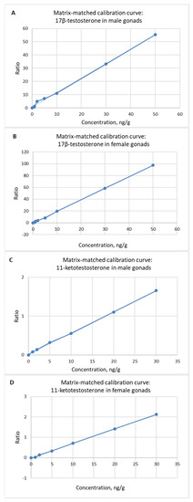
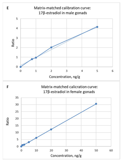
Figure 6.
Linearity of the method evaluated in male and female gonad matrix-matched calibration: (A,B)—17β-T, (C,D)—11-KT, (E,F)—17β-E2.
Experiment on dilution integrity, performed by quantification of samples containing 17β-T, 11-KT and 17β-E2 concentrations higher than the upper calibration level, diluted at 1:10 and 1:20, gave acceptable results (5 repetitions).
The obtained coefficients of determination (R2) were higher than 0.99 for 17β-T, 11-KT and 17β-E2 in female and male gonad samples (Table 3).
Table 3.
Calibration parameters a.
3.3.3. Precision, Relative Recovery, Matrix Effect
The evaluation of repeatability, within-laboratory reproducibility and relative recoveries for samples spiked target analytes at the levels 1.0, 2.0 and 5.0 ng/g are listed in Table 4 for male gonad and in Table 5 for female gonad samples, respectively.
Table 4.
Repeatability, within-laboratory reproducibility and relative recovery in evaluated male gonad samples.
Table 5.
Repeatability, within-laboratory reproducibility and relative recovery in evaluated female gonad samples.
The repeatability values ranged from 1.7 to 27.2%, within-lab reproducibility from 2.2 to 37.1%, and relative recovery from 94.0 to 110.0% in the evaluated male gonad samples (Table 4). Additionally, in the female gonad samples were from 2.0 to 14.9%, from 2.7 to 20.2%, and from 98.3 to 103.5%, respectively (Table 5). Matrix effect was not observed in samples of male as well as female gonads.
3.3.4. LOD and LOQ
The obtained LOD and LOQ for 17β-T, 11-KT and 17β-E2 were from 0.15 to 0.28 ng/g and from 0.41 to 0.76 ng/g in male gonads from 0.15 to 0.24 ng/g and 0.32 to 0.64 ng/g, in female gonads (Table 6). LOD and LOQ were found to be low enough to detect expected values of hormones in the female and male gonads of sea trout.
Table 6.
LOD and LOQ.
3.3.5. Measurement Uncertainty
In this study, the expanded uncertainty (U) ranged from 21.2 to 41.2% for the analysed hormones in samples of male gonads and from 22.2 to 37.7% in female gonads (Table 7). The estimated uncertainty was below the recommended threshold volume of 50%, which shows suitability for the developed method’s purpose.
Table 7.
Uncertainty.
3.4. Application to Real Samples
The usefulness of the developed method was verified by the determination of the concentrations of 17β-T, 11-KT and 17β-E2 in the female (n = 8) and male (n = 22) gonads of healthy sea trout. The mean body weight of female and male fish was 600 g (from 400 to 1200 g and 800 g (from 300 to 1600 g), and the mean female and male gonad weight was 58 g (from 40 to 80 g) and 52 g (from 35 to 72 g), respectively. The mean female fish GSI was 9.6%, and for the male was 6.5%.
In male gonads, the determined concentration of 11-KT ranged from 7.4 to 36.6 ng/g, 17β-T from 1.9 to 16.6 ng/g, and 17β-E2 was not detected. In female gonads, the concentration of 11-KT ranged from 3.4 to 9.2 ng/g, 17β-T ranged from 2.1 to 9.6 ng/g, and 17β-E2 ranged from 9.4 to 40.7 ng/g, respectively (Table 8). The obtained results indicate that the concentrations of 17β-T and 11-KT were twice higher in the analysed samples of male gonads than in female ones.
Table 8.
Concentrations of 17β-T, 11-KT and 17β-E2 (mean ± s.d. and range, ng/g) in the real samples of gonads of sea trout.
4. Conclusions
In-house validated analytical procedure was found applicable for quantitation of 17β-T, 11-KT and 17β-E2 in the female and male gonads of sea trout. The proposed procedure characterizes good sensitivity, linearity, accuracy, and precision. Using the QuEChERS method with the pre-weighted amounts of sorbents and salt pre-constituted in a propylene tube allowed us to prepare samples relatively quickly.
This method is currently being applied to determine 17β-T, 11-KT and 17β-E2 concentrations in gonads of wild sea trout in order to explain the causes of pathomorphological changes on the skin (ulcerative dermal necrosis) formed during the migration of fish from the Baltic Sea to rivers.
Author Contributions
Conceptualization, A.P. and I.M.-Ż.; Formal analysis, A.K.; Investigation, A.K., S.W. and I.M.-Ż.; Methodology: A.P. and I.M.-Ż.; Sample preparation, A.P.-S.; Project administration, A.P.; Writing—original draft: A.P.; Writing—review, I.M.-Ż. All authors have read and agreed to the published version of the manuscript.
Funding
This work was supported by the National Veterinary Research Institute in Puławy, Poland (Project No S/439).
Data Availability Statement
The data presented in this study are available on request from the corresponding author. The data are not publicly available due to data privacy protection.
Conflicts of Interest
The authors declare no conflict of interest.
References
- Scholz, S.; Kluver, N. Effects of endocrine disruptors on sexual, gonadal development in fish. Sex Dev. 2009, 3, 136–151. [Google Scholar] [CrossRef] [PubMed]
- Nearns, A.J.; Bissell, M.; Morrison, A.M.; Rempel-Hester, M.A.; Courney, A.; Rutherford, N. Effects of pollution on marine organisms. Water Environ. Res. 2019, 91, 1229–1252. [Google Scholar]
- Milla, S.; Depiereux, S.; Kestemont, P. The effects of estrogenic and androgenic endocrine disrupters on the immune system in fish: A revive. Ecotoxicology 2011, 20, 305–319. [Google Scholar] [CrossRef]
- Cabas, I.; Chaves-Pozo, E.; Mulero, V.; Garcia-Ayala, A. Role of estrogen in fish immunity with special emphasis on GPER1. Dev. Comp. Immunol. 2018, 89, 102–110. [Google Scholar] [CrossRef]
- Jobling, S.; Tyler, C.R. Endocrine disruption, parasites and pollutants in wild freshwater fish. Parasitology 2003, 126, S103–S108. [Google Scholar] [CrossRef]
- Pottinger, T.G. Seasonal variation in specific plasma- and target-tissue binging of androgens, relative to plasma steroid levels in the brown trout, Salmo trutta L. Gen. Comp. Endocrinol. 1988, 70, 334–344. [Google Scholar] [CrossRef]
- Debowski, P. The largest Baltic population of sea trout (Salmo trutta L.): Its decline, restoration attempts, and current status. Fish. Aquat. Life 2018, 26, 81–100. [Google Scholar] [CrossRef]
- Conclin, S.E.; Knezevic, C.E. Advancements in the gold standard: Measuring steroid sex hormones by mass spectrometry. Clin. Biochem. 2020, 82, 21–32. [Google Scholar] [CrossRef] [PubMed]
- Woźniak, B.; Matraszek-Żuchowska, I.; Żmudzki, J. Determination of 17β-Oestradiol and testosterone in bovine serum with gas chromatography-mass spectrometry. Bull. Vet. Inst. Pulawy 2011, 55, 755–759. [Google Scholar]
- Matraszek-Żuchowska, I.; Woźniak, B.; Posyniak, A. Determination of hormones residues in milk by gas chromatography-mass spectrometry. Food Anal. Methods 2017, 10, 727–739. [Google Scholar] [CrossRef]
- Matraszek-Żuchowska, I.; Woźniak, B.; Sielska, K.; Posyniak, A. Determination of selected testosterone esters in blood serum of slaughter animals by liquid chromatography with tandem mass spectrometry. Steroids 2020, 163, 108723. [Google Scholar] [CrossRef] [PubMed]
- Boggs, A.S.P.; Bowden, J.A.; Galligan, T.M.; Guillette, L.J., Jr.; Kucklick, J.R. Development of a mult-class steroid hormone screen method using liquid chromatography/tandem mass spectrometry. Anal. Bioanal. Chem. 2016, 408, 4179–4190. [Google Scholar] [CrossRef] [PubMed]
- Gravitte, A.; Archibald, T.; Cobble, A.; Kennard, B.; Brown, S. Liquid chromatography-mass spectrometry applications for quantification of endogenous sex hormones. Biomed. Chromatogr. 2021, 35, e5036. [Google Scholar] [CrossRef] [PubMed]
- Nouri, M.-Z.; Kroll, K.J.; Webb, M.; Denslow, N.D. Quantification of steroid hormones in low volume plasma and tissue homogenates of fish using LC-MS/MS. Gen. Comp. Endocrinol. 2020, 296, 113543. [Google Scholar] [CrossRef]
- Zhang, F.; Bartels, M.J.; Geter, R.D.; Carr, M.S.; McClymount, L.E.; Mario, T.A.; Klecka, G.M. Simultaneous quantitation testosterone, estradiol, ethinyl estradiol, and 11-ketotestorenon in fatheadminnow fish plasma by liquid chromatography/positive atmospheric pressure photoionization tandem mass spectrometry. Rapid Commun. Mass Spectrom. 2009, 23, 3637–3646. [Google Scholar] [CrossRef]
- Dang, V.D. Simultaneous measured of sex steroid hormones in largemouth bass (Micropterus salmoides) plasma by application of liquid chromatography-tandem mass spectrometry. J. Chromatogr. Sci. 2020, 59, 7–14. [Google Scholar] [CrossRef]
- Errico, S.; Chioccarelli, T.; Moggio, M.; Diano, N.; Cobellis, G. A new LC-MS/MS method of simultaneous and quantitative detection of bisphenol-A and steroids in target tissues: A power tool of characterize the interference of bisphenol-A exposure on steroid levels. Molecules 2020, 25, 48. [Google Scholar] [CrossRef]
- Hoga, C.A.; Almeida, F.L.; Reyes, F.G.R. A review on the use hormones in fish farming: Analytical methods to determine their residues. J. Food 2018, 16, 679–691. [Google Scholar] [CrossRef]
- Yuan, T.-F.; Le, J.; Cui, Y.; Peng, R.; Wang, S.-T.; Li, Y.J. An LC-MS/MS analysis for seven sex hormones in serum. Pharm. Biomed. Anal. 2019, 162, 34–40. [Google Scholar] [CrossRef]
- Blasco, M.; Carrquiriborde, P.; Marino, D.; Ronco, A.E.; Somoza, G.M. A quantitative HPLC-MS for the simultaneous determination of testosterone, 11-ketotestosterone and 11-β hydroxyandrostenedione in fish serum. J. Chromatogr. B 2009, 877, 1509–1515. [Google Scholar] [CrossRef]
- Hoga, C.A.; Reche, V.G.; Almeida, F.L.; Reis, V.R.; Cordeido, R.P.; Anadon, A.; Reyes, F.G.R. Development and validation of an analytical method for the determination of 17β-estradiol residues in muscle of tambaqui (Colossoma macropomum Cuvier, 1818) by LC-MS/MS and its application in samples from a fish sexual reversion study. J. Chromatogr. B 2019, 1128, 121774. [Google Scholar] [CrossRef] [PubMed]
- Yoo, H.S.; Napoli, J.L. Quantification of dehydroepiandrosterone, 17β-estradiol, testosterone, and their sulfates in mouse tissues by LC-MS/MS. Anal. Chem. 2019, 91, 14624–14630. [Google Scholar] [CrossRef] [PubMed]
- Navarro, P.; Bustamante, J.; Vallejo, A.; Prieto, A.; Usobiaga, A.; Arrasate, S.; Anakabe, E.; Puy-Azurmendi, E.; Zuloaga, O. Determination of alkylphenols and 17β-estradiol in fish homogenate. Extraction and clean-up strategies. J. Chromatogr. A 2010, 1217, 5890–5895. [Google Scholar] [CrossRef]
- McNamara, K.M.; Harwood, D.T.; Simanainen, U.; Walters, K.A.; Jimenez, M.; Handelsman, D.J. Measurement of sex steroids in murine blood and reproductive tissues by liquid chromatography-tandem mass spectrometry. J. Steriod Biochem. Mol. Biol. 2010, 1211, 611–618. [Google Scholar] [CrossRef] [PubMed]
- Xu, C.L.; Chu, X.G.C.; Peng, C.F.; Jin, Z.Y.; Wang, L.Y. Development of a faster determination of 10 anabolic steroids residues in animal muscle tissues by liquid chromatography tandem mass spectrometry. J. Pharm. Biomed. Anal. 2006, 41, 616–621. [Google Scholar] [CrossRef] [PubMed]
- Anastassiades, M.; Lehotay, S.-J.; Stajnbaher, D.; Schenck, F.-J. Fast and easy multi-residue method employing acetonitrile extraction/partitioning and “dispersive solid-phase extraction” for the determination of pesticide residues in produce. JAOAC Int. 2003, 86, 412–431. [Google Scholar] [CrossRef]
- European Commission. Commission Decision 2002/657/EC. Off. J. Eur. Commun. 2002, L221, 8–36. [Google Scholar]


Publisher’s Note: MDPI stays neutral with regard to jurisdictional claims in published maps and institutional affiliations. |
© 2022 by the authors. Licensee MDPI, Basel, Switzerland. This article is an open access article distributed under the terms and conditions of the Creative Commons Attribution (CC BY) license (https://creativecommons.org/licenses/by/4.0/).