Review

Recent Advances on the Development of Chemosensors for the Detection of Mercury Toxicity: A Review

by
Shiva Prasad Kollur
1,
Chandan Shivamallu
2,
Shashanka K. Prasad
2,
Ravindra Veerapur
3,
Sharanagouda S. Patil
4,
Charley A. Cull
5,
Johann F. Coetzee
6 and
Raghavendra G. Amachawadi
7,*
1
Department of Sciences, Amrita School Arts and Sciences, Amrita Vishwa Vidyapeetham, Mysuru Campus, Mysuru 570 026, India
2
Division of Biotechnology and Bioinformatics, School of Life Sciences, Department of Water and Health, JSS Academy of Higher Education and Research, Mysuru 570 015, India
3
Department of Metallurgy and Materials Engineering, Malawi Institute of Technology, Malawi University of Science and Technology, Limbe P.O. Box 5916, Malawi
4
Pathoepidemiology Laboratory, ICAR—National Institute of Veterinary Epidemiology and Disease Informatics (NIVEDI), Post Box No.6450, Ramagondanahalli, Yelahanka, Bengaluru 560064, India
5
Midwest Veterinary Services, Inc., Oakland, NE 68045, USA
6
Department of Anatomy & Physiology, College of Veterinary Medicine, Kansas State University, Manhattan, KS 66506, USA
7
Department of Clinical Sciences, College of Veterinary Medicine, Kansas State University, Manhattan, KS 66506, USA
*
Author to whom correspondence should be addressed.
Separations 2021, 8(10), 192; https://doi.org/10.3390/separations8100192
Submission received: 15 August 2021 / Revised: 8 October 2021 / Accepted: 9 October 2021 / Published: 18 October 2021
(This article belongs to the Section Materials in Separation Science)

Abstract

:
The harmful impact of mercury on biological systems is of great concern. Regardless of the efforts made by the regulating agencies, a decrease in Hg2+ concentration has not been realized, and hence mercury accumulation in the environment remains of utmost concern. Designing novel and efficient probes for recognition and detection of toxic metals in environmental samples has been of primary importance. Among the available techniques, probe designs involving the study of spectral properties has been preferred because of its obvious ease of instrumentation. Furthermore, occurrence of significant changes in the visible portion of electronic spectra enables detection by the naked eye, thereby endorsing the preference for development of probes with off-on binary responses to aid in the in-field sample analysis. The prominence is further streamlined to the use of fluorescence to help characterize on-response the cellular detection of Hg2+ with ease. In order to overcome the problem of developing efficient probes or sensors bearing fluorescence on-response mechanism that can work effectively in physiological conditions, various methodologies, such as chemo-dosimetric reaction mechanisms for the designing of new luminescent ligands, are being adopted. Additionally, modified charge transfer processes are also being considered for optical detection of the mercury (II) ion. In this review, all such possible techniques have been discussed in detail.

1. Introduction

Mercury, previously named hydrargyrum (Hg) and commonly called quicksilver, is a chemical element with the atomic number 80 and a relatively high atomic mass of 200.59 u. This element, which is also the only known liquid metal, is the second most toxic element on earth preceded by plutonium. Mercury spike in water is relatively rare with the sources of the metal as common as fossil fuels. With the expansive use in hydroelectric, mining, pulp and paper industries, the levels of mercury in the environment has increased over time. Mercury is a common pollutant of drinking water and can cause kidney complications if present in amounts of more than 2 ppb [1,2,3]. However, vaporization of liquid mercury leads to gaseous mercury, which is poisonous due to its nature of being absorbed into the blood [4]. With regard to the bioaccumulation prospect, mercury enters the top of the food chain via aquatic bacteria in the form of methylmercury, leading to a neurological condition, “minamata disease” [5,6,7]. Elemental mercury is considered more lethal form and usually enters the system via the cutaneous and respiratory routes [8]. Elemental mercury is known to cause cardiovascular, neural, and renal complications by inflicting DNA damage [9,10]. Mercury methylation increases its lipophilic properties, posing a threat to the central nervous system [11]. Taken together, the high risk of toxicity associated with mercury exposure has incentivized global efforts toward potable water purification and mercury toxicity reduction. Concerns about the deleterious effects of mercury poisoning have also motivated researchers to develop novel, affordable, and rapid detection tests that may be applicable to both the environmental and biological systems [12,13,14]. Therefore, a simple and convenient sensor that can solve the situation at hand rapidly, have sufficient sensitivity, and be resistant to interference by other metal ions is timely. The developments thus far have accounted for the expensive, sophisticated instruments, enabling complicated procedures or low sensitivity and selectivity approaches such as atomic absorption, emission spectroscopy, or individually coupled plasma mass spectroscopy [15]. Despite efforts from the various regulatory agencies for reducing mercury emission in the environment, mercury contamination from natural and industrial processes across the globe has remained a serious threat to the human race in the last few decades. Therefore, monitoring mercury is of paramount importance to both environmental and human health.
In recent years, sensors for the recognition of heavy metal ions have been developed, which has attracted much attention from environmental scientists and chemists. Colorimetric receptors were developed for the selective recognition of heavy metal ions that received attention for several decades due to their ability to permit naked eye detection of color change and simplify the whole method. However, chemosensors remained a reliable technique for the effective detection of mercury toxicity. Fluorescent chemo-sensors are preferred for a ratiometric response due to the ratio between two emission intensities that can help correcting the sensor analytic concentration, as well as environmental effects such as polarity, photo-bleaching and temperature. The expectations lead to the development of reversible ratiometric chemosensors capable of detecting Hg2+ [16]. Chemosensors with selectivity for specific targets of metal ions are continuously demanded. Those targeting the toxic heavy metal ions remain a vital category of chemosensors. Recognition of the detrimental effects of certain transition and post-transition metal ions on humans and animals has, in part, inspired work to develop compounds that selectively respond to specific metal ions for use as ion sensors. Research on fluorescence sensor capable of detecting the heavy and transition metal ions has gained attention due to significant progress in synthesis of novel fluorophores and the development of cost-effective yet efficient methods. Advantages such as high sensitivity, selectivity, short response time, naked-eye detection, and fluorescence detection have been regarded as promising candidates in the molecular recognition and current applications of chemosensors. Furthermore, in biological and environmental systems, Hg2+ sensor interactions commonly occur as aqueous solutions. Hence, enough attention has been paid into developing Hg2+ sensors, which can work in the aqueous phase. Despite the preference, water-soluble Hg2+ sensors are still not common. Recently Benzothiazole-derived chemosensor that contain sulphur moiety have a sensing process involving the coordination of Hg2+ to the S atom, which is not recyclable. Further, in this work, they observed that the pink-colored chemosensor turns blue when reacted with mercury (II) ions due to the formation of a 2:1 coordination complex [17]. Wen-Juan Qu et al., 2014, reported a fluorescent sensor, (E)-1-((5-(4-nitrophenyl)furan-2-yl)methylene) semicarbazone, which is highly sensitive and selective for Hg(II) ions. The observations from the above work indicate that the chemosensor bonded to mercury ions leading to the supramolecular self-assembly breaking and the cooperation reaction occurrence, which led to the decrease in intra-molecular charge density in the chemosensor [18].

2. Types of Probing Agents for Mercury
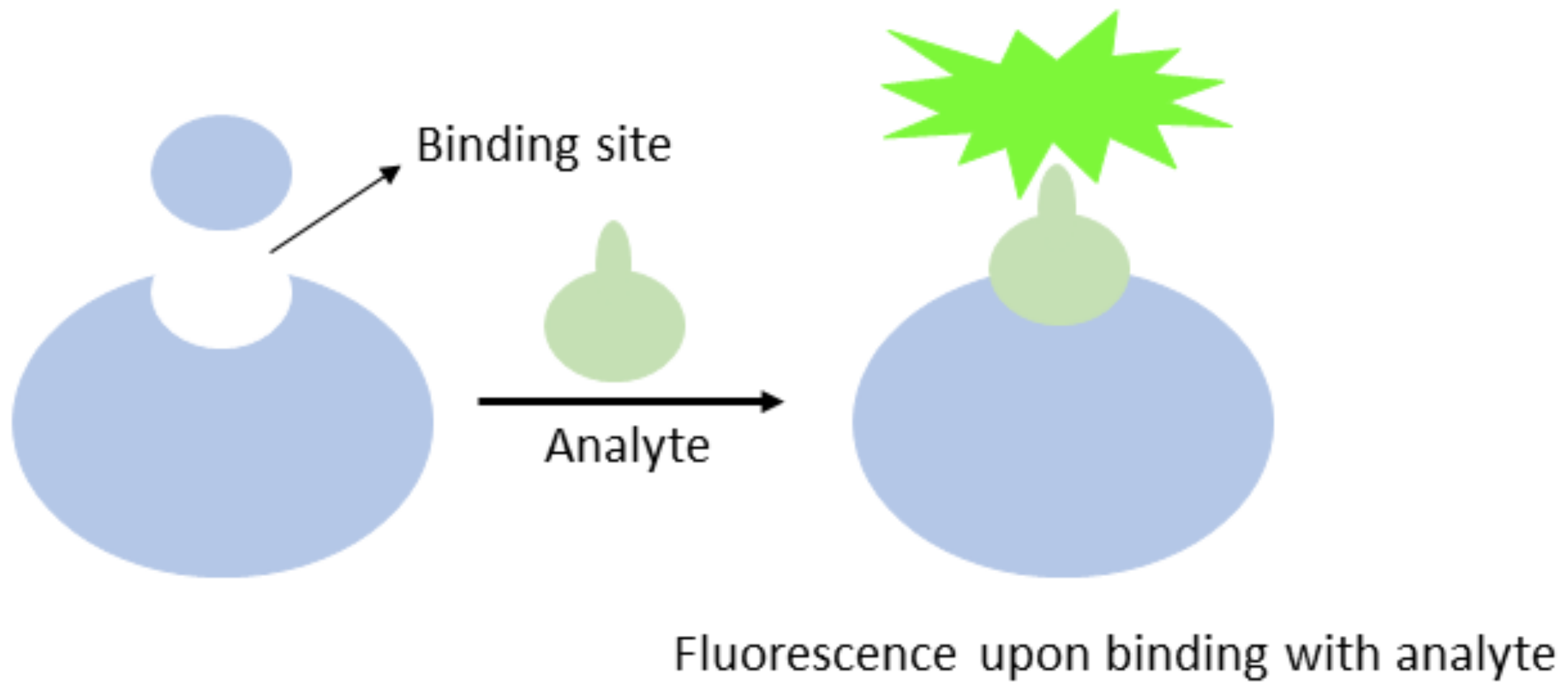
2.1. Chemosensors

A chemoreceptor, also referred to as chemosensor, is a sensory receptor that transfers a chemical signal into action potential. Additionally, a chemosensor detects chemical stimuli within the environment. Basically, it detects the presence of a required analyte in the solution/environment and gives a detectable signal to certify the presence of the analyte (Figure 1).
Based on the type of signal generation, these chemosensors are categorized into four types: (1) redox potential, (2) absorbance (or color), (3) luminescence (or fluorescence) and (4) NMR relaxation times [19]. This review will focus on fluorescent chemosensors, or those which give fluorescence signals in the presence of the analyte. These fluorescent chemosensors are categorized (as depicted in Figure 2) on the basis of their synthesis, into the intrinsic, conjugate and auto-assembled varieties. The luminescent chemosensors are used as a binding site in case of intrinsic sensors or as an optical signaling unit covalently linked to a receptor unit in case of conjugated sensor. Auto-assembled chemosensors operate through a signal transduction pathway, causing dimerization of low affinity ligands and resulting in a complex with a high affinity for a multivalent monolayer protected nanomaterials. However, the basic mechanism commonly followed by the fluorescent chemosensors is primarily by photo-induced electron transfer (PET), where the donor moiety gains energy from the incoming light used for excitation of its electron(s) to the higher state, facilitating easy donation to the acceptor moiety. In due course of the process, energy released can be detected as a fluorescence signal. Internal electron transfer (ICT) is an alternate mechanism where a fraction of the electron charge is transferred between the molecular entities [20].

2.2. Mercury Chemosensors

Over the period of modifications on the mercury detecting probes, research has made significant progress. Starting from the use of the thio-group containing organic solvents to checking the black coloration generated by HgS, there is a preference of metal complexing with suitable ligands and for the validation of Hg2+ occurrence (Figure 3). According to the current field of exploration, most chemosensors now being synthesized are of the fluorescent nature, owing to their ease of detection, cost effective synthesis, and easy instrumentation. The use of fluorophores in detection of this toxic metal ion can now be seen as a common approach.

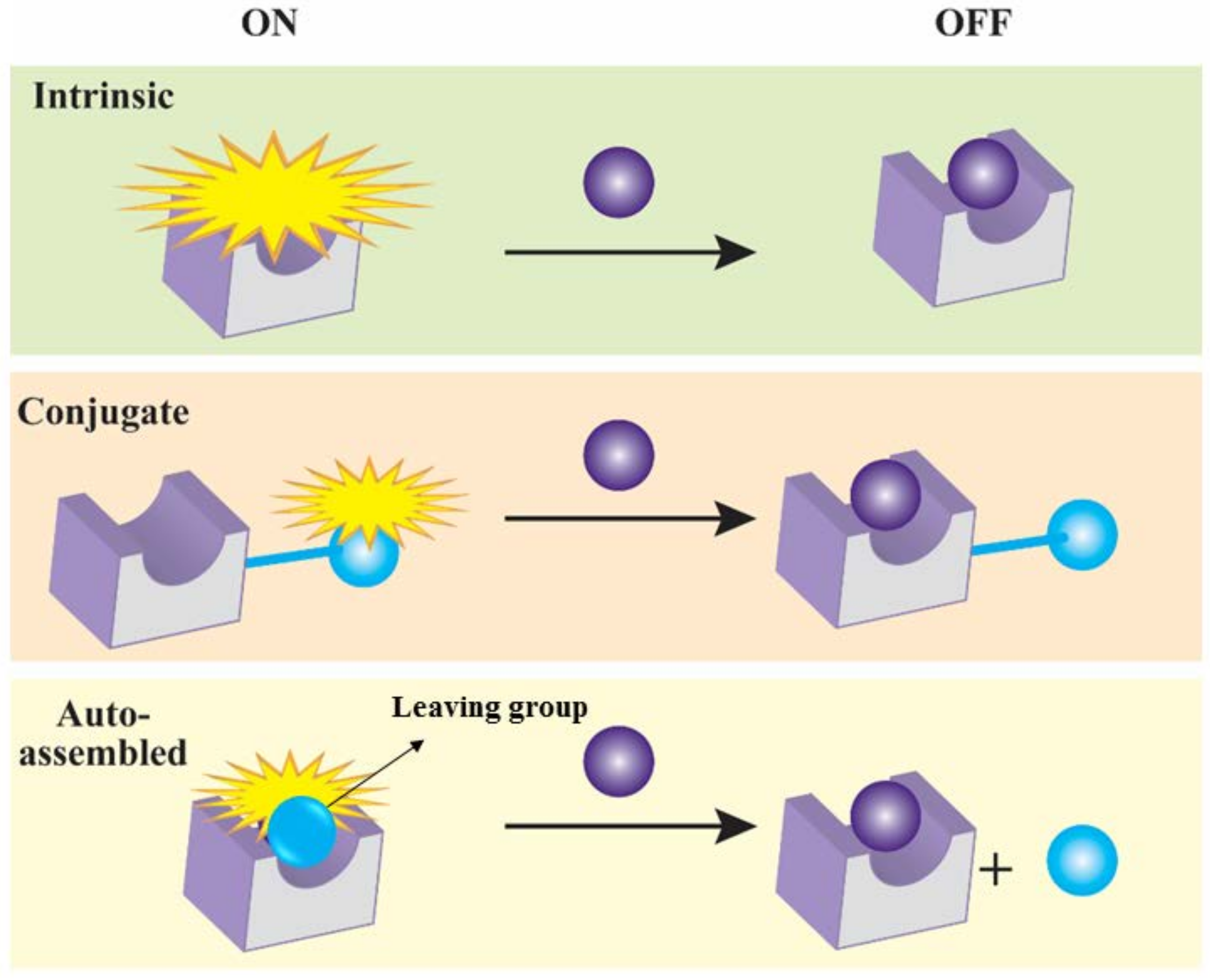
2.3. Types of Mercury Detecting Chemosensors

2.3.1. Molecular Sensors

The most important strategy that needs to be understood for Hg2+ sensing is due to the fact that the chemical bond formation occurs using the fourth-, fifth-, and sixth-group elements as electron acceptors. During this bond formation, a strong affinity persists between electron acceptors and donors. It is the selective recognition that increases the stability and sensitivity of a sensor. This so-formed chemical bond can either be Hg-O, Hg-C, Hg-N or Hg-S bonds as described below [21].
Hg-O: These involve the cleavage of C=O bonds in the rhodamine derivatives, which are excellent fluorophores used in sensors. The ring-closed form of spirolactam is colorless and is not fluorescent. However, upon addition of analytes, structural change occurs and the closed spirolactam changes to the ring-opened form and results in change from colorless to an intense coloration, along with the presence of strong fluorescence. This principle has been used to design numerous mercury sensors. Hg2+ is known to have been able to promote the hydrolysis of isopropenyl acetate under mild conditions. In 2012, Das et al. synthesized a probe that incorporated a quinolone unit into a rhodamine 6G derivative in order to obtain different wavelengths along the fluorescence spectrum [22]. They also synthesized two rhodamine derivatives (sensor 1 and 2 in Figure 4) for recognition of mercury and copper ions. The change in the structural framework of rhodamine led to the visible change in emission intensities. The experiment was performed in CH3CN aqueous media, and absorption was studied after reacting with Hg2+. Absorption was observed around 527 nm. As the rhodamine derivative was made to interact with Hg2+, the luminescence intensity observed at 557 nm was enhanced, with a detection limit of 0.35 ppb for Hg2+. The ratiometric sensor enabled the interaction of rhodamine with 1,8-naphthalimide derivatives as two fluorophores for the detection of Hg2+. The non-radiative transfer of excitation energy was induced due to the binding of Hg2+ from the donor naphthalimide to the acceptor xanthene moiety.
The ability of the sensor to detect the presence of different divalent ions using the UV-visible and fluorescence spectroscopy tool is shown below (Figure 5). The change in the emission spectra occurred at 336 nm when 15 equivalents of the metal ions were excited in CH3CN containing 5% DMSO. It was observed that the sensor had a strong interaction with Hg2+ and a little interaction with Zn2+. Fluorescence data confirmed emission at 336 nm, which became clearly visible when Hg(ClO4)2 was added through titration, and a red shift of 5 nm was observed. This red shift was caused by the stabilization of the fluorophore in its excited state compared to the ground state on binding of the cation (Figure 6).
Hg-S: Formation of Hg-S occurs by C=S or C-S-C bond cleavage. Chemical properties of sulfur and oxygen are somewhat similar because they belong to the same group on the periodic table. As discussed above, extensive studies were performed for Hg2+ detection by Hg-O bond formation and the cleavage of C=O bonds. Hence, Figure 7 depicts the synthesis of chemosensors with the Hg-S bond formation and its role in the detection of Hg2+ ions.
Yang’s group designed and synthesized a fluorescein-based chemodosimeter for Hg2+ detection [23]. In addition to this probe of (I) by Hg2+ in aqueous methanol solution, the thiosemicarbazide group undergoes a desulfurization reaction that involves irreversible cleavage of C=S bond and forms the corresponding oxadiazole (Figure 8). Herein, the Hg2+ become stable by the removed sulfur through Hg-S bond formation. Hence, the visible coloration and fluorescence emission caused by the oxadiazole states that the maximum linear response of the sensor to Hg2+ is 0.8 µM, with a detection limitation of 9.4 nM in methanol-H2O. Changing the substitution with a multi-nitro moiety made this probe a reversible sensor (II), which is highly fluorescent (Figure 8).
The design and synthesis of Schiff base with rhodamine B thiohydrazide and a quinoline moieties was reported in a recent literature [24]. When probe I was made to react with Hg2+, a clear pink color turn-on appeared, which caused a 106-fold fluorescence enhancement with an absorption maximum at 565 nm. This probe had a response range of 0.1 to 10 µM and detection limit of 8.5 nM. Similarly, a fluorescent chemosensor (Figure 9) with a receptor composed of S atom and an alkene moiety when reacted with Hg2+ metal ion showed a large fluorescent enhancement response (approximately 1000-fold). There was a maximum absorption at 561 nm with high selectivity and sensitivity, while having an LOD of 27.5 nM [25].
Hg-N: Hancock’s research group found a “turn-off” fluorescent sensor for Hg2+ ions in solution [26]. They basically explored through the tendency of Hg2+ ions to quench the fluorescence of a potential fluorescent sensor. Hancock’s group worked with multiple divalent ions and studied the chelation-enhanced fluorescent (CHEF) effect [27] and observed that Hg2+ showed way lesser chelation effect in comparison to Zn2+ and Cd2+. This reflects the heavy atom effect, which could have been caused by increasing spin-orbit coupling constants (Figure 10). Unlike the other divalent ions, mercury is usually good at quenching the fluorescence, hence causing a negative “turn-off” chelation enhanced quenching (CHEQ) effect [28]. They worked with coordinated and tethered fluorophores such as N,N-bis(2-methylquinoline)-2-(2-aminomethyl)pyridine (DQPMA) and (N-(9-anthracenylmethyl)-N,N-di-(picolyl)amine) (ADPA).
As manifested in Figure 11, Lee et al. showed that Hg2+ contained in a solution prominently quenches the fluorescence of ADPA, which was investigated using crystallography for Hg-ADPA complexes, and the conclusion was supported by DFT calculations [29]. It was also found that coordination of halide ions or hydroxide to the Hg2+-ADPA complex can displace the anthracenyl group coordinated to Hg2+ and restore its fluorescence, hence forming a novel-type anion sensor. The addition of more covalent binding ligands to the mercury ion tends to lengthen Hg-N bonds to the saturated N-donor atom of the dipicolylamine part of ADPA. The information was supported by crystallography and DFT calculations. Moreover, DFT calculations also show that the quenching effect observed in Hg2+-ADPA complex is the cause of π-interaction of mercury ions with the fluorophore, leading to a decrease of the electronic transition probability to ground state from excited state. It was concluded that for designing a turn-on sensor for mercury ions, it is important to have a covalently bonded ion such as an S-donor atom for chelating.
Hg interaction with both N and S moiety
Lippolis’ group reported that the 1,10-phenanthroline unit was an integral part of the macrocyclic structure, which includes prominent thioether donors that are potential fluorescent chemosensors for soft metal sensors. These phenanthroline derivatives were then used to study the toxicity of divalent ions of lead, cadmium, and mercury [30].
Formation of Hg-Se bond
This occurs by the cleavage of the C-Se-C bond. Being of the same group as sulfur, selenium does show strong affinity to Hg2+, similar to S [31].
It was observed that the selonacton-based sensor exhibited intensive fluorescence enhancement at 580 nm over the test range of concentration from 0 to 30 nM. Moreover, this sensor showed an observable optical response to methylmercury species in HEPES buffer as well as in cells/zebrafish [32,33,34]. The unusual fluorescence enhancement is induced due to the formation of Hg-Se bond followed by eventual deselenation reaction (Figure 12).

2.3.2. Formation of Hg-C Bond

The need for this type of sensor occurred because it was observed that Hg-S binding had drawbacks, such as the oxidation of sulfide over long-term storage. Hence, binding of Hg2+ using the Hg-C bond can be considered a fair alternative. As reported by Koide’s group, an oxidation-resistant fluorescent sensor for mercury was based on an alkyne oxymercuration mechanism [35]. Results from this study suggested that the binding of Hg2+ to dissolved organic matter (DOM; hydrophobic acids isolated from the Florida Everglades by XAD-8 resin) under ambient conditions (with low Hg/DOM ratios) is controlled by a small number of DOM molecules containing a highly reactive thiol functional group (Figure 13). Thus, the distribution coefficients of Hg/DOM that were used for studying the biogeochemical behavior of Hg in natural systems need to be determined at low Hg/DOM ratios. With the addition of Hg2+ to this sensor system, hydration of alkynes occurs to form ketones.

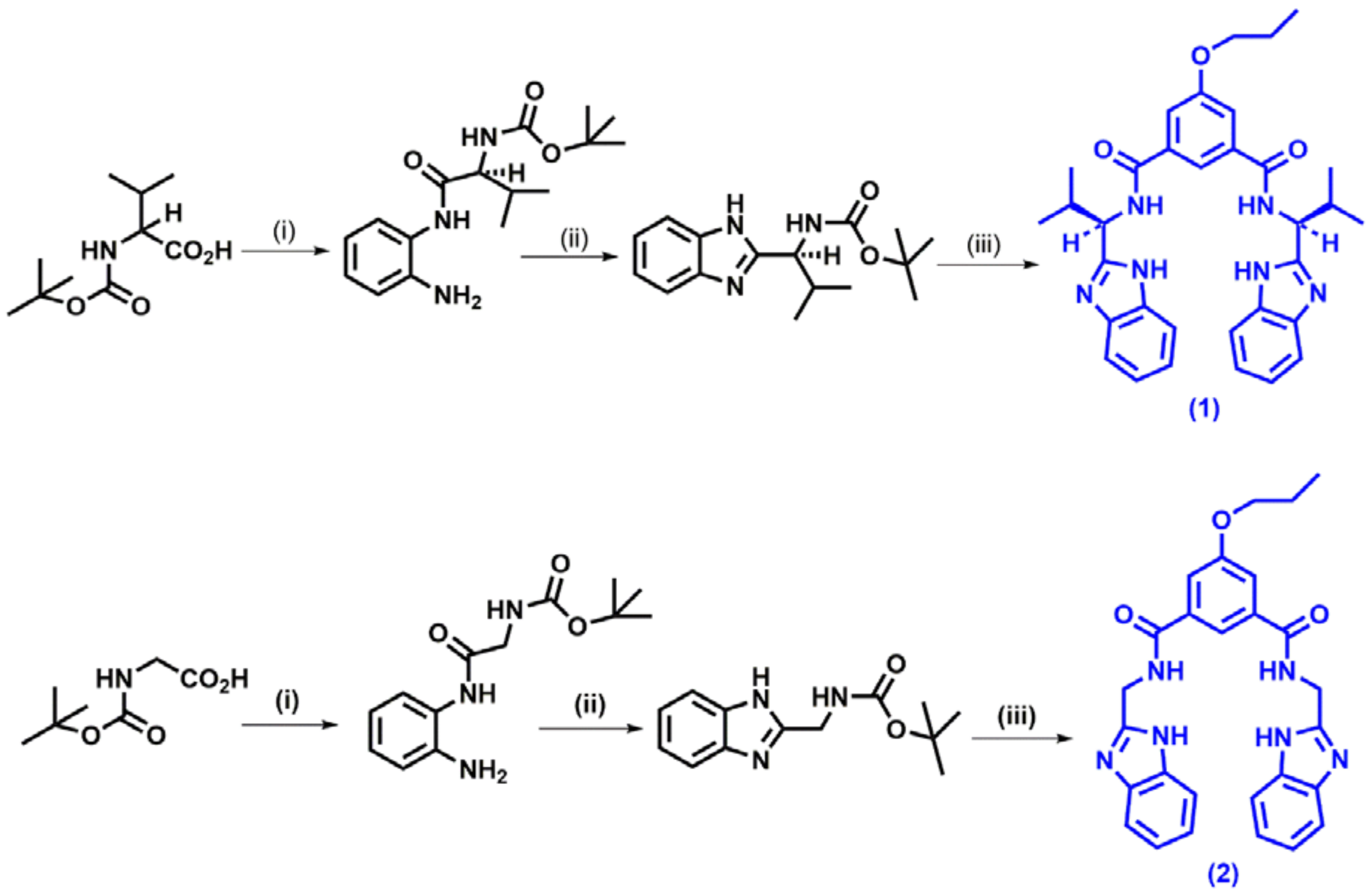
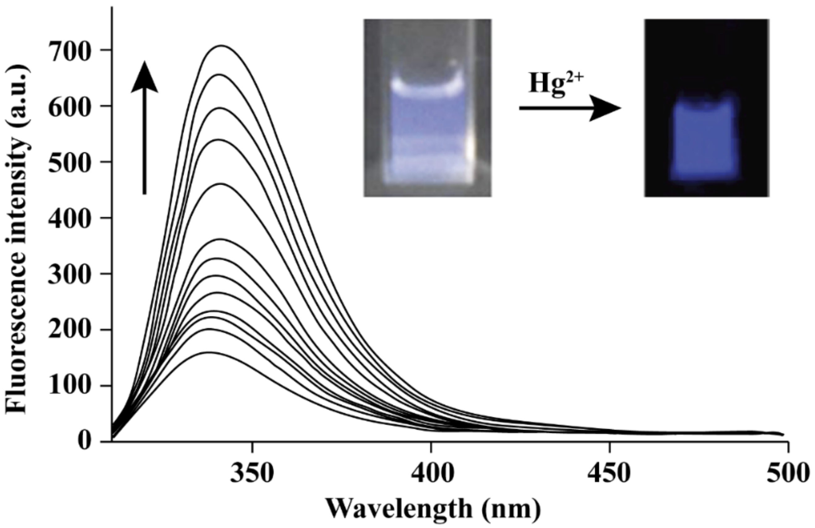
3. Hg2+ Detection via Amide Binding Site

It is well known that amide contains nitrogen and oxygen and that mercury (II) ions have a profound biding affinity toward amides. For instance, rhodamine B phenyl hydrazide selectively recognizes and coordinates with Hg2+ [36]. Similarly, in a recent report, a rhodamine-based chemosensor was synthesized to detect mercury ions in zebrafish tissue and organs [37]. This chemosensor responded rapidly and readily to mercury ions in aqueous media at room temperature. Real time monitoring showed that the mercury ion uptake by cells reaches the saturation limit in roughly 20–30 min. They uncovered a rhodamine-based chemosensor, which unlike any other, could react irreversibly with mercury ions (Figure 14).

4. Hg2+ Detection through Sulfonamide Binding Site

A new pyrenyl-appended triazole system (Figure 15) for fluorescent recognition in acetonitrile aqueous solution was reported [38]. It was also a selective and sensitive chemosensor. In the presence of mercury ion in the environment and in acetonitrile aqueous solution, the fluorescence intensity of the complex decreased by 80%. Complex formation with mercury (II) salt was observed with a distinctive fluorescent selectivity for Hg2+ ion.

5. Schiff Base Ligands in Hg2+ Ion Detection

A double naphthalene-based Schiff base with mercury detecting properties was recently reported [39]. Authors developed a fluorescent chemosensor that was cheap, sensitive, and highly selective to mercury ions. The imine present in the ligand could be oxidized to an amide with DMSO, and this could confer the coordination capacity required for coordination with Hg2+. Later, spectroscopic analysis was performed in order to study the sensitivity and selectivity of the sensor. The sensor was synthesized by a simple and low-cost Schiff base reaction of α-napthaldehyde and α-napthylamine, with a catalytic amount of acetic acid in hot absolute ethanol for 4 h. The proposed mechanism is shown in Figure 16.
The probable mechanism of detection can be explained as follows. The presence of DMSO and Hg2+ led to the formation of an O-Hg2+-N bond, which eventually was converted into C=N of the ligand and was oxidized to O=C-NH of the sensor (G1). A distinct blue coloration was visible under 365 nm UV lamp on allowing the Schiff base to form a complex with mercury ions. The ligand was found to be highly selective due to mercury divalent ions and did not bind with any other cation, including after a 10-fold increase in cation concentration. The LOD was recorded to be 5.595 × 10⁻⁸ M, hence the sensor can serve as a fluorescent sensor for Hg2+ ion.

6. With Porphyrine Binding Group

A unveiled a novel napthalimide–porphyrin hybrid-based fluorescent probe that could detect the presence of Hg2+ ratiometrically in aqueous solution and living cells [40]. It was designed by keeping two independent Hg-sensors at their maximum excitation wavelengths. The ultimate probe hence generated has a limit of detection of 2.0 × 10⁻⁸ M. Moreover, the ratiometric fluorescence change of the sensor changes significantly and is highly specific for mercury ions, even in the presence of cellular metal ions such as Na+, K+, Mg2+, or Ca2+. The results are similar in the presence of essential metal ions such as Zn2+, Fe3+, Fe2+, Cu2+, Mn2+, Co2+, and Ni2+, or heavy metal ions such as Ag+, Pb2+, Cr3+, and Cd2+, which enable the selective requirements for environmental and biomedical monitoring application. The recovery of Hg2+ from water samples collected from any random location proves the feasibility of the above-designed probe and justifies its practicality. Because of its ratiometric imaging of the Hg2+ in cells with enough resolution, it indicates its efficiency as a novel sensor. Mostly on–off probes are reported to be favorable for bioimaging applications. Most fluorescent probes based on single emission intensity changes are usually affected by a variety of factors, such as efficiency of the instrumentation, concentration of sensor molecule, stability under photo-illumination, and the microenvironment around the sensor molecule. These drawbacks can be overcome by using ratiometric probes that are not prone to such issues. Conversely, they involve observation of changes in the ratio of intensities of emission at two wavelengths when adding the target, which is beneficial for increasing the variable range and provides alarm for environmental hazards.

7. Nanostructures as Efficient Hg2+ Probes

In recent years, extending the chemosensor research to a direction with a constructive approach is setting in, particularly 1D assemblies of nanoparticles, i.e., chain of attached molecules can help understand the vivid range of phenomenon, starting from processes in living organisms’ bodies to quantum mechanism of nanometer-scale systems. The sensing mechanism of AuNPs originates from the aggregation of in the presence of Hg2+ [41,42,43,44,45,46,47,48,49].
Two main reasons that support the existence of binding mode is (i) the negative superficial charge of the silver nanoparticle, and (ii) the ionic radium of this metal ion is small enough to favor the accommodation of metal ions in the inner cavity. A similar effect was also observed in the recently reported systems. The chelation interaction between Hg2+ ions and the carboxylate groups of chemosensor L located on the surface of AuNPs and AgNPs is responsible for the selective formation of chains between the NPs modulated for Hg2+ ions (Figure 17) [50,51,52,53,54,55].
A label-free colorimetric sensor for Hg2+ quantitation using gold nanostar (GNS) was recently developed. The mechanism of action is based on the formation of Au-Hg amalgamate that leads to shape-evolution of the GNS and further changes in its absorbance. Addition of ascorbic acid (AA) to GNS solution is important for quantitation of Hg2+, mainly because it can reduce Hg2+ to Hg to enhance amalgamation on the GNSs and stabilize GNSs. Herein, the change in morphology of GNS alters the longitudinal localized surface plasmonic resonance (LSPR) absorbance of the material remarkably, leading to a dynamic response for Hg2+ in the range of 1–4,000 nM with a detection limit of 0.24 nM. The study also demonstrated that increasing Hg2+ concentration causes the solution to change its color from greenish-blue to purple to red, which can be visualized by the naked eye when the Hg2+ concentration is higher than 250 nM [56]. Similarly, the Tyndall effect of gold nanoparticles (GNPs) acts as a light-scattering signaling system. The ultrasensitive detection of the GNPs was significantly targeted with a LOD of up to 0.13 nM [57]. Table 1 presents the various nanostructures that have been reported as efficient fluorescent chemosensors.

8. Conclusions

Because of the dedicated efforts by researchers over many years, multiple chemo-sensors have been developed, and a pathway toward controlling the toxicity can be constructed. Most traditional chemosensors include a sulfur moiety involving mechanism that is driven by mercury’s affinity for thio-groups, hence yielding Hg-S formation, ring opening of spirocyclic systems (rhodamine and fluorescein, etc.), conversion of thiocarbonyl compounds into their carbonyl analogues, or a sequential desulfurization reaction. This thiophilic approach is not completely reliable. Hence, formation of metal complexes with fluorophores is a better approach. Furthermore, the “heavy atom” effect for Hg(II) favors an enhanced spin-orbit coupling constant (ζ) and induces a strong luminescence quenching of the bound luminophore. Moreover, the high hydration enthalpy (1824 kcal mol−1) for Hg2+ ions is another challenging fact for scientists to achieve the seemingly simple but tricky issue of Hg2+ recognition with a luminescence on or enhancement response, either in an aqueous environment or in physiological conditions. Luminescence enhancement is crucial for designing the reagent for imaging application and detection of the cellular uptake of this ion. To sum up, a brief but extensive literature survey is carried out to account on the recent developments on the design and synthesis of chemical sensors, along with the reports on colorimetric reagents that are conducive for infield sample analysis with yes–no-type binary response.

Author Contributions

Author Contributions: Conceptualization, S.P.K. and C.S.; methodology, S.P.K. and R.G.A.; software, R.G.A. and R.V.; validation, S.S.P., C.A.C. and J.F.C.; formal analysis, S.K.P. and C.S.; investigation, S.P.K.; resources, R.G.A.; data curation, R.V.; writing—original draft preparation, S.K.P. and S.K.P.; writing—review and editing, C.S. and R.G.A.; visualization, R.V.; supervision, R.G.A.; project administration, S.P.K.; funding acquisition, R.G.A. All authors have read and agreed to the published version of the manuscript.

Funding

This work was supported in part by the USDA National Institute of Food and Agriculture, Hatch/Multistate Project 1014385.

Acknowledgments

The efforts of the researchers working in the field of developing chemical sensors for mercury detection are highly appreciated. The authors would also like to thank Mal Hoover (College of Veterinary Medicine, Kansas State University, USA) for her assistance with the images. S.P.K. gratefully thanks Amrita Vishwa Vidyapeetham (Mysuru) for providing infrastructural facilities. C.S., and S.K.P. thankfully acknowledge the support and infrastructure provided by the JSS Academy of Higher Education and Research (JSSAHER, Mysuru).

Conflicts of Interest

Authors declare no conflict of interests.

References

  1. Momidia, B.K.; Tekuria, V.; Trivedi, D.R. Selective detection of mercury ions using benzothiazole based colorimetric chemosensor. Inorgan. Chem. Comm. 2016, 74, 1–5. [Google Scholar] [CrossRef]
  2. Bernard, V.; Isabelle, L. Design principles of fluorescent molecular sensors for cation recognition. Coord. Chem. Rev. 2000, 205, 3–40. [Google Scholar]
  3. Yu, Y.; Lin, L.R.; Yang, K.B.; Zhong, X.; Huang, R.B.; Zheng, L.S. A fluorescent probe for rapid detection of low concentration mercury ions and its application in biological cells. Talanta 2006, 69, 103–106. [Google Scholar] [CrossRef] [PubMed]
  4. Amin-Zaki, L.; Elhassani, S.; Majeed, M.A.; Clarkson, T.W.; Doherty, R.A.; Greenwood, M. Intra-uterine methylmercury poisoning in Iraq. Pediatrics 1974, 54, 587–595. [Google Scholar] [PubMed]
  5. Liu, D.B.; Qu, W.S.; Chen, W.W.; Zhang, W.; Wang, Z.; Jiang, X.Y. Highly sensitive, colorimetric detection of mercury(II) in aqueous media by quaternary ammonium group-capped gold nanoparticles at room temperature. Anal. Chem. 2010, 82, 9606–9610. [Google Scholar] [CrossRef]
  6. Xu, Y.; Deng, L.; Wang, H.; Ouyang, X.; Zheng, J.; Li, J.; Yang, R. Metal-induced aggregation of mononucleotides-stabilized gold nanoparticles: An efficient approach for simple and rapid colorimetric detection of Hg (II). Chem. Comm. 2011, 47, 6039–6041. [Google Scholar] [CrossRef] [PubMed]
  7. Wu, J.; Li, L.; Zhu, D.; He, P.; Fang, Y.; Cheng, G. Nanomaterial-based optical sensors for mercury ions. Anal. Chim. Acta 2011, 694, 115–119. [Google Scholar] [CrossRef] [PubMed]
  8. Jung-Duck, P.; Wei, Z. Human Exposure and Health Effects of Inorganic and Elemental Mercury. J. Prev. Med. Public Health 2012, 45, 344–352. [Google Scholar]
  9. Björkman, L.; Lundekvam, B.F.; Laegreid, T.; Bertelsen, B.I.; Morild, I.; Lilleng, P. Mercury in human brain, blood, muscle and toenails in relation to exposure: An autopsy study. Environ. Health 2007, 6, 30. [Google Scholar] [CrossRef] [Green Version]
  10. Clarkson, T.W.; Magos, L. The toxicology of mercury and its chemical compounds. Crit. Rev. Toxicol. 2006, 36, 609–662. [Google Scholar] [CrossRef]
  11. Cariccio, V.L.; Samà, A.; Bramanti, P.; Mazzon, E. Mercury and Alzheimer’s disease: A look at the links and evidence. Biol. Trace Elem. Res. 2019, 187, 341–356. [Google Scholar] [CrossRef] [PubMed]
  12. Kevin, M.R.; Ernest, M.W., Jr.; Miaozong, W.; Chris, G.; Eric, R.B. Environmental mercury and its toxic effects. J. Prev. Med. Public Health 2014, 47, 74–83. [Google Scholar]
  13. Byeong-Jin, Y.; Byoung-Gwon, K.; Man-Joong, J.; Se-Yeong, K.; Hawn-Cheol, K.; Tae-Won, J.; Hong-Jae, C.; Won-Jun, C.; Mi-Na, H.; Young-Seoub, H. Evaluation of mercury exposure level, clinical diagnosis and treatment for mercury intoxication. Ann. Occup. Environ. Med. 2016, 28, 5. [Google Scholar]
  14. Ying, G.; Zeming, S.; Zhou, L.; Peng, W.; Chengbin, Z.; Xiandeng, H. Determination and speciation of mercury in environmental and biological samples by analytical atomic spectrometry. Microchem. J. 2012, 103, 1–14. [Google Scholar]
  15. Lakshmi, N.S.; Young-Kyo, S.; Sung-Ok, B. Speciation and determination of mercury by various analytical techniques. Rev. Anal. Chem. 2013, 32, 225–245. [Google Scholar]
  16. Qisong, Z.; Jian, Z.; Hujin, Z.; Chengyun, W.; Shen, Y. A novel near-infrared chemosensor for mercury ion detection based on DA structure of triphenylamine and benzothiadiazole. Tetrahedron 2017, 73, 2824–2830. [Google Scholar]
  17. Mahnaz, D.G.; Sergei, M.; Prashant, S.; Godwin, A.A.; Emad, L.I. Dual chemosensor for the rapid detection of mercury(II) pollution and biothiols. Analyst 2019, 144, 4908–4916. [Google Scholar]
  18. Qu, W.J.; Gao, G.Y.; Shi, B.B.; Wei, T.B.; Zhang, Y.M.; Lin, Q.; Yao, H. A highly selective and sensitive fluorescent chemosensor for mercury ions based on the mechanism of supramolecular self-assembly. Sens. Actuators B Chem. 2014, 204, 368–374. [Google Scholar] [CrossRef]
  19. Jia, H.; Yang, M.; Meng, Q.; He, G.; Wang, Y.; Hu, Z.; Zhang, R.; Zhang, Z. Synthesis and Application of an Aldazine-Based Fluorescence Chemosensor for the Sequential Detection of Cu2+ and Biological Thiols in Aqueous Solution and Living Cells. Sensors 2016, 16, 79. [Google Scholar] [CrossRef] [Green Version]
  20. Kyle, P.C.; Alexandra, M.Y.; Amy, E.P. Fluorescent Sensors for Measuring Metal Ions in Living Systems. Chem. Rev. 2014, 114, 4564–4601. [Google Scholar]
  21. Bruno, B.C.; Manuel, A.; Beatriz, A.; Carmen, M.C.; Joaquim, C.G.; da Silva, E. Mercury(ii) sensing based on the quenching of fluorescence of CdS–dendrimer nanocomposites. RSC Adv. 2009, 134, 2447–2452. [Google Scholar]
  22. Prasenjit, M.; Sukdeb, S.; Priyadip, D.; Hridesh, A.; Amitava, D. An overview of the recent developments on Hg2+ recognition. RSC Adv. 2014, 4, 36140–36174. [Google Scholar]
  23. Yang, X.F.; Li, Y.; Bai, Q. A highly selective and sensitive fluorescein-based chemodosimeter for Hg2+ ions in aqueous media. Anal. Chim. Acta 2006, 584, 95–100. [Google Scholar] [CrossRef]
  24. Guiqiu, C.; Zhi, G.; Guangming, Z.; Lin, T. Fluorescent and colorimetric sensors for environmental mercury detection. Analyst 2015, 140, 5400–5443. [Google Scholar]
  25. Lin, W.Y.; Cao, X.W.; Ding, Y.D.; Yuan, L.; Yu, Q.X. A reversible fluorescent Hg2+ chemosensor based on a receptor composed of a thiol atom and an alkene moiety for living cell fluorescence imaging. Org. Biomol. Chem. 2010, 8, 3618–3620. [Google Scholar] [CrossRef]
  26. Wei, G.; Bart, J.S.; Joseph, H.R.; Robert, D.H. A fluorescent ligand rationally designed to be selective for zinc(II) over larger metal. Inorg. Chim. Acta 2005, 358, 3958–3966. [Google Scholar]
  27. Michael, E.H.; Engin, U.A.; Anthony, W.C. Chelation enhanced fluorescence detection of non-metal ions. JACS 1989, 111, 8735–8737. [Google Scholar]
  28. Engin, U.A.; Michael, E.H.; Anthony, W.C. Spectral shifts in acid-base chemistry. 1. van der Waals contributions to acceptor numbers. JACS 1990, 112, 3590–3593. [Google Scholar]
  29. Hyunjung, L.; Hee-Seung, L.; Joseph, H.R.; Robert, D.H. Mechanism of “Turn-on” Fluorescent Sensors for Mercury(II) in Solution and Its Implications for Ligand Design. Inorg. Chem. 2012, 51, 10904–10915. [Google Scholar]
  30. Andrea, B.; Vito, L. 1,10-Phenanthroline: A versatile building block for the construction of ligands for various purpose. Coord. Chem. Rev. 2010, 254, 2096–2180. [Google Scholar]
  31. Parthiban, V.; Natesan, T.; Shu-Pao, W. A rhodamine-based chemosensor with diphenylselenium for highly selective fluorescence turn-on detection of Hg2+ in vitro and in vivo. RSC Adv. 2017, 7, 21733–21739. [Google Scholar]
  32. Cesar, S.H.; Olalla, C.L.; Arnan, M.; Laura, M.L. Advanced Evanescent-Wave Optical Biosensors for the Detection of Nucleic Acids: An Analytic Perspective. Front. Chem. 2019, 7, 724. [Google Scholar]
  33. Sudibya, H.G.; He, Q.; Zhang, H.; Chen, P. Electrical de-tection of metal ions using field-effect transistors based on micropatterned reduced graphene oxide films. ACS Nano 2011, 5, 1990–1994. [Google Scholar] [CrossRef]
  34. Marimuthuu, P.; Umamahesh, B.; Baskaran, S.; Sathiyanarayanan, K.I.; Venkatachalapathy, B.; Ravichandran, C.; Karthikeyan, N.S. Development of paper-based chemosensor for the detection of mercury ions using mono- and tetra-sulfur bearing phenanthridines. New J. Chem. 2018, 42, 8530–8536. [Google Scholar]
  35. Ando, S.; Koide, K. Development and Applications of Fluorogenic Probes for Mercury(II) Based on Vinyl Ether Oxymercuration. JACS 2011, 133, 2556–2566. [Google Scholar] [CrossRef] [Green Version]
  36. Krishnendu, P.; Priyabrata, S.; Dipankar, B. Semi-quantitative colorimetric and supersensitive electrochemical sensors for mercury using rhodamine b hydrazide thio derivative. J. Mol. Liq. 2019, 276, 141–152. [Google Scholar]
  37. Sung-Kyun, K.; Young-Keun, Y.; Jinsung, T.; Injae, S. In Vivo Monitoring of Mercury Ions Using a Rhodamine-Based Molecular Probe. JACS 2006, 128, 14150–14155. [Google Scholar]
  38. Qian-Yong, C.; Yuan-Ming, H.; Hong-Ming, W.; Yu, X. A new pyrenyl-appended triazole for fluorescent recognition of Hg2+ ion in aqueous solution. Dye. Pigment. 2013, 99, 798–802. [Google Scholar]
  39. Tai-bao, W.; Guo-ying, G.; Wen-juan, Q.; Bing-bing, S.; Qi, L.; Hong, Y.; You-ming, Z. Selective fluorescent sensor for mercury(II) ion based on an easy to prepare double naphthalene Schiff base. Sens. Actuators B Chem. 2014, 199, 142–147. [Google Scholar]
  40. Chun-Yan, L.; Xiao-Bing, Z.; Li, Q.; Yan, Z.; Chun-Mei, H.; Shuang-Yan, H.; Li-Min, L.; Li-Xin, J.; Guo-Li, S.; Ru-Qin, Y. Naphthalimide−Porphyrin Hybrid Based Ratiometric Bioimaging Probe for Hg2+: Well-Resolved Emission Spectra and Unique Specificity. Anal. Chem. 2009, 81, 9993–10001. [Google Scholar]
  41. Alivisatos, P. The use of nanocrystals in biological detection. Nat. Biotech. 2004, 22, 47–52. [Google Scholar] [CrossRef] [PubMed]
  42. Xi, Z.; Lei, D.; Chong, W.; Lan, M.; Sai-Feng, X.; Zhu, T. Highly sensitive chemosensor for Cu(II) and Hg(II) based on the tripodal rhodamine receptor. Sens. Actuators B Chem. 2009, 141, 506–510. [Google Scholar]
  43. Da-Hye, K.; Junho, S.; Hyunsook, L.; Keun-Hyeung, L. Ratiometric fluorescence detection of Hg(II) in aqueous solutions at physiological pH and live cells with a chemosensor based on tyrosine. Sens. Actuators B Chem. 2014, 196, 421–428. [Google Scholar]
  44. Nuriman, B.K.; Willem, V. Selective chemosensor for Hg(II) ions based on tris[2 -(4-phenyldiazenyl)phenylaminoethoxy]cyclotriveratrylene in aqueous samples. Anal. Chim. Acta 2009, 655, 75–79. [Google Scholar] [CrossRef]
  45. Zhuo, W.; Deqing, Z.; Daoben, Z. A sensitive and selective “turn on” fluorescent chemosensor for Hg(II) ion based on a new pyrene–thymine dyad. Anal. Chim. Acta 2005, 549, 10–13. [Google Scholar]
  46. Hossein, A.Z.; Elaheh, R. A novel chemosensor based on graphitic carbon nitride quantum dots and potassium ferricyanide chemiluminescence system for Hg(II) ion detection. Sens. Actuators B Chem. 2016, 225, 258–266. [Google Scholar]
  47. Xiaohong, P.; Yujiao, W.; Xiaoliang, T.; Weisheng, L. Functionalized magnetic core–shell Fe3O4@SiO2 nanoparticles as selectivity-enhanced chemosensor for Hg(II). Dye. Pigment. 2011, 91, 26–32. [Google Scholar]
  48. Nai-bo, Z.; Jian-jun, X.; Chen-guang, X. Core–shell structured mesoporous silica nanoparticles equipped with pyrene-based chemosensor: Synthesis, characterization, and sensing activity towards Hg(II). J. Lumin. 2011, 131, 2021–2025. [Google Scholar]
  49. Venkatesan, M.; Sathiyanarayanan, K. Twisted pyrene with perfect hetero atomic cavity optical sensor for Hg22+ and Pb2+. Inorgan. Chem. Comm. 2020, 121, 108187. [Google Scholar]
  50. Nguyen, K.H.; Nguyen, C.B.; Nguyen, T.A.N.; Nguyen, T.T. Pham Cam Nam, Tran Duong, Jong Seung Kim, Duong Tuan Quang, A highly sensitive fluorescent chemosensor for simultaneous determination of Ag(I), Hg(II), and Cu(II) ions: Design, synthesis, characterization and application. Dye. Pigment. 2015, 116, 89–96. [Google Scholar]
  51. Juyoung, Y.; Norman, E.O.; David, H.V.; Wade, D.A.; Anthony, W.C. A fluorescent chemosensor signalling only Hg(II) and Cu(II) in water. Tetrahedron. Lett. 1997, 38, 3845–3848. [Google Scholar]
  52. Ali, C.; Deniz, Y.M.; Engin, U.A. Bis(2-pyridyl)-Substituted Boratriazaindacene as an NIR-Emitting Chemosensor for Hg(II). Org. Lett. 2007, 9, 607–609. [Google Scholar]
  53. Zhi-Xiang, H.; Hong-Yuan, L.; Xiao-Bing, Z.; Rong-Mei, K.; Guo-Li, S.; Ru-Qin, Y. A ratiometric chemosensor for fluorescent determination of Hg2+ based on a new porphyrin-quinoline dyad. Spectrochim. Acta Part A Mol. Biomol. Spectrosc. 2009, 72, 1084–1088. [Google Scholar]
  54. Reham, A.; Ibrahim, A.I.A.; Sabri, M.; Fahad, M.A.; Sayed, M.S. An effective optical chemosensor film for selective detection of mercury ions. J. Mol. Liq. 2021, 336, 116122. [Google Scholar]
  55. Buddhadeb, S.; Manjira, M.; Siddhartha, P.; Koushik, D.; Sushil Kumar, M.; Anisur, R.K.; Pabitra, C. A water soluble FRET-based ratiometric chemosensor for Hg(II) and S2− applicable in living cell staining. RSC Adv. 2014, 4, 14919–14927. [Google Scholar]
  56. Xu, D.; Yu, S.; Yin, Y.; Wang, S.; Lin, Q.; Yuan, Z. Sensitive Colorimetric Hg2+ Detection via Amalgamation-Mediated Shape Transition of Gold Nanostars. Front. Chem. 2018, 6, 566. [Google Scholar] [CrossRef] [PubMed]
  57. Zihao, D.; Wenying, J.; Qiaobo, Y.; Jinkun, H.; Ziheng, H.; Haiyan, F.; Yali, Y.; Jianmei, Z.; Jinfang, N.; Yun, Z. Ultrasensitive visual detection of Hg2+ ions via the Tyndall effect of gold nanoparticles. Chem. Comm. 2021, 57, 2613–2616. [Google Scholar]
  58. Tao, Y.; Lin, Y.; Huang, Z.; Ren, J.; Qu, X. Poly(acrylic acid)-templated silver nanoclusters as a platform for dual fluorometric turn-on and colorimetric detection of mercury (II) ions. Talanta 2012, 88, 290–294. [Google Scholar] [CrossRef]
  59. Wang, C.; Xu, L.; Wang, Y.; Zhang, D.; Shi, X.; Dong, F.; Yu, K.; Lin, Q.; Yang, B. Fluorescent silver nanoclusters as effective probes for highly selective detection of mercury(II) at parts-per-billion levels. Chem—Asian J. 2012, 7, 1652–1656. [Google Scholar] [CrossRef]
  60. Zhao, Q.; Chen, S.; Zhang, L.; Huang, H.; Liu, F.; Liu, X. Synthesis of biocompatible AuAgS/Ag2S nanoclusters and their applications in photocatalysis and mercury detection. J. Nanopar Res. 2014, 16, 1–10. [Google Scholar] [CrossRef]
  61. Zhang, N.; Si, Y.; Sun, Z.; Chen, L.; Li, R.; Qiao, Y.; Wang, H. Rapid, selective, and ultrasensitive fluorimetric analysis of mercury and copper levels in blood using bimetallic gold–silver nanoclusters with “silver effect”-enhanced red fluorescence. Anal. Chem. 2014, 86, 11714–11721. [Google Scholar] [CrossRef] [PubMed]
  62. Ding, S.-N.; Guo, Y.-X. One-pot synthesis of dual-emitting BSA-Pt-Au bimetallic nanoclusters for fluorescence ratiometric detection of mercury ions and cysteine. Anal. Meth. 2015, 7, 5787–5793. [Google Scholar] [CrossRef]
  63. Koneswaran, R. Narayanaswamy, Mercaptoacetic acid capped CdS quantum dots as fluorescence single shot probe for mercury(II). Sens. Actuators B Chem. 2009, 139, 91–96. [Google Scholar] [CrossRef]
  64. Xie, W.Y.; Huang, W.T.; Luo, H.Q.; Li, N.B. CTAB-capped Mn-doped ZnS quantum dots and label-free aptamer for room-temperature phosphorescence detection of mercury ions. Analyst 2012, 137, 4651–4653. [Google Scholar] [CrossRef]
Figure 1. Schematic representation depicting the mechanism of action of chemosensor detecting an analyte in the solution and giving rise to a detectable signal.
Separations 08 00192 g001
Figure 2. Types of fluorescent chemosensors based on functionality. Top panel: intrinsic; middle panel: conjugate; bottom panel: auto-assembled chemosensors. On arrow: sphere represents an “analyte”.
Separations 08 00192 g002
Figure 3. Common approaches using thio- and nitrogen-based ligands as chemosensors for Hg2+ detection. The mechanism of detection is mainly through metal complex formation.
Separations 08 00192 g003
Figure 4. Synthesis of rhodamine derivatives (1 and 2) as the sensors for the detection of Hg2+ and Cu2+. (i) o-phenylenediamine, N,N’-Dicyclohexylcarbodiimide (DCC), 4-Dimethylaminopyridine (DMAP), stirred in CH2Cl2 for 19 h; (ii) AcOH, heat; (iii) 50% trifluoroacetic acid (TFA) in CH2Cl2, stirred for 3 h. Reproduced with permission from [22] by Royal Society of Chemistry.
Separations 08 00192 g004
Figure 5. Graph manifesting the change in emission of ligand 1 upon gradual addition of Hg2+ ion at 336 nm. Inset shows the switch-on fluorescence upon interaction of chemosensor with Hg2+ ion (re-drawn from [22]).
Separations 08 00192 g005
Figure 6. Change in fluorescence ratio of ligand 1 upon addition of 15 equivalents of cations at 336 nm. This red shift was caused due to the stabilization of the fluorophore in its excited state (re-drawn from [22]).
Separations 08 00192 g006
Figure 7. Proposed sensing mechanism of Hg2+ ion through Hg-S bond formation from fluorescein-based thiosemicarbazide group (re-drawn from [22]).
Separations 08 00192 g007
Figure 8. Desulphurization of I by Hg2+ ion, leading to the highly fluorescent oxadiazole (II). Reproduced with permission from [23] by Elsevier.
Separations 08 00192 g008
Figure 9. Fluorescent chemosensor with S and alkene moiety showing turn-on response upon addition of Hg2+ ion. Reproduced with permission from [25] by Royal Society of Chemistry.
Separations 08 00192 g009
Figure 10. Possible structure of a mercury (II) complex of the ligand that leads to quenching of the fluorescence via formation of a π complex between Hg2+ and the anthracenyl fluorophore. Reproduced with permission from [26] by Elsevier.
Separations 08 00192 g010
Figure 11. Formation of stable Hg2+/ADPA complex, which helps in restoring its fluorescence. Reproduced with permission from [29] by American Chemical Society.
Separations 08 00192 g011
Figure 12. Sensing mechanism of rhodamine-based chemosensor for Hg2+ detection.
Separations 08 00192 g012
Figure 13. Non-fluorescent DOM upon reacting with a thio-based mercury compound in the presence of an oxidant becomes a highly fluorescent system. Reproduced with permission from [35] by American Chemical Society.
Separations 08 00192 g013
Figure 14. Conversion of non-fluorescent to strongly fluorescent rhodamine-based system by mercury ions. Reproduced with permission from [37] by American Chemical Society.
Separations 08 00192 g014
Figure 15. Synthetic route for obtaining a novel pyrenyl-appended triazole system for the detection of Hg2+ ions. Reproduced with permission from [38] by Elsevier.
Separations 08 00192 g015
Figure 16. Double-naphthalene Schiff base chemosensor (G) toward Hg2+ probing in DMSO solution. Reproduced with permission from [39] by Elsevier.
Separations 08 00192 g016
Figure 17. The surface coat of AuNPs and AgNPs using ligand (L) and their use in probing of Hg2+ ions.
Separations 08 00192 g017
Table 1. Characteristic features of fluorescent nanosensors for Hg(II) sensing.
NanosensorProbing MechanismLinear RangeLimit of DetectionAnalytical ApplicationReference
AgNCsHg2+-induced the changes of poly(acrylic acid)-templated the Ag NCs states.0–20 μM2 nMRiver water and tap water[58]
Au NCsThe addition of Hg2+ caused the aggregation-induced fluorescence quenching of glutathione-capped Ag NCs.0.1 nM–10 μM0.1 nMDrinking water[59]
AuAgS/Ag2S NCsHg2+ closed to the AuAgS/Ag2S NCs could form HgS resulting to the quenching of fluorescent nanoclusters.-10−13 MFish samples[60]
Au-Ag NCsThe fluorescence quenching of Au-AgNCs was resulted from the interactions between Hg2+ ions and Au of Au-AgNCs by metallophilic bonding of 5d10 centers.0.20–2500 nM0.10 nMBlood samples[61]
Pt-Au NCsThe d10-d10 metallophilic interaction between Hg2+ and Au+ induced the quenching of fluorescent BSA-Pt-Au NCs.0.5 nM–22 μM0.3 nMUrine and serum[62]
CdS QDsHg2+ could bind carboxyl and carbonyl groups on the surfaces of mercaptoacetic acid capped CdS QDs.5–400 nM4.2 nM-[63]
Mn-doped ZnS QDsThe bind between Hg2+ and thymine bases induced electron transfer quenching of the QDs.50–800 nM1.5 nMTap water[64]
Publisher’s Note: MDPI stays neutral with regard to jurisdictional claims in published maps and institutional affiliations.

