Article

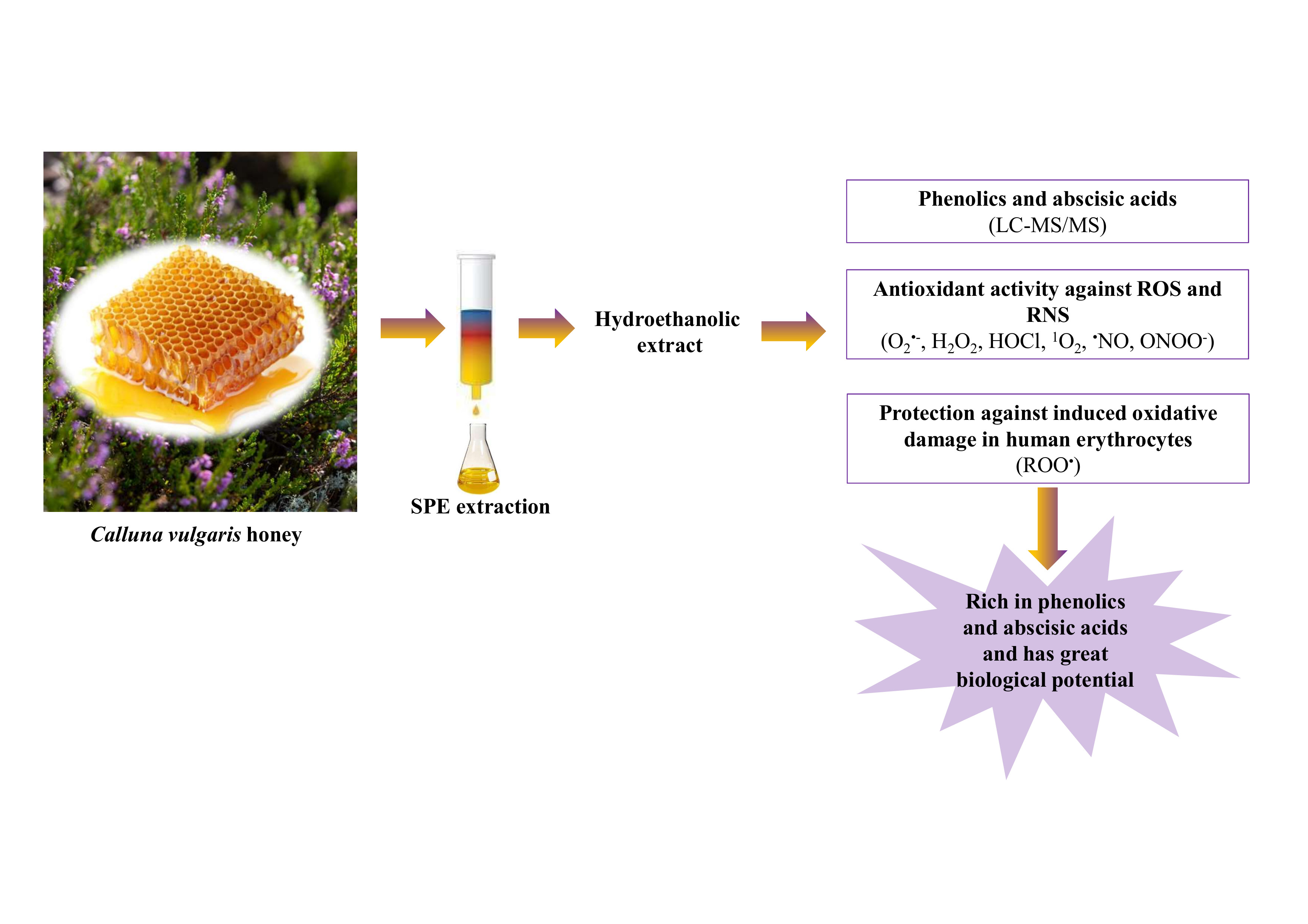
Chemical and Antioxidant Characterization of the Portuguese Heather Honey from Calluna vulgaris

1
Center for Potential and Innovation of Natural Resources, CPIRN-UDI-IPG–Research Unit for Inland Development, Polythecnic of Guarda, 6300-554 Guarda, Portugal
2
LAQV, REQUIMTE, Laboratório de Química Aplicada, Departamento de Ciências Químicas, Faculdade de Farmácia, Universidade do Porto, 4050-313 Porto, Portugal
3
Faculty of Food Engineering, Institute of Technology, Federal University of Pará (UFPA), Belém 66075-110, PA, Brazil
*
Authors to whom correspondence should be addressed.
Separations 2021, 8(10), 177; https://doi.org/10.3390/separations8100177
Submission received: 16 September 2021 / Revised: 24 September 2021 / Accepted: 3 October 2021 / Published: 8 October 2021
(This article belongs to the Section Analysis of Food and Beverages)

Abstract

:
The Calluna vulgaris honey produced in Portugal, concerning its phenolic compounds and abscisic acids profiles, as well as its antioxidant activity and the protective effect against oxidative damage in human erythrocytes were herein performed for the first time. The phenolic and abscisic acid profiles were tentatively identified by LC-MS/MS (17 compounds). The total content of phenolics and abscisic acids was 15,446.4 µg/g of honey extract, with catechin derivatives and abscisic acids being major constituents. The highest scavenging capacity was found against reactive nitrogen species. Additionally, the honey extract prevented ROO-induced oxidative damage in erythrocytes collected from human blood, by inhibiting hemolysis, lipid peroxidation and hemoglobin oxidation. In conclusion, C. vulgaris honey contains high content of catechin derivatives and abscisic acids that may be responsible for its biological activity, characterized by a strong antioxidant capacity, which adds up to the nutritional value of this delicacy.

Graphical Abstract

1. Introduction

Heather is the common designation of honeys obtained from plants that belongs to Ericaceae family. Calluna vulgaris L. Hull (known as common heather) is the only species described for the genus Calluna in the Ericaceae family [1]. Honeybees essentially collecting pollen from Calluna flowers to produce this honey, being relatively rare and very appreciated in Portugal due to its sensorial properties and potential health benefits. Several ethnobotanical uses of this plant have been reported, such as disinfectant, antiseptic, astringent, cholagogue, depurative, diuretic, diaphoretic, expectorant, sedative, antirheumatic, vasoconstrictor and used for treatment of gout [2,3].
In the last years, several works have been dedicated to identifying floral markers in heather honeys. Triterpenoids (free or aglycones of saponins) were described, with special importance for the high concentrations of oleanolic and ursolic acids, associated with their low toxicity and biological effects, such as antioxidant, anti-inflammatory and anticancer activities, among others [4,5].
Furthermore, honeys from heather are characterized by high amounts of phenolic acid derivatives and few contents of flavonoids [6]. Several phenolics were identified in heather honey reported as potential botanical markers, such as the flavonoids myricetin, myricetin 3-methyl ether, myricetin 3′-methyl ether and tricetin [7]. The floral authentication is achieved with the presence of higher levels of abscisic acid [8,9] and ellagic acid [9,10]. Phenylethyl caffeate, dimethyl allyl caffeate, (±)-abscisic, chlorogenic, syringic, p-coumaric, vanillic, 3-hydroxybenzoic, 3,4-hydroxybenzoic, p-hydroxybenzoic, 2,4-dihydroxybenzoic, caffeic, ferulic and rosmarinic and acids, quercetin, kaempferol, chrysin and galangin were reported in C. vulgaris honey from Spain and Poland [6,9]. In respect to volatile compounds, heather honeys were previously described by a relative high content of phenolics, namely guaiacol, p-cresol, propylanisole and p-anisaldehyde [11]. Additionally, isophorone and 2-methylbutyric acid were reported only in heather honeys [12], and phenylacetic acid was identified only in honey from C. vulgaris [13].
Several reports regarding the Portuguese heather honeys from Erica are available in the literature [6,14,15]. As far as we know, there are few reports about the phenolic profile, antioxidant activity against reactive oxygen species (ROS) and reactive nitrogen species (RNS), and no reports exist about the protection of erythrocytes relatively to oxidative damage of the C. vulgaris honey produced in Portugal. So, the objetives of the present work were to determine the phenolic composition of a Portuguese heather honey from C. vulgaris, by high-performance liquid chromatography coupled to diode array and mass spectrometry detectors (HPLC/DAD-MS/MS), to assess the antioxidant potential of honey extracts to scavenging ROS and RNS, such as superoxide radical (O2•−), hydrogen peroxide (H2O2), hypochlorous acid (HOCl), singlet oxygen (1O2), nitric oxide (NO) and peroxynitrite anion (ONOO). Furthermore, the capacity of the honey extract to inhibit peroxyl radical (ROO)-induced oxidative damage in human erythrocytes was also evaluated.

2. Materials and Methods

2.1. Standards and Reagents

All chemicals used were of analytical grade. 2-Amino-2-(hydroxymethyl)-1,3-propanediol (TRIS) α,α′-azodiisobutyramidine dihydrochloride (AAPH), dimethyl sulfoxide (DMSO), dihydrorhodamine 123 (DHR), 5,5′-dithiobis(2-nitrobenzoic acid) (DTNB), 30% hydrogen peroxide solution, fluorescein sodium salt, β-nicotinamide adenine dinucleotide phosphate (β-NADPH), perchloric acid (HClO4), phosphate-buffered saline (PBS), potassium bicarbonate (KHCO3), sodium hydroxide (NaOH), sodium hypochlorite solution with chlorine at 4%, tert-butyl hydroperoxide (tBHP), thiobarbituric acid (TBA), trichloroacetic acid (TCA), trypan blue, 2-vinylpyridine, and the standards syringic acid, caffeic acid, p-coumaric acid, catechin, chrysin, quercetin, (±)-abscisic acid and kaempferol were obtained from Sigma-Aldrich (St. Louis, MO, USA). Ultrapure water was obtained by a Milli-Q system (Billerica, MA, USA). The samples and solvents were filtered through Millipore membranes with a pore size of 0.22 and 0.45 μm, respectively.

2.2. Honey Sample

The honey sample (1 kg) was collected in Castelo Branco (Portugal) during November, 2014 (after flowering of C. vulgaris). It was conserved in a place without light and dry at 20 °C. The analysis were carried out in a time period less than 2 months after your collection from the hives. The sample was characterized as a honey with pH = 5.3, moisture = 24.2%, electrical conductivity = 663.1 µs.cm−1 and Brix = 75.8%. In respect to the pollen spectrum, it was determined according to a previously described technique [16]- The honey is composed by C. vulgaris (66.3%), Cytisus spp. (9.8%), Rubus spp. (5.1%), Castanea sativa (3.7%), Cistus ladanifer (3.7%), Cistacea (3.1%), Lavandula spp. (1.8%), honey elements (1.8%) and Anthemia arvenses (1.2%).

2.3. Preparation of Honey Extracts

The honey extract was prepared following a previously described procedure [17]. Sample aliquots (20 g) were dissolved in five parts of acid water (with the pH adjusted to 2 with HCl). Then, 100 mL of each extract were purified in a SPE column (Chomabond C18 SPE column 70 mL/10,000 mg, Macherey® Nagel, Duren, Germany). The column was previously conditioned with 30 mL of ethanol and then with 70 mL of acid water (pH = 2). Then, the column was washed with 10 mL of acid water, the phenolics remained adsorbed in the column, while sugars and other polar compounds were eluted with the aqueous solvent. Then, the phenolic fraction was eluted with 50 mL of ethanol. The obtained honey fraction was evaporated to dryness under low pressure (40 °C).

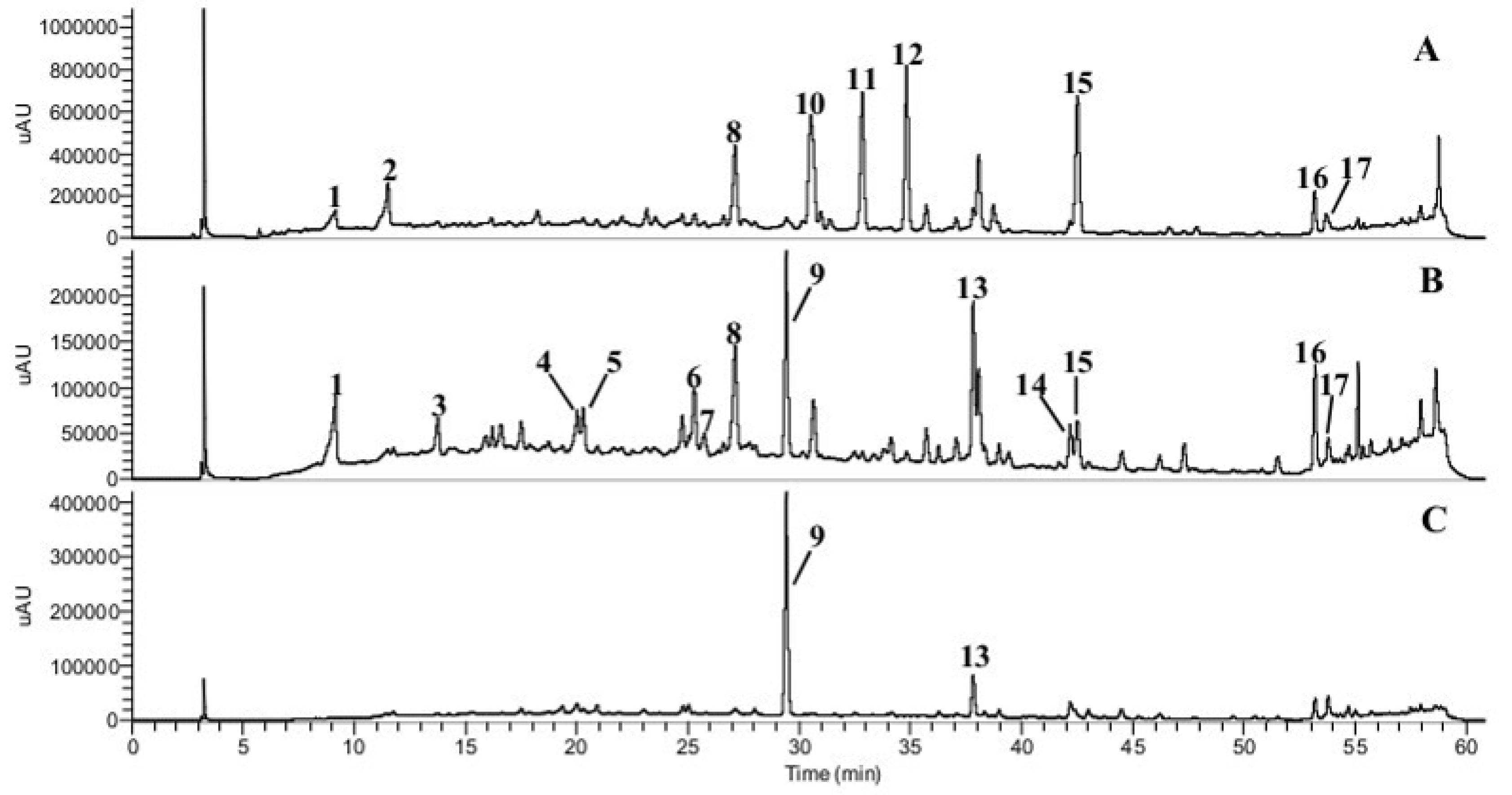
2.4. HPLC–DAD–MS/MSn Analysis of Phenolic Compounds

The phenolic profile of the honey dried extract was analysed after solubilizing 1 mg of extract in 1 mL of methanol/water (8:2, v/v) and filtered with 0.22 µm membranes. The extract was analysed in a LC (Thermo Fisher Scientific, San Jose, CA, USA) equipped with a DAD detector, quaternary pumps and an auto-sampler cooled to 5 °C. The LC was also connected in series to a LTQ Obritrap™ XL mass spectrometer (Thermo Fisher Scientific, San Jose, CA, USA) with an electrospray ionisation source (ESI) (Bruker Daltonics, model Esquire 4000, Bremen, Germany), and a hybrid system combining linear ion-trap and Orbitrap mass analysers. The identification and quantification of phenolics was carried out using a method previously described [18]. Briefly, the phenolic compounds were separated using a C18 Synergi Hydro column (4 µm, 250 × 4.6 mm, Phenomenex) heated at 29 °C, with a mobile phase in a linear gradient of water/formic acid (99.5:0.5, v/v) and acetonitrile/formic acid (99.5:0.5, v/v) using a flux of 0.9 mL/min. The mass spectra were obtained with a scan range from m/z 100 to 1000. The MS parameters used were as follows: ESI source in negative ion mode; capillary voltage at 2.5 kV and capillary temperature at 275 °C. The sheath gas and the auxiliary gas flow rate were set to 40 and 10, respectively. The phenolic compounds were tentatively identified based on the elution order, peak retention time, and UV–Vis and mass spectra features as compared to authentic standards analysed under the same conditions and data available in the literature [18,19,20]. The levels of phenolics were evaluated by HPLC-DAD (analytical curves with six-points ranging from 3.125 to 100 µg/mL, r2 > 0.9955) of syringic acid, caffeic acid, p-coumaric acid, quercetin, (±)-abscisic acid, catechin and chrysin. The results were expressed as µg/g of extract (dry matter), considering three independent extraction procedures.

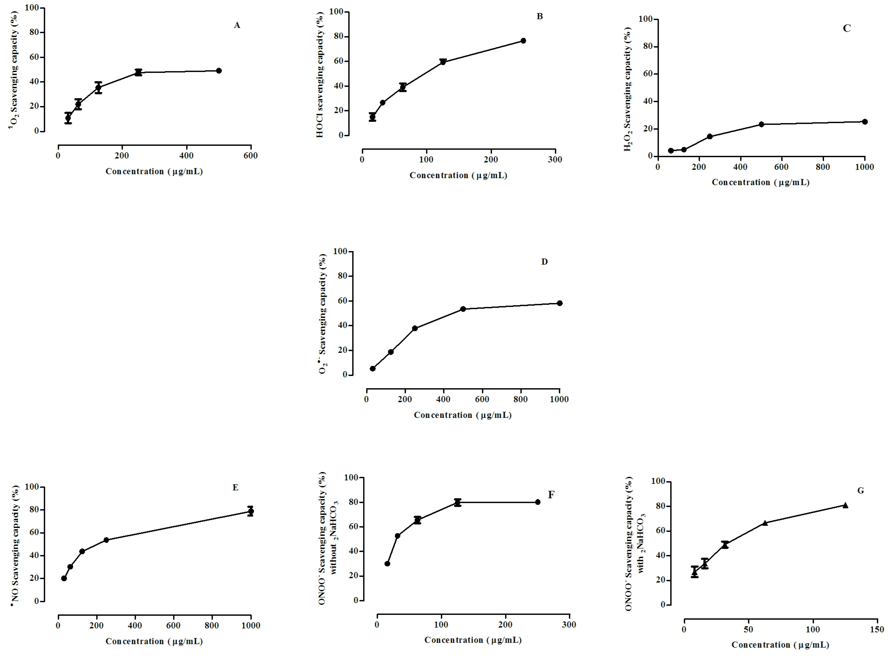
2.5. ROS- and RNS-Scavenging Assays

The scavenging assays of ROS and RNS were carried out in a microplate reader. The honey extract was dissolved in Milli-Q water. Ascorbic acid was used as positive control for O2ˉ, H2O2, HOCl, 1O2 NO, ONOOˉ scavenging assays. For each assay, four independent experiments were performed, using six different concentrations. The results were expressed as IC50 values (µg/mL) using GraphPad Prism 5.03 software (Graph Pad Software Inc., San Diego, CA, USA).

2.5.1. O2ˉ-Scavenging Assay

O2ˉ was generated by a non-enzymatic system NADH/PMS/O2. This radical produces a purple-coloured reduces by the reduction of the NBT into formazan [21], and the O2ˉ scavenging capacity was determined spectrophotometrically at 560 nm. The effects are presented as percentage inhibition of NBT reduction into formazan.

2.5.2. H2O2-Scavenging Assay

The scavenging activity of the H2O2 was determined as a methodology previously described [22]. The effect of the honey extract on the H2O2-induced oxidation of lucigenin was monitored. The luminescence increase resulting from H2O2-induced oxidation of lucigenin. The results are presented as percentage inhibition of H2O2-induced oxidation of lucigenin.

2.5.3. HOCl-Scavenging Assay

HOCl-scavenging activity was evaluated according to a previously described text [21]. The assay assessed the effect of the honey extract on HOCl-induced oxidation of DHR to rhodamine 123. The concentration of HOCl was obtained in a spectrophotometer at 235 nm [21]. The results are expressed as percentage inhibition of HOCl-induced oxidation of DHR.

2.5.4. 1O2-Scavenging Assay

1O2-Scavenging capacity was measured by evaluating the effect of the honey extract on the oxidation of the non-fluorescent DHR to the fluorescent rhodamine observed by the reaction with 1O2, generated by thermal decomposition (37 °C) of a previously synthesised water-soluble endoperoxide (disodium 3,30-(1,4-naphthalene) bispropionate, NDPO2) [23]. The results are expressed as percentage inhibition of 1O2-induced oxidation of DHR.

2.5.5. NO-Scavenging Assay

The capacity to scavenge the NO was evaluated by the monitorization of the effect of honey extract on NO-induced oxidation of DAF-2 (non-fluorescent) to rhodamine triazolofluorescein (DAF-2T) (fluorescent) [23]. NO was generated by decomposition of NOC-5. The results are presented as the percentage inhibition of NO-induced oxidation of DAF-2.

2.5.6. ONOOˉ-Scavenging Assay
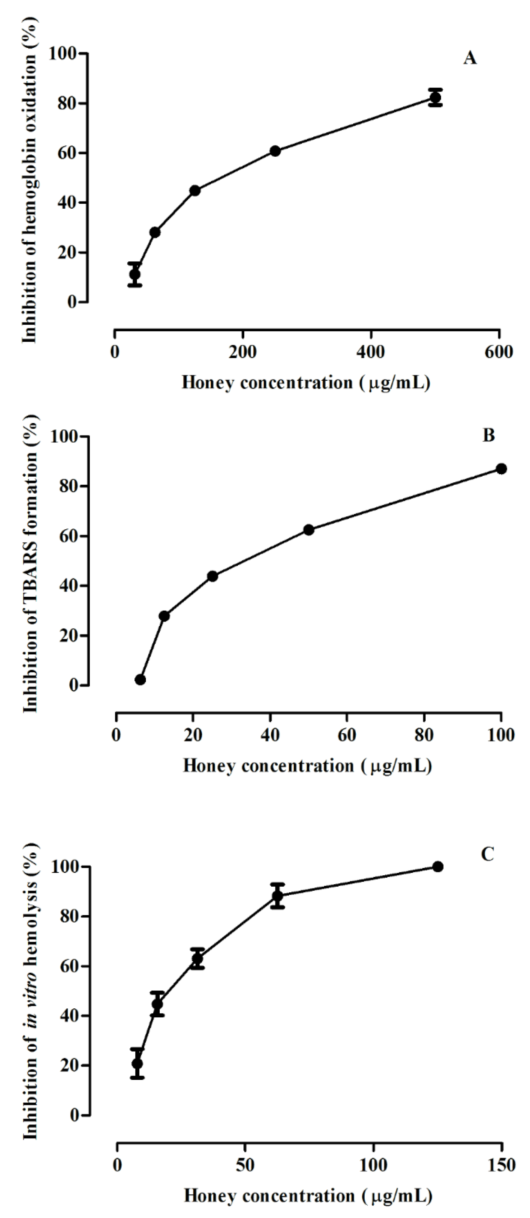
ONOOˉ-Scavenging capacity was determined by the monitorization of the effect of the honey extract on ONOOˉ-induced oxidation DHR (non-fluorescent) to rhodamine 123 (fluorescent) [23]. The results are presented as the percentage inhibition of ONOOˉ-induced oxidation of DHR.

2.6. In Vitro Evaluation of the ROO-Induced Oxidative Damage in Erythrocytes from Human Blood

Erytrocytes were isolated according to a described procedure [24], being the blood collected from healthy. The honey extract was dissolved in PBS and six different concentrations were evaluated. Each result used for calculations corresponds to four experiments (n = 4), performed in duplicate in each microplate. The results of IC50 were calculated from the curves as the percentage of inhibition of hemoglobin oxidation, lipid peroxidation and hemolysis versus concentration of the honey, using the GraphPad Prism Software.

2.6.1. Inhibition of ROO-Induced Hemoglobin Oxidation

The measurement of inhibition ROO-induced hemoglobin (Hb) oxidation was performed by the monitorization of the effects of the honey extract against the formation of methemoglobin (metHb) after the reaction of oxyhemoglobin (HbO2) with ROO generated by AAPH [24]. The honey extract was dissolved in PBS (final concentrations ranging from 200 to 0.78 μg/mL), then was mixed with the suspension of erythrocytes from human blood, and was incubated in a water bath at 37 °C during a time period of 30 min, under slow agitation (≈50 rpm). Then, AAPH (50 mM) was added to the media and incubated under slow agitation, during a period time of 4 h at 37 °C. The total volume resulting from the reaction mixture was centrifuged at 1500× g during 5 min at 4 °C. The vis-spectra of the supernatant were determined and the absorbance was evaluated at 630 nm. The spectra were measured and the oxidation products were predominantly metHb (absorption band = 630 nm). The results were expressed as IC50 values (μg/mL).

2.6.2. Inhibition of Hemolysis

The prevention of ROO-induced hemolysis of human erythrocytes was determinated by the monitorization of the release of hemoglobin after membrane disruption provoked by the hemolytic [18]. A total of six different concentrations of the honey extract, dissolved in PBS (0.78 -200μg/mL), and the suspension of human erythrocytes, were incubated at 37 °C in a water bath, for 30 min, with slow agitation (≈50 rpm). Then, the AAPH solution was added and incubated for more 3 h as described above. Then, the total volume of the reaction mixture was inserted to 1.5 mL-conic microtubes and subjected to centrifugation at 1500× g for 5 min at 4 °C. A total of 300 μL of the supernatant were inserted in a 96-well plate and the absorbance was measured at 540 nm. The results are presented as IC50 values (μg/mL).

2.6.3. Inhibition of Lipid Peroxidation

The determination of the lipid peroxidation in human erythrocytes was measured by the formation of thiobarbituric acid-reactive substances (TBARS), according a previously procedure [18]. A total of six different concentrations of honey extract were prepared with PBS (0-78-200 μg/mL, final concentration). The extract was added to the suspension of erythrocytes at 37 °C, during a period time of 30 min, with slow agitation (≈50 rpm). After incubation, tBHP at 0.2 mM was added to the media, which was then further incubated at 37 °C during 30 min. After incubation, the total volume was collected and inserted to 1.5 mL-connic eppendorfs. Then, trichloroacetic acid 28% (w/v) was added to precipitate the proteins. Then, the mixture was centrifugated (16,000× g for 10 min at 18 °C). Then, the supernatant was inserted in a 2 mL-conical test tube, followed by the addition of TBA 1% (w/v). Next, the solution was heated during a period time of 15 min at 100 °C in a water bath. After this, the test tubes were cooled at room temperature and the absorbance was determined at 532 nm. The final results were expressed as IC50 value (μg/mL).

3. Results and Discussion

3.1. Phenolic Compounds and Abscisic Acids

The phenolics and abscisic acids of C. vulgaris honey extract produced in Portugal were herein determined for the first time by HPLC-DAD/ESI-Msn analysis (Table 1). The UV-Vis and mass spectra obtained for the honey sample allowed the assignment and quantification of 17 compounds, which can be grouped into four groups: hydroxybenzoic acids (peaks 1 and 2), hydroxycinnamic acids (peaks 3 to 7), abscisic acids (peaks 10 to 12) and flavonoids (peaks 8, 9, 13 to 17). The total sum of the phenolic compounds and abscisic acids contents was 15,446.4 µg/g of honey extract (Table 1 and Figure 1).
The hydroxybenzoic acids corresponded to ca. 1.78% of the total phenolic compounds content, the benzoic acid derivative (peak 2) being the major one. Hydroxycinnamic acids represented 0.28% of the phenolic compounds quantified in the honey extract, with the hydroxycinnamic acid derivative (peak 4) found in higher amounts than the other compounds from the same class. Regarding the flavonoids, they corresponded to ca. 67% and a catechin derivative (peak 15) was the most abundant flavonoid (Table 1).
Phenolics occur in nature as sugar conjugates, mainly in the form of O-glycosides, however, in this work, the identification of sugar moiety could not be performed by the applied technique. A total of seven compounds were identified based on comparisons with UV–Vis and MS spectrum characteristics of authentic standards and by co-chromatography: caffeic acid (5), p-coumaric acid (7), (±)-abscisic acid (11), pinobanksin (14), chrysin+pinocembrin (16) and galangin (17). All these compounds were previously reported in heather honey from Erica and C. vulgaris [6,7,8,9,10], except for kaempferol-3-O-rutinoside, which was reported in several other honeys.
Peaks 1 and 2 were tentatively identified as hydroxybenzoic acid derivatives according to your UV spectra, revealing a maximum wavelength between 284 and 295 nm; and deprotonated molecule ([M-H]-) at m/z 176 and at m/z 326, respectively (Table 1). Hydroxybenzoic acids usually exhibit a neutral loss of 44 u as the main fragments in MS/MS experiments, which represents a loss from the carboxylic acid moiety of a CO2 group [19].
Five hydroxycinnamic acids and derivatives were identified: hydroxycinnamic acid derivative 1, hydroxycinnamic acid derivative 2, caffeic acid, p-coumaric acid derivative and p-coumaric acid (peaks 3–7) (Table 1). The peaks 3 and 4 were tentatively identified according to their characteristic UV spectra, showing a maximum wavelength around 325 and 309, and [M-H]- at m/z 465 and 327, respectively. Caffeic (5) and p-coumaric (7) acids produced [M-H]- at m/z 179 and at m/z 163, respectively, and the MS2 spectra showed fragment ions at m/z 135 and at m/z 119, respectively, due to loss of CO2 group from the carboxylic acid moiety ([M-H-44]-) [19]. The compound 6 was tentatively identified as p-coumaric acid derivative according to their characteristic UV and MS spectra, which showed a maximum wavelength at 307 nm, [M-H]- at m/z 379 with fragments at m/z 333 in the MS2 spectra probably due to the neutral loss of H2O (-18 u) and a CO (-28 u) moieties ([M-H-CO-H2O]-) (Table 1).
The compound 10 was tentatively identified as abscisic acid derivative and revealed [M-H]- m/z 250, the maximum wavelength being observed at 280 nm. The peaks 11 (cis-trans-abscisic acid) and 12 (trans-trans-abscisic acid) showed the same UV-Vis spectrum with a maximum wavelength obtained at 270 nm. The analysis by HPLC-DAD-ESI/MS gave the same m/z 263 for each peak. The confirmation was obtained with the injection of pure standard of cis-trans-abscisic acid. According to Tuberoso et al. [20], both the isomers should be also differentiated by their mass fragmentation, since only cis-trans-abscisic acid are supposed to form the fragment at m/z 153.
Heather honeys from Erica and C. vulgaris are characterized by high amounts of abscisic acid, and this compound was reported to be used for floral authentication [6,8,9]. Abscisic acid isomers were also reported in strawberry tree honey, but they are normally found in honeys in levels <100 µg/g [20]. Abscisic acids are norisoprenoids, being products of cyclohexene formed by the degradation of the carotenoid. The abscisic acids act as a hormone in plants, principally in higher plants, inhibiting the growth, promoting seed dormancy and their germination. Additionally, these compounds can help plants better tolerate the conditions of stressful and controlling stomal closure [8,9]. Additionally, abscisic acid is naturally found in fruits and vegetables, and it plays an important role in managing glucose homeostasis in humans [25].
Previous studies reported the presence of quercetin, kaempferol, chrysin and galangin in C. vulgaris honeys [6,9]. In the sample studied in this work, a total of seven flavonoids were tentatively identified and quantified (Figure 1 and Table 1).
In particular, it was possible to identify the propolis-derived flavonoids in their aglycones forms: pinobanksin, chrysin+pinocembrin and galangin, which were found in very small amounts (Table 1). The presence of such compounds indicates that during elaboration/ripening process, these compounds migrate from beeswax toward honey [26]. Another possibility consists in the incorporation of these phenolics from propolis into honey by bee secretions [27]. The HPLC-DAD-ESI-MS/MSn evaluation of the standards of pinobanksin (m/z 271), chrysin (m/z 253), pinocembrin (m/z 255) and galangin (m/z 269) presented the same fragmentation patterns at peaks 14, 16 and 17, respectively, and these behaviors can be also found in the literature [28].
The compound 8 was identified as an unknown flavonoid and revealed [M-H]- at m/z 267, the maximum wavelength being observed at 270 nm, and possess 249 (loss of H2O), 221 (loss of a CO group), 206 (loss of CH3) and 179 (loss of an acetyl group) as the main fragments. The compound 9 was assigned as quercetin acetyl hexoside (m/z 505), and showed the maximum wavelength at 350 nm [26,28]. Peak 13 was tentatively identified as kaempferol-3-O-rutinoside, which the major fragments obtained for kaempferol-3-O-rutinoside (m/z 593) were 447 (loss of rhamnose), 285 (loss of rutinose), 257 (loss of a CO group from m/z 285) and 229 (CO group loss from m/z 257). Finally, peak 15 was assigned as an unknown flavonoid derivative 2 (m/z 329), and possessed 311 (loss of OH group), 293 (loss of two OH groups), 229, 211 and 171 as the major fragments with a maximum wavelength at 281 nm.

3.2. Scavenging of ROS and RNS by Calluna vulgaris Honey

Some compounds originating from plants are known to exhibit antioxidant capacity. The most effective are plant phenolics, namely phenolic acids, flavonoids and tannins [29]. Honey have been described to possess several phenolic compounds associated with health benefits due to their antioxidant potential [17,26,27,30,31,32].
The antioxidant activity of the heather honey extract from C. vulgaris was tested for the first time in this work against O2•˗, HOCl, H2O2, 1O2, NO and ONOOˉ and a concentration-dependent effect was observed in all assays (Figure 2) at low µg/mL range, except for 1O2 and H2O2 (Table 2).
Regarding O2•˗, this radical reveals an important role relatively to the formation of other reactive species, such as 1O2, HO and H2O2 [30]. Despite the low reactivity of O2•˗, it can react with NO to form ONOO, another reactive species with very high reactivity that contributes to oxidative stress [31]. The results obtained with C. vulgaris honey extract against O2•˗ revealed a scavenging activity with an IC50 = 394 ± 3µg/mL, being more active than ascorbic acid (IC50 = 1200 µg/mL) and less active than trolox (IC50 = 20 µg/mL) [33]. Previous studies proved that the Manuka honey showed strong scavenging capacity against O2•˗, the authors suggest that this activity could be attributed to the presence of methyl syringate [34]. Moreover, Küçük et al. [33] reported the same effect with aqueous extracts of chestnut (IC50 = 5.2 mg/mL), Rhododendron (IC50 = 7.6 mg/mL) and heterofloral (IC50 = 7.2 mg/mL) honeys, and the differences observed between honey samples were justified by the position and type of the substituents of phenolic compounds, such as flavonoids and anthocyanidins.
The H2O2 has the capacity to decompose rapidly into oxygen and water, and being able to produce hydroxyl radicals (HO). These can start the peroxidation of the lipids and produces DNA damage. Furthermore, the H2O2 should be produced by the action of various oxidase enzymes, namely xanthine oxidase and monoamine oxidase. The honey extract seems to present low scavenging capacity against H2O2, since it inhibits 23.25% with 500 µg/mL of honey extract, being their activity lower than ascorbic acid (188 µg/mL) (Figure 2C, Table 2), quercetin (IC50 = 526.4 µg/mL), gallic acid (IC50 = 214.8 µg/mL).
In biological systems, HOCl is formed by the enzyme myeloperoxidase in activated neutrophils. When HOCl is in the presence of H2O2 it oxidizes chlorine ions. The honey extract revealed high antioxidant capacity against HOCl with an IC50 = 95.0 ±1.64 µg/mL (Figure 2B, Table 2). The activity of honey samples against HOCl was previously reported in Ginger, Eucaliptus, Lithci and Longan honeys (IC50 = 36.4; 24.0, 26.5 and 21.7 mg/mL expressed as honey sample, respectively) [35]. Additionally, its activity is lower than the control ascorbic acid (IC50 = 0.054 µg/mL) (Table 2).
Unlike other oxidants, 1O2 is a non-radical and is the excited state from molecular oxygen, with higher energy than the previous. In biological systems, 1O2 is a main agent produced by several different cell types by a range of peroxidase enzymes and during lipoxygenase-catalyzed reactions, especially by neutrophil granulocytes [21,31]. In our study, the C. vulgaris honey extract inhibited the oxidizing effect of 1O2 by 47.8% at an extract concentration of 250 µg/mL (Figure 2A, Table 2), 1O2 has been described as being involved in the oxidation of the cholesterol, and it is believed to have a major role in many photooxidation processes (being important in photodynamic therapy, and may react rapidly with electron-rich substances, namely phenols, unsaturated hydrocarbons, amines or sulfides, commonly located in membrane lipids, as well as with DNA and proteins [31]).
In biological tissues, NO is formed from the oxidation of L-arginine to citrulline through nitric oxide synthase. It is a very important bio-regulatory molecule, being associated with several physiological effects even when present in low concentrations, such as platelet function, control of blood pressure, neural signal transduction, antimicrobial and anticancer activity [32]. Throughout the infection and inflammation processes, high concentrations of NO may be formed, leading to several undesirable effects, such as reduction and deleterious effects on cell viability [32]. The C. vulgaris honey extract showed interesting scavenging activity against NO with an IC50 = 180.8 ± 2.53 µg/mL, being less effective than ascorbic acid (IC50 = 0.28 ± 0.02 µg/mL) (Table 2). Our results are in agreement with those obtained by Kassim et al. [36], that reported the inhibitory effects of Gelam honey and its extracts against NO and PGE in rat paws, that were induced with carrageenan in the non-immune inflammatory and nociceptive model, and lipopolysaccharide (LPS) in the immune inflammatory model. In another work performed with honey from Mauritius (Ginger, Eucalyptus, Litchi and Longan) were reported IC50 values that ranging between 2.8 to 47.3 mg/mL expressed as honey sample [35].
Recently, some reports indicates that great part of the cytotoxicity attributed to NO is due to ONOO. ONOO interacts with proteins, lipids and DNA leading to oxidative injury, inducing cells to necrosis or apoptosis [37]. The C. vulgaris honey extract revealed a high scavenging capacity against ONOO˗ with an IC50 = 29.2 ±2.10 µg/mL and 32.3 ±0.86 µg/mL in absence and in the presence of NaHCO3, respectively, however, was less active than ascorbic acid positive control (IC50 = 1.7 ±0.1 µg/mL and 2.1 ±0.4 µg/mL in absence and in the presence of NaHCO3, respectively) (Figure 2F, G, Table 2). These results were expected, since the capacity of Gelam honey to scavenge peroxynitrite in RAW 264.7 cells induced with LPS/IFN-γ in vitro was reported in a previous study [36]. Furthermore, Küçük et al. [33] also reported the scavenging capacity of aqueous extract from Chestnut (IC50 = 28.2 mg/mL), Rhododendron (IC50 = 282.6 mg/mL) and heterofloral (IC50 = 20.5 mg/mL) honeys against ONOO˗. Many phenolic compounds, such as caffeic acid, kaempferol, ferulic acid, p-coumaric, quercetin and flavan-3-ols have been shown to inhibit ONOO˗. Monohydroxylated phenolics, such as p-coumaric acid, act as ONOO˗ scavengers by nitration. Furthermore, compounds with a catechol moiety, such as caffeic acid, reduce ONOO˗ by electron donation [38]. Interestingly, all of the above-mentioned phenolic compounds were also identified in C. vulgaris honey (Table 1); thus, the mechanisms for the scavenging capacity of the honey extract include those referred above.
The major phenolic compounds in C. vulgaris honey extract were benzoic acid derivative 2, chrysin derivative, quercetin acetyl hexoside, abscisic acids, catechin derivative, chrysin+pinocembrin and galangin (Table 1). It is well known that the radical scavenging capacity correlates positively with the total phenolic content [29]. Therefore, these results demonstrated the antioxidant potential of honey, a readily available natural product, against oxidative damage induced by ROS/RNS, such as inflammatory-based diseases. Honey may contain many other flavonoids, such as chrysin, galangin, kaempferol, pinocembrin and quercetin; phenolic acids, namely caffeic, p-coumaric, ellagic, and ferulic acids; other bioactive compounds, such as ascorbic acid, catalase, tocopherols, superoxide dismutase, reduced glutathione, Maillard reaction products and peptides, where most of them have a joint synergistic antioxidant effect [29,30].

3.3. Protective Effect against ROO-Induced Oxidative Damage in Human Erythrocytes

Few studies were reported concerning the scavenging capacity of honey extracts against the oxidative damage in erythrocytes from human blood [39,40]. Hemolysis and lipid peroxidation could be inhibited by C. vulgaris honey extract in a concentration-dependent manner (Figure 3). In this work, as far as we know, this is the first report about the preventive effect of C. vulgaris honey extract against ROO-induced oxidative damage in erythrocytes from human blood.

3.3.1. Inhibition of Hemoglobin Oxidation in Human Erythrocytes

C. vulgaris honey extract prevented the oxidation of hemoglobin, in a concentration-dependent manner (Figure 3A), with an IC50 = 158.52 ± 2.31 µg/mL (Table 3). The extract was 50 times less active than quercetin (IC50 = 3.1 µg/mL) [24], evaluated at the same experimental conditions. Quercetin is a very hydrophobic molecule reported to possesses an important anti-hemolytic capacity [41], which is selectively partitioned into hydrophobic regions or modified lipid–protein components of the erythrocyte’s membrane [38].
One study reported the scavenging capacity of honey extracts (aqueous and ether fractions of multifloral honey from Italy) against oxidative damage in human erythrocytes, including hemoglobin oxidation, but H2O2 was ROS evaluated [40]. The results obtained by these authors proved that both extract fractions protect erythrocytes against lysis induced by peroxyl radicals generated by AAPH (a synthetic free radical generator) that may knock the membrane of the erythrocytes from the outside, the extent of hemolysis being proportional to their amount. Additionally, only the ether fraction proved to be active to inhibit the hemolysis, but not the formation of ferrylhemoglobin and methemoglobin from H2O2 [40].

3.3.2. Inhibition of Lipid Peroxidation in Human Erythrocytes

The C. vulgaris honey extract was efficient in inhibiting lipid peroxidation in a concentration-dependent manner with an IC50 of 35.59 ± 1.03 µg/mL (Figure 3B and Table 3). As far as we know, this is the first report about the protective effect of C. vulgaris honey extract against oxidative damage in human erythrocytes induced by lipid peroxidation.
In a previous study performed with multifloral honey from Italy, the authors reported that the ether fraction of honey prevented the lipid peroxidation induced by tBHP in entire erythrocytes and in isolated membranes [40]. This extract prevented the production of malondialdehyde. The protective action depends on the interaction between the bioactive compounds with erythrocytes cell membrane, which is probably due to the binding of the flavonoids with membrane. This action may explain the activity observed for lipid peroxidation by methanolic extract of C. vulgaris honey.
Another study performed with two monofloral honeys from Cuba (Christmas vine and Liven vine) showed that both honey extracts lead to a significant reduction in TBARS values in rat liver homogenates by 79% and 62% for Liven vine and Christmas vine, respectively [39].
Several studies reported the protective effect of several phenolic-rich extracts against the damage of lipids membrane caused by free radicals [42]. One of the positive effects associated to flavonoids are their potential to stabilize cell membranes by scavenging free radicals and by reducing lipid peroxidation [39,40,42,43]. Additionally, flavonoids could lie in their localization in the domains of the lipoproteins and cell membranes, serving as target for lipid peroxidation, explaining a protective effect between flavonoids and lipid bilayers [42].

3.3.3. Inhibition of Hemolysis in Human Erythrocytes

C. vulgaris honey extract inhibited hemolysis in a concentration-dependent manner and showed an IC50 = 19 ± 1. µg/mL (Figure 3C and Table 3), but not as active as quercetin (IC50 = 0.7 µg/mL) [26]. The decrease in the hemolysis effect suggests that the phenolic compounds (exogenous antioxidant) from C. vulgaris honey may efficiently scavenge reactive species to protect the cell against the ROO-induced hemolysis. This result and behavior are in accordance with previous ones for Linen vine extracts (Gouania polygama (Jack) Urb (IC50 = 24.68 ± 6.73 µg/mL) and Christmas vine (Turbina corymbosa (L.) Raf (IC50 = 62.56 ± 12.07 µg/mL) monofloral honeys from Cuba [39].
Several works reported the potential protective effects of phenolics against oxidative damage in human erythrocytes [39,40,41,43]. The phenolic compounds identified in C. vulgaris honey are known to enhanced erythrocytes resistance to oxidative stress, such as hydroxycinnamic acids [44], quercetin [41,42], catechins [45], among others.

4. Conclusions

Our results showed that C. vulgaris honey extract is composed by high levels of phenolics, namely hydroxybenzoic, hydroxycinnamic, abscisic acids and flavonoids. The extract was able to scavenge ROS and RNS, as well as to protect human erythrocytes against hemoglobin oxidation, lipid peroxidation and hemolysis. Summarizing, the obtained results demonstrated that C. vulgaris honey contains high contents of phenolic compounds that may be responsible for its biological activity, characterized by a strong antioxidant capacity, which adds up to the nutritional value of this delicacy.

Author Contributions

Conceptualization, L.R.d.S.; methodology, L.R.d.S. and R.C.C.; formal analysis, L.R.d.S.; investigation, L.R.d.S. and R.C.C.; resources, L.R.d.S., R.C.C. and E.F.; data curation, L.R.d.S. and R.C.C.; writing—original draft preparation, L.R.d.S.; writing—review and editing, L.R.d.S., R.C.C. and E.F.; visualization, L.R.d.S. and R.C.C.; supervision, E.F.; project administration, E.F.; funding acquisition, E.F. All authors have read and agreed to the published version of the manuscript.

Funding

This research was funded by LAQV, REQUIMTE, Faculty of Pharmacy, University of Porto, Porto, Portugal.

Institutional Review Board Statement

Not applicable.

Informed Consent Statement

Not applicable.

Data Availability Statement

All data is contained within the article.

Acknowledgments

This work received financial support from PT national funds (FCT/MCTES, Fundação para a Ciência e Tecnologia and Ministério da Ciência, Tecnologia e Ensino Superior) through the project UIDB/50006/2020 and from the European Union [FEDER funds through the Operational Competitiveness Program (COMPETE2020) POCI-01-0145-FEDER-029253—Project PTDC/MED-QUI/29253/2017]. The authors also thank to Vítor Claro, a beekeeper in Castelo Branco, for providing honey samples.

Conflicts of Interest

The authors declare no conflict of interest.

References

  1. Filip, G.A.; Postescu, I.D.; Tatomir, C.; Muresan, A.; Clichici, S. Calluna vulgaris extract modulates NF-kappaB/ERK signaling pathway and matrix metallo-proteinase expression in SKH-1 hairless mice skin exposed to ultraviolet B irradiation. J. Physiol. Pharmacol. 2002, 63, 423–432. [Google Scholar]
  2. Chie, R. Encyclopaedia of Medicinal Plants; MacDonald Press: London, UK, 1984. [Google Scholar]
  3. Mabey, R. Plants with a Purpose; Fontana Presshouse: London, UK, 1979. [Google Scholar]
  4. Liu, J. Pharmacology of oleanolic acid and ursolic acid. J. Ethnopharmacol. 1995, 49, 57–68. [Google Scholar] [CrossRef]
  5. Simon, A.; Najid, A.; Chulia, A.J.; Delage, C.; Rigaud, M. Inhibition of lipoxygenase activity and HL60 leukemic cell proliferation by ursolic acid isolated from heather flowers (Calluna vulgaris). Biochim. Biophys. Acta Lipids Lipid Metab. 1992, 1125, 68–72. [Google Scholar] [CrossRef]
  6. Andrade, P.; Ferreres, F.; Gil, M.I.; Tomás-Barberán, F.A. Determination of phenolic compounds in honeys with different floral origin by capillary zone electrophoresis. Food Chem. 1997, 60, 79–84. [Google Scholar] [CrossRef]
  7. Ferreres, F.; Andrade, P.; Tomás-Barberán, F.A. Flavonoids from Portuguese heather honey. Z Lebensm Unters Forsch 1994, 1991, 32–37. [Google Scholar] [CrossRef]
  8. Ferreres, F.; Andrade, P.; Tomás-Barberán, F.A. Natural occurrence of abscisic acid in heather honey and floral nectar. J. Agric. Food Chem. 1996, 44, 2053–2056. [Google Scholar] [CrossRef]
  9. Jasicka-Misiak, I.; Poliwoda, A.; Dereń, M.; Kafarski, P. Phenolic compounds and abscisic acid as potential markers for the floral origin of two Polish unifloral honeys. Food Chem. 2012, 131, 1149–1156. [Google Scholar] [CrossRef]
  10. Ferreres, F.; Paula, A.; Gil, M.I.; Tomás-Barberán, F.A. Floral nectar phenolic as biochemical markers of the botanic origin of heather honey. Z. Lebensm Unters Forsch 1996, 202, 40–44. [Google Scholar] [CrossRef]
  11. Castro-Vázquez, L.; Díaz-Maroto, M.C.; González-Viñas, M.A.; Pérez-Coello, M.S. Differentiation of monofloral citrus, rosemary, eucalyptus, lavender, thyme and heather honeys based on volatile composition and sensory descriptive analysis. Food Chem. 2009, 112, 1022–1030. [Google Scholar] [CrossRef]
  12. Seisonen, S.; Kivima, E.; Vene, K. Characterisation of the aroma profiles of different honeys and corresponding flowers using solid-phase microextraction and gas chromatography-mass spectrometry/olfactometry. Food Chem. 2015, 169, 34–40. [Google Scholar] [CrossRef]
  13. Guyot, C.; Scheirman, V.; Collin, S. Floral origin markers of heather honeys: Calluna vulgaris and Erica arborea. Food Chem. 1999, 64, 3–11. [Google Scholar] [CrossRef]
  14. Andrade, P.B.; Amaral, M.T.; Isabel, P.; Carvalho, J.C.M.F.; Seabra, R.M.; Proença da Cunha, A. Physicochemical attributes and pollen spectrum of Portuguese heather honeys. Food Chem. 1999, 66, 503–510. [Google Scholar] [CrossRef] [Green Version]
  15. Martins, R.C.; Lopes, V.V.; Valentão, P.; Carvalho, J.C.; Isabel, P.; Amaral, M.T.; Batista, M.T.; Andrade, P.B.; Silva, B.M. Relevant principal component analysis applied to the characterisation of Portuguese heather honey. Nat. Prod. Res. 2008, 22, 1560–1582. [Google Scholar] [CrossRef] [PubMed]
  16. Lutier, P.M.; Vaissière, B.E. An improved method for pollen analysis of honey. Rev. Palaeobot. Palynol. 1993, 78, 129–144. [Google Scholar] [CrossRef]
  17. Escuredo, O.; Silva, L.R.; Valentão, P.; Seijo, M.C.; Andrade, P.B. Assessing Rubus honey value: Pollen and phenolic compounds content and antibacterial capacity. Food Chem. 2012, 130, 671–678. [Google Scholar] [CrossRef]
  18. Chisté, R.C.; Mercadante, A.Z. Carotenoids are effective inhibitors of in vitro hemolysis of human erythrocytes, as determined by a practical and optimized cellular antioxidant assay. J. Agric. Food Chem. 2012, 60, 5884–5892. [Google Scholar] [CrossRef]
  19. Sun, J.; Liang, F.; Bin, Y.; Li, P.; Duan, C. Screening non-colored phenolics in red wines using liquid chromatography/ultraviolet and mass spectrometry/mass spectrometry libraries. Molecules 2007, 12, 679. [Google Scholar] [CrossRef] [Green Version]
  20. Tuberoso, C.I.G.; Bifulco, E.; Caboni, P.; Cottiglia, F.; Cabras, P.; Floris, I. Floral markers of strawberry tree (Arbutus unedo L.) honey. J. Agric. Food Chem. 2010, 58, 384–389. [Google Scholar] [CrossRef]
  21. Gomes, A.; Fernandes, E.; Silva, A.M.S.; Santos, C.M.M.; Pinto, D.C.G.A.; Cavaleiro, J.A.S.; Lima, J.L.F.C. 2-Styrylchromones: Novel strong scavengers of reactive oxygen and nitrogen species. Bioorg. Med. Chem. 2007, 15, 6027–6036. [Google Scholar] [CrossRef] [PubMed]
  22. Costa, D.; Gomes, A.; Reis, S.; Lima, J.L.; Fernandes, E. Hydrogen peroxide scavenging activity by non-steroidal anti-inflammatory drugs. Life Sci. 2005, 76, 2841–2848. [Google Scholar] [CrossRef] [PubMed]
  23. Costa, D.; Fernandes, E.; Santos, J.L.M.; Pinto, D.C.G.A.; Silva, A.M.S.; Lima, J.L.F.C. New noncellular fluorescence microplate screening assay for scavenging activity against singlet oxygen. Anal. Bioanal. Chem. 2007, 387, 2071–2081. [Google Scholar] [CrossRef] [PubMed]
  24. Chisté, R.C.; Freitas, M.; Mercadante, A.Z.; Fernandes, E. Carotenoids inhibit lipid peroxidation and hemoglobin oxidation, but not the depletion of glutathione induced by ROS in human erythrocytes. Life Sci. 2014, 99, 52–60. [Google Scholar] [CrossRef] [PubMed]
  25. Zocchi, E.; Hontecillas, R.; Leber, A.; Einerhand, A.; Carbo, A.; Bruzzone, S.; Tubau-Juni, N.; Philipson, N.; Zoccoli-Rodriguez, V.; Sturla, L.; et al. Abscisic acid: A novel nutraceutical for glycemic control. Front. Nutr. 2017, 4, 24. [Google Scholar] [CrossRef] [Green Version]
  26. Truchado, P.; Ferreres, F.; Bortolotti, L.; Sabatini, A.G.; Tomás-Barberán, F.A. Nectar flavonol rhamnosides are floral markers of Acacia (Robinia pseudacacia) honey. J. Agric. Food Chem. 2008, 56, 8815–8824. [Google Scholar] [CrossRef]
  27. Falcão, S.I.; Vilas-Boas, M.; Estevinho, L.M.; Barros, C.; Domingues, M.R.M.; Cardoso, S.M. Phenolic characterization of Northeast Portuguese propolis: Usual and unusual compounds. Anal. Bioanal. Chem. 2010, 396, 887–897. [Google Scholar] [CrossRef] [Green Version]
  28. Barros, L.; Dueñas, M.; Carvalho, A.M.; Ferreira, I.C.F.; Santos-Buelga, C. Characterization of phenolic compounds in flowers of wild medicinal plants from Northeastern Portugal. Food Chem. Toxicol. 2012, 50, 1576–1582. [Google Scholar] [CrossRef] [Green Version]
  29. Roche, A.; Ross, E.; Walsh, N.; O’Donnell, K.; Williams, A.; Klapp, M.; Fullard, N.; Edelstein, S. Representative literature on the phytonutrients category: Phenolic acids. Crit. Rev. Food Sci. Nutr. 2017, 57, 1089–1096. [Google Scholar] [CrossRef]
  30. Silva, L.R.; Valentão, P.; Andrade, P.B. Phenolic compounds in honey as health promoters. In Honey: Produxtion, Consumption and Health Benefits; Bondurand, G., Bosch, H., Eds.; Nova Science Publishers, Inc.: New York, NY, USA, 2012; pp. 81–111. [Google Scholar]
  31. Stief, T.W. Physiology and pharmacology of singlet oxygen. Med. Hypotheses 2003, 60, 567–572. [Google Scholar] [CrossRef]
  32. Halliwell, B. Antioxidants and human disease: A general introduction. Nutr. Rev. 1997, 55, S44–S49. [Google Scholar] [CrossRef]
  33. Küçük, M.; Kolaylı, S.; Karaoğlu, Ş.; Ulusoy, E.; Baltaci, C.; Candan, F. Biological activities and chemical composition of three honeys of different types from Anatolia. Food Chem. 2007, 100, 526–534. [Google Scholar] [CrossRef]
  34. Inoue, K.; Murayama, S.; Seshimo, F.; Takeba, K.; Yoshimura, Y.; Nakazawa, H.J. Identification of phenolic compound in manuka honey as specific superoxide anion radical scavenger using electron spin resonance (ESR) and liquid chromatography with coulometric array detection. Sci. Food Agric. 2005, 85, 872–878. [Google Scholar] [CrossRef]
  35. Dor, G.O.L.M.; Mahomoodally, M.F. Chemical profile and in vitro bioactivity of tropical honey from Mauritius. Asian Pac. J. Trop. Dis. 2014, 4, S1002–S1013. [Google Scholar] [CrossRef]
  36. Kassim, M.; Achoui, M.; Mansor, M.; Yusoff, K.M. The inhibitory effects of Gelam honey and its extracts on nitric oxide and prostaglandin E2 in inflammatory tissues. Fitoterapia 2010, 81, 1196–1201. [Google Scholar] [CrossRef]
  37. Pacher, P.; Beckman, J.; Liaudet, S.L. Nitric oxide and peroxynitrite in health and disease. Physiol. Rev. 2007, 87, 315–424. [Google Scholar] [CrossRef] [Green Version]
  38. Pannala, A.; Razaq, R.; Halliwell, B.; Singh, S.; Rice-Evans, C.A. Inhibition of peroxynitrite dependent tyrosine nitration by hydroxycinnamates: Nitration or electron donation? Free Radic. Biol. Med. 1998, 24, 594–606. [Google Scholar] [CrossRef]
  39. Alvarez-Suarez, J.M.; Giampieri, F.; González-Paramás, A.M.; Damiani, E.; Astolfi, P.; Martinez-Sanchez, G.; Bompadre, S.; Quiles, J.L.; Santos-Buelga, C.; Battino, M. Phenolics from monofloral honeys protect human erythrocyte membranes against oxidative damage. Food Chem. Toxicol. 2012, 50, 1508–1516. [Google Scholar] [CrossRef]
  40. Blasa, M.; Candiracci, M.; Accorsi, A.; Piacentini, M.P.; Piatti, E. Honey flavonoids as protection agents against oxidative damage to human red blood cells. Food Chem. 2007, 104, 1635–1640. [Google Scholar] [CrossRef]
  41. Ferrali, M.; Signorini, C.; Caciotti, B.; Sugherini, L.; Ciccoli, L.; Giachetti, D.; Comporti, M. Protection against oxidative damage of erythrocyte membrane by the flavonoid quercetin and its relation to iron chelating activity. FEBS Lett. 1997, 416, 123–129. [Google Scholar] [CrossRef] [Green Version]
  42. Chaudhuri, S.; Banerjee, A.; Basu, K.; Sengupta, B.; Sengupta, P.K. Interaction of flavonoids with red blood cell membrane lipids and proteins: Antioxidant and antihemolytic effects. Int. J. Biol. Macromol. 2007, 41, 42–48. [Google Scholar] [CrossRef]
  43. Mariutti, L.R.B.; Rodrigues, E.; Chisté, R.C.; Fernandes, E.; Mercadante, A.Z. The Amazonian fruit Byrsonima crassifolia effectively scavenges reactive oxygen and nitrogen species and protects human erythrocytes against oxidative damage. Food Res. Int. 2014, 64, 618–625. [Google Scholar] [CrossRef] [PubMed]
  44. Youdim, K.A.; Shukitt-Hale, B.; MacKinnon, S.; Kalt, W.; Joseph, J.A. Polyphenolics enhance red blood cell resistance to oxidative stress: In vitro and in vivo. Biochim. Biophys Acta Gen. Subj. 2000, 1523, 117–122. [Google Scholar] [CrossRef]
  45. Rizvi, S.I.; Zaid, M.A.; Anis, R.; Mishra, N. Protective role of tea catechins against oxidation-induced damage of type 2 diabetic erythrocytes. Clin. Exp. Pharmacol. Physiol. Suppl. 2005, 32, 70–75. [Google Scholar] [CrossRef] [PubMed]
Figure 1. Chromatogram of phenolic compounds from Calluna vulgaris honey extract obtained by HPLC-DAD. Peak characterization is given in Table 1. Detection at (A) 280 nm, (B) 320 nm and (C) 350 nm.
Separations 08 00177 g001
Figure 2. Scavenging capacity of Calluna vulgaris honey extract against (A) singlet oxygen (1O2), (B) hypochlorous acid (HOCl), (C) hydrogen peroxide (H2O2), (D) superoxide radical (O2•˗), (E) nitric oxide (NO) and (F,G) peroxynitrite (ONOO˗) in the absence and presence of 2NaHCO3.
Separations 08 00177 g002
Figure 3. Inhibition of (A) hemoglobin oxidation, (B) lipid peroxidation and (C) hemolysis of the erythrocytes by Calluna vulgaris honey extract.
Separations 08 00177 g003
Table 1. Phenolic compounds of Calluna vulgaris honey extract obtained from Castelo Branco, as tentatively identified by its chromatographic UV-Vis spectra and mass spectroscopy characteristics (HPLC-DAD-ESI-MSn).
PeaksCompoundsConcentration (μg/g) aHPLC-DAD-ESI-MSn Characteristics
tR bλmax[M-H] (m/z)Fragments (m/z) c
1Benzoic acid derivative 162 ± 59.1295175.9583MS2 [175]: 157, 147, 131, 115, 97, 89.
MS3 [175→131]: 119, 103, 87.
2Benzoic acid derivative 2170 ± 111.5284326.1218MS2 [326]: 308, 278, 235, 164.
MS3 [326→235]: 164.
3Hydroxycinnamic acid derivative 114 ± 213.7300(sh), 325465.1665MS2 [465]: 447, 419, 303.
MS3 [465→419]: 391, 359, 257, 221, 179, 161
4Hydroxycinnamic acid derivative 217 ± 320.0309, 342(sh)327.1117MS2 [327]:309, 165, 147
MS3 [327→147]: 147.
5Caffeic acid13 ± 220.3290, 320179.0371MS2 [179]: 161, 151, 135.
MS3 [179→135]: 135, 107, 91.
6p-Coumaric acid derivative<LOQ25.3280(sh), 307379.2022MS2 [379]: 333, 291, 179.
MS3 [379→333]: 315, 241, 161, 143, 101.
7p-Coumaric acid<LOQ25.56299, 309163.04MS2[163]: 145
MS3[163→145]: 119, 103
8Unknown flavonoid derivative 1711 ± 1927.1254, 270(sh), 310267.1267MS2 [267]: 249, 221, 199
MS3 [267→221]: 206, 179, 160.
9Quercetin acetyl hexoside632 ± 2829.4255, 350, 386505.1408MS2: nd
MS3: nd
10Abscisic acid derivative1015 ± 2830.6280(sh)249.9634MS2 [249]: 231, 205, 153.
MS3 [249→205]: 181, 155.
11trans-trans-Abscisic acid942 ± 3232.8270263.1318MS2 [263]: 245, 219, 201, 153.
MS3 [263→219]: 201, 186, 163.
265.1434MS2 [265]: 247, 229, 208, 205, 181, 151.
MS3 [265→208]: 191, 163, 149, 119, 93.
12cis-trans-Abscisic acid1011 ± 3634.8270263.1318MS2 [263]: 245, 219, 201, 153.
MS3 [263→153]: 138.
13Kaempferol-3-O-rutinoside79 ± 637.8267, 290(sh), 315, 365593.1370MS2 [593]: 447, 285, 257, 229.
MS3 [593→285]: 257, 241, 213, 151.
14Pinobanksin1.5 ± 0.142.2270, 290, 325271.0642MS2 [271]: 253, 225, 197.
MS3 [271→253]: 225, 207, 197.
15Unknown flavonoid derivative 210,370 ± 42242.5281329.2370MS2 [329]: 311, 293, 229, 211, 171.
MS3 [329→229]: 211, 165.
16Chrysin + pinocembrin?238 ± 953.2270, 283, 325253.0536
255.0690
MS2 [253]: 235, 225, 209, 181, 165, 151, 143.
MS3 [253→209]: 181, 165, 153, 139, 121
MS2 [255]: 213, 187, 169, 151, 145, 107.
MS3 [255→213]: 195, 185, 169, 145.
17Galangin171 ± 653.77225, 245, 276269.05MS2 [269]: 241, 197
MS3 [269→241]: 197, 169, 141
Total sum of identified phenolic compounds15,446.5
a Mean ± standard deviation (n = 3, dry basis). b Retention time on the C18 Synergi Hydro (4 μm) column. c In the MS2 and MS3, the most abundant ion is shown in boldface. sh = shoulder. < LOQ = lower than the limit of quantification (2.97 µg/mL); nd = not detected.
Table 2. Scavenging activities of Calluna vulgaris honey extract against singlet oxygen (1O2), hypochlorous acid (HOCl), hydrogen peroxide (H2O2), superoxide radical (O2•˗), nitric oxide (NO) and peroxynitrite (ONOO˗).
Reactive SpeciesIC50 (µg/mL)
Honey ExtractAscorbic Acid (Positive Control)
ROS
  1O248 ± 1% at 250 µg/mL1.1 ± 0.1
  HOCl95 ± 20.05 ± 0.03
  H2O223 ± 2% at 500 µg/mL188 ± 2
  O2•˗394 ± 3NA
RNS
  NO181 ± 20.28 ± 0.02
  ONOO˗29 ± 21.7 ± 0.1
  ONOO˗ *32 ± 12.1 ± 0.4
IC50 = inhibitory concentration, in vitro, to decrease in 50% the amount of reactive species in the tested media (mean ± standard error of the mean). NA = no activity was found up to the highest tested concentration (1 mg/mL). * In the presence of 25 mM NaHCO3 to simulate physiological conditions.
Table 3. Inhibition of hemoglobin oxidation, hemolysis and lipid peroxidation (TBARS formation) by Calluna vulgaris honey extract.
Erythrocytes AssayIC50 (µg/mL) a
Inhibition of hemoglobin oxidation158 ± 2
Inhibition of lipid peroxidation36 ± 1
Inhibition of hemolysis19 ± 1
a Values are expressed as mean ± standard deviation of four assays.
Publisher’s Note: MDPI stays neutral with regard to jurisdictional claims in published maps and institutional affiliations.

