Article

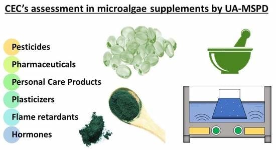
Screening of Contaminants of Emerging Concern in Microalgae Food Supplements

1
Agro-Energy Group, Department of Producción Agraria, Universidad Politécnica de Madrid, Av. Puerta de Hierro 2, 28040 Madrid, Spain
2
Departamento de Medio Ambiente y Agronomía, Instituto Nacional de Investigación y Tecnología Agraria y Alimentaria (INIA), Ctra. de la Coruña, Km. 7, 28040 Madrid, Spain
3
Teagasc Food Research Centre, Food Chemistry and Technology, Ashtown, 15 Dublin, Ireland
*
Author to whom correspondence should be addressed.
Separations 2020, 7(2), 28; https://doi.org/10.3390/separations7020028
Submission received: 20 April 2020 / Revised: 2 May 2020 / Accepted: 14 May 2020 / Published: 20 May 2020
(This article belongs to the Special Issue Analysis of Organic Micropollutants)

Abstract

:
The frenetic lifestyle in the developed countries has driven us to be deficient in some nutrients, which may be overcome by supplements. Microalgae, like spirulina (Arthrospira platensis) and chlorella (Chlorella ssp.) are widely used as supplements due to their high contents of macro- and micronutrients. Chlorella and spirulina can be grown naturally in a range of water bodies, showing their high adaptability to harsh environments. They are mainly produced in countries with poor water quality and sometimes inexistent water legislation, which can be a vector of micropollutant introduction into the food chain. Thus, a method for the simultaneous determination of 31 emerging contaminants commonly found as micropollutants in freshwater (pharmaceutical and personal care products, hormones, flame retardants and biocides) in two microalgae is presented. Target contaminants were extracted from the microalgae employing ultrasound-assisted matrix solid-phase dispersion followed by gas chromatography-mass spectrometry analysis. The method was validated for chlorella and spirulina with recoveries ranging from 70% to 111% at concentrations of 25 and 100 ng·g−1, and good linearity in the range from 5 to 400 ng·g−1 with limits of detection below 2.5 ng·g−1, in both microalgae. The method validated was applied to a range of microalgae supplement foods and the results proved that the compounds studied were below limits of detection.

Graphical Abstract

1. Introduction

The human population is increasing and by 2050 it will probably be larger by 2 to 4 billion people [1]. As the population continues to rise, the demand for high nutritive food and healthy products will increase as well. Due to the diverse nutritional components found in microalgae, its rapid growth and environmental and health benefits [2], the demand for microalgae is on rise. Microalgae have been postulated to improve the nutritional content of conventional foods, as food or dietary supplements, prebiotic agents or in therapeutic applications with a positive effect on human health. This is mainly due to the presence of compounds such as fiber, carbohydrates, lipids, unsaturated fatty acids (with double bonds in ω-3 and ω-6), vitamins, pigments, polyphenols and minerals [3,4]. Particularly, chlorella (Chlorella ssp.), a unicellular green alga found in fresh and marine water, is widely sold as a healthy food and generally recognized as safe (GRAS) by the U.S. Food and Drug Administration. Studies carried out on pure extracts or isolated molecules of chlorella (mainly in vitro) have demonstrated its potential benefits to treat and prevent many diseases due to its anti-inflammatory [5] and antimicrobial activities [6], antitumor activity [7], cholesterol-lowering properties [8], antiproliferative activity [3] or a higher antioxidant activity in comparison with other microalgae [9]. Spirulina (Arthrospira platensis) is a prokaryotic blue-green microalga (cyanobacteria) that is grown naturally in warm climates [10,11]. Spirulina has been used as food for centuries in Mexico, Chad and Myanmar [12]. It was the first cyanobacterium to be commercially cultivated and has been produced to be used as food supplements, due to the numerous potential benefits to human health, such as diabetes treatment [13], hypertension treatment [14], antiviral activity [15], anticancer properties [16] and to treat certain allergies and inflammatory processes [17].
Microalgae are mainly produced in countries where the legislation regarding water quality is very poor and contaminants may be introduced into the human food chain. On the other hand, because the price of biomass is high, ranging from 30 to 300 € kg−1 depending on the strain, the biorefinery industry of microalgae tries to minimize the inputs of the process, using wastewater as the cultivation medium [18]. However, several contaminants of emerging concern (CECs) such as pharmaceutical compounds, hormones, personal care products, biocides and flame retardants have been detected in wastewaters all over the world [19,20,21] and can be introduced into the food chain [22,23]. Moreover, the use of manure to substitute the nutrients input, such as poultry, pig and dairy manures [24,25,26,27], may be a path to introduce toxic compounds into the microalgae as CECs have been already reported in these matrices [28,29].
Since supplements containing microalgae are on the rise, the European Union found the necessity to harmonize and regulate the vitamin and mineral content of food supplements and fortified foods [30,31]. However, this directive misses targeting undesirable compounds such as CECs that may be present in the microalgae. Thus, Directive 37/2010 [32] on pharmacologically active substances in food of animal origin and SANTE/11813/2017 Guideline [33] on pesticide residues in food can be used as a reference. Hence, a methodology for the simultaneous determination of 31 CECs in chlorella and spirulina, based on a previous work where these analytes were determined in aquatic plants [34], was tested, validated and applied to assess the presence of these contaminants in commercialized microalgae food supplement products.

2. Materials and Methods

2.1. Standards and Reagents

Standards of methyl triclosan, triclosan and pyrethroids (bifenthrin, fenpropathrin, λ- cyhalothrin, permethrin, cyfluthrin, α-cypermethrin, τ-fluvalinate, esfenvalerate and deltamethrin) (all purity >99%) were supplied by Riedel-de Haën (Seelze, Germany). Tris(2-carboxyethyl)phosphine (TCEP) and tris(2-chloroisopropyl)phosphate (TCPP), were obtained from Dr. Ehrenstorfer (Augsburg, Germany). Standards of methyl and propyl paraben, ibuprofen, nonylphenol, gemfibrozil, fenoprofen, benzophenone-3 (BP3), naproxen, mefenamic acid, ketoprofen, carbamazepine, 2,2′,4,4′-tetra-bromodiphenyl ether (BDE-47), fenofibrate, bisphenol A (BPA), 2,2′,4,4′,6-penta-bromodiphenyl ether (BDE-100), hexestrol, diethylstilbestrol and estrone (all purity > 97%), were purchased from Sigma-Aldrich (St Louis, MO, USA). Individual stock solutions of each compound were made up at 50 µg mL−1 in acetonitrile (ACN) and stored in amber flasks at −18 °C. A stock mixture solution of 1 µg mL−1 containing all analytes was prepared by dilution with ACN. A working solution at 500 ng mL−1 was prepared weekly by dilution with ACN of the stock mixture solution and stored at 4 °C up to 8 weeks to ensure their stability.
Ethyl acetate (EtAc) and ACN residue analysis grade, ammonium hydroxide (NH4OH) ≥32% and Silica Bondesil-C18 (particle diameter of 40 µm) were purchased from Varian (Palo Alto, CA, USA). Florisil 150–250 µm (60–100 mesh) was supplied by Aldrich (Steinheim, Germany) and magnesium sulfate anhydrous (MgSO4) was purchased from Merck (Darmstadt, Germany). The derivatization agent N-(tert-butyldimethylsilyl)- N-methyl-trifluoroacetamide (MTBSTFA, purity ≥95%) with 1% tert-butyldimethylchlorosilane (TBDMCS) and formic acid were obtained from Sigma-Aldrich (St Louis, MO, USA).

2.2. Microalgae

Chlorella (Chlorella sorokiniana, strain 0002) was obtained from the Spanish Bank of Algae, Marine Biotechnology Center, University of Las Palmas de Gran Canaria, Spain and spirulina (Arthospira platensis) was provided by Professor Luis Mª Lubián from the Andalusian Institute of Marine Science, CSIC, Puerto Real, Cádiz, Spain. Both microalgae were cultivated aseptically in 1 L conical flasks to provide biomass to select a suitable method for the detection of CECs and validation. Both cultures were initiated with an optical density of 0.1 (measured with a spectrophotometer Pharmacia Biotech Ultrospec 2000 UV/Vis at 650 nm wavelength, pellet was centrifugated and dried during 48 h in a suitable dryer at 100 °C), corresponding to a density of 15 mg dry weight L−1. Spirulina medium was kept at pH 8 with the needed nutrients [35] whereas chlorella medium used was kept at pH 7.3 [36]. Both microalgae were kept in an environmental chamber at a temperature controlled at 25 °C, illuminated with cool white fluorescent tubes (F58W/GRO) (SYLVANIA GRO-LUX F58W/GRO 5FT T8 58W, Erlangen, Germany) with an intensity of 132 µmol s−1m−2 and a light/dark cycle of 16/8 h. The culture was aerated with filtered air (0.2 µm vent filter) at a rate of 2 L min−1 through a mechanical air pump (KNF LABOPORT Mini Diaphragm Vacuum Pump N 86 ProfiLab24, Freiburg, Germany). The cells were harvested at exponential phase, around 14 days of cultivation, with a concentration of 1004.8 mg dry weight L−1 (0.1% dry matter and 99.9% moisture content) calculated from an optical density of 0.70 for spirulina and 1392.77 mg dry weight L−1 (0.14% dry matter and 99.86% moisture content) calculated from an optical density of 0.93 for chlorella. Then cells were centrifuged at 4000 rpm (2630× g) for 15 min at 25 °C. The supernatant was discarded, and the algae pellets were collected.
Seven products acquired from shops specialized in food supplements (four containing pure spirulina and the rest pure chlorella) were used as real samples to assess the presence of contaminants of emerging concern.

2.3. Sample Preparation

2.3.1. Method Extraction Selection

Two extraction methods, one classical and the other a more novel procedure, were tested to evaluate their extraction efficiency of CECs from spirulina. The best performing method was then validated with both microalgae (spirulina and chlorella) and consequently applied to microalgae food supplements. The extraction yields obtained with both methods was evaluated spiking spirulina with a mixture of 17 representative CECs.
Spirulina material pelleted was weighed (1.0 g) and spiked with 200 µL of a working mixture solution of targeted compounds (500 ng·mL−1), reaching a final concentration of 100 ng·g−1, allowing 24 h of rest at 4 °C to reach equilibrium before extraction with methods described below.
A classic solid–liquid extraction (SLE) was performed placing the spiked 1.0 g pelletized spirulina into a 15 mL screw-cap glass tube that contained 4.0 g of Florisil and 4.0 g of MgSO4. EtAc with 3% NH4OH (10 mL) was added and stirred intensively by magnetic agitation for one hour. Then, it was centrifuged at 4000 rpm (2630× g) for 4 min. The supernatant was transferred to a graduated glass tube and evaporated to dryness using a Genevac EZ-2 evaporator (NET Interlab, S.A.L., Madrid, Spain). A second extraction cycle was performed to the remaining pellet with 10.0 mL ACN containing 4% formic acid. After centrifuging, the supernatant was transferred to the same graduated tube in which the extract of the previous extraction step was collected and dried. The extract was evaporated to a final volume of 1.0 mL.
Ultrasound assisted-matrix solid-phase dispersion (UA-MSPD) was performed as described in Figure 1 carrying out three extraction cycles. In a glass mortar, the spiked 1.0 g of spirulina pellet was mixed with 4.0 g of Florisil and 2.0 g of MgSO4 and blended with a glass pestle for 5 min to reach complete homogenization. The sample is then placed in a 20 mL glass column (10 cm × 20 mm I.D., from Becton-Dickinson, Madrid, Spain) with 2 paper filters (Whatman No. 1 paper circles of 2 cm diameter, Maidstone, UK) at the end and 2.0 g of MgSO4. In a first extraction cycle, EtAc with 3% NH4OH (8.0 mL) was added to the column and 2.0 mL were used to wash the glassware and added to the column. The column was closed with a one-way stopcock and sonicated at room temperature for 15 min in an ultrasonic water bath (Branson Ultrasonics, 40 Hz, Carouge, Switzerland). Extracts were collected in tubes using a multiport vacuum manifold (Supelco, Visiprep, Madrid, Spain) and evaporated to dryness. The second extraction cycle was carried out adding 5.0 mL ACN containing 4% formic acid to the column and sonicated for 15 min before collecting the extract in the same tube where the extract from the first extraction cycle was evaporated to dryness. The extraction was done with another 5.0 mL ACN containing 4% formic acid and sonicated 15 min. The solvent was collected in the same tube combining the extracts and evaporated to 1.0 mL (same volume as SLE to be able to compare results).

2.3.2. Clean-Up

All algae extracts (1.0 mL), using the above described extraction techniques, were purified through a 5 mL glass column (Normax, Lisbon, Portugal) with 2 paper filters (Whatman No. 1, Maidstone, UK) containing 1.0 g of MgSO4 and 1.0 g of C18. Analytes were eluted with 5.0 mL of ACN and extracts were collected in tubes using a multiport vacuum manifold, evaporated to dryness and reconstituted to 0.5 mL with ACN before their derivatization.

2.3.3. Derivatization

Prior to the gas chromatographic analysis, some of the studied analytes need to be derivatized to increase their volatility and stability. The derivation agent MTBSTFA: TBDMCS (99:1, v/v) was selected following previous experience with the target contaminants [37,38]. Thus, an aliquot (100 µL) of the microalgae extract was transferred to a 250 µL micro insert placed within a 2 mL glass vial and 50 µL of derivatization agent were added. Then, vials were closed, vortexed and the mixture left to react for 1 h at 70 °C before analysis.

2.4. Chromatographic Analysis

Gas chromatography-mass spectrometry (GC-MS) analysis was performed on an Agilent 7890A (Waldbronn, Germany) gas chromatograph coupled to a mass spectrometer (HP 5977A) equipped with an automatic injector. Separations were carried out using a ZB-5MS column, (30 m × 0.25 mm i.d. and 0.25 µm film thickness) from Phenomenex (Torrance, CA, USA). Helium (purity 99.995%) was used as carrier gas at a flow-rate of 1.2 mL/min. Solvent-vent mode operating conditions were as follows: 2 μL of plant extracts were injected in a simple-taper glass liner with glass wool. The injection port temperature was programmed to start at 50 °C (held 0.1 min) and reach 300 °C at 600 °C min−1 (held 5 min). The split vent was open for 0.1 min with an inlet pressure of 5 psi and a flow rate of 100 mL min−1 and then closed for analyte transfer into the column. After 2.6 min, the purge value was activated at a 60 mL min−1 flow rate. The column temperature was maintained at 50 °C for 2.6 min, then programmed at 20 °C min−1 to 300 °C and held for 5 min. The total analysis time was 20.1 min.
The mass spectrometric detector was operated in electron impact ionization mode with an ionizing energy of 70 eV. Ion source and transfer line temperatures were 230 and 280 °C, respectively. Retention time and mass spectra of all analytes were acquired in the full scan mode (mass range from 50 to 600 m/z). Selected Ion Monitoring (SIM) mode was employed for quantitative analysis, using one target and two qualifier ions to identify each analyte. Table 1 lists the compounds with their retention times and ions selected for the analysis. The compounds were confirmed by their retention times, the identification of target and qualifier ions and the determination of qualifier to target ratio. Retention times must be within ±0.1 min of the expected time and qualifier-to-target ratio within a 20% range for positive confirmation. The quantification was accomplished by matrix-matched calibration to overcome the matrix effect produced in GC-MS by complex matrices [39].

2.5. Method Validation

Several quality parameters were assessed through the process of method validation: recovery, precision, limits of detection (LOD), limits of quantification (LOQ), linearity and matrix effect [33]. Both microalgae (chlorella and spirulina) were spiked with the target contaminants at two levels (25 and 100 ng·g−1; n = 4), to study the extraction efficiency. The precision of the analytical procedure was evaluated as the relative standard deviation (RSD) of the recovery test. The limits of detection (LODs) and limit of quantification (LOQs) of the selected method were determined analyzing ten replicates of blank extracts of both microalgae, spiked at 5 ng·g−1. The equation to calculate the LOD was as follows (1):
LOD = t99 × SD
where t99 is the Students’ value for a 99% confidence level with n-1 degrees of freedom and SD is the standard deviation of the replicate analyses. The LOQ was calculated as 10 times the SD of the results of the replicate analysis used to determine LOD. The linearity and matrix effect were studied by analyzing two sets of seven calibration points each, one set was prepared in neat solvent (ACN) and the other was prepared spiking blank microalgae extracts (400 µL) with 100 µL of the corresponding standard solution in ACN to reach the same concentration range (5 to 400 ng·g−1).

2.6. Statistical Analysis

The overall datasets are expressed as mean values of four replicates. ANOVA test and Tukey–Kramer’s HSD test were used to determine significant differences (>95%) among treatments using the XLSTAT 2016 software (Addinsoft, Paris, France).

3. Results and Discussion

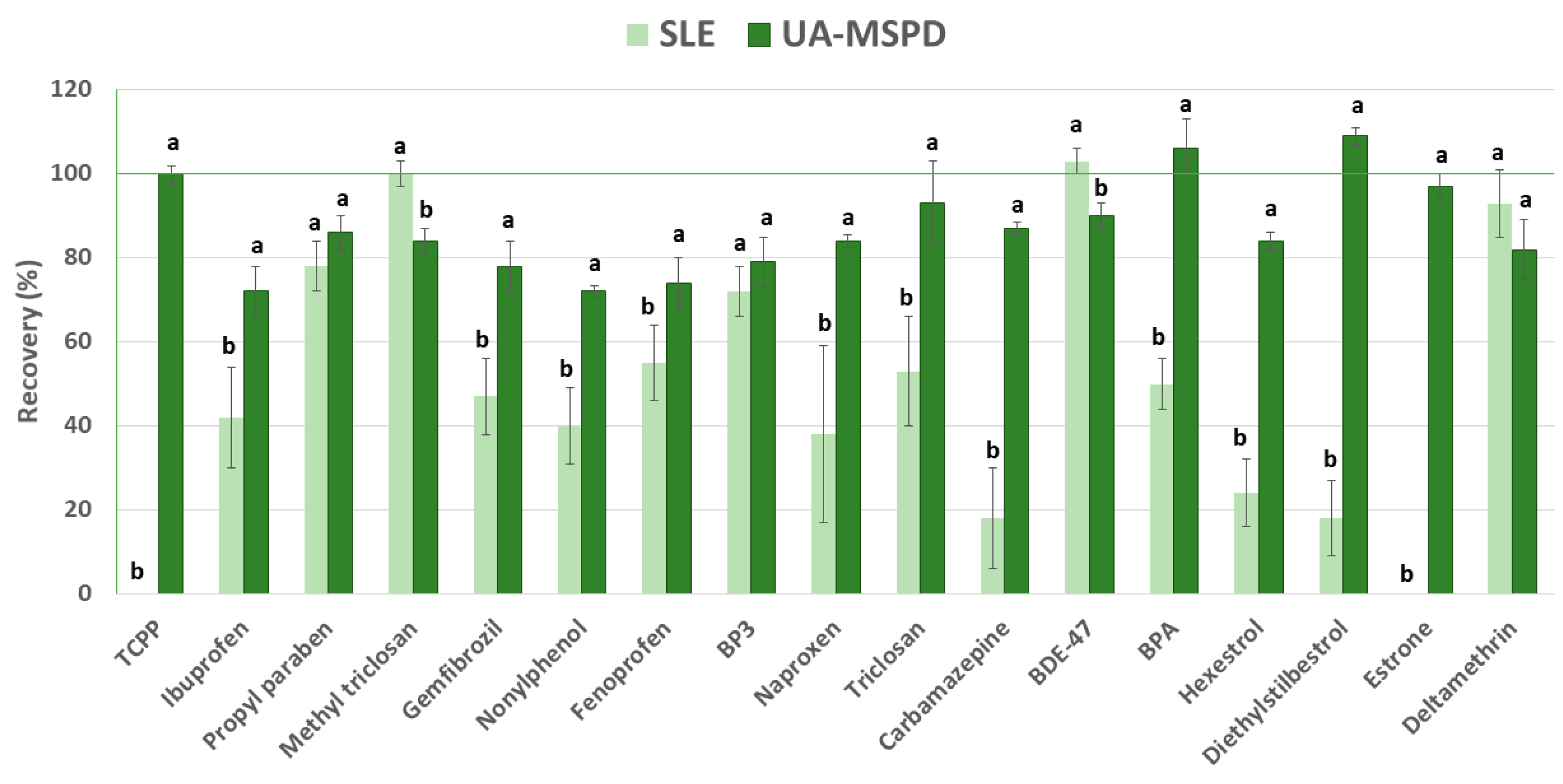
3.1. Method Selection

An UA-MSPD extraction method developed in our laboratory for the determination of CEC in four different aquatic plants was selected to assay its applicability to other aquatic organisms such as microalgae [34]. In addition, a conventional extraction procedure, SLE was tested employing the same extraction solvents, salts and sorbents as in the UA-MSPD method. In this assay, spirulina was spiked with a mixture containing 17 compounds representative of all the families of targeted compounds, reaching a final concentration of 100 ng·g−1, allowing 24 h of rest at 4 °C to reach equilibrium before performing the extraction. The results of this assay are shown in Figure 2.
Compounds such as methyl triclosan, BDE-47 and deltamethrin were easily extracted with recoveries close to 100% using SLE. However, other compounds, such as TCPP and estrone were missing or very poor recoveries were obtained for carbamazepine or diethylstilbestrol, which may be explained due to the lack of penetration into the cell as SLE is a non-disrupting cell technique.
Ultrasound-assisted extraction is a mild technique that uses the cavitation effect that produces the ultrasound bath to penetrate into the cell [40], helping the transfer of the target contaminants to the extraction solvent. In addition, the sorbent used in MSPD acts as an abrasive that promotes the disruption of the physical structure of the sample matrix facilitating the extraction of the analytes. Thus, UA-MSPD performed the best results, having recoveries that ranged from 70% to 111%. Hence, UA-MSPD, depicted in Figure 1, was selected to perform method validation with chlorella and spirulina and apply it to food supplements.

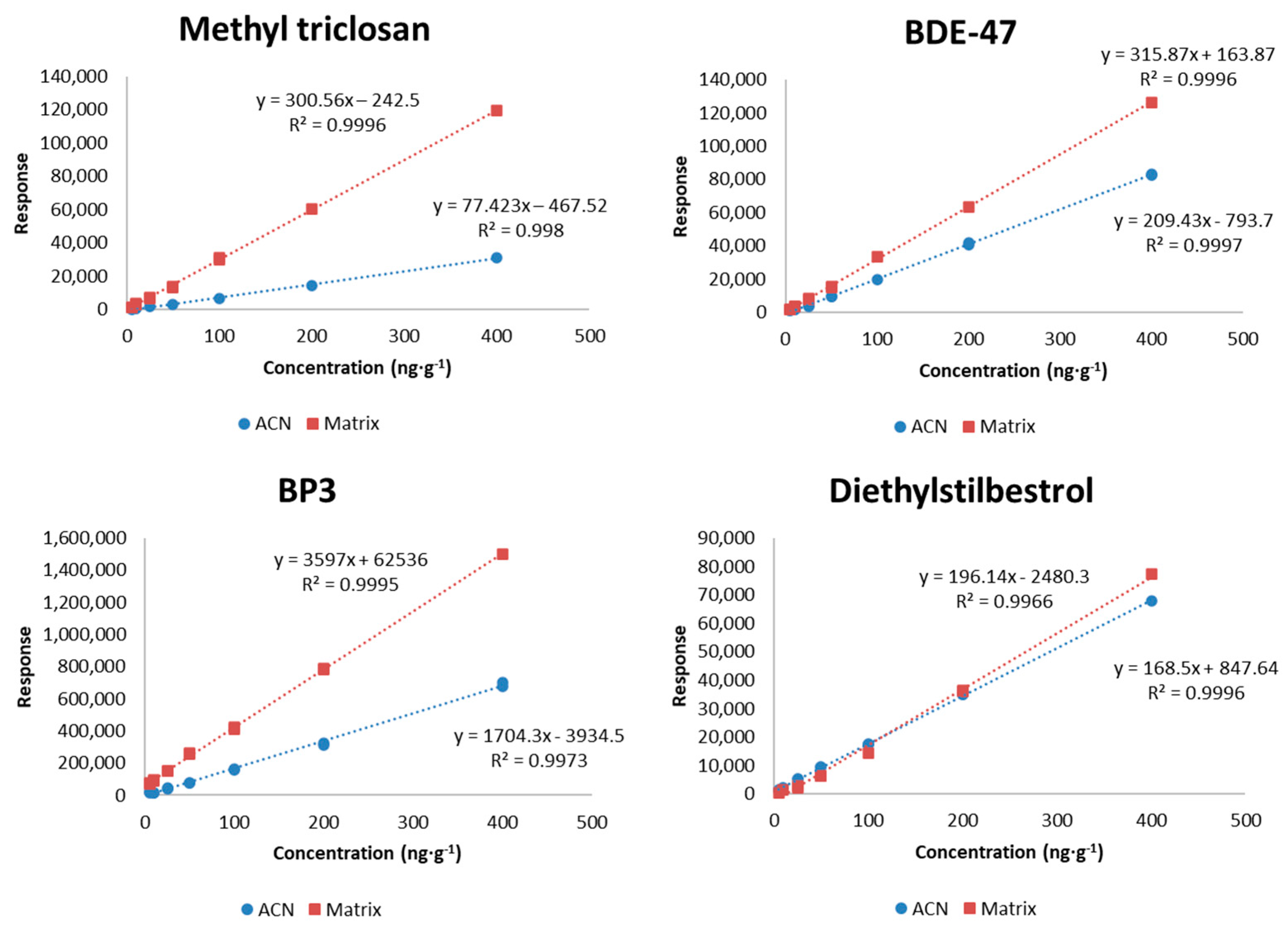
3.2. Method Validation

The method was validated using UA-MSPD extraction for each matrix (chlorella and spirulina) in terms of recovery, precision, LODs, LOQs, linearity and matrix effect.
The accuracy of the method was assessed by determining the recovery of 31 CECs from spirulina and chlorella samples spiked with standard solutions at two concentration levels (100 and 25 ng·g−1). Satisfactory recoveries were achieved ranging from 70% to 103% and from 70% to 111% for chlorella and spirulina, respectively (Table 2). The precision was determined by analyzing four spiked samples, where the RSDs were lower than 11% in both cases (Table 2).
Microalgae are a complex matrix and the different physicochemical properties of the target analytes make the development of a method for the simultaneous determination of 31 CECs difficult. To the best of our knowledge, there are no published papers determining levels of a high number of CECs in microalgae so the results included in this study could not be compared with other obtained using other methods. In a very recent work, BDE-47 was extracted from Chlorella spp. after a 24 h Sohxlet extraction, a time-consuming technique, but no information on the performance of the method was provided [41]. Research has focused mainly on the ecotoxicological effects of these compounds in algae [42] or on the application of microalgae on the bioremediation of contaminated water [43]. Regarding the uptake of CECs by microalgae, most of the studies have determined what remains in the aqueous phase rather than the amount taken up by the algae [44,45].
Low limits were obtained due to the high selectivity and sensitivity of mass spectrometry, allowing the determination of these compounds at trace levels in microalgae food supplement. As shown in Table 2, LODs and LOQs for chlorella ranged from 0.3 to 1.5 ng·g−1 and from 1.0 to 4.0 ng·g−1, respectively. Similar results were obtained for spirulina matrix, ranging from 0.3 to 2.4 ng·g−1 and from 1.2 to 5.4 ng·g−1 for LODs and LOQs, respectively.
The linearity of the method was evaluated by comparing the curves obtained by injecting standards in a neat solvent (ACN) and spiked microalgae extracts ranging from 5 to 400 ng·g−1 for all studied compounds. Good linearity was obtained for the 31 CECs studied with correlation coefficients equal to or higher than 0.992. The chromatographic response of target analytes may be affected by the presence of co-extracted components that produce an enhancement transferring analytes from the inlet to the column, having a negative impact on the correct quantification. Figure 3 shows how diethylstilbestrol does not present matrix effect as both calibration curves match perfectly (ACN and spiked matrix) on the opposite, as expected several compounds such as methyl triclosan, BDE-47 and BP3, presented an enhancement of the chromatographic response when injected in matrix extract.
Hence, to overcome matrix effect there are several approaches, but due to the high price and the nonexistence of isotope-labeled standards for all targeted contaminant studied, matrix-matched calibration was selected [37].

3.3. Food Supplements

The UA-MSPD described above was applied to different food supplements containing pure chlorella and spirulina. Samples were hydrated and centrifuged as it was described in Section 2.1 to reproduce the same conditions assayed. The levels in the food supplements analyzed were found to be below the LODs (presented in Table 2). The supplements assessed have shown no risk for human consumption however the use of regained waters or the use of manure during microalgae production will increase over the years as this industry expands so it is necessary to keep screening CECs in these food supplements.

4. Conclusions

A method, based on UA-MSPD, was successfully validated for the determination of 31 CECs in supplements of two different microalgae. The method showed satisfactory recovery results for all the studied compounds and low LODs (<2.5 ng·g−1). After the validation of the recovery, precision, LOD, LOQ, linearity and matrix effect parameters, the method was applied to commercial supplements of chlorella and spirulina, showing that all the compounds were below the LODs of the proposed method and consequently are safe to be taken. With the expanding market of microalgae, the current method could be used to assess the safety of microalgae supplements due to the increasing use of reclaimed waters and manure that can lead to the introduction of CECs into the food chain. Although this work has provided a sensitive method to detect and quantify CECs in chlorella and spirulina, more studies involving new species that will be accepted for human consumption will be necessary to ensure food safety in the near future.

Author Contributions

Conceptualization, I.M.-G., R.A. and B.A.; methodology, I.M.-G., R.A. and B.A.; validation, E.M., R.A. and B.A.; data analysis, E.M., R.A. and B.A.; writing—original draft preparation, I.M.-G. and R.A.; writing—review and editing, R.A.; B.A. and B.K.T.; project administration, R.A. All authors have read and agreed to the published version of the manuscript.

Funding

Authors wish to thank EU INTERREG Atlantic Area European programme Enhance microalgae project (EAPA_338/2016) for financial support.

Acknowledgments

Authors wish to thank Consuelo Sanchez-Brunete and José Luis Tadeo for their advice in this project.

Conflicts of Interest

The authors declare no conflict of interest.

References

  1. Cohen, J.E. Human Population: The Next Half Century. Science 2003, 302, 1172–1175. [Google Scholar] [CrossRef] [PubMed]
  2. Bishop, W.M.; Zubeck, H.M. Evaluation of Microalgae for use as Nutraceuticals and Nutritional Supplements. J. Nutr. Food Sci. 2012, 2, 2. [Google Scholar] [CrossRef] [Green Version]
  3. Wu, L.-C.; Ho, J.A.-A.; Shieh, M.-C.; Lu, I.-W. Antioxidant and antiproliferative activities of spirulina and Chlorella water extracts. J. Agric. Food Chem. 2005, 53, 4207–4212. [Google Scholar] [CrossRef] [PubMed]
  4. Gábor, V. Microalgae as the source of natural products. Orv. Hetil. 2018, 159, 703–708. [Google Scholar]
  5. Guzmán, S.; Gato, A.; Lamela, M.; Freire-Garabal, M.; Calleja, J.M. Anti-inflammatory and immunomodulatory activities of polysaccharide from Chlorella stigmatophora and Phaeodactylum tricornutum. Phytherapy Res. 2003, 17, 665–670. [Google Scholar] [CrossRef]
  6. Hasegawa, T.; Tanaka, K.; Ueno, K.; Ueno, S.; Okuda, M.; Yoshikai, Y.; Nomoto, K. Augmentation of the resistance against Escherichia coli by oral administration of a hot water extract of Chlorella vulgaris in rats. Int. J. Immunopharmacol. 1989, 11, 971–976. [Google Scholar] [CrossRef]
  7. Wang, X.; Zhang, X. Separation, antitumor activities, and encapsulation of polypeptide from Chlorella pyrenoidosa. Biotechnol. Prog. 2013, 29, 681–687. [Google Scholar] [CrossRef]
  8. Kim, S.; Kim, J.; Lim, Y.; Kim, Y.J.; Kim, J.Y.; Kwon, O. A dietary cholesterol challenge study to assess Chlorella supplementation in maintaining healthy lipid levels in adults: A double-blinded, randomized, placebo-controlled study. Nutr. J. 2016, 15, 54. [Google Scholar] [CrossRef] [Green Version]
  9. Rodríguez-García, I.; Guil-Guerrero, J.L. Evaluation of the antioxidant activity of three microalgal species for use as dietary supplements and in the preservation of foods. Food Chem. 2008, 108, 1023–1026. [Google Scholar] [CrossRef]
  10. Danxiang, H.; Yonghong, B.; Zhengyu, H. Industrial production of microalgal cell-mass and secondary products—Species of high potential:Nostoc. In Handbook of Microalgal Culture; Blackwell Publishing Ltd.: Oxford, UK, 2007; pp. 304–311. [Google Scholar]
  11. Handbook of Microalgal Culture; Richmond, A.; Hu, Q. (Eds.) John Wiley & Sons, Ltd.: Oxford, UK, 2013. [Google Scholar]
  12. Farrar, W.V. Tecuitlatl; A glimpse of Aztec food technology. Nature 1966, 211, 341–342. [Google Scholar] [CrossRef]
  13. Lee, J.; Park, A.; Kim, M.J.; Lim, H.J.; Rha, Y.A.; Kang, H.G. Spirulina extract enhanced a protective effect in type 1 diabetes by anti-apoptosis and anti-ROS production. Nutrients 2017, 9, 1363. [Google Scholar] [CrossRef] [PubMed] [Green Version]
  14. Szulinska, M.; Gibas-Dorna, M.; Miller-Kasprzak, E.; Suliburska, J.; Miczke, A.; Walczak-Gałezewska, M.; Stelmach-Mardas, M.; Walkowiak, J.; Bogdanski, P. Spirulina maxima improves insulin sensitivity, lipid profile, and total antioxidant status in obese patients with well-treated hypertension: A randomized double-blind placebo-controlled study. Eur. Rev. Med. Pharmacol. Sci. 2017, 21, 2473–2481. [Google Scholar] [PubMed]
  15. Hernández-Corona, A.; Nieves, I.; Meckes, M.; Chamorro, G.; Barron, B.L. Antiviral activity of Spirulina maxima against herpes simplex virus type 2. Antivir. Res. 2002, 56, 279–285. [Google Scholar] [CrossRef]
  16. Girardin-Andréani, C. Spiruline: Système sanguin, système immunitaire et cancer*. Phytothérapie 2005, 3, 158–161. [Google Scholar] [CrossRef]
  17. Chamorro, G.; Salazar, M.A.; Araújo, K.G.D.L.; Dos Santos, C.P.; Ceballos, G.A.; Castillo, L.H.F. Update on the pharmacology of Spirulina (Arthrospira), an unconventional food. Arch Lat. Nutr. 2002, 52, 232–240. [Google Scholar]
  18. Gao, F.; Yang, Z.H.; Li, C.; Zeng, G.M.; Ma, D.H.; Zhou, L. A novel algal biofilm membrane photobioreactor for attached microalgae growth and nutrients removal from secondary effluent. Bioresour. Technol. 2015, 179, 8–12. [Google Scholar] [CrossRef]
  19. Jasinska, E.J.; Goss, G.G.; Gillis, P.L.; Van Der Kraak, G.J.; Matsumoto, J.; de Souza Machado, A.A.; Giacomin, M.; Moon, T.W.; Massarsky, A.; Gagné, F.; et al. Assessment of biomarkers for contaminants of emerging concern on aquatic organisms downstream of a municipal wastewater discharge. Sci. Total Environ. 2015, 530–531, 140–153. [Google Scholar] [CrossRef]
  20. Barreca, S.; Busetto, M.; Colzani, L.; Clerici, L.; Marchesi, V.; Tremolada, L.; Daverio, D.; Dellavedova, P. Hyphenated high performance liquid chromatography–tandem mass spectrometry techniques for the determination of perfluorinated alkylated substances in Lombardia region in Italy, profile levels and assessment: One year of monitoring activities during 2018. Separations 2020, 7, 17. [Google Scholar] [CrossRef] [Green Version]
  21. Tran, N.H.; Reinhard, M.; Gin, K.Y.H. Occurrence and fate of emerging contaminants in municipal wastewater treatment plants from different geographical regions-a review. Water Res. 2018, 133, 182–207. [Google Scholar] [CrossRef]
  22. Hurtado, C.; Domínguez, C.; Pérez-Babace, L.; Canameras, N.; Comas, J.; Bayona, J.M. Estimate of uptake and translocation of emerging organic contaminants from irrigation water concentration in lettuce grown under controlled conditions. J. Hazard. Mater. 2016, 305, 139–148. [Google Scholar] [CrossRef]
  23. Wang, F.; Guo, X.Y.; Zhang, D.N.; Wu, Y.; Wu, T.; Chen, Z.G. Ultrasound-assisted extraction and purification of taurine from the red algae Porphyra yezoensis. Ultrason. Sonochemistry 2015, 24, 36–42. [Google Scholar] [CrossRef] [PubMed]
  24. De Godos, I.; Vargas, V.A.; Blanco, S.; González, M.C.G.; Soto, R.; García-Encina, P.A.; Becares, E.; Muñoz, R. A comparative evaluation of microalgae for the degradation of piggery wastewater under photosynthetic oxygenation. Bioresour. Technol. 2010, 101, 5150–5158. [Google Scholar] [CrossRef] [PubMed]
  25. Molinuevo-Salces, B.; García-González, M.C.; González-Fernández, C. Performance comparison of two photobioreactors configurations (open and closed to the atmosphere) treating anaerobically degraded swine slurry. Bioresour. Technol. 2010, 101, 5144–5149. [Google Scholar] [CrossRef] [PubMed]
  26. Levine, R.B.; Costanza-Robinson, M.S.; Spatafora, G.A. Neochloris oleoabundans grown on anaerobically digested dairy manure for concomitant nutrient removal and biodiesel feedstock production. Biomass Bioenergy 2011, 35, 40–49. [Google Scholar] [CrossRef]
  27. Ungsethaphand, T. Production of Spirulina platensis using dry chicken manure supplemented with urea and sodium bicarbonate. Maejo Int. J. Sci. Technol. 2009, 3, 379–387. [Google Scholar]
  28. Albero, B.; Sánchez-Brunete, C.; Miguel, E.; Aznar, R.; Tadeo, J.L. Rapid determination of natural and synthetic hormones in biosolids and poultry manure by isotope dilution GC-MS/MS. J. Sep. Sci. 2014, 37, 811–819. [Google Scholar] [CrossRef]
  29. Aznar, R.; Albero, B.; Sánchez-Brunete, C.; Miguel, E.; Tadeo, J.L. Multiresidue analysis of insecticides and other selected environmental contaminants in poultry manure by gas chromatography/mass spectrometry. J. AOAC Int. 2014, 97, 978–986. [Google Scholar] [CrossRef]
  30. Directive 2002/46/EC on the approximation of the laws of the Member States relating to food supplements. Off. J. Eur. Commun. 2002, L183/51.
  31. Flynn, A.; Hirvonen, T.; Mensink, G.B.M.; Ocké, M.C.; Serra-Majem, L.; Stos, K.; Szponar, L.; Tetens, I.; Turrini, A.; Fletcher, R.; et al. Intake of selected nutrients from foods, from fortification and from supplements in various European countries. Food Nutr. Res. 2009, 53. [Google Scholar] [CrossRef]
  32. Commision Regulation (EU) No. 37/2010 on pharmacologically active substances and their classification regarding maximum residue limits in foodstuffs of animal origin. Off. J. 2009, L15/1.
  33. SANTE/11813/2017 Guidance Document on Analytical Quality Control and Method Validation Procedures for Pesticide Residues and Analysis in Food and Feed. Available online: https://ec.europa.eu/food/sites/food/files/plant/docs/pesticides_mrl_guidelines_wrkdoc_2017-11813.pdf (accessed on 1 May 2020).
  34. Aznar, R.; Albero, B.; Sánchez-Brunete, C.; Miguel, E.; Martín-Girela, I.; Tadeo, J.L. Simultaneous determination of multiclass emerging contaminants in aquatic plants by ultrasound-assisted matrix solid-phase dispersion and GC-MS. Environ. Sci. Pollut. Res. 2017, 24, 7911–7920. [Google Scholar] [CrossRef] [PubMed] [Green Version]
  35. Guillard, R.R.L. Culture of Phytoplankton for Feeding Marine Invertebrates. In Culture of Marine Invertebrate Animals; Springer: Berlin/Heidelberg, Germany, 1975; pp. 29–60. [Google Scholar]
  36. Rodríguez-López, M. Influence of the inoculum and the medium on the growth of Chlorella pyrenoidosa. Nature 1964, 203, 666–667. [Google Scholar] [CrossRef]
  37. Aznar, R.; Sánchez-Brunete, C.; Albero, B.; Rodríguez, J.A.; Tadeo, J.L. Occurrence and analysis of selected pharmaceutical compounds in soil from Spanish agricultural fields. Environ. Sci. Pollut. Res. 2014, 21, 4772–4782. [Google Scholar] [CrossRef] [PubMed] [Green Version]
  38. Albero, B.; Sánchez-Brunete, C.; Miguel, E.; Pérez, R.A.; Tadeo, J.L. Analysis of natural-occurring and synthetic sexual hormones in sludge-amended soils by matrix solid-phase dispersion and isotope dilution gas chromatography-tandem mass spectrometry. J. Chromatogr. A 2013, 1283, 39–45. [Google Scholar] [CrossRef]
  39. Gil García, M.D.; Galera, M.M.; Uclés, S.; Lozano, A.; Fernández-Alba, A.R. Ultrasound-assisted extraction based on QuEChERS of pesticide residues in honeybees and determination by LC-MS/MS and GC-MS/MS. Anal. Bioanal. Chem. 2018, 410, 5195–5210. [Google Scholar] [CrossRef]
  40. Phong, W.N.; Show, P.L.; Ling, T.C.; Juan, J.C.; Ng, E.P.; Chang, J.S. Mild cell disruption methods for bio-functional proteins recovery from microalgae—Recent developments and future perspectives. Algal Res. 2018, 31, 506–516. [Google Scholar] [CrossRef]
  41. Lv, M.; Tang, X.; Zhao, Y.; Li, J.; Zhang, B.; Li, L.; Jiang, Y.; Zhao, Y. The toxicity, bioaccumulation and debromination of BDE-47 and BDE-209 in Chlorella sp. under multiple exposure modes. Sci. Total Environ. 2020, 723, 138086. [Google Scholar] [CrossRef]
  42. Chen, S.; Zhang, W.; Li, J.; Yuan, M.; Zhang, J.; Xu, F.; Xu, H.; Zheng, X.; Wang, L. Ecotoxicological effects of sulfonamides and fluoroquinolones and their removal by a green alga (Chlorella vulgaris) and a cyanobacterium (Chrysosporum ovalisporum). Environ. Pollut. 2020, 263, 114554. [Google Scholar] [CrossRef]
  43. García-Galán, M.J.; Arashiro, L.; Santos, L.H.M.L.M.; Insa, S.; Rodríguez-Mozaz, S.; Barceló, D.; Ferrer, I.; Garfí, M. Fate of priority pharmaceuticals and their main metabolites and transformation products in microalgae-based wastewater treatment systems. J. Hazard. Mater. 2020, 390, 121771. [Google Scholar] [CrossRef] [Green Version]
  44. Muñoz, R.; Alvarez, M.T.; Muñoz, A.; Terrazas, E.; Guieysse, B.; Mattiasson, B. Sequential removal of heavy metals ions and organic pollutants using an algal-bacterial consortium. Chemosphere 2006, 63, 903–911. [Google Scholar] [CrossRef]
  45. Matamoros, V.; Gutiérrez, R.; Ferrer, I.; García, J.; Bayona, J.M. Capability of microalgae-based wastewater treatment systems to remove emerging organic contaminants: A pilot-scale study. J. Hazard. Mater. 2015, 288, 34–42. [Google Scholar] [CrossRef] [PubMed] [Green Version]
Figure 1. Workflow of the ultrasound assisted-matrix solid-phase dispersion (UA-MSPD) method used to extract contaminants of emerging concern from microalgae.
Separations 07 00028 g001
Figure 2. Recovery in percentage of the studied compounds in spirulina spiked at 100 ng·g−1 employing two extraction procedures, solid–liquid extraction (SLE) and UA-MSPD. Different letters indicate significant differences (p ≤ 0.05).
Separations 07 00028 g002
Figure 3. Comparison of seven-point calibration curves of methyl triclosan, BDE-47, BP3 and diethylstilbestrol, obtained by injection of standards in neat solvent ACN and spiked spirulina extracts, ranging from 5 to 400 ng·g−1.
Separations 07 00028 g003
Table 1. Use, retention times and mass spectrometric parameters of the studied analytes.
NameUsetRSIM Parameters
T Q1Q2
Methyl Paraben (tBDMS)Preservative11.27209210266
TCEPPlasticizer11.3824925063
TCPPPlasticizer11.5712599277
Ibuprofen (tBDMS)NSAID11.85263264117
Propyl Paraben (tBDMS)Preservative12.15237238294
Methyl TriclosanAntifungal13.38302304252
Nonylphenol (tBDMS)Surfactant13.38334277278
Gemfibrozil (tBDMS)Lipid regulator13.45243179307
Fenoprofen (tBDMS)NSAID13.68299197206
Benzophenone-3 (BP3) (tBDMS)Sunscreen14.11285242286
Naproxen (tBDMS)NSAID14.13287185288
Triclosan (tBDMS)Antifungal14.34347345200
Mefenamic acid (tBDMS)NSAID14.64298224355
Ketoprofen (tBDMS)NSAID14.65311295105
BifenthrinPesticide14.80181165166
FenpropathrinPesticide14.87125181265
Carbamazepine (tBDMS)Antiepileptic14.95193194293
BDE-47Flame retardant15.16486326488
FenofibrateLipid regulator15.19121273139
λ-CyhalothrinPesticide15.24197181208
PermethrinPesticide15.74183163165
BPA (tBDMS)Plasticizer15.70441442456
BDE-100Flame retardant15.92404406566
CyfluthrinPesticide16.06163206226
Hexestrol (tBDMS)Hormone16.35249250337
α-CypermethrinPesticide16.41163165181
Diethylstilbestrol (tBDMS)Hormone16.50496497498
τ-FluvalinatePesticide17.09250252181
Estrone (tBDMS)Hormone17.10327384328
EsfenvaleratePesticide17.20125167181
DeltamethrinPesticide17.74181253251
tBDMS, tert-butyldimethylsilyl ethers group formed after derivatization of -OH groups; NSAID, nonsteroidal anti-inflammatory drug; tR = retention time, min; T = target ion, m/z; Q1 and Q2 = qualifier ions, m/z.
Table 2. Recoveries (%) and relative standard deviations (RSD (n = 4), % in parenthesis), limits of detection (LOD) and limits of quantification (LOQ) (n = 10) obtained for target compounds in chlorella and spirulina.
Chlorella spp.Arthrospira platensis
100 ng·g−125 ng·g−1LOD ng·g−1LOQ ng·g−1100 ng·g−125 ng·g−1LOD ng·g−1LOQ ng·g−1
Methyl Paraben70 (8)82(8)0.41.276 (5)72 (9)1.83.6
TCEP84 (4)80 (9)1.53.274 (3)94 (6)1.53.2
TCPP94 (9)81 (8)0.82.4100 (3)96 (9)0.91.7
Ibuprofen100 (3)71 (3)0.82.770 (6)72 (5)1.32.6
Propyl Paraben 89 (3)80 (6)0.31.086 (2)80 (5)1.52.9
Methyl Triclosan88 (3)74 (7)1.34.087 (4)83 (8)2.34.6
Gemfibrozil86 (6)81 (5)0.51.577 (7)79 (10)0.82.6
Nonylphenol87 (2)83 (8)0.41.570 (2)77 (5)1.32.6
Fenoprofen78 (9)71 (3)0.92.672 (8)78 (2)2.14.1
BP397 (4)98 (6)0.82.578 (7)80 (4)0.81.7
Naproxen82 (8)98 (2)1.03.186 (3)79 (5)0.81.8
Triclosan89 (4)84 (2)0.31.094 (11)80 (7)2.14.0
Mefenamic acid91 (8)94 (8)0.41.470 (9)70 (3)1.32.8
Ketoprofen73 (2)72 (2)0.31.075 (10)78 (8)0.71.9
Bifenthrin86 (3)89 (7)0.41.394 (5)82 (5)0.31.2
Fenpropathrin84 (3)89 (7)1.23.690 (6)73 (5)2.13.9
Carbamazepine90 (2)83 (2)0.51.588 (3)75 (9)2.44.9
BDE-4776 (8)84 (9)0.72.589 (3)82 (6)1.34.5
Fenofibrate83 (3)82 (8)1.02.789 (5)83 (5)1.83.9
λ-Cyhalothrin83 (11)80 (5)0.93.085 (6)90 (11)1.93.9
Permethrin83 (2)83 (8)1.13.691 (5)79 (6)1.64.8
BPA77 (7)77 (4)1.53.2107 (10)105 (3)1.01.9
BDE-10086 (2)80 (9)1.13.688 (4)79 (9)1.85.4
Cyfluthrin80 (4)84 (7)1.03.187 (5)82 (8)1.23.6
Hexestrol90 (0)80 (7)0.51.885 (3)78 (6)1.93.7
α-Cypermethrin81 (3)79 (8)1.13.583 (4)87 (10)1.13.7
Diethylstilbestrol97 (6)101 (2)0.31.0111 (4)106 (6)0.72.1
τ-Fluvalinate74 (3)85 (8)0.92.882 (6)86 (6)1.22.6
Estrone103 (11)97 (9)0.51.895 (5)74 (10)1.23.1
Esfenvalerate82 (11)74 (6)0.62.095 (6)89 (2)1.54.8
Deltamethrin80 (5)73 (11)1.33.383 (7)95 (7)1.43.8

