Article

Sunflower Seed Oil Enriched with Compounds from the Turmeric Rhizome: Extraction, Characterization and Cell Viability

by
Késia Corsato de Oliveira Segantini
1,
Oscar de Oliveira Santos Junior
2,
Vitor Augusto Dos Santos Garcia
3,
Djéssica Tatiane Raspe
4 and
Camila da Silva
1,*
1
Programa de Pós-GraduaçãoemSustentabilidade, UniversidadeEstadual de Maringá, Av. Angelo Moreira da Fonseca, 1800, Umuarama 87506-370, PR, Brazil
2
Laboratório de Química de Alimentos, Programa de Pós-GraduaçãoemQuímica, UniversidadeEstadual de Maringá (UEM), Av. Colombo, 5790, Maringá 87020-900, PR, Brazil
3
Faculdade de CiênciasAgronômicas, UniversidadeEstadualPaulista“Júlio de MesquitaFilho”, Botucatu 18618687, SP, Brazil
4
Programa de Pós-GraduaçãoemEngenhariaQuímica, UniversidadeEstadual de Maringá, Av. Colombo, 5790, Maringá 87020-900, PR, Brazil
*
Author to whom correspondence should be addressed.
Separations 2025, 12(5), 121; https://doi.org/10.3390/separations12050121
Submission received: 31 March 2025 / Revised: 28 April 2025 / Accepted: 7 May 2025 / Published: 11 May 2025
(This article belongs to the Special Issue Application of Sustainable Separation Techniques in Food Processing)

Abstract

:
The present work aimed to obtain and characterize sunflower seed oil (SO) enriched with compounds from turmeric rhizome (TR). For this purpose, the enriched oil was obtained from two strategies: extraction of the compounds from TR using SO as solvent (ESO) and simultaneous extraction of SO and TR compounds using ethyl acetate as solvent (ESOS). In these strategies, the effect of time (15 and 30 min) and temperature (60 and 70 °C) on the enrichment in relation to the curcuminoids content was determined. Evaluation of phytochemicals such as total phenolic compounds (TPCs), phenolic compound profile and fatty-acid profile and bioactivity by antioxidant potential (AP) was carriedoutin the enriched oils and in the SO;mean while, oxidative stability and cytotoxicity were evaluated using HaCaT (human immortalized keratinocyte) cells. From the results obtained, higher contents of curcuminoids (510 mg/100 g oil) were observed in the oil obtained from simultaneous extraction (ESOS) in a shorter time and lower temperature (15 min and 60 °C), and similar behavior was found for the content of phenolic compounds and antioxidant potential. The profile of phenolic compounds revealed the presence of phenolic acids, curcuminoids and terpenes in the composition of the enriched oils, which increased oxidative stability. The oils obtained did not show any cytotoxic effect against the cells tested, confirmed by the high survival rate (>88%) after 48 h of exposure.

Graphical Abstract

1. Introduction

The use of vegetable oils in dermatology indicates an effective healing function for the treatment of skin diseases, wounds or burns and skin conditions [1]. Their anti-inflammatory and antioxidant action [2] favors cell proliferation, which increases collagen synthesis, stimulating dermal reconstruction and restoring the skin’s lipid barrier [3].
Sunflower (Helianthus annuus L.) is an oilseed that contains phenolic compounds, flavonoids, polyunsaturated fatty acids and vitamins. Turmeric (Curcuma longa L.), known as saffron, is a yellowish rhizome widely used in cooking and folk medicine due to its main compound, curcumin. Both matrices have antioxidant [4,5], cardiovascular [6,7] and anti-inflammatory [8,9] potential. In the case of oil, its unsaturated fatty acids, as well as triglycerides, glycerol and components of unsaponifiable matter, contribute to the advancement of polymorphonuclear leukocyte chemotaxis after tissue injury and may influence healing and accelerate the repair of damaged tissue [10]. Curcumin, on the other hand, has been demonstrated to haveanticancer [11], antidiabetic [12] and antithrombotic effects [7,13]. Reports mention that in skin lesions, this compound has a regulatory effect on inflammatory cytokines, exertingan inhibitory action on the potassium channels of these cells, reducing the erythema, scaling and hardening of wounds [14]. In addition, this active compound helps to relieve the pain caused by inflammatory skin diseases [15], and the use of turmeric gel has been shown to be effective in the treatment of psoriasis lesions, reducing redness and scaling and improving the overall appearance of the skin [16].
Sunflower oil is predominantly composed of oleic fatty acid (~48.0 wt%) [7], a constituent reported as beneficial for being an excellent topical precursor [17]. Although its use is already a reality in dermal treatments, the low melting point of this predominant fatty acid (~13.4 °C) results in limitations in the use of the oil. This is due to the average body temperature of human beings being 37 °C, a condition sufficiently capable of reducing the density of the oil, making it difficult to handle on the body. Curcumin, in turn, is reported to be a compound with low water solubility [18], which limits its use in aqueous treatments. Inovercoming these drawbacks, a joint formulation of sunflower oil and curcumin has shown promise from a technological point of view, emerging as an alternative to conventional treatments; exploring the simultaneous therapeutic potential of both compounds mentioned may add research value since to date, there are no reports on the subject.
Therefore, formulating products that have intrinsic therapeutic activity, as one of the vehicles for use in the incorporation of active substances, has been reported to have benefits [19]. This incorporation has been driven by the advantages of its high therapeutic efficacy, low side effects and, in some cases, lower cost compared to synthetic products. This fact justifies the use of plant species for the development of potential products for the market. However, the enrichment of vegetable oils with compounds from natural sources, which have the potential for substance release and dermal delivery, is still considered a considerable challenge, mainly because achieving the appropriate combination of the proportions of their substrates is directly related to the properties of the final topical preparation, often decisive for the maintenance of the active compounds.
It is worth mentioning that topical administration is used to deliver local therapeutic agents due to its convenience and economy. A topical drug delivery system is primarily designed to treat local diseases by applying therapeutic agents to parts of the body surface [20]. In contrast to oral administration, topical administration avoids passage through the liver and possible changes generated by gastric pH in the stomach. In addition, it involves ease and convenience of application, the formulation of a non-invasive system, increased bioavailability of compounds, better physiological and pharmacological action and minimal systemic toxicity [21]. This type of therapy is usually the first line of treatment for common inflammatory skin disorders such as atopic dermatitis, psoriasis and seborrheic dermatitis [22]. In addition to research on treating these lesions, which can include wounds or burns and skin conditions, there is also compelling evidence regarding the mechanisms of action responsible for the healing effects of vegetable oils [3].
The general objective of this work wasto obtain sunflower seed oil enriched with compounds from turmeric rhizomeandto evaluate its composition to obtain a potential topical formulation for application to skin lesions. To achieve this objective, two strategies were adopted: (a) obtaining enriched oil from the extraction of turmeric rhizome compounds with sunflower seeds oil and (b) obtaining enriched oil from the simultaneous extraction of sunflower seed oil and turmeric rhizome compounds using ethyl acetate as a solvent.

2. Materials and Methods

2.1. Materials

Sunflower seeds purchased from a local store (Umuarama, PR, Brazil) and turmeric rhizome obtained from Cruzeiro do Oeste, PR, Brazil (23°50′45.8″ S 53°04′19.6″ W) were used. The authenticity of the turmeric rhizome was verified and registered in the herbarium of the State University of Maringá (HUEM) under number 40,703 (Maringá, PR, Brazil). Ethyl acetate (purity ≥ 99.5%, Synth, Diadema, SP, Brazil) was used as the solvent in the extractions.
The following were used to quantify the curcuminoids content: methanol (purity of ≥99.9%, Panreac, Castellar del Vallès, BCN, Espanha), dichloromethane (purity of ≥98.0%,Anidrol, Diadema, SP, Brazil) and curcumin (purity of ≥65.0%, Sigma-Aldrich, St. Louis, MO, USA). The total phenolic compound content and antioxidant capacity were determined using gallic acid (purity ≥ 97.0%, Sigma-Aldrich), Folin-Ciocalteu reagent (Dinâmica®, Indaiatuba, SP, Brazil), sodium carbonate (purity ≥ 99.5%, Anidrol), 2,2-diphenyl-1-picrylhydrazyl (DPPH•, purity ≥ 100%, Sigma-Aldrich), 6-hydroxy-2,5,7,8-tetramethylchroman-2-carboxylic acid (Trolox, purity 97%, Sigma-Aldrich), ethanol (purity 99.5%, Anidrol), sodium acetate (QuímicaModerna, Santana de Parnaíba, SP, Brazil), acetic acid (Anidrol), hydrochloric acid (Anidrol), 2,4,6-tris[2-pyridyl]-1,3,5-triazine (TPTZ, Sigma-Aldrich), chloride ferric acid (Dinâmica), 2,2-azinobis (3-ethylbenzothiazoline-6-sulfuric acid (ABTS•+, Sigma-Aldrich), potassium persulfate (Synth) and distilled water (Tecnal R-TE-4007/20, Piracicaba, SP, Brazil). To determine the profile of phenolic compounds, methanol (Merck, Darmstadt, HE, Alemanha) and formic acid (HPLC grade, Sigma-Aldrich) were used as solvents. The following were used to determine the fatty-acid profile: sodium hydroxide (≥97.0% purity, Anidrol), sulfuric acid (>95.0% purity, Anidrol) and heptane (99.0% purity, Synth).
For the cell viability analysis, the following materials were used: HaCaT cells, Dulbecco’s Modified Eagle Medium (DMEM, Life Technologies, Waltham, MA, USA), fetal bovine serum (FBS, Gibco, São Paulo, Brazil), penicillin (Nova Biotecnologia, São Paulo, Brazil), streptomycin (Gibco) and 3-(4,5-dimethylthiazol-2-yl)-2,5-diphenyltetrazolium bromide (MTT, Sigma-Aldrich).

2.2. Preparation of Raw Materials

Sunflower seeds (moisture content of 3.2 ± 0.6%) were crushed (Cadence, model BLD300, Balneário Piçarras, SC, Brazil) and subsequently classified by particle size (ASTM series sieves, Tyler, Bertel, Caieiras, SP, Brazil) to obtain particles with an average diameter of 0.60 mm. The turmeric rhizome was washed with water, then cut and dried to a moisture content of 12.8 ± 0.6 and ground and sieved (average diameter of ≥0.15 mm).

2.3. Extraction Procedure

Solid–liquid extraction tests aimed to obtain (1) sunflower seed oil (SO) and (2) enriched oil by two strategies: extraction of compounds from turmeric rhizome using sunflower seed oil obtained in step 1 (ESO) and simultaneous extraction of sunflower seed oil and compounds from turmeric rhizome using ethyl acetate as solvent (ESOS). Table 1 summarizes the steps performed and the experimental conditions adopted.
Extractions were carried out in a metabolic shaking bath (Marconi, MA093/1, Piracicaba, SP, Brazil) at 150 rpm. The extraction conducted only with sunflower seeds resulted in 35.60 ± 1.5 of oil. To determine the extraction time to obtain the enriched oils, extraction kinetics were obtained for times ranging from 15 to 60 min at 60 °C (Table S1, Supplementary Materials), with times of 15 and 30 min being defined for these extractions in order to reduce the processing time. The experimental conditions from temperature (60 and 70 °C) were selected according to previous studies [23,24], with the responses of the extraction yield and composition of the oil obtained under these conditions being considered. After the extraction time, the samples were filtered (UNIFFIL, Curitiba, PR, Braziland the solvent recovered in a rotary evaporator (Marconi, MA120). To obtain the ESO sample, after the extraction time, the samples were centrifuged (3500 rpm for 15 min), with removal of the supernatant. Figure S1 (Supplementary Materials) presents further details of the procedures adopted.

2.4. Analytical Methods

2.4.1. Curcuminoids Content

The curcuminoids content in the samples was determined by colorimetric assay as described by Silva-Buzanello et al. [25]. For this purpose, a calibration curve was constructed from the curcumin solution in dichloromethane and methanol (1:1 v/v) at seven different concentration levels ranging from 0.4 to 8.0 mg/L (R2 > 0.99). The samples were diluted (1.25 mg/mL) in a solution of dichloromethane and methanol (1:1 v/v), the absorbance of the solutions was read using a UV-visible spectrophotometer (Shimadzu UV-1900, Japan, Tokyo) at a wavelength of 425 nm [26], and the results were expressed in mg of curcuminoids per g of extract.

2.4.2. Total Phenolic Compound Content, Antioxidant Potential and Phenolic Compound Profile

Phenolic compounds from the sample were extracted in a manner described by Haiyan et al. [27], wherein 200 mg of oil (SO, ESO and ESOS) was solubilized in 1.5 mL of n-hexane. Then, methanol (3 × 1 mL) was added, and stirring was carried out for 2 min for each sample. After storage in a dark environment for 16 h, distilled water (0.5 mL) was added, the mixture was centrifuged (3500 rpm for 5 min), the supernatant was removed, and the hydromethanolic extract was washed with n-hexane (2.5 mL) and centrifuged again. This procedure was performed to ensure the total elimination of oil from the sample. After removal of the supernatant, hydromethanolic extract remained, which was analyzed according to the Folin–Ciocalteu method [28]. The absorbance of the samples was determined at 760 nm and quantified using a calibration curve prepared with gallic acid at eight different concentration levels ranging from 0.006 to 0.6 mM (R2 > 0.99).
Antioxidant potential was determined in hydromethenolic extract viaferric reducing antioxidant power (FRAP) [29] and free-radical DPPH• [30] and ABTS•+ [31] assays. The absorbance of the samples was determined at 593, 517 and 734 nm, respectively, and quantified using calibration curves prepared with Trolox (R2 ≥ 0.99). The following solutionconcentrations of Troloxwere used in the calibration curves: 52.7 to 896.5 µM for FRAP, 50 to 950 µM for DPPH• and 49.94 to 1997.68 µM for ABTS•+.
The profile of active compounds present in the samples was determined through ananalysis of the hydromethanolic extract in an ultra-high performance liquid chromatograph coupled to a triple-quadrupole mass spectrometer (UHPLC-MS/MS, Waters ZTQD Acquity LCMS, Milford, MA, USA), equipped with a direct infusion electrospray ionization source (ESI-MS XevoAcquity R, Waters) operating in negative and positive mode (ES- and ES+), using the multiple reaction monitoring (MRM) technique. Separation was conducted on a BEH C18 column (50 × 2.1 mm, 1.7 µm) using isocratic elution with acetonitrile and ultrapure water (70:30, v/v), both acidified with 0.1% formic acid, at a 0.15 mL/min flow rate, 30 ± 1 °C, and a 1.5 µL injection volume. The MS system operated in ESI mode (positive or negative depending on the analyte) with optimized voltages and gas flows under the following parameters: capillary voltage 3.0 kV, cone voltage 17 V, extractor 3.0 V, source at 150 °C and desolvation at 350 °C. Nitrogen and argon were used as cone/desolvation and collision gases, respectively. The system was controlled via MassLynx™ software version number 4.1. Compound monitoring and identification were performed in multiple reaction monitoring (MRM) mode, optimized by direct infusion of standards where available.

2.4.3. Fatty-Acid Profile

To obtain the fatty-acid profile, samples were analyzed on a gas chromatograph (Shimadzu, GC-2010 Plus) equipped with a flame ionization detector (FID). The oils were derivatized to obtain the methyl esters corresponding to each fatty acid, and then the prepared sample was analyzed via gas chromatography. The procedures for derivatization and analysis are described by Stevanato and Silva [32]. The samples were injected using a split ratio of 40 with the following heating ramp: 130 °C increasing at 10 °C/min to 180 °C and then at 4 °C/min to 240 °C. The injector and ion source temperatures were maintained at 250 °C and 260 °C, respectively. The results were reported as a percentage of normative area obtained from the area of each peak and the sum of the area of all peaks.

2.4.4. Induction Time

The oxidative stability of the oils was determined in a Rancimatinstrument (model 873, Metrohm, Switzerland) at 110 °C and with a forced air flow of 20 L/h [33]. The volatile compounds released during the degradation process were collected in a receiving flask containing distilled water. The induction time of this solution, determined by the conductivity, was measured and recorded automatically by the instrument, using StabNet software (version 1.1).

2.4.5. Cell Viability

Cell viability was determined using HaCat cells [34]. For the assay, cells were cultured in a medium containing a negative control of 70% alcoholic solution (ethanol) (100.01 ± 9.62%). Oil samples (100 µg/mL) were diluted in the culture medium and DMSO. The solution was incubated in a shaker at room temperature for 48 h. After incubation, 200 μL of the solution was added to each well, followed by the removal of the culture medium. An MTT solution (5 mg/mL) was then added, and the presence of formazan crystals was measured using a microplate reader (Agilent, Santa Clara, CA, USA at 550 nm.

2.5. Statistical Analysis

To verify the influence of the parameters evaluated, at each stage, on the results obtained, analysis of variance (Statistica® software version 8.0) and the Tukey test were performed, with a 95% confidence interval.The results obtained refer to the extraction and analysis carried out in duplicate (n = 4).

3. Results

3.1. Curcuminoids Content in Enriched Oils

Table 2 presents data on the curcuminoids content in oils enriched through a process conducted at temperatures of 60 and 70 °C.
Analysis of the data in Table 2indicated that the temperature favored the enrichment of curcuminoids in the ESO, representing a percentage increase of 22.4% and ~26% with an increase in temperature of 15 min and 30 min, respectively. In the extractions for obtaining ESOS, the temperature did not influence the curcuminoids content in the oils. Temperature in the extraction processes is the variable that most influences the extraction rate, as it is responsible for opening the pores and releasing the compounds bound to the plant matrix, resulting from the transfer and migration of biocomponents [35]. The effect of this variable on extractions using SO as the extracting solvent can also be explained by the reduction in oil viscosity, which facilitates its diffusion into the pores of the solid matrix. In the case of extraction using ethyl acetate as a solvent, under the conditions evaluated, it reached the plateau, and the effect of this variable was not observed.
In general, increasing the processing time did not provide greater curcuminoids extraction, with similar values being obtained from the two strategies adopted; therefore, 15 min is enough time to extract the curcuminoids. Therefore, to characterize the enriched oils in terms of phytochemicals, samples extracted under conditions of 60 and 70 °C for ESO and 60 °C for ESOS were selected.

3.2. Characterization of the Oils Obtained

Table 3 presents the data obtained through the analysis of total phenolic compounds (TPCs) and antioxidant potential (AP) of sunflower oil and enriched oils.
As can be seen in Table 3, the evaluated properties increased notably with the enrichment of SO through the addition of rhizome compounds. The phenolic compounds in turmeric rhizome are known for their antioxidant properties and free-radical control [21]. The ESOS showed a higher percentage increase in enrichment in relation to the TPC content than did ESO at 60 and 70 °C, withan average proportion of 43% and ~47%, respectively, which was directly reflected in the AP presented by each sample. Likewise, it is noteworthy that the enriched oils presented superior results when compared to the SO. Through a comparison ofthe data from this study with reports on curcumin nanoemulsions, for example, it is possible to observe that the enrichment proposed here demonstrated greater efficiency in terms of TPC, reaching almost twice the values of a nanoemulsion [36]. In this case, it is important to highlight that regardless of the strategy, all the tests were efficient in relation to the enrichment proposal, demonstrating effectiveness in the transfer of the compounds to the final oil.
Regarding AP, in the comparison ofthe enriched oils, the ESO sample obtained at 70 °C presented the highest contentbutremainedbelow the ESOS content, which presented a percentage increase for the FRAP, DPPH• and ABTS•+ tests of ~100%, 78.2% and 44%, respectively. When we compared the different strategies (ESO and ESOS) at the same temperature (60 °C), this percentage increased, being 220.6%, ~139% and ~67%, for the FRAP, DPPH• and ABTS•+ tests, respectively, possibly due to the same extractive behavior mentioned above for curcuminoids (Table 2).
Table 4 presents the phenolic compound profile of the SO and enriched oils that presented the highest quality in relation to the properties already evaluated, with the presence of three classes of compounds in the enriched oils beingobserved: phenolic acids, curcuminoids and terpenes.
For the phenolic acids class, four metabolites were identified whose peak intensities totaled the order of magnitude of 9 × 10⁹, ~7 × 10⁹ and 21.5 × 10⁹ for SO, ESO and ESOS, respectively. Among the metabolites, ferrulic acid was found in ESOS, corresponding to 40% of its total phenolic acids. The increase of ~440% in the magnitude scale of the intensity of the phenolic acids peaks in the samples is consistent with the TPC content presented in Table 3, where the order ESOS > ESO > SO is confirmed.
The presence of curcuminoids and terpenes in turmeric rhizome is already known; however, the increased addition of these compounds to an oil suggests beneficial properties for use. It is possible to verify in Table 4 that twice the intensity of the peaks of the ESO composition was observed in the ESOS, both for curcuminoids (~207.4 × 10⁹ and ~425.4 × 10⁹, respectively) and for terpenes (~8.7 × 10⁹ and 16.6 × 10⁹, respectively). Curcumin and demethoxycurcuminare considered the most characteristic active secondary metabolites found in the turmeric rhizome [37], and are compounds that act as potent antioxidants, neutralizing free radicals or reactive oxygen species. This information refutes the data regarding the antioxidant capacity of the samples mentioned above (Table 3) and, in general, suggests that enrichment of the oil can enhance its applicability, providing, for example, stability to this product.
Table 5 shows the fatty-acid profile of the oils obtained, in which it is found that, in general, SO and ESO presented similar composition. However, the strategy adopted to obtain the ESOS sample, which allowed for greater removal of compounds (evidenced by the results presented previously), caused a modification of the fatty-acid profile of the oil obtained. This modification occurred mainly for oleic and linoleic acids and can be explained by the higher percentages of these fatty acids in the oil obtained from the rhizome of turmeric [38] compared to the oil from sunflower seeds.
Figure 1 shows the conductivity curves of the SO, ESO 70 °C and ESOS 60 °C samples. The ESOS sample led to a longer induction period and therefore showed higher oxidative stability. This behavior can be attributed to the composition of this sample, which showed prominence in terms of active compounds (Table 4), which led to a 42% increase in the induction time in relation to ESO and >275% in relation to SO.
According to the literature, the oxidative stability of different oils enriched with plant extractsis associated with their phenolic composition and antioxidant potential [39,40,41]. However, the better oxidative stability of the aforementioned oils, corresponding to the ESOS sample, may be the result not only of their higher TPC contents, which showed high antioxidant potential in the FRAP, DPPH• and ABTS•+ tests (Table 3) and were confirmed by the spectral intensity of the phytochemical composition (Table 4), but especially of the stability promoted by curcuminoidssomatized to terpenes under accelerated oxidation conditions (Figure 1).Additionally, this oil presented a different fatty-acid profile, which may be correlated with the values obtained. ESOS demonstrated high oleic acid content and reduced linoleic acid content compared to SO and ESO, which may have prolonged the induction period [42,43], thus delaying lipid oxidation.
The phenolic fraction present in the sample acts as a stabilizer in a mechanism that interrupts the initiation and propagation stages of the oxidative reaction, after reacting with free radicals to produce more stable compounds [44]. However, it has been shown that in the long term, the structural characteristics of phenolic acids may not play a sufficient role in maintaining this property [45], gradually decomposing due to instability and thus giving rise to secondary oxidation products [46].
Curcuminoids, in turn, constitute hydrophobic metabolites that are highly soluble in oil [47], which contribute significantly to the progression of the induction time (Figure 1). This phenomenon can be attributed to the efficiency in the removal of reactive oxygen and nitrogen species, including superoxide anion, hydroxyl radical and nitrogen dioxide [48]. This was previously confirmed in studies that increased and maintained the oxidative stability of soybean [49] and perilla [50] oils. Added to terpenes, the enriched oils had their biological effects enhanced, which contributed to the extension of the induction period, since the mechanism of action of these metabolites involves good radical-scavenging properties.
The enrichment of oil matrices with phytochemicals is a recent practice that has shown promising results. Jaski et al. [51] evaluated the simultaneous extraction of bioactive compounds from Olea europaea L. leaves and chia and sesame seed oils. In addition to reducing lipid oxidation, the enriched oils presented better lipid profiles and bioactivity when compared to pure oil. Similarly, Rosa et al. [52] evaluated the incorporation of active compounds from jambolan leaves into sunflower seed oil. In this case, the enriched oil provided a higher content of flavonoids, TPC, phenolic acids, bioactivity, phytosterol content, α-tocopherol and squalene, reducing the lipid oxidation of the proposed product. Regardless of the strategy proposed here, what is clear is that obtaining an enriched oil was effective since there was synergy in the removal of the compounds from the matrices in question, where phenolic acids, curcuminoids and terpenes are active compounds attributed to the turmeric extract.

3.3. Cell Viability

Figure 2 shows the cell viability data for sunflower oil and enriched oils. The results obtained for HaCaT cells indicated that none of the oils presented cytotoxicity at the concentration tested according to the safety criteria established by ISO 10993-5 [53], which considers cell viability values above 70% as non-toxic. The safety and lack of toxicity of the oils were confirmed by the high survival rate of the treated cells after 48 h of exposure (>88%).
Direct comparison of the results was not possible since no reports on the application of sunflower oil enriched with compounds from the turmeric rhizome in HaCaTcells were found; thus, we could only compare the results of sunflower oil and curcuminoids (compounds with greater prominence in the rhizome). Lundvig et al. [54] evaluated the cytotoxic effects of curcuminoids (dissolved in 100% ethanol) on HaCaT cells and reported that concentrations of 30 µM or more of the extract resulted in a reduction in cell viability (~40%), inducing apoptosis. Oliveira et al. [55] evaluated the viability of keratinocytes (HaCat) after 24 and 48 h in contact with nanoemulsions based on sunflower and rosehip oils and found no interference in cell viability. Puxeddu et al. [56] demonstrated that over 24 h of treatment, ozonated sunflower oil did not exert any cytotoxic effect on HaCaT cells at a concentration of 12.5 mg/mL.

4. Conclusions

In this study, the objective was to evaluate the enrichment of SO with TR compounds through two extraction strategies, including(i) the use of SO as a solvent (ESO) and (ii) the simultaneous extraction of TR and OS compounds (ESOS). The shorter operational time provided the highest curcuminoids contents in the oils resulting from both strategies investigated; however, the reduction in the process temperature influenced only the ESOS results. The TPC and AP contents (DPPH•, FRAP and ABTS•+) confirmed that the maximization of extraction was achieved by the simultaneous extraction strategy (ESOS). Together with the profile of phenolic acids, curcuminoids, terpenes and fatty acid, these data proved to be a significant parameter for predicting the oxidative stability of the oils, which was increased by 42% in ESOS>ESO in the SO enrichment. Cytotoxicity data indicated that none of the oils showed toxicity at the concentration tested, confirming the safety and potential of the enriched oils. These findings represent an initial step, but improvement and developments are needed, mainly in relation to the study of the effect of the sample/solvent ratio and particle-size-seeking scalability of the extraction methods. However, we can preliminarily suggest that the simultaneous production of compounds for SO enrichment can be considered a precursor to the emergence of a potential natural product that can prevent the oxidation of cells and their products, presenting constituents with pharmacological effects that can aid in the treatment of various pathologies, including the stimulation of dermal reconstruction and restoration.

Supplementary Materials

The following supporting information can be downloaded at: https://www.mdpi.com/article/10.3390/separations12050121/s1: Table S1: Effect of time on obtaining curcuminoids content from the enriched oil for extractions at 60 °C; Figure S1. Steps involved in obtaining samples.

Author Contributions

Conceptualization, K.C.d.O.S. and C.d.S.; methodology, K.C.d.O.S., D.T.R., V.A.D.S.G.and O.d.O.S.J.; software, C.d.S. and O.d.O.S.J.; validation, K.C.d.O.S., D.T.R. and C.d.S.; formal analysis, D.T.R., V.A.D.S.G. and O.d.O.S.J.; investigation, K.C.d.O.S., D.T.R. and C.d.S.; resources, K.C.d.O.S., D.T.R. and C.d.S.; data curation, K.C.d.O.S., D.T.R., V.A.D.S.G. and O.d.O.S.J.; writing—original draft preparation, D.T.R. and C.d.S.; writing—review and editing, C.d.S.; visualization, C.d.S.; supervision, C.d.S.; project administration, C.d.S.; funding acquisition, C.d.S. All authors have read and agreed to the published version of the manuscript.

Funding

The authors would like to thank the CAPES (Coordenação de Aperfeiçoamento de Pessoal de Nível Superior/Process 88887.658985/2021-00).

Data Availability Statement

The datasets supporting the conclusions of this article are included within the manuscript.

Acknowledgments

The authors would like to thank the UniversidadeEstadual de Maringá (UEM).

Conflicts of Interest

The authors declare no conflicts of interest.

Abbreviations

The following abbreviations are used in this manuscript:
SOsunflower seed oil
TRturmeric rhizome
ESOenriched oil obtained using sunflower seed oil as solvent
ESOSenriched oil obtained from the simultaneous extraction of sunflower seed oil and compounds from turmeric rhizome
TPCtotal phenolic compounds
APantioxidant potential
HaCaThuman immortalized keratinocyte

References

  1. Moore, E.M.; Wagner, C.; Komarnytsky, S. The Enigma of Bioactivity and Toxicity of Botanical Oils for Skin Care. Front. Pharmacol. 2020, 11, 785. [Google Scholar] [CrossRef] [PubMed]
  2. Santos, J.S.; Conto, J.C.K.; Silva, C.M.; Scheneider, R.C.S.; Müller, C.R. Análisis de nanoformulaciones de aceite de aguacate para aplicacióncutánea. Ars. Pharm. 2021, 62, 66–74. [Google Scholar] [CrossRef]
  3. Wang, D.; Fan, W.; Guan, Y.; Huang, H.; Yi, T.; Ji, J. Oxidative stability of sunflower oil flavored by essential oil from Coriandrumsativum L. during accelerated storage. Food Sci. Technol. 2018, 98, 268–275. [Google Scholar] [CrossRef]
  4. Poljšak, N.; Kreft, S.; KočevarGlavač, N. Vegetable butters and oils in skin wound healing: Scientific evidence for new opportunities in dermatology. Phytother. Res. 2020, 34, 254–269. [Google Scholar] [CrossRef]
  5. Lira, A.L.; Vesoloski, J.F.; Peruzzolo, M.; Flôres, D.Z.; Cansian, R.L.; Paroul, N. Atividades antioxidante, antimicrobiana e compostos fenólicos de extratos comercial e in natura de Curcuma longa. Rev. Perspec. 2021, 45, 107–114. [Google Scholar] [CrossRef]
  6. Petraru, A.; Ursachi, F.; Amariei, S. Nutritional Characteristics Assessment of Sunflower Seeds, Oil and Cake. Perspective of Using Sunflower Oilcakes as a Functional Ingredient. Plants 2021, 10, 2487. [Google Scholar] [CrossRef]
  7. Keihanian, F.; Saeidinia, A.; Bagheri, R.K.; Johnston, T.P.; Sahebkar, A. Curcumin, hemostasis, thrombosis, and coagulation. J. Cell. Physiol. 2018, 233, 4497–4511. [Google Scholar] [CrossRef] [PubMed]
  8. Eli, S.; Orluwene, C.G.; Oku, I.Y.; Bob-Manuel, M.; Enyinnaya, S.O.; Iyama, A.C.; Nnoka, V.N.; Emeghara, G.I. Proposed treatment of COVID-19 infection in poor resource setting—The use of medicinal extract from sunflower seed (Helianthus annuus). Int. J. Sci. Res. Publ. 2022, 5, 9–15. [Google Scholar] [CrossRef]
  9. Hung, C.-T.; Huang, S.-M.; Cheng, H.-C.; Liu, S.-T.; Chang, Y.-L.; Liu, Y.-C.; Wang, W.-M. The inhibitory mechanism by curcumin on the Zac1-enhanced cyclin D1 expression in human keratinocytes. J. Dermatol. Sci. 2015, 79, 262–267. [Google Scholar] [CrossRef]
  10. Torres, S.B.; Queiroz, A.L.F.G.; Santos, A.N.A.; Alves, G.Q.; Silva, I.A.; Brito, J.K.C.; Sultanun, R.F.S.; Monteiro, A.C.S. Sunflower oil (Helianthus annus L.) As a wound healer in diabetic elderly people. Braz. J. Rev. 2021, 4, 4692–4703. [Google Scholar] [CrossRef]
  11. Lateh, L.; Yuenyongsawad, S.; Chen, H.; Panichayupakaranant, P. A green method for preparation of curcuminoid-rich Curcuma longa extract and evaluation of its anticancer activity. Pharmacogn. Mag. 2019, 15, 730. [Google Scholar] [CrossRef]
  12. Silva, Á.D.F.; Ferreira, G.L.; Dias, A.J.A.; Barros, N.B.; Silva, Z.P.N. Uso e eficácia de plantasmedicinais com açõesemdoençascardiovasculares e em Diabetes Tipo 2: Panax ginseng, Curcuma longa, Adonis vernalis. Braz. J. Dev. 2021, 7, 86526–86549. [Google Scholar] [CrossRef]
  13. Kanjana, S.; Piya, K.; Kanitta, J.; Wannee, J. Antithrombotic activity of turmeric (Curcuma longa): A review. Indian J. Agric. Res. 2016, 50, 101–106. [Google Scholar] [CrossRef]
  14. Lee, G.; Chung, H.-S.; Lee, K.; Lee, H.; Kim, M.; Bae, H. Curcumin attenuates the scurfy-induced immune disorder, a model of IPEX syndrome, with inhibiting Th1/Th2/Th17 responses in mice. Phytomedicine 2017, 33, 1–6. [Google Scholar] [CrossRef]
  15. Razavi, B.M.; GhasemzadehRahbardar, M.; Hosseinzadeh, H. A review of therapeutic potentials of turmeric (Curcuma longa) and its active constituent, curcumin, on inflammatory disorders, pain, and their related patents. Phytother. Res. 2021, 35, 6489–6513. [Google Scholar] [CrossRef] [PubMed]
  16. Draelos, Z.D. The Efficacy and Tolerability of Turmeric and Salicylic Acid in Psoriasis Treatment. Psoriasis 2022, 12, 63–71. [Google Scholar] [CrossRef]
  17. Atef, B.; Ishak, R.A.H.; Badawy, S.S.; Osman, R. Exploring the potential of oleic acid in nanotechnology-mediated dermal drug delivery: An up-to-date review. J. Drug Deliv. Sci. Technol. 2022, 67, 103032. [Google Scholar] [CrossRef]
  18. Pelikh, O.; Pinnapireddy, S.R.; Keck, C.M. Dermal Penetration Analysis of Curcumin in an ex vivo Porcine Ear Model Using Epifluorescence Microscopy and Digital Image Processing. Skin Pharmacol. Physiol. 2021, 34, 281–299. [Google Scholar] [CrossRef] [PubMed]
  19. Scomoroscenco, C.; Teodorescu, M.; Raducan, A.; Stan, M.; Voicu, S.N.; Trica, B.; Ninciuleanu, C.M.; Nistor, C.L.; Mihaescu, C.I.; Petcu, C.; et al. Novel Gel Microemulsion as Topical Drug Delivery System for Curcumin in Dermatocosmetics. Pharmaceutics 2021, 13, 505. [Google Scholar] [CrossRef]
  20. Almoshari, Y. Novel Hydrogels for Topical Applications: An Updated Comprehensive Review Based on Source. Gels 2022, 8, 174. [Google Scholar] [CrossRef]
  21. Malik, D.S.; Mital, N.; Kaur, G. Topical drug delivery systems: A patent review. Expert Opin. Ther. Pat. 2016, 26, 213–228. [Google Scholar] [CrossRef] [PubMed]
  22. Guo, J.-W.; Jee, S.-H. Strategies to Develop a Suitable Formulation for Inflammatory Skin Disease Treatment. Int. J. Mol. Sci. 2021, 22, 6078. [Google Scholar] [CrossRef] [PubMed]
  23. Castejón, N.; Luna, P.; Señoráns, F.J. Alternative oil extraction methods from Echiumplantagineum L. seeds using advanced techniques and green solvents. Food Chem. 2018, 244, 75–82. [Google Scholar] [CrossRef]
  24. Yusuff, A.S. Parametric optimization of solvent extraction of Jatropha curcas seed oil using design of experiment and its quality characterization. S. Afr. J. Chem. Eng. 2021, 35, 60–68. [Google Scholar] [CrossRef]
  25. Silva-Buzanello, R.A.; Ferro, A.C.; Bona, E.; Cardozo-Filho, L.; Araújo, P.H.H.; Leimann, F.V.; Gonçalves, O.H. Validation of an Ultraviolet–visible (UV–Vis) technique for the quantitative determination of curcumin in poly (l-lactic acid) nanoparticles. Food Chem. 2015, 172, 99–104. [Google Scholar] [CrossRef]
  26. Algahtani, M.S.; Ahmad, M.Z.; Ahmad, J. Nanoemulsion loaded polymeric hydrogel for topical delivery of curcumin in psoriasis. J. Drug Deliv. Sci. Technol. 2020, 59, 101847. [Google Scholar] [CrossRef]
  27. Haiyan, Z.; Bedgood, D.R.; Bishop, A.G.; Prenzler, P.D.; Robards, K. Endogenous biophenol, fatty acid and volatile profiles of selected oils. Food Chem. 2007, 100, 1544–1551. [Google Scholar] [CrossRef]
  28. Singleton, V.L.; Orthofer, R.; Lamuela-Raventós, R.M. Analysis of total phenols and other oxidation substrates and antioxidants by means of Folin-Ciocalteu reagent. Methods Enzymol. 1999, 299, 152–178. [Google Scholar] [CrossRef]
  29. Benzie, I.F.F.; Strain, J.J. The Ferric Reducing Ability of Plasma (FRAP) as a Measure of “Antioxidant Power”: The FRAP Assay. Anal. Biochem. 1996, 239, 70–76. [Google Scholar] [CrossRef]
  30. Gu, C.; Howell, K.; Dunshea, F.R.; Suleria, H.A.R. LC-ESI-QTOF/MS Characterisation of Phenolic Acids and Flavonoids in Polyphenol-Rich Fruits and Vegetables and Their Potential Antioxidant Activities. Antioxidants 2019, 8, 405. [Google Scholar] [CrossRef]
  31. Pellegrini, N.; Colombi, B.; Del Rio, D.; Salvatore, S.; Bianchi, M.; Brighenti, F.; Serafini, M. Total Antioxidant Capacity of Plant Foods, Beverages and Oils Consumed in Italy Assessed by Three Different In Vitro Assays. J. Nutr. 2003, 133, 2812–2819. [Google Scholar] [CrossRef]
  32. Stevanato, N.; Silva, C. Radish seed oil: Ultrasound-assisted extraction using ethanol as solvent and assessment of its potential for ester production. Ind. Crops Prod. 2019, 132, 283–291. [Google Scholar] [CrossRef]
  33. Pattnaik, M.; Mishra, H.N. Oxidative stability of ternary blends of vegetable oils: A chemometric approach. LWT 2021, 142, 111018. [Google Scholar] [CrossRef]
  34. Malich, G.; Markovic, B.; Winder, C. The sensitivity and specificity of the MTS tetrazolium assay for detecting the in vitro cytotoxicity of 20 chemicals using human cell lines. Toxicology 1997, 124, 179–192. [Google Scholar] [CrossRef] [PubMed]
  35. Park, C.Y.; Lee, K.Y.; Gul, K.; Rahman, M.S.; Kim, A.N.; Chun, J.; Choi, S.G. Phenolics and antioxidant activity of aqueous turmeric extracts as affected by heating temperature and time. LWT 2019, 105, 149–155. [Google Scholar] [CrossRef]
  36. Adnanet, N.A.; Kassim, A.S.M.; Razak, A.H.A. Evaluation of Antioxidant Property of Curcumin-loaded Nanoemulsion. PEAT 2022, 3, 70–79. Available online: https://publisher.uthm.edu.my/periodicals/index.php/peat/article/view/6397 (accessed on 10 March 2025).
  37. Burapan, S.; Kim, M.; Paisooksantivatana, Y.; Eser, B.E.; Han, J. Thai Curcuma Species: Antioxidant and Bioactive Compounds. Foods 2020, 9, 1219. [Google Scholar] [CrossRef]
  38. Paul, B.; Munshi, M.; Ahmed, M.; Saha, G.; Roy, S. The Fatty Acid Composition and Properties of Oil Extracted from Fresh Rhizomes of Turmeric (Curcuma longa Linn.) Cultivars of Bangladesh. Bangladesh J. Sci. Ind. Res. 2011, 46, 127–132. [Google Scholar] [CrossRef]
  39. Yang, Y.; Song, X.; Sui, X.; Qi, B.; Wang, Z.; Li, Y.; Jiang, L. Rosemary extract can be used as a synthetic antioxidant to improve vegetable oil oxidative stability. Ind. Crop Prod. 2016, 80, 141–147. [Google Scholar] [CrossRef]
  40. Franco, D.; Rodriguez-Amado, I.; Agregán, R.; Munekata, P.E.; Vázquez, J.A.; Barba, F.J.; Lorenzo, J.M. Optimization of antioxidants extraction from peanut skin to prevent oxidative processes during soybean oil storage. LWT 2018, 88, 1–8. [Google Scholar] [CrossRef]
  41. Pedro, A.C.; Maurer, J.B.B.; Zawadzki-Baggio, S.F.; Ávila, S.; Maciel, G.M.; Haminiuk, C.W.I. Bioactive compounds of organic goji berry (Lyciumbarbarum L.) prevents oxidative deterioration of soybean oil. Ind. Crop Prod. 2018, 112, 90–97. [Google Scholar] [CrossRef]
  42. Almoselhy, R.I.M. Comparative Study of Vegetable Oils Oxidative Stability using DSC and Rancimat Methods. Egypt. J. Chem. 2021, 64, 1. [Google Scholar] [CrossRef]
  43. Symoniuk, E.; Ksibi, N.; Wroniak, M.; Lefek, M.; Ratusz, K. Oxidative Stability Analysis of Selected Oils from Unconventional Raw Materials Using Rancimat Apparatus. Appl. Sci. 2022, 12, 10355. [Google Scholar] [CrossRef]
  44. Chiou, A.; Kalogeropoulos, N. Virgin Olive Oil as Frying Oil. Compr. Rev. Food Sci. Food Saf. 2017, 16, 632–646. [Google Scholar] [CrossRef]
  45. Trajkovska, M.; Derwiche, F.; Grigorakis, S.; Makris, D.P. Natural Phenolic Acids as Effective Bulk Oil Antioxidants: Oxidative Stability Modeling Using Olive Kernel Oil as a Case Study. Appl. Sci. 2024, 14, 6508. [Google Scholar] [CrossRef]
  46. Choe, E.; Min, D.B. Mechanisms of antioxidants in the oxidation of foods. Compr. Rev. Food Sci. Food Saf. 2009, 8, 345–358. [Google Scholar] [CrossRef]
  47. Eshghi, N.; Asnaashari, M.; Khodaparast, M.H.H.; Hosseini, F. Evaluating the potential of natural curcumin for oxidative stability of soybean oil. Nat. Prod. Res. 2014, 28, 1375–1378. [Google Scholar] [CrossRef]
  48. Liczbiński, P.; Michałowicz, J.; Bukowska, B. Molecular mechanism of curcumin action in signaling pathways: Review of the latest research. Phytother. Res. 2020, 34, 1992–2005. [Google Scholar] [CrossRef]
  49. Hussein, Y.M.; El-Masry, R.A.; Al-Gaby, A.M.; Osman, A. Antioxidant activity of curcumin as a natural antioxidant on the oxidative stability of soybean oil under thermoxidation. Zagazig J. Agric. Res. 2023, 50, 529–537. [Google Scholar] [CrossRef]
  50. Lee, K.-Y.; Gul, K.; Kim, A.-N.; Rahman, M.S.; Lee, M.H.; Kim, J.I.; Kwak, D.; Shin, E.-C.; Kim, H.-J.; Kerr, W.L.; et al. Impact of supercritical carbon dioxide turmeric extract on the oxidative stability of perilla oil. Int. J. Food Sci. Technol. 2020, 55, 183–191. [Google Scholar] [CrossRef]
  51. Jaski, J.M.; Cruz, R.M.S.; Pimentel, T.C.; Stevanato, N.; Silva, C.; Barão, C.E.; Cardozo-Filho, L. Simultaneous Extraction of Bioactive Compounds from Olea europaea L. Leaves and Healthy Seed Oils Using Pressurized Propane. Foods 2023, 12, 948. [Google Scholar] [CrossRef] [PubMed]
  52. Rosa, A.C.S.; Costa, A.J.N.; Santos Júnior, O.O.; Silva, C. Ultrasound-Assisted Extraction of Sunflower Seed Oil Enriched with Active Compounds from Jambolan Leaf. J. Braz. Chem. Soc. 2025, 36, e-20240116. [Google Scholar] [CrossRef]
  53. ISO 10993-5:2009; Biological Evaluation of Medical Devices—Part 5: Tests for In Vitro Cytotoxicity. International Organization for Standardization: Geneva, Switzerland, 2009.
  54. Lundvig, D.M.S.; Pennings, S.W.C.; Brouwer, K.M.; Mtaya-Mlangwa, M.M.; Mugonzibwa, E.; Kuijpers-Jagtman, A.M.; Wagener, F.A.D.T.G.; Von den Hoff, J.W. Cytoprotective responses in HaCaT keratinocytes exposed to high doses of curcumin. Exp. Cell Res. 2015, 336, 298–307. [Google Scholar] [CrossRef] [PubMed]
  55. Oliveira, C.N.P.; Leite, M.N.; Paula, N.A.P.; Martins, A.; Figueiredo, S.A.; Frade, M.A.C.; Lopez, R.F.V. Nanoemulsions Based on Sunflower and Rosehip Oils: The Impact of Natural and Synthetic Stabilizers on Skin Penetration and an Ex Vivo Wound Healing Model. Pharmaceutics 2023, 15, 999. [Google Scholar] [CrossRef]
  56. Puxeddu, S.; Scano, A.; Scorciapino, M.A.; Delogu, I.; Vascellari, S.; Ennas, G.; Manzin, A.; Angius, F. Physico-Chemical Investigation and Antimicrobial Efficacy of Ozonated Oils: The Case Study of Commercial Ozonated Olive and Sunflower Seed Refined Oils. Molecules 2024, 29, 679. [Google Scholar] [CrossRef]
Figure 1. Typical Rancimat conductivity curves for sunflower seed oil (SO), oils obtained by extraction of curcuminoids from rhizome powder using sunflower seed oil (ESO 70 °C) and simultaneous extraction of sunflower seed oil and curcuminoids from rhizome (ESOS 60 °C). The dashed line indicates the mean induction time of the SO (Separations 12 00121 i001), ESO 70 °C (Separations 12 00121 i002) and ESOS 60 °C (Separations 12 00121 i003) samples, which was 2.19 h ± 0.05, 5.81 h ± 0.13 and 8.25 h ± 0.11, respectively.
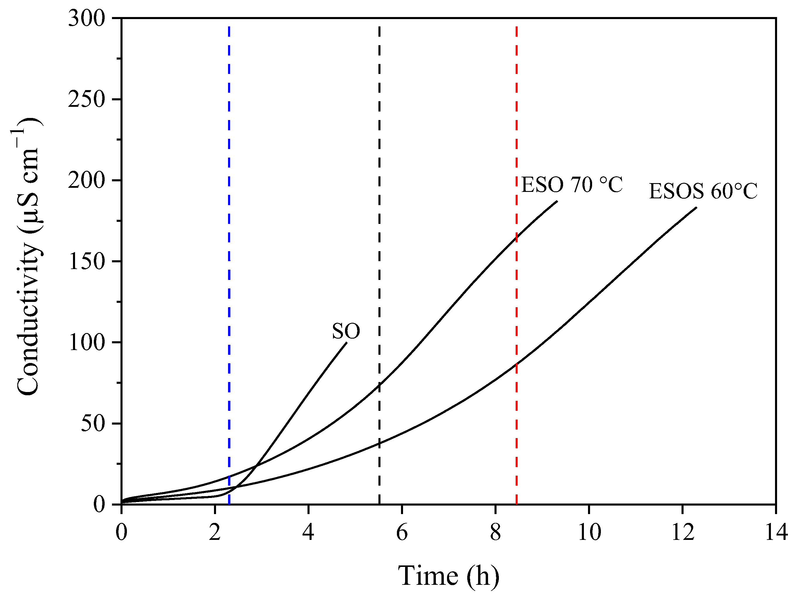
Separations 12 00121 g001
Figure 2. Cell viability assessment for HaCaT cells for sunflower seed oil (SO), oils obtained by extraction of curcuminoids from rhizome powder using sunflower seed oil (ESO 70 °C) and simultaneous extraction of sunflower seed oil and curcuminoids from rhizome (ESOS 60 °C). The different letters above the columns indicate statistically significant differences (p < 0.05).
Separations 12 00121 g002
Table 1. Experimental conditions adopted to obtain sunflower seed oil and enriched oils.
Product/AcronymMaterialsExperimental Conditions
Temperature (°C)ProportionTime (min)
Sunflower seed oil/SOSunflower seeds + ethyl acetate601:8 (g/mL)60
Enriched oil/ESOTurmeric rhizome + sunflower seed oil60 and 701:2 (g/g)15 and 30
Enriched oil/ESOSTurmeric rhizome + sunflower seeds + ethyl acetate1:4.5:8 (g/g/mL)15 and 30
Table 2. Effect of temperature and time on curcuminoids content in enriched sunflower seed oil.
Sample 1Temperature (°C)
6070
15 min30 min15 min30 min
ESO (mg/100 g oil)352.8 aA ± 0.1355.6 aA ± 0.6431.8 aB ± 0.3447.7 aB ± 2.8
ESOS (mg/100 g oil)512.5 aA ± 1.2497.3 bA ± 4.7509.6 aA ± 6.2511.5 aA ± 7.3
1 as Table 1. Means followed by the same lowercase letters (time effect for each temperature) and uppercase letters (temperature effect for each time) did not differ statistically (p > 0.05).
Table 3. Characterization of the oils obtained in terms of total phenolic compounds content and antioxidant potential.
Property Sample 1
SOESOESOS
60 °C/15 min70 °C/15 min60 °C/15 min
Total phenolic compound content (mg GAE/100 g oil)13.8 a ± 0.1437.7 b ± 5.4449.5 c ± 8.0643.4 d ± 18.4
Antioxidant potential (µmolTrolox/100 g oil)FRAP5.8 a ± 0.21098.5 b ± 118.51763.9 c ± 3.83522.2 d ± 163.2
DPPH•178.0 a ± 8.51016.7 b ± 15.01361.6 c ± 259.92427.1 d ± 81.2
ABTS•+79.6 a ± 3.4991.8 b ± 112.81150.7 c ± 114.01657.6 d ± 71.7
1 as Table 1. Means followed by the same lowercase letters (in each row) did not differ statistically (p > 0.05).
Table 4. Phenolic compound profile of the enriched oils obtained.
ClassIdentified CompoundPrecursor Ion (m/z)Fragment (m/z)Ionization ModeSample 1
(Peak Intensity/1.00 × 109)
SOESOESOS
70 °C/15 min60 °C/15 min
Phenolic acidsCinnamic147103.0Negative0.350.270.57
Gallic169125.04.392.341.46
Ferulic193134.90.282.8215.33
Caffeic178135.04.011.654.18
CurcuminoidsCurcumin367177.1Negativend50.11134.00
Demethoxycurcumin337217.1nd20.83134.00
Bisdemethoxycurcumin307179.1nd134.00134.00
Dihydroxycurcumin369191.1nd2.3721.39
Tetrahydrocurcumin371137.1nd0.071.96
TerpenesBisaccumol229137.1Positivend0.097.46
Dehydrocurdione235191.2ndnd1.31
Linalool13795.1nd6.131.95
α-turmerone216161.1nd1.150.29
β-turmerone218161.1nd1.325.63
1 as Table 1. nd: not detected.
Table 5. Fatty-acid profile of the enriched oils obtained.
Fatty Acid (%) 1SOESOESOS
70 °C/15 min60 °C/15 min
Palmitic6.76 a±0.17.75 b±0.15.72 c±0.04
Palmitoleic0.11 a±0.010.10 a±0.010.07 a±0.01
Stearic4.06 a±0.14.25 a ± 0.073.77 c±0.01
Oleic34.89 a±0.332.07 b ± 0.0141.80 c±0.3
Linoleic54.17 a±0.355.90 b ± 0.148.64 c±0.3
1 percentage of normative area. Means followed by the same lowercase letters (in each row) did not differ statistically (p > 0.05).
Disclaimer/Publisher’s Note: The statements, opinions and data contained in all publications are solely those of the individual author(s) and contributor(s) and not of MDPI and/or the editor(s). MDPI and/or the editor(s) disclaim responsibility for any injury to people or property resulting from any ideas, methods, instructions or products referred to in the content.

