Article

Phytochemical Content and Anticancer Activity of Jamaican Dioscorea alata cv. White Yam Extracts

by
Kenroy Wallace
1,2,*,
Racquel Wright
3,4,*,
Melisa Williams-Longmore
1,
Sasha-Gay Wright
1,5 and
Helen Asemota
1,3
1
Department of Basic Medical Sciences, Biochemistry Section, Faculty of Medical Sciences, The University of the West Indies, Mona, Kingston, Jamaica
2
College of Natural and Applied Science Allied Health and Nursing, Northern Caribbean University, Mandeville, Jamaica
3
Biotechnology Centre, Faculty of Science & Technology, The University of the West Indies, Mona, Kingston, Jamaica
4
Faculty of Engineering and Applied Technology, Caribbean Maritime University, Kingston, Jamaica
5
Faculty of Engineering, The University of the West Indies, Mona, Kingston, Jamaica
*
Authors to whom correspondence should be addressed.
Separations 2024, 11(2), 44; https://doi.org/10.3390/separations11020044
Submission received: 29 June 2023 / Revised: 23 October 2023 / Accepted: 24 October 2023 / Published: 30 January 2024
(This article belongs to the Special Issue Advances in Chromatographic Analysis of Bioactive Compounds)

Abstract

:
Dioscorea spp. is known for its myriad medicinal properties. D. alata, specifically crude extracts, have displayed potent anticancer properties. However, the chemical constituents of these extracts have not been examined. The aim of this study is to determine the chemical composition and antioxidant characteristics of the active extracts from D. alata tuber. Chemoinformatic profiling of the Jamaican Dioscorea alata cultivar white yam tuber was generated by a sequential Soxhlet extraction of dried milled tuber, producing five crude extracts: hexane (E-1), diethyl ether (E-2), acetone (E-3), ethanol (E-4) and water (E-5). The analytes within the five extracts were dissolved in 0.1% DMSO and their anticancer activity was determined using DU145 prostate cancer cells. Both the acetone and the ethanolic extract were able to induce greater than 50% cell death at 50 µg/mL. The order of growth inhibition of the extracts in DU-145 cell is E3 (IC50, 10.81 µg/mL) > E-4 (IC50 24.17 µg/mL) > E-1 (IC50 > 100 µg/mL) ≥ E-2 (IC50 > 100 µg/mL) ≥ E-5 (IC50 > 100 µg/mL). Phytochemical screening of both E-3 and E-4 revealed the presence of all major classes of secondary metabolites except tannins. Resins were also absent in the E-3 extract. Phenolic quantification indicated that E-3 and E-4 possessed GAEs of 31 ± 1.1 and 72 ± 1.8 mg per g of sample, respectively. Inversely, E-3 displayed greater antioxidant capability with IC50 of 82.9 µg/mL compared to E-4 (166.9 µg/mL); however, neither was comparable to citric acid (33.6 µg/mL). The extract E-3 was further isolated by HPLC into 11 fractions. Fractions 4 and 5 possessed potent cell growth inhibitory effects. GCMs of fractions 4 and 5 showed they possessed numerous saturated fatty acids with pharmacological relevance. The presence of these compounds shows potential for exploitation of D. alata extracts for pharmacological purposes.

1. Introduction

Yams are monocotyledonous plants belonging to the family Dioscoreaceae with the Dioscorea being the largest genus. They are known for their production of tubers, a storage organ composed mostly of starch. Yams are mainly found in tropical regions and are considered important staple crops in West Africa, Southeast Asia and the Caribbean [1,2]. Despite the large number of Dioscorea species, only about 11 species are cultivated or economically important, including the very popular Dioscorea alata or white yam [2].
Most commonly, yam is consumed boiled, baked, or fried [2]. Occasionally, yam is ingested in the form of raw yam, cooked soup, powder, or flour [2]. When compared to cassava or sweet potato, yam boasts a greater caloric value and contains relatively high levels of protein, vitamin C, and vitamin E [3]. Mucin, dioscin, dioscorin, allantoin, choline, polyphenols, diosgenin, and vitamins including carotenoids and tocopherols are only a few of the bioactive substances found in yam tubers [3] Mucilage of the yam tuber contains soluble glycoprotein and dietary fiber [3].
Dioscin, diosgenin, dioscorin, and anthocyanins are a few of the bioactive substances that have been linked to the anticancer effects of the Dioscorea species. Even though these compounds are prolific and anti-cancer—anti-proliferative, based on our results they are not the cytotoxic agents responsible for the antiproliferative activity in D. alata acetonic and ethanolic extracts.
These substances have been demonstrated to have strong cytotoxic effects against numerous cancer cell lines, including those from the breast, lungs, colon, and prostate, among others [4,5,6]. There are various cultivars of D. alata with the white yam cultivar (D. alata cv white yam), being very popular and widely eaten in Jamaica. Other cultivars of D. alata grown in Jamaica, such as the purple yam, commonly called St. Vincent, Dark Night, and Moonshine yam, have been shown to possess anticancer activities in vitro and in vivo [2]. The properties observed are thought to be due mainly to the presence of potent antioxidants, namely brightly coloured anthocyanins, which are responsible for the purple color in these yams [1]. It is possible that other cultivars may possess similar properties. We postulated that D. alata cv. white yam grown in Jamaica possesses anti-proliferative properties due to its phytochemical content. Studies have shown that many illnesses, including certain cancers and atherosclerosis, are linked to redox imbalance/oxidative stress [7,8]. Oxidative stress results from an imbalance in the reactive oxidative species (ROS) in cells. ROS are formed as by-products of metabolism but can also be generated from external sources. When there is a high concentration of ROS in cells, this may lead to uncontrolled cellular oxidation which can result in cellular damage and apoptosis. Conversely, studies have also indicated the stimulative effect of antioxidants in cancer cell growth [9].
Antioxidants are chemicals that are designed to reduce or neutralize ROS. Antioxidants are generated internally in the body; however, antioxidant sources are not limited only to compounds produced in the body as they can be obtained exogenously [10,11]. There has therefore been an effort to extract antioxidants from plants to determine their benefits. Yams possess a variety of compounds including saponins, polyphenols and vitamins; these components are thought to contribute to their bioactivity in some efficacy studies. The literature shows that these components have displayed antioxidant properties in other plants.
The effects of antioxidants on cancer cells are complex and can depend on several factors, such as the type of antioxidant, the type of cancer, and the stage of the cancer. Some studies have suggested that antioxidants may be beneficial in preventing cancer development and reducing cancer-related mortality. For example, a meta-analysis of randomized controlled trials found that supplementation with beta-carotene, vitamin C, and vitamin E was associated with a decreased risk of cancer incidence and mortality [12]. Additionally, several preclinical studies have suggested that certain antioxidants, such as curcumin and resveratrol, have anticancer properties and can inhibit the growth and metastasis of cancer cells [13,14].
Some studies have demonstrated that antioxidants can stimulate the growth of cancer cells in vitro and in vivo, as well as promote metastasis [15]. One potential mechanism by which antioxidants may promote cancer growth is through their ability to reduce oxidative stress in cancer cells, which may make cancer cells less vulnerable to chemotherapy and radiation therapy. These treatment options rely on the generation of ROS to induce cell death [16].
Studies have shown that yam plants possess antioxidant properties particularly in brightly colored cultivars such as the St. Vincent yam. There has been limited research on the antioxidant content of yams with white coloration such as the white yam. This is the first research known to the authors which focuses on the anticancer properties of yams with white pigmentation in Jamaica. This study therefore seeks to screen for phytochemical components present in the D. alata, cv white yam and identify the possible compounds contributing to the anticancer properties observed. The result of this study will provide the foundations for further investigation into the chemical compositions of the medicinal extracts of D. alata.

2. Materials and Methods

2.1. Collection of Dioscorea Alata Tuber Samples

Dioscora alata cv white yam tubers were gathered in the Jamaican parish of St. Catherine and shipped to the University of the West Indies’ Biotechnology Center in Mona, Jamaica. The tubers underwent a 6-h sun-drying process after a tap-water wash. After being dried at 50 °C to a constant weight, the tubers were ground into a fine powder and kept in sample bottles at room temperature.

2.2. Preparation of Extracts

The milled sample (1.2 kg) was placed in a large thimble constructed with steel mesh wire and filter paper. A continuous extraction was performed via Soxhlet extraction using 4 L of 100% hexane, diethyl ether, acetone, ethanol and water sequentially. Each solvent was used under reflux for 1 week before removal and subsequent addition of the next solvent. Each extract was concentrated using a rotary evaporator at 120 rpm at the solvents’ melting point. The water extract was vacuum filtered and dried using a lyophilizer. The five extracts were dried and stored at room temperature.

2.3. Phytochemical Analysis

The presence of the following phytochemicals was assessed using the acetone and ethanolic extracts: Phenols, alkaloids, tannins, sterols, glycosides, flavonoids, saponins, terpenoids, carboxylic acid and resins. Presence or absence was indicated by a plus (+) or minus (−) sign, respectively. Strong reactivity (that is, a large phytochemical content) was indicated by two pluses (++).

2.3.1. Test for Phenols

The E-3 and E-4 solutions were treated with five drops of 5% (w/v) glacial acetic acid and 5% (w/v) sodium nitrite solution. Muddy brown precipitate developed in the test tube; hence, the presence of phenols was confirmed [17,18].

2.3.2. Test for Alkaloids

A small quantity of extract was warmed in 2% hydrochloric acid for 2 min. The mixture was filtered, and a few drops of Mayer’s reagent and Wagner’s reagent were added separately. The Mayer’s reagent produced creamy-white colored precipitation giving a positive result. The Wagner’s reagent produced a reddish-brown precipitate, which indicated the presence of alkaloids [17,18].

2.3.3. Test for Tannins

A small quantity of extract was boiled in 5 mL of 45% ethanol solution. The mixture was then cooled and filtered. Two drops of ferric chloride (FeCl3) were added to 1 mL of filtrate. A greenish black or dark blue color indicated the presence of tannins [17,18,19].

2.3.4. Test for Glycosides

Extract (2 mL) was mixed with glacial acetic acid (1 mL), one drop of ferric chloride and one drop of concentrated sulphuric acid. The presence of a reddish-brown color at the junction of two layers with the upper layer having a bluish green color would confirm the presence of glycosides [18].

2.3.5. Test for Flavonoids

Extracts (10 mg) were boiled in 5 mL of ethyl acetate for 3 min. The mixture was filtered and the filtrate was mixed in 1 mL of diluted ammonium chloride solution (1%) by shaking. The layers were allowed to separate. A yellow coloration indicated the presence of flavonoids [17,20].

2.3.6. Test for Sterols

Extract (2 mL) was mixed with 2 mL of chloroform. To the mixture, 2 mL of acetic anhydride was added followed by two drops of concentrated sulphuric acid to the mixture via the side of the test tube. If sterol was present, there would be a red, then blue and finally green colour change [21].

2.3.7. Test for Saponins

Extract (0.2 mg) was dissolved in boiling distilled water. The extract was allowed to cool. The extract was then shaken vigorously and the formation of froth indicated the presence of saponin. The procedure was repeated using 5 g of dried yam material. Following the boiling in distilled water, the extract was allowed to cool and the filtrate was collected. The filtrate was then shaken vigorously for the formation of froth. Formation of steady froth indicated the presence of saponins [18,22,23].

2.3.8. Test for Terpenoids

Extract (2 mL) was mixed with 2 mL of chloroform. To this mixture 3 mL of concentrated H2SO4 was added and a layer of reddish-brown coloration was formed at the interface, thus indicating the presence of terpenoids [17,19]

2.3.9. Test for Resins

Acetonic extract (1 mL) was treated with few drops of acetic anhydride. Sulfuric acid (2 mL) was added to the mixture. A color change to yellow orange was observed; this indicated the presence of resins [24].

2.3.10. Test for Carboxylic Acid

Acetonic extract (0.5 mL) of extract was treated separately with 1 mL of saturated sodium bicarbonate solution (0.3 g solid to 4.5 mL distilled water). The liberation of carbon dioxide (visualized by effervescence or bubbling in the sample) confirmed the presence of carboxylic acid [24,25].

2.4. Cell Culture of DU145 Cells

All cell lines used in this study were obtained from the American Type Culture Collection (ATCC; Rockville, MD, USA). DU145 prostate cancer cells were cultured at the Environmental Toxicology Department at Southern University, Baton Rouge in an RPMI-1640 medium. Media were supplemented with 10% (v/v) heat inactivated fetal bovine serum (50 mL) and 1% (v/v) penicillin-streptomycin (5 mL). Cells were harvested to perform various assays once 80–85% confluency was attained. Cells were incubated at 37 °C with 5% CO2.

2.5. Cell Viability of DU145 Cells

Cells were seeded in 96-well plates at 5 × 10³ cell/well and allowed to grow for 24 h. Then, extracts were applied to the cells in five separate twofold dilutions, ranging from 100 g/mL to 6.5 g/mL. Each extract’s final dimethyl sulfoxide (DMSO) content in the growing medium did not go above 0.5% (v/v). Cell viability and cytotoxicity were measured using 10% (v/v) CellTiter 96® aqueous one solution reagent after cells had been exposed to the extract for 24 h. The absorbance was measured at 490 nm with an ELX800UV universal microplate reader following a 60-min incubation with (4,5-dimethyl-2-yl)-5-(3-carboxymethoxyphenyl)-2-(4-sulfophenyl)-2H-tetrazolium. A GraphPad Prism 8 was used to analyze the findings [26].

2.6. Antioxidant Capacity of Extracts Using 2,2-Diphenyl-1-picrylhydrazyl (DPPH)

Standards and extracts were diluted in methanol to the proper quantities in order to determine each compound’s antioxidant capability within a linear range. Totals of 100, 200, 400, 800, and 1000 µg/mL concentrations of the extracts were used. The ascorbic acid standard was made daily and diluted to concentrations of 2.5, 5, 10, 20, 40, 80, 160, and 320 µg/mL. A quantity of 0.1 mM DPPH in methanol was also utilized. The DPPH (1000 µL each) was reacted with the controls and extracts (200 µL each) in test tubes for 30 min in the dark. Additionally, a blank was created and incubated alongside the samples using methanol in place of the sample. Following incubation, the samples’ absorbance was measured at 517 nm using a Unico scanning spectrophotometer (Unico, Dayton, NJ, USA) [27].
The mean optical density of the sample was utilized to determine the DPPH reduction (inhibition), representing the percentage of DPPH that was neutralized by the antioxidants present in the added samples.
The equation below was used to calculate this percentage:
A o A s A o × 100
  • AO: absorbance blank
  • AS: sample absorbance
The findings were organized into a table and illustrated through graphs. The IC50 values were determined based on the DPPH reduction percentages and represent the concentration of the sample (in µg/mL) needed to neutralize 50% of the DPPH in the assay.

2.7. Polyphenol Quantification

Standards containing 1 µg, 2 µg, 4 µg, 6 µg, 8 µg and 10 µg of gallic acid dissolved in a volume of 200 µL were prepared. To each well, 20 µL of standard was added in quadruplicates. Some 80 µL of 7% calcium carbonate was added to each well containing 20 µL of standards and unknown samples. The Folin–Ciocalteu reagent was diluted 1:10 in deionized water and 100 µL was added to each well. The absorbance was measured at 700 nm after the addition of the Folin–Ciocalteu reagent. The total phenolic content of the extract was expressed as mg gallic acid equivalents (GAE) per gram of sample in dry weight (mg/g) [28].
The total phenolic content was calculated according to the equation:
C = c V m
  • C : Total phenolic content mg GAE/g dry extract
  • c : Concentration of gallic acid derived from calibration curve in mg/mL
  • V : Volume of extract in mL
  • m : Mass of extract in grams

2.8. High Performance Liquid Chromatography (HPLC)

The E-3 extract was purified prior to HPLC separation using solid phase extraction possessing a carbon 18 column (C18-E, 55 µm, 70A, 8B-S001-LEG). Methanol (10%) was used to wash the column; then, 25%, 75%, and 100% methanol was used to elute it. The samples were subsequently dried using a combination of rotary evaporation and straightforward distillation. Before being injected into the HPLC apparatus, the 100% methanol elute was dissolved in 20% DMSO and filtered through a 0.25 µm filter disc.
The E-3 extract was prepared by performing a 1:100 dilution of E-3 stock solution (1.46 × 10⁵ µg/mL); therefore, the sample injected had a concentration of 1.46 × 10³ µg/mL The experiment was carried out using an Agilent HPLC 1200 series and Chemstation for LC systems Rev. B.04.03 SP2 with a diode array detector. The method employed a Phenonmenex-250 × 21.20 nm, 10 u, C18(2), 100 Å column. The chromatography was monitored at 210 nm, 230 nm, and 280 nm. The mobile phase contained two solvents (A: distilled deionized water; B: acetonitrile); the gradient profile is described in Table 1 below.
The fractions collection was conducted according to the table below (Table 2).
Fraction collection was initiated at 3 min into the HPLC run. The Fr. 1 and Fr. 2 fractions were collected within a time interval of 3.5 min. The following eight fractions had a time interval of 2 min. The final fraction had a time interval of 5 min. The fractionation protocol was repeated 13 times. Fractions were dried using a vacuum concentrator and dissolved in 20% DMSO. DU145 cells were treated with 10 µL of each fraction and their cell growth inhibition activity was assessed using an MTT assay.

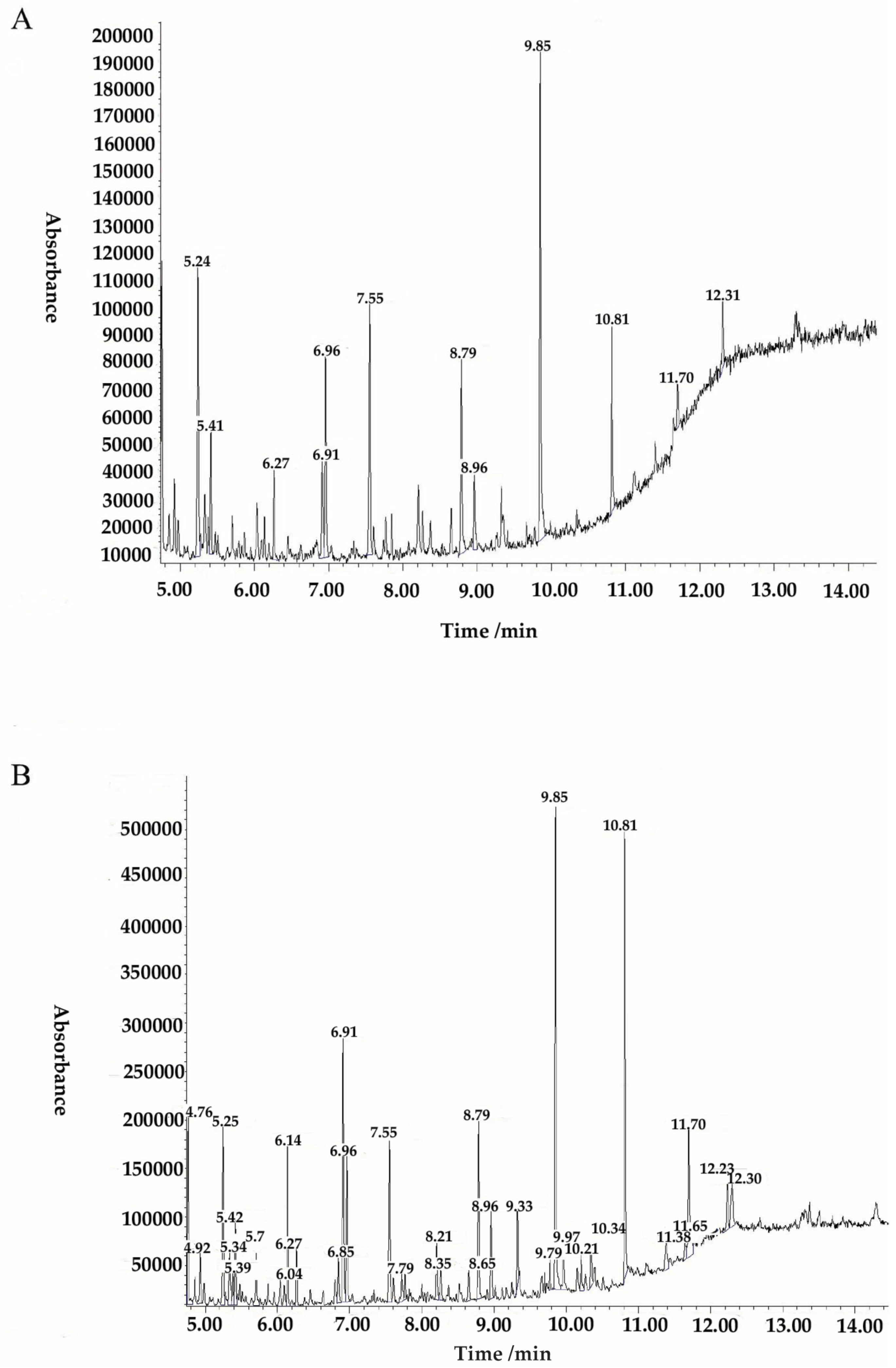
2.9. Gas Chromatography Mass Spectrometry

The analysis was conducted using an electron impact mode at 70 eV. The splitless mode was employed with an injection purge duration of 0.75 min. The injector temperature was set to 250 °C, while the transfer line temperature was maintained at 280 °C. The column used was a DB-1701 with dimensions of 30 m length, 0.25 mm inner diameter, and 0.25 mm film thickness. A ramped temperature program was employed beginning at 80 °C and held for 2 min, followed by an increase of 20 °C per minute for a duration of 10 min. Subsequently, the temperature was held for an additional 10 min. A solvent delay of 3 min was implemented, and helium was used as the carrier gas at a flow rate of 1.2 mL min−1. The operations were conducted in a Scan mode.

3. Results

Natural products commonly found in our diet present promising possibilities for identifying potent anticancer compounds. Dioscorea spp. have been extensively studied by various researchers and have been recognized for their diverse nutritional and medicinal properties [29,30].

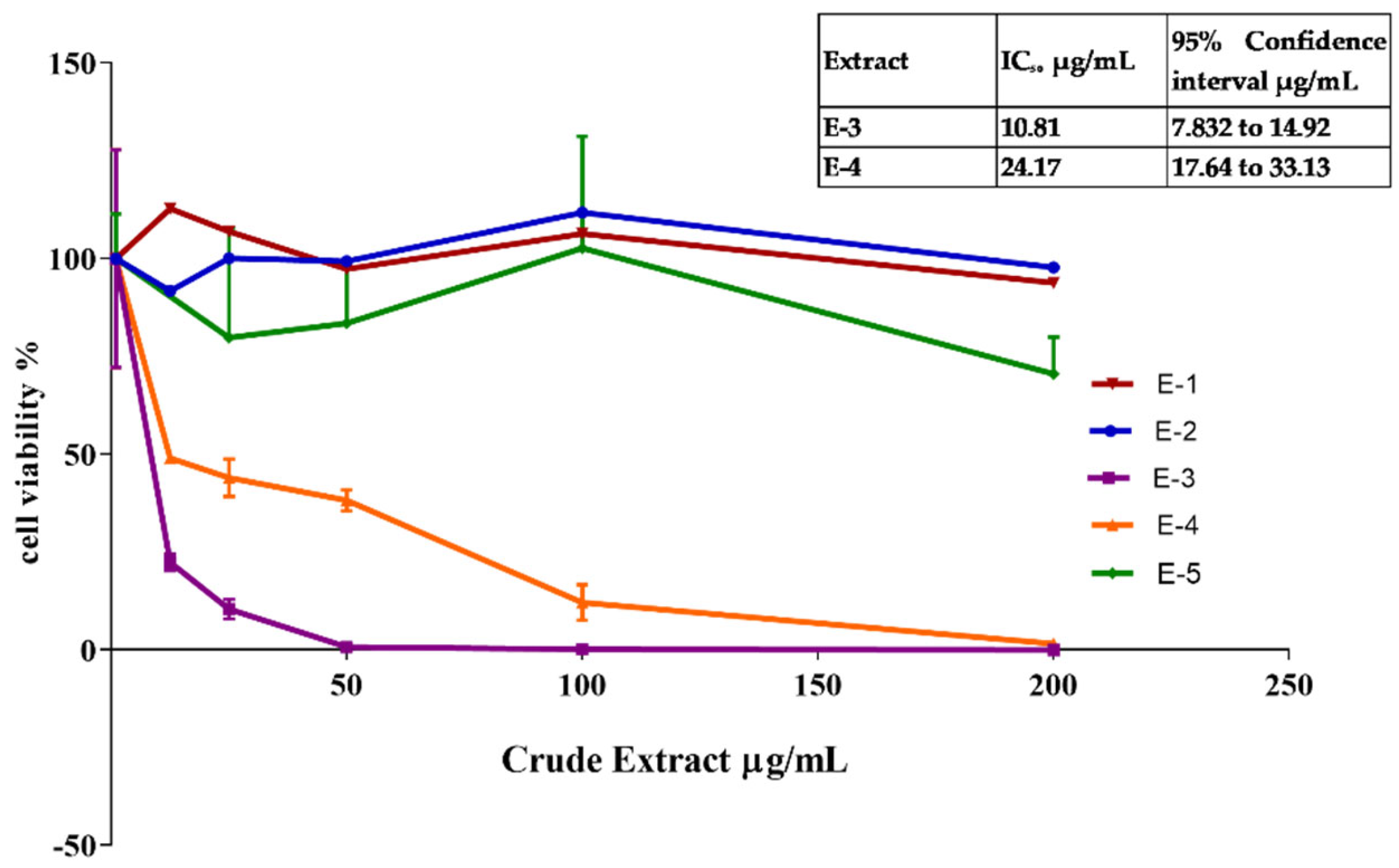
3.1. Dioscorea alata Solvent Extraction

D. alata contains a wide array of active and non-active compounds distributed throughout its different parts. To extract these compounds, solvents with varying polarities were utilized. The data obtained from these extractions allowed us to create a fingerprint of the bioactive analytes present in D. alata. For the extraction process, D. alata (18.802 g) from Jamaica, W. I., was sequentially extracted using hexane (E-1), diethyl ether (E-2), acetone (E-3), ethanol (E-4), and water (E-5) through Soxhlet extraction for 24 h at their respective boiling points. Subsequently, a combination of simple distillation and rotary evaporation was employed to remove the solvents, and the gravimetric method was used to determine the dry weights of the extracted materials. Solid–liquid extraction of D. alata with water for 24 h resulted in the largest number of extractable analytes (5.12% 0.96/18 g). We obtained (1.68% 0.32 g/18 g), (1% 0.19 g/18 g) and (0.29% 0.05/18 g) for hexane (E-1), diethyl ether (E-2), acetone (E-3), ethanol (E-4) or water (E-5), respectively. The initial analytical extraction was used to conduct a cytotoxicity screen using DU145 cells (Figure 1). The screen showed that extracts E-3 and E-4 displayed the lowest cell viability in DU145 cells when comparing all the extracts.

3.2. Preliminary Phytochemical Assessment of Acetonic and Ethanoic Extracts of Dioscorea alata

White yams have been attributed various medicinal properties owing to the existence of specific secondary metabolites in the tuber [31]. The bioactive compounds found in plants are secondary metabolites that can elicit pharmacological or toxicological effects in humans and animals. The Jamaican Dioscorea alata extracts were subjected to phytochemical evaluation. Various plant constituents present were identified in the acetone and ethanol extract using both qualitative and quantitative methods. The qualitative phytochemical assessment revealed the presence of various classes of secondary metabolites present within the acetonic and ethanolic extract (Table 3). Both extracts tested positive for the presence of phenols, alkaloids, sterols, alkaloids, glycosides, terpenoids, and phytosteroids. Both extracts tested negative for tannins. The ethanolic extract tested positive for resins; however, the acetonic extract tested negative. Both extracts possessed compounds that are known to exhibit antioxidant properties.
To corroborate the phytochemical screen, the total phenol content of E-3 and E-4 extracts was determined. The Folin–Ciocalteu assay method, with a Gallic acid standard curve, was used to determine the total phenolic content of the extract. The content was expressed as milligrams of Gallic acid equivalents (GAE) per gram of extract. Table 4 reveals that the total phenolic content in E-4 was approximately twice as high as E-3, with values of 72 ± 1.8 mg GAE per g and 31 ± 1.1 mg GAE per g, respectively.

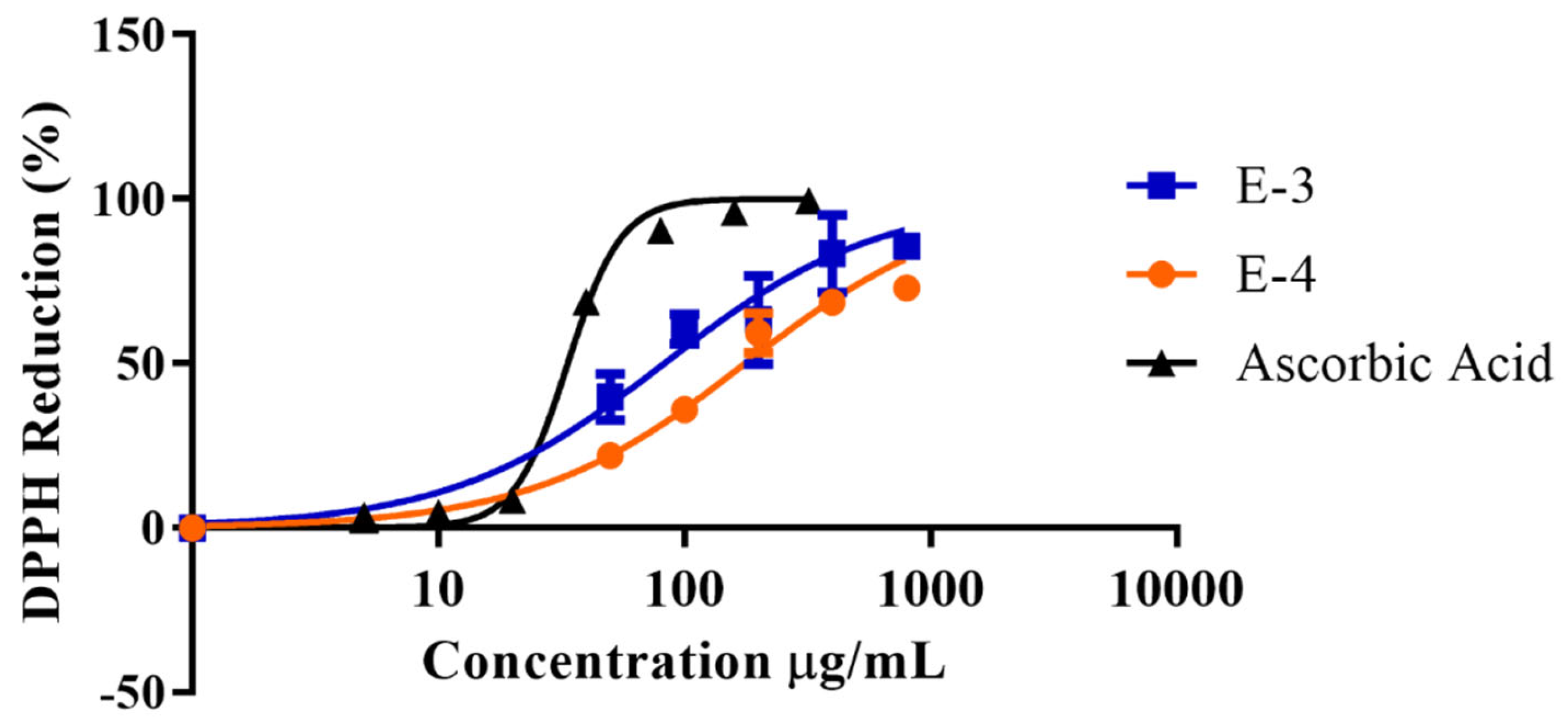
3.3. Dioscorea alata Acetonic and Ethanolic Extracts Possess Antioxidant Properties

Free radical scavenging ability when assessed showed that E-3 was more potent than E-4 (Figure 2). The order of increasing antioxidant potency of the yam extract was E-3, IC50 of 83 µg/mL, then E-4 with an IC50 value of 169.4 µg/mL (Table 5). It was observed that the DPPH reagent was reduced with increased concentration of both extracts. Ascorbic acid displayed more than double the potency of acetonic extract with an IC50 value of 33.6 ± 0.1 µg/mL and was five times more potent than that of E-4, an IC50 value of 167 µg/mL. The data highlighted the likelihood that the protective properties of Dioscorea alata tuber may not be due to its free radical scavenging abilities.
The E-3 extract displayed greater cytotoxic activity towards DU145 cells as well as more favorable antioxidant capabilities. Further investigations focused on identifying the compounds present within the E-3 extract. The E-3 extract was subjected to solid phase extraction. It was washed with 10% acetone and eluted with 25, 75 and 100% methanol (MeOH). The 100% MeOH elute, in a single dose assessment, inhibited 97.9% cell growth of the DU145 cells as compared to the crude extract’s 98.5% cell growth inhibition. The resulting sample was further separated into 10 fractions via high performance liquid chromatography (HPLC) (Figure 3). The HPLC protocol was developed using the time fractionation method.
Major peaks were collected in different fractions; this was enhanced by using two detection wavelengths, 210 nm and 230 nm. The protocol was designed using three different time intervals. Three time intervals were used; fractionation was initiated at a 2 min retention time. The first two fractions were collected within a time interval of 3.5 min. This was used to remove the major peak observed within the retention time of 3.4 min and 8.7 min detected using 230 nm and 210 nm. The following eight subsequent fractions had a time interval of 2 min. The final fraction had a time interval of 5 min, which collected all the smaller peaks at the end of the chromatogram.
Each HPLC fraction was concentrated and evaluated for proliferative activity using an MTT viability assay in DU145 cancer cells (Figure 4). Six fractions displayed positive inhibitory activity against DU145 cells; however, fraction 4 (Fr4) and fraction 5 (Fr5) possessed the greatest inhibitory effect at 92% and 93% growth inhibition. This was comparable to the load and docetaxel (positive control), which had a growth inhibitory activity of 94% and 97%, respectively. Both Fr4 and Fr5 were further assessed for their chemical composition using gas chromatography mass spectrometry (GC-MS).
The compounds identified in Fraction 4 (Fr4), and Fraction 5 (Fr5) are listed in Table 5 and Table 6. Figure 5 highlights the gas chromatogram of the extract. The compounds were identified through the NIST08 L database as listed in Table 5 and Table 6. The major compound identified by GC-MS analysis was azelaic acid RT 9.85 min in Fr4 and Fr5. However, the best matches from the database were hexadecanoic acid at RT 10.81 minutes in Fraction 4 with an abundance of 6.73% and RT 10.81 min with an abundance of 8.58% in Fraction 5. Other components also identified in the fractions are highlighted in Table 6 and Table 7. Octanoic acid was present in both fractions but was more abundant in Fraction 4. Fractions 4 and 5 possessed the highest azelaic acid content. They also contained nonanoic acid RT 6.96 min, dodecanoic acid RT 8.79 min, hexadecanoic acid RT 10.81 min, and octadecanoic acid RT 9.33 min in relatively great abundance. Additionally, 1,2-Bis(trimethylsiloxy)ethane RT 5.24 min was identified in large quantities in Fraction 4.

4. Discussion

D. alata is a widely consumed staple crop in Jamaica and the Caribbean; however, there are limited studies locally of its medicinal properties, particularly its antiproliferative capacity. Previous studies have shown the anti-proliferative capacity of the purple yam (D alata cultivar) but not of the generally consumed water yam. This study sought to evaluate the antiproliferative potential of components form fibers of locally grown D. alata cv water yams [26]. Sequential continuous extraction using solvents of increasing polarities allowed for separation of compounds in the yam tubers. This preliminary level of extraction has proven highly beneficial over the years (in natural compound isolation) because it allows for an initial separation of components for further analysis. The resulting extracts were tested for their cell inhibition activity against DU-145, an androgen-insensitive prostate cancer cell line. Androgen-insensitive prostate cells are the most difficult to test and cytotoxic compounds towards this cellular model would prove extremely beneficial. Extracts E3 and E4 were the most promising extracts as seen in Figure 1. These extracts displayed potent toxicity against androgen-insensitive prostate cancer cells (DU145) producing IC50 values of 10.81 µg/mL by E-3 and 24.17 µg/mL by E-4.
Despite the higher phenol content in E-4, E-3 (IC50 of 82.9 µg/mL) displayed superior antioxidant activity to E-4 (IC50 of 166.9 µg/mL). The use of solvents with varying polarity in the solid–liquid extraction allowed for the solubilizing of cytotoxic compounds using acetone (E-3) and ethanol (E-4). The extracts when screened for their phytochemical content had several phytochemicals of interest. Given the polarity of the extraction solvents used, many phytochemicals were polar in nature including phenolic compounds such as flavonoids, phenols, and tannins. The general classes of phytochemicals identified in our extracts, as seen in Table 2, have also been identified in various Dioscorea tuber species and cultivars from various locations [32,33]. Terpenoids, alkaloids, sterols, steroids, and glycosides were also present in the acetonic (E3) and ethanolic (E4) extracts [26,34]. Qualitative analysis additionally indicated a strong presence of carboxylic acid, which can be associated with high fatty acid content present within the tuber [35]. Phenolic compounds such as flavonoids have been implicated as antiproliferative agents. They are also known as antioxidants as they are able to scavenge free radicals [36]. The antioxidant capacity of the extracts E3 and E4 were evaluated as well as their phenol content. The total phenol content of E-3 and E-4 extracts was expressed in Gallic acid equivalents (GAE) and showed that E-4 possessed greater phenol content (72 ± 1.8 mg per g of sample) than E-3 (31 ± 1.1 mg per gram of sample). Even though the phenol and Gallic acid content of E4 was greater than E3, the antioxidant capacity was greater than that of E3. This may indicate that the phenolics present in the extracts may not be responsible for the antiproliferative activity displayed by E-3 and E-4 extracts.
Extract E3 (acetonic extract) was the most potent cytotoxic extract, as seen in Figure 1; and was subjected to SPE and HPLC fractionation and subsequently to cytotoxic testing. A total of 11 fractions was fractionated and evaluated for cellular inhibitory activity with Fraction 4 (Fr4) and Fraction 5 (Fr5) showing the greatest potency (over 90% inhibition).
The lack of discernible peaks in the HPLC chromatograms of Fr4 and Fr5 (Figure 3) suggests the potential presence of organic fatty acids. Due to the absence of significant chromophores, these fatty acids cannot be detected at UV wavelengths [37]. Therefore, to determine the chemical content of the fractions, GCMS was used. Fr4 and Fr5 were treated with BSFTA to increase the volatility of the samples by substituting –NH, –OH, –COOH, and –SH bonds with a dimethysilyl group. This derivatization reduced hydrogen bonding, which further reduced the polarity, increased the thermal stability and improved separation and detection by the GCMS system [38]. The major compounds identified are summarized in Table 5 and Table 6 and the chromatograms are presented in Figure 5. The derivatives of some of the most abundant organic acid compounds in Fr4 and Fr5 were identified as octanoic acid (caprylic acid), propanedioic acid (malonic acid), butanedioic acid (succinic acid), nonanoic acid (pelargonic acid), dodecanoic acid (lauric acid), benzoic acid, hexadecenoic acid (palmitoleic acid), octadecanoic acid (stearic acid), azelaic acid and heptadecanoic acid (margaric acid).
Therapeutic relevant hexadecenoic acid has been shown to display a cell growth inhibitory effect against prostate cancer in vitro and in vivo [26,34]. Similar responses were observed with the treatment of E-3 extract against androgen-insensitive DU145 cells. Other relevant compounds include azelaic acid, ocatanoic, and octadecanoic acid, have been shown to display anticancer activity against leukemia, colon cancer, neuroblastoma human skin epidermoid (A-431), and human mammary gland adenocarcinoma cells [39,40,41,42]. Lauric acid prompts the demise of colon cancer cells by means of the downgrading of the epidermal growth factor receptor by 1.33- and 1.58-fold, leading to cell death [43].
The presence of these compounds suggests that the antiproliferative effects of the extract are possibly due to the individual or synergistic effects of these compounds.
Previous studies have shown the anticancer properties of dioscin, diosgenin, dioscorin, and anthocyanin. However, our research indicates that these compounds were not the primary agents responsible for the observed anticancer activity in Fractions 4 and 5. This finding supports the presence of other anticancer compounds in the D. alata tuber.
The diversity of bioactive compounds present in the D. alata tuber is an indicator of its potential use as a source of natural anticancer agents. Other studies indicate that these therapeutic relevant compounds are not localized to the tuber but are also found in the leaves and stems of D. alata plant [44]. Future studies will assess the effectiveness of the identified compounds on an in vivo prostate cancer model.

5. Conclusions

There is an ongoing demand for the development of alternative cancer treatment methods sourced from natural origins. Through the conducted experiments, it was discovered that compounds found in the D. alata tuber exhibit potent growth inhibitory effects against androgen-insensitive prostate cancer cells. Phytochemical screening successfully identified the chemical constituents of the D. alata tuber; however, no significant antioxidant properties were detected. The isolation of the bioactive compounds using techniques such as solid-phase extraction (SPE), high-performance liquid chromatography (HPLC), and gas chromatography-mass spectrometry (GC-MS) generated a chemical fingerprint that unveiled the presence of bioactive compounds within the D. alata tuber. These findings point towards a potential synergistic relationship between these compounds, contributing to the observed pharmacological properties.

Author Contributions

Conceptualization, K.W. and H.A.; methodology, K.W. and R.W.; software, K.W. and R.W.; validation, S.-G.W. and K.W.; formal analysis, K.W.; investigation, K.W., R.W. and S.-G.W.; resources K.W., H.A., M.W.-L. and S.-G.W.; data curation, K.W.; writing—original draft preparation, K.W.; writing—review and editing, S.-G.W., R.W. and M.W; visualization, KW., S.-G.W. and M.W.-L.; supervision, H.A.; project administration, K.W. and H.A.; funding acquisition, K.W. and H.A. All authors have read and agreed to the published version of the manuscript.

Funding

This research was funded by the University of the West Indies, Mona, Research and Publication grant.

Data Availability Statement

Data is contained within the article.

Acknowledgments

The authors express their gratitude to Wesley Gray from Southern University Baton Rouge for facilitating the cell culture aspect of this research and providing valuable guidance. Additionally, the authors would like to acknowledge the Office of Graduate Studies and Research and the Department of Basic Medical Sciences at the University of the West Indies for their funding support, which facilitated other components of this research.

Conflicts of Interest

The authors declare no conflicts of interest.

References

  1. UWI Mona Discovers Anti Cancer Properties in Yams | Marketing and Communications Office, The University of West Indies at Mona. Available online: https://www.mona.uwi.edu/marcom/newsroom/entry/5775 (accessed on 6 January 2022).
  2. Lebot, V.; Lawac, F.; Legendre, L. The Greater Yam (Dioscorea Alata L.): A Review of Its Phytochemical Content and Potential for Processed Products and Biofortification. J. Food Compos. Anal. 2023, 115, 104987. [Google Scholar] [CrossRef]
  3. Bhandari, M.R.; Kasai, T.; Kawabata, J. Nutritional Evaluation of Wild Yam (Dioscorea Spp.) Tubers of Nepal. Food Chem. 2003, 82, 619–623. [Google Scholar] [CrossRef]
  4. Srinivasan, S.; Koduru, S.; Kumar, R.; Venguswamy, G.; Kyprianou, N.; Damodaran, C. Diosgenin Targets Akt-Mediated Prosurvival Signaling in Human Breast Cancer Cells. Int. J. Cancer 2009, 125, 961–967. [Google Scholar] [CrossRef] [PubMed]
  5. Hong, S.H.; Cha, H.-J.; Hwang-Bo, H.; Kim, M.Y.; Kim, S.Y.; Ji, S.Y.; Cheong, J.; Park, C.; Lee, H.; Kim, G.-Y.; et al. Anti-Proliferative and Pro-Apoptotic Effects of Licochalcone A through ROS-Mediated Cell Cycle Arrest and Apoptosis in Human Bladder Cancer Cells. Int. J. Mol. Sci. 2019, 20, 3820. [Google Scholar] [CrossRef] [PubMed]
  6. Karar, J.; Maity, A. PI3K/AKT/mTOR Pathway in Angiogenesis. Front. Mol. Neurosci. 2011, 4, 51. [Google Scholar] [CrossRef] [PubMed]
  7. Dufie, W.-M.F.; Oduro, I.; Ellis, W.O.; Asiedu, R.; Maziya-Dixon, B. Potential Health Benefits of Water Yam (Dioscorea Alata). Food Funct. 2013, 4, 1496–1501. [Google Scholar] [CrossRef]
  8. Reuter, S.; Gupta, S.C.; Chaturvedi, M.M.; Aggarwal, B.B. Oxidative Stress, Inflammation, and Cancer: How Are They Linked? Free Radic. Biol. Med. 2010, 49, 1603–1616. [Google Scholar] [CrossRef]
  9. Mendelsohn, A.R.; Larrick, J.W. Paradoxical Effects of Antioxidants on Cancer. Rejuvenation Res. 2014, 17, 306–311. [Google Scholar] [CrossRef]
  10. Saha, S.K.; Lee, S.B.; Won, J.; Choi, H.Y.; Kim, K.; Yang, G.-M.; Dayem, A.A.; Cho, S.-G. Correlation between Oxidative Stress, Nutrition, and Cancer Initiation. Int. J. Mol. Sci. 2017, 18, E1544. [Google Scholar] [CrossRef]
  11. Singh, U.; Jialal, I. Oxidative Stress and Atherosclerosis. Pathophysiology 2006, 13, 129–142. [Google Scholar] [CrossRef]
  12. Bjelakovic, G.; Nikolova, D.; Simonetti, R.G.; Gluud, C. Antioxidant Supplements for Preventing Gastrointestinal Cancers. Cochrane Database Syst. Rev. 2008, CD004183. [Google Scholar] [CrossRef] [PubMed]
  13. Aggarwal, B.B.; Sundaram, C.; Malani, N.; Ichikawa, H. Curcumin: The Indian Solid Gold. Adv. Exp. Med. Biol. 2007, 595, 1–75. [Google Scholar] [CrossRef] [PubMed]
  14. Singh, R.P.; Tyagi, A.K.; Dhanalakshmi, S.; Agarwal, R.; Agarwal, C. Grape Seed Extract Inhibits Advanced Human Prostate Tumor Growth and Angiogenesis and Upregulates Insulin-like Growth Factor Binding Protein-3. Int. J. Cancer 2004, 108, 733–740. [Google Scholar] [CrossRef] [PubMed]
  15. Sayin, V.I.; Ibrahim, M.X.; Larsson, E.; Nilsson, J.A.; Lindahl, P.; Bergo, M.O. Antioxidants Accelerate Lung Cancer Progression in Mice. Sci. Transl. Med. 2014, 6, 221ra15. [Google Scholar] [CrossRef] [PubMed]
  16. Ristow, M. Unraveling the Truth about Antioxidants: Mitohormesis Explains ROS-Induced Health Benefits. Nat. Med. 2014, 20, 709–711. [Google Scholar] [CrossRef] [PubMed]
  17. Sheel, R.; Nisha, K. Qualitative Phytochemical Analysis for Isolation of Terpenes from Clerodendron Infortunatum Leaves. ISOR J. Appl. Chem. 2014, 7, 14–18. [Google Scholar] [CrossRef]
  18. Ali, S.; Khan, M.R.; Irfanullah; Sajid, M.; Zahra, Z. Phytochemical Investigation and Antimicrobial Appraisal of Parrotiopsis Jacquemontiana (Decne) Rehder. BMC Complement. Altern. Med. 2018, 18, 43. [Google Scholar] [CrossRef] [PubMed]
  19. Das, N.; Islam, M.E.; Jahan, N.; Islam, M.S.; Khan, A.; Islam, M.R.; Parvin, M.S. Antioxidant Activities of Ethanol Extracts and Fractions of Crescentia Cujete Leaves and Stem Bark and the Involvement of Phenolic Compounds. BMC Complement. Altern. Med. 2014, 14, 45. [Google Scholar] [CrossRef]
  20. Gizaw, A.; Marami, L.M.; Teshome, I.; Sarba, E.J.; Admasu, P.; Babele, D.A.; Dilba, G.M.; Bune, W.M.; Bayu, M.D.; Tadesse, M.; et al. Phytochemical Screening and In Vitro Antifungal Activity of Selected Medicinal Plants against Candida Albicans and Aspergillus Niger. in West Shewa Zone, Ethiopia. Adv. Pharmacol. Pharm. Sci. 2022, 2022, e3299146. [Google Scholar] [CrossRef]
  21. Shaikh, J.R.; Patil, M. Qualitative Tests for Preliminary Phytochemical Screening: An Overview. Int. J. Chem. Stud. 2020, 8, 603–608. [Google Scholar] [CrossRef]
  22. Bankole, A.E.; Adekunle, A.A.; Sowemimo, A.A.; Umebese, C.E.; Abiodun, O.; Gbotosho, G.O. Phytochemical Screening and in Vivo Antimalarial Activity of Extracts from Three Medicinal Plants Used in Malaria Treatment in Nigeria. Parasitol. Res. 2016, 115, 299–305. [Google Scholar] [CrossRef]
  23. Onwukaeme, D.N.; Ikuegbvweha, T.B.; Asonye, C.C. Evaluation of Phytochemical Constituents, Antibacterial Activities and Effect of Exudate of Pycanthus Angolensis Weld Warb (Myristicaceae) on Corneal Ulcers in Rabbits. Trop. J. Pharm. Res. 2007, 6, 725–730. [Google Scholar] [CrossRef]
  24. Kumar, S.; Venkateshwar, C.; Samuel, G.; Rao, S.G. Phytochemical Screening of Some Compounds from Plant Leaf Extracts of Holoptelea Integrifolia (Planch.) and Celestrus Emarginata (Grah.) Used by Gondu Tribes at Adilabad District, Andhrapradesh, India. Int. J. Eng. Sci. Invent. 2013, 2, 65–70. [Google Scholar]
  25. Siddique, A.B.; Rahman, S.M.M.; Hossain, M.A.; Hossain, M.A.; Rashid, M.A. Phytochemical Screening and Comparative Antimicrobial Potential of Different Extracts of Stevia Rebaudiana Bertoni Leaves. Asian Pac. J. Trop. Dis. 2014, 4, 275–280. [Google Scholar] [CrossRef]
  26. Wallace, K.; Asemota, H.; Gray, W. Acetone Extract of Dioscorea Alata Inhibits Cell Proliferation in Cancer Cells. Am. J. Plant Sci. 2021, 12, 300. [Google Scholar] [CrossRef]
  27. Wright, R.J.; Lee, K.S.; Hyacinth, H.I.; Hibbert, J.M.; Reid, M.E.; Wheatley, A.O.; Asemota, H.N. An Investigation of the Antioxidant Capacity in Extracts from Moringa Oleifera Plants Grown in Jamaica. Plants 2017, 6, 48. [Google Scholar] [CrossRef] [PubMed]
  28. Durazzo, A.; Lucarini, M. Extractable and Non-Extractable Antioxidants. Molecules 2019, 24, 1933. [Google Scholar] [CrossRef]
  29. Stennett, D.; Oladeinde, F.; Wheatley, A.; Bryant, J.; Dilworth, L.; Asemota, H. Effects of Dioscorea Polygonoides (Jamaican Bitter Yam) Supplementation in Normocholesterolemic and Genetically Modified Hypercholesterolemic Mice Species. J. Food Biochem. 2014, 38, 28–37. [Google Scholar] [CrossRef]
  30. Grindley, P.B.; Omoruyi, F.O.; Asemota, H.N.; St. A. Morrison, E.Y. Asemota Effect of Yam (Dioscorea cayenensis) and Dasheen (Colocassia esculenta) Extracts on the Kidney of Streptozotocin-Induced Diabetic Rats. Int. J. Food Sci. Nutr. 2001, 52, 429–433. [Google Scholar] [CrossRef] [PubMed]
  31. Chandrasekara, A.; Josheph Kumar, T. Roots and Tuber Crops as Functional Foods: A Review on Phytochemical Constituents and Their Potential Health Benefits. Available online: https://www.hindawi.com/journals/ijfs/2016/3631647/ (accessed on 14 September 2020).
  32. Das, A.; Chaudhuri, D.; Ghate, N.; Chatterjee, A.; Mandal, N. Phytochemical Analysis, Antioxidant and Anticancer Potential of Leaf Extracts from Edible Greater Yam, Dioscorea Alata L., from North-East India. Int. J. Phytopharm. 2014, 5, 109–119. [Google Scholar]
  33. Maithili, V.; Dhanabal, S.P.; Mahendran, S.; Vadivelan, R. Antidiabetic Activity of Ethanolic Extract of Tubers of Dioscorea Alata in Alloxan Induced Diabetic Rats. Indian J. Pharmacol. 2011, 43, 455–459. [Google Scholar] [CrossRef] [PubMed]
  34. Zhu, S.; Jiao, W.; Xu, Y.; Hou, L.; Li, H.; Shao, J.; Zhang, X.; Wang, R.; Kong, D. Palmitic Acid Inhibits Prostate Cancer Cell Proliferation and Metastasis by Suppressing the PI3K/Akt Pathway. Life Sci. 2021, 286, 120046. [Google Scholar] [CrossRef] [PubMed]
  35. Muzac-Tucker, I.; Asemota, H.N.; Ahmad, M.H. Biochemical Composition and Storage of Jamaican Yams (Dioscorea Sp). J. Sci. Food Agric. 1993, 62, 219–224. [Google Scholar] [CrossRef]
  36. Sakthidevi, G.; Mohan, V.R. Total Phenolic, Flavonoid Contents and In Vitro Antioxidant Activity of Dioscorea Alata l. Tuber. J. Pharm. Sci. Res. 2013, 5, 115. [Google Scholar]
  37. De Baere, S.; Eeckhaut, V.; Steppe, M.; De Maesschalck, C.; De Backer, P.; Van Immerseel, F.; Croubels, S. Development of a HPLC–UV Method for the Quantitative Determination of Four Short-Chain Fatty Acids and Lactic Acid Produced by Intestinal Bacteria during in Vitro Fermentation. J. Pharm. Biomed. Anal. 2013, 80, 107–115. [Google Scholar] [CrossRef] [PubMed]
  38. Wettersten, H.I.; Ganti, S.; Weiss, R.H. Chapter Fourteen-Metabolomic Profiling of Tumor-Bearing Mice. In Methods in Enzymology; Galluzzi, L., Kroemer, G., Eds.; Cell-Wide Metabolic Alterations Associated with Malignancy; Academic Press: Cambridge, MA, USA, 2014; Volume 543, pp. 275–296. [Google Scholar]
  39. Narayanan, A.; Baskaran, S.A.; Amalaradjou, M.A.R.; Venkitanarayanan, K. Anticarcinogenic Properties of Medium Chain Fatty Acids on Human Colorectal, Skin and Breast Cancer Cells in Vitro. Int. J. Mol. Sci. 2015, 16, 5014–5027. [Google Scholar] [CrossRef] [PubMed]
  40. Aiyelaagbe, O.O.; Negi, A.S.; Hamid, A.A.; Luqman, S.; Kumar, S.B.; Kaneez, F. Chemical Constituents from Alafia Barteri Oliv. Leaves with Cytotoxic Activity. J. Chin. Chem. Soc. 2015, 62, 751–755. [Google Scholar] [CrossRef]
  41. Mericli, F.; Becer, E.; Kabadayı, H.; Hanoglu, A.; Yigit Hanoglu, D.; Ozkum Yavuz, D.; Ozek, T.; Vatansever, S. Fatty Acid Composition and Anticancer Activity in Colon Carcinoma Cell Lines of Prunus Dulcis Seed Oil. Pharm. Biol. 2017, 55, 1239–1248. [Google Scholar] [CrossRef]
  42. Atasever-Arslan, B.; Yilancioglu, K.; Bekaroglu, M.G.; Taskin, E.; Cetiner, E.A.S. Cytotoxic Effect of Extract from Dunaliella Salina against SH-SY5Y Neuroblastoma Cells. Gen. Physiol. Biophys. 2015, 34, 201–207. [Google Scholar] [CrossRef]
  43. Sheela, D.L.; Narayanankutty, A.; Nazeem, P.A.; Raghavamenon, A.C.; Muthangaparambil, S.R. Lauric Acid Induce Cell Death in Colon Cancer Cells Mediated by the Epidermal Growth Factor Receptor Downregulation: An in Silico and in Vitro Study. Hum. Human. Exp. Toxicol. 2019, 38, 753–761. [Google Scholar] [CrossRef]
  44. Dey, P.; Chaudhuri, T.K. Phytochemical Characterization of Dioscorea alata Leaf and Stem by Silylation Followed by GC-MS Analysis. J. Food Biochem. 2016, 40, 630–635. Available online: https://onlinelibrary.wiley.com/doi/abs/10.1111/jfbc.12235 (accessed on 24 January 2021).
Figure 1. Cytotoxic effect of Jamaican Dioscorea alata crude extracts on DU145 cells. E−1—Hexane, E−2—diethyl ether, E−3 acetone, E−4 ethanol and E−5 water extract. Wells were seeded with 10,000 cells and were treated with crude extracts. Cells were harvested after 24 h and their viability was measured using a CellTiter 96® non-radioactive cell proliferation assay (MTS). Assays were performed in quadruplicate.
Separations 11 00044 g001
Figure 2. Antioxidant screening of bioactive acetonic and ethanolic extract. In vitro antioxidant screening of E-3 and E-4, using vitamin C (ascorbic acid) as a positive control. Assay was done in triplicate.
Separations 11 00044 g002
Figure 3. High performance liquid chromatography (HPLC) and fractionation design for E-3 extract gradient run of sample with detection using 240 nm HPLC fractionation design. Major peaks detected using time split.
Separations 11 00044 g003
Figure 4. Single dose assessment of anticancer properties of HPLC fractions on DU145 cells. Fractions were assayed using DU145 cells with 5000 cell/well. Cells were harvested after 24 h and their viability was measured using a CellTiter 96® non-radioactive cell proliferation assay (MTT). Numbers 1-10 refer to the fractions from the HPLC.
Separations 11 00044 g004
Figure 5. Chromatogram of gas chromatography for Fraction 4 (A) and Fraction 5 (B).
Separations 11 00044 g005
Table 1. Gradient profile method of HPLC separation of E-3.
Time
(min)
Flow Rate
(mL/min)
Deionized Water (%)
A
Acetonitrile (%)
B
017525
515050
1012575
201.50100
Sample separation for 30 min. Column washed and regenerated with 95% acetonitrile (5 min, 1 mL/min) followed by 100% acetonitrile (5 min, 1.5 mL/min).
Table 2. Fraction collection of HPLC separation for E3.
FractionsTime
(min)
FractionsTime
(min)
0362
13.572
23.582
3292
42102
52115
Table 3. Phytochemical Screening of E-3 and E-4 Extract from Dioscorea alata.
PhytochemicalsAcetone Extract
(E-3)
Ethanol Extract
(E-4)
Phenols++++
Alkaloids++
Tannins
Sterols+++
Glycosides++++
Flavonoids++
Saponins+++
Terpenoids+++
Carboxylic acid+++
Resins+
Steroids++++
(+) indicates present, (++) indicates strongly present, (−) indicates absence.
Table 4. Total Phenol Content in Acetonic and Ethanolic extracts of Jamaican Dioscorea alata.
ExtractsGallic Acid Equivalents (GAE) mg/g
E-331 ± 1.1
E-472 ± 1.8
Table 5. IC50 Values of Free radical Scavenging Properties of Dioscorea alata Acetonic and Ethanolic extracts.
SampleIC50 (µg/mL)95% Confidence Interval
Ascorbic Acid33.631.98 to 35.34
E-382.970.56 to 93.95
E-4166.9140.4 to 204.3
Table 6. GC-MS analysis of the HPLC Fraction 4 of the E3 Extract of Dioscorea alata.
Retention Time (min)Abundance %Name of CompoundsMolecular Mass g/mol
5.249.591,2-Bis(trimethylsiloxy)ethane206.43
6.262.8Octanoic acid, trimethylsilyl ester216.39
6.913.62Butanedioic acid, bis(trimethylsilyl) ester262.45
6.966.37Nonanoic acid, trimethylsilyl ester230.42
7.558.574H-1-Benzopyran-4-one, 5,7-dihydroxy-2-methyl-192.17
8.795.82Dodecanoic acid, trimethylsilyl ester272.5
8.962.71Benzoic acid, 4-[(trimethylsilyl)oxy]-, trimethylsilyl ester282.48
9.8516.18Azelaic acid, bis(trimethylsilyl)ester332.58
10.816.73Hexadecanoic acid, trimethylsilylester328.6
11.71.9Octadecanoic acid, trimethylsilylester356.7
12.312.74Dodecanamide199.33
11.71.9Heptadecanoic acid, trimethylsilylester342.6
Table 7. GC-MS analysis of the HPLC Fraction 5 of E3 Extract of Dioscorea alata.
Retention Time (min)Abundance %Name of CompoundsMolecular g/mol
4.763.34Propanoic acid, 2-(methoxyimino)-,trimethylsilyl ester189.28
5.253.24Ethanedioic acid, bis(trimethylsilyl) ester234.4
5.341.22Dithioerythritol, tetrakis(trimethylsilyl)-443
5.421.45Butanoic acid, 2-[(trimethylsilyl)amino]-, trimethylsilyl ester247.48
6.142.61Trimethylsilyl ether of glycerol308.64
6.260.92Octanoic acid, trimethylsilyl este216.39
6.914.96Butanedioic acid, bis(trimethylsilyl) ester262.45
6.963.04Nonanoic acid, trimethylsilyl este230.42
7.553.54Anthracene, 1-methyl-192.25
8.793.3Dodecanoic acid, trimethylsilyl ester272.5
8.961.73Benzoic acid, 4-[(trimethylsilyl)oxy]-, trimethylsilyl ester282.48
9.331.75Octanedioic acid, bis(trimethylsilyl) ester318.56
9.859.74Azelaic acid, bis(trimethylsily) ester 332.58
9.961.55Arabinofuranose, 1,2,3,5-tetrakis-O-(trimethylsilyl)-150.13
10.210.98Trimethylsilyl ether of glycerol308.64
10.341.38.alpha.-D-Glucopyranoside, methyl2,3,4,6-tetrakis-O-(trimethylsilyl)-194.18
10.818.58Hexadecanoic acid, trimethylsilylester328.6
11.381.05Dodecanamide199.33
11.640.89Oleic acid282.5
11.74.03Octadecanoic acid, trimethylsilylester356.7
12.233.39.beta.-D-Galactopyranose, 1,2,3,4,6-pentakis-O-(trimethylsilyl)-541.1
12.33.54Benzeneethanamine, 2-fluoro-.beta.,3,4-trihydroxy-N-isopropyl-229.25
14.291.421H+R6:R27ido [1,2-a]quinoline-2-carboxylic acid, 5,6-dihydro-1-oxo-, methyl ester256.26
Disclaimer/Publisher’s Note: The statements, opinions and data contained in all publications are solely those of the individual author(s) and contributor(s) and not of MDPI and/or the editor(s). MDPI and/or the editor(s) disclaim responsibility for any injury to people or property resulting from any ideas, methods, instructions or products referred to in the content.

