Article

Isolation of Osteoblastic Differentiation-Inducing Constituents from Peanut Sprouts and Development of Optimal Extraction Method

1
Laboratory of Pharmacognosy, College of Pharmacy, Dankook University, Cheonan 31116, Republic of Korea
2
Host Defense Modulation Laboratory, College of Pharmacy, Chung-Ang University, Seoul 06974, Republic of Korea
*
Authors to whom correspondence should be addressed.
These authors contributed equally to this work.
Separations 2023, 10(8), 435; https://doi.org/10.3390/separations10080435
Submission received: 22 June 2023 / Revised: 26 July 2023 / Accepted: 28 July 2023 / Published: 1 August 2023
(This article belongs to the Special Issue Separation, Analysis and Biological Evaluation of Bioactive Compounds)

Abstract

:
Osteoporosis, one of the most common bone diseases, results from an imbalance between bone formation and resorption. Osteoblasts are primarily involved in bone formation, whereas osteoclasts are involved in bone resorption. Therefore, any substances that can increase osteoblastic differentiation would be beneficial for the prevention or treatment of osteoporosis. In this study, peanut sprouts, a germinated product of peanuts, significantly enhanced osteoblastic differentiation of pre-osteoblastic MC3T3-E1 cells, as determined by the alkaline phosphatase (ALP) assay. The ethyl acetate fraction of peanut sprout extract was subjected to diverse column chromatographies using silica gel, ODS, and Sephadex LH-20 as stationary phases, and three nucleobases, namely, adenine, uracil, and thymine, and three phenolic acids, including caffeic acid, coumaric acid, and ferulic acid, were isolated as active constituents. In particular, adenine at 4 μg/mL and caffeic acid at 1 μg/mL increased ALP activity by 40 and 30%, respectively, compared with the osteoblastic differentiation medium-treated group, and these two compounds were set as marker compounds. Furthermore, extraction of peanut sprouts grown for 14–17 days with 60% ethanol was the best condition to obtain a high quantity of extract for peanut sprouts by analyzing the contents of marker compounds through HPLC. Together, these results suggest that peanut sprouts and their isolated compounds significantly enhance osteoblastic differentiation. Therefore, peanut sprouts have the potential to be developed as functional foods against osteoporosis.

Graphical Abstract

1. Introduction

Osteoporosis is the most common bone disease, manifesting as pores in the bones and leading to the loss of skeleton strength and increased fracture risk [1,2,3,4]. Imbalance in bone formation and resorption plays an important role in this disease, especially with increasing age. With age, the formation of new bones consistently decreases, whereas bone destruction remains the same or increases [5,6]. Osteoblasts are mostly involved in bone formation, whereas osteoclasts are primarily involved in bone resorption [7,8]. Hence, the promotion of osteoblastic functions or inhibition of osteoclastic actions could be a prophylactic or therapeutic strategy against osteoporosis.
To date, osteoporosis drugs used in clinical settings have been classified into two categories [4]: antiresorptive agents, which inhibit osteoclasts, including bisphosphonate, receptor activators of nuclear factor kappa-B ligand antibodies, selective estrogen receptors, and calcitonin; and anabolic medicine, which induces osteoblastic performance, leading to bone renewal, including para-thyroidal hormone [4]. One of the treatment strategies for osteoporosis is inhibiting bone resorption through treatment with commercially available drugs that can suppress the function of osteoclasts; however, this strategy is not feasible for elderly patients with osteoporosis, who already have low bone density at treatment initiation [6]. As a result, stimulation of osteoblastic functions needs to be accompanied by inhibition of osteoclastic actions for bone remodeling in elderly patients with osteoporosis.
Osteoblasts need to be mature to function properly through differentiation. Osteoblastic differentiation is a crucial process regulated by many transcription factors, such as runt-related transcription factor 2 (Runx2) and ostrix (Osx), as well as bone matrix proteins, such as alkaline phosphatase (ALP) and type 1 collagen (Col1) [6]. This mechanism facilitates calcium accumulation, that is, mineralization, and completes bone formation [9]. ALP is a representative enzyme expressed in the early phase of osteoblastic differentiation and observed on the cell surface and in matrix vesicles [10]. After the initiation of the bone developmental process, ALP expression decreases. Thus, ALP is a key biomarker for the initiation of osteoblastic differentiation.
Many studies have been performed or are underway to discover natural resources with significant effects on bone health [11,12]. Because osteoporosis is a degenerative disease, patients with the disease must receive the medication until the symptoms dissipate, sometimes for their lifetime. Thus, natural products might be an attractive resource because they have relatively low side effects [11]. For example, Paeonia lactiflora, Scytosiphon lomentaria, Dalbergia odorrifera, curcumin derivatives, Eurycoma longifolia, and Psoralea corylifolia have been reported as natural products that induce osteoblastic differentiation [13,14,15,16,17,18]. Peanut sprouts are a novel product obtained a few days after the germination of peanuts, and they contain high polyphenol content, including gallic acid, protocatechuic acid, resveratrol, and caffeic acid [19,20]. It has been reported that peanut sprouts have antioxidant, anti-inflammatory, and anti-obesity activities [21,22,23,24], and these pharmacological effects are correlated with the high polyphenol content in peanut sprouts [25]. Even though peanut sprouts are reported as a good natural resource with many benefits for human health, the effects of peanut sprouts on bone health remain unknown.
Therefore, the aim of the present study was to evaluate the effects of peanut sprout extract on osteoblastic differentiation, to identify the active components, and to develop optimal extraction conditions.

2. Materials and Methods

2.1. Preparation of Peanut Sprout Extract

Fresh peanut sprouts (3 kg) purchased from Bioresver Co., Ltd. (Chuncheon-si, Gangwon-do, Republic of Korea) were ground (Shinil Electronics, SMX-MX4000DW, Cheonan, Republic of Korea). Then, the fatty components were removed from the peanut sprouts through extraction with n-hexane, followed by 100% ethanol. The filtrate was concentrated in vacuo to obtain 110 g of peanut sprout extract (PSE), which was used for further studies.
In order to determine the optimal extraction solvent, the fresh peanut sprouts (100 g) were ground (Shinil Electronics) and extracted with 500 mL of 30, 50, 60, 70, 80, 90, and 100% ethanol twice for 12 h each. The filtrate was concentrated in vacuo and was used for further studies.
In order to determine the best growth days of peanut sprouts, fresh peanut sprouts (100 g) grown for 2, 4, 7, 9, 10, 12, 14, 17, and 20 days (Supplementary Figure S1) were ground (Shinil Electronics) and extracted with 500 mL of 100% ethanol. Extraction was performed twice under the same conditions for 12 h. The filtrate was concentrated in vacuo and was used for further studies.

2.2. Isolation of Active Components

PSE was partitioned with dichloromethane (DCM), ethyl acetate (EA), and water (DW) to obtain DCM, EA, and DW layers. The EA layer, which significantly increased ALP activity, was subjected to open column chromatography (CC) using silica gel, ODS, and sephadex LH-20 as stationary phases (Supplementary Figure S2). Six compounds were isolated from the EA layer of PSE, and their structures were elucidated based on NMR data (the Center for Bio-Medical Engineering Core Facility (Dankook University, Republic of Korea)). All solvents were purchased from Samchun Chemical (Pyoungtaek, Republic of Korea), and nucleobases were purchased from Sigma (St. Louis, MO, USA).

2.3. Cell Culture and Viability

A rodent pre-osteoblastic cell line, MC3T3-E1 cells were cultured in α-MEM supplemented with 10% fetal bovine serum, and 1% penicillin and streptomycin. The cells were incubated in a humidified 5% CO2 atmosphere at 37 °C. Cell viability was measured using the MTT assay. Briefly, the MC3T3-E1 cells (5 × 10³ cells/well) were seeded into a 96-well plate. After 24 h, the cells were treated with test samples in osteoblastic differentiation media (ODM) containing 10 mM glycerophosphate and 50 μg/mL ascorbic acid (Sigma) for 5 days. The test samples in the ODM were replaced every 2 or 3 days. Next, MTT solution (5 mg/mL) was added to the media for 3 h, and DMSO was added to each well to dissolve the formazans formed after the media were removed. Absorbance was measured at 540 nm (Biotek, Winooski, VT, USA).

2.4. Alkaline Phosphatase (ALP) Assay

ALP is an early marker for osteoblastic differentiation, and high ALP activity indicates a higher proportion of differentiated cells. Thus, the osteoblastic differentiation of MC3T3-E1 cells by the test samples was determined using the ALP assay. Briefly, the MC3T3-E1 cells (3 × 10⁴ cells/well) were seeded into a 12-well plate. After 24 h, PSE dissolved in ODM at a given concentration was added to each well, and the treatment was refreshed every 2 or 3 days. After 5 days, the media were removed, and the cells were lysed with 1% NP-40. Cell lysates were collected and centrifuged at 13,000 rpm for 10 min. The supernatants were then made to react with pNPP (Sigma) for 30 min at 37 °C. Absorbance was measured at 405 nm (BioTek, Winooski, VT, USA).

2.5. Analysis of Nucleobase Content in Peanut Sprouts

To efficiently extract nucleobases, PSE was suspended in distilled water at pH 4 and partitioned with EA. Next, the DW layer was collected and partitioned with EA again after adjusting the pH of the DW layer to 9. The EA layer was used for the analysis of nucleobase content. A Waters HPLC system and Empower software (Waters Corp., Milford, MA, USA) were employed, and separation was performed using a CAPCELL PAK C18 column (250 mm × 4.6 mm, 5 μm particle size; Osaka Soda, Osaka, Japan). The gradient mobile phase system comprised 1% NaH2PO4 (A) and acetonitrile (B) in a gradient manner (0–10 min: 98% (A), 10–12 min: 95% (A), and 12–15 min: 95% (A)). The flow rate was 0.8 mL/min and the sample injection volume was 20 μL.

2.6. Analysis of Phenolic Acid Content in Peanut Sprouts

PSE was used for the analysis of phenolic acid content. A Waters HPLC system and Empower software (Waters Corp, Milford, MA, USA) were employed, and separation was performed using a CAPCELL PAK C18 column (250 mm × 4.6 mm, 5 μm particle size; Osaka Soda, Osaka, Japan). The gradient mobile phase system comprised 0.5% trifluoroacetic acid in water (A) and acetonitrile (B) in a gradient manner (0–15 min: 85% (A), 15–20 min: 15% (A), 20–28 min: 15% (A)). The flow rate was 0.8 mL/min and the sample injection volume was 20 μL.

2.7. Statistical Analysis

Data are presented as mean ± standard deviation. Two or more group comparisons were evaluated using one-way analysis of variance followed by the Tukey post hoc test (SPSS version 17.0, Armonk, NY, USA). Differences between values were considered statistically significant when the p value was below 0.05 (* p < 0.05).

3. Results

3.1. PSE and Its EA Layer Upregulated ALP Activity

The possible toxicity of PSE on MC3T3-E1 cells was determined using the MTT assay. No significant cytotoxicity was observed in the groups treated with up to 100 μg/mL PSE compared with the ODM-treated group (over 80% cell viability) (Figure 1A). Next, the effect of PSE on the osteoblastic differentiation of MC3T3-E1 cells was determined using the ALP assay. As shown in Figure 1B, 100 μg/mL PSE, compared with ODM, significantly increased ALP activity.
The solvent-partitioned layers of PSE were also subjected to MTT and ALP assays. The viability of MC3T3-E1 cells treated with the three partitioned layers (50 and 100 μg/mL) were over 80% compared with that of cells treated with ODM, which is considered to have no cytotoxicity (Figure 2A). Among the three layers, the ethyl acetate (EA) layer at 100 μg/mL significantly upregulated ALP activity compared with that in the ODM-treated group (Figure 2B). Thus, the EA layer was subsequently subjected to CCs to isolate the active components.

3.2. Nucleobases and Phenoli Acids as Active Components Isolated from PSE

The bioassay-guided isolation method was employed to isolate active constituents from the EA layer of the PSE using diverse CCs. As a result, six compounds were isolated as pure compounds. Their structures were elucidated through the comparison of their NMR and MS data with those from the literature [26,27,28,29,30,31], and they were identified as nucleobases including adenine (1), thymine (2) and uracil (3), and phenolic acids including caffeic acid (4), ferulic acid (5), and coumaric acid (6) (Figure 3).
These six compounds were subjected to MTT and ALP assays to determine their possible cytotoxicity and effects on osteoblastic differentiation. Compared with ODM alone, none of the compounds exhibited cytotoxicity after treatment of up to 10 μg/mL (over 80%) (Figure 4A,B). Treatment of cells with ODM alone significantly increased ALP activity compared with that of the normal media (NM)-treated group. Furthermore, adenine (1) at 4 μg/mL significantly increased ALP activity, whereas thymine (2) and uracil (3) at 4 and 10 μg/mL significantly upregulated ALP activity (Figure 4C) compared with that in the ODM-treated group. On the other hand, caffeic acid (4), ferulic acid (5), and coumaric acid (6) at 1 and 4 μg/mL significantly increased ALP activity (Figure 4D) compared with that in the ODM-treated group.
To determine whether the osteoblastic differentiation of MC3T3-E1 cells was induced only by the three nucleobases isolated from peanut sprouts or all five nucleobases, guanine (G) and cytosine (C) were also tested for their osteogenic effects. As shown in Figure 5B, all five nucleobases at 4 μg/mL significantly upregulated ALP activity compared with that of the ODM-treated groups. Among the five nucleobases, adenine (A) and uracil (U) were more efficient than thymine (T), guanine (G), and cytosine (C).

3.3. Qualitative Analyses of Nucleobases and Phenolic Acids in PSE

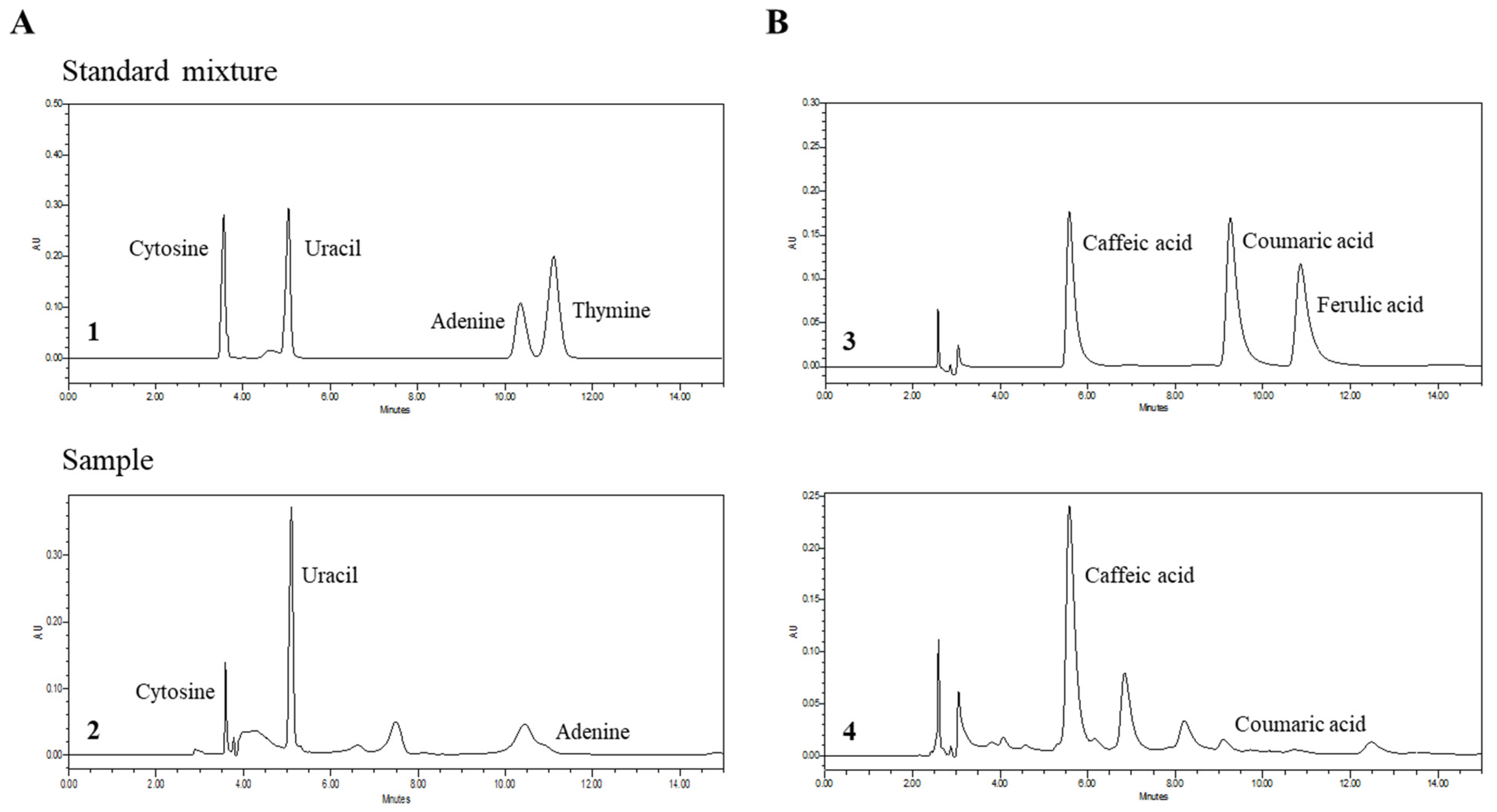
All three nucleobases isolated from PSE were successfully separated using high-pressure liquid chromatography (HPLC) within 15 min with a gradient elution of 1% NaH2PO4 and acetonitrile. Uracil, adenine, and thymine were analyzed by comparing them with a standard compound mixture. The HPLC analysis of the EA layer of PSE at 3 mg/mL (Figure 6A) under the same conditions revealed the presence of uracil and adenine, but thymine was barely detected. The nucleobases that were not isolated from PSE, cytosine, and guanine, were not detected (Figure 6A).
All three phenolic acids isolated from PSE were successfully separated using HPLC within 15 min with a gradient elution of 0.5% trifluoroacetic acid in water and acetonitrile. Coumaric acid, ferulic acid, and caffeic acid were analyzed by comparing them with a standard compound mixture. The HPLC analysis of PSE under the same conditions revealed the presence of caffeic acid, coumaric acid, and ferulic acid (Figure 6B).
Based on the results of the HPLC analysis, adenine and caffeic acid were set as marker compounds of PSE. The optimal extraction methods were determined based on the yield of the extracts and the contents of marker compounds.

3.4. Determination of Optimal Extraction Conditions for PSE

3.4.1. The Yield of PSE

In order to determine optimal extraction conditions for PSE, peanut sprouts were extracted with different extraction solvents, 30% to 100% ethanol, and the yields of the extracts were measured (Figure 7A). The yields of the extracts were between 3.3% and 3.8%, and 70% ethanol yielded better extraction efficiency. In addition, the yields of the extracts were also compared based on the days that peanut sprouts grew. The yields of extracts were between 1.5% and 4.5%, and peanut sprouts that grew 7 days gave better extraction efficiency in the case of 100% ethanol extraction (Figure 7B).

3.4.2. Contents of Marker Compounds with Different Extraction Solvents

The contents of adenine and caffeic acid in PSE extracted with 30–100% ethanol were determined using HPLC. The PSE extracted with a lower % of ethanol showed a higher amount of adenine. The content of adenine was the highest in the 30% ethanol extract at 84.1 μg/g (Figure 8A,C). In the case of caffeic acid, extraction with a higher % of ethanol allowed a higher isolation amount of caffeic acid, but the contents of caffeic acid were similar in PSE extracted with 60–100% ethanol. The highest content of caffeic acid was 1198.4 μg/g in the 100% extract (Figure 8B,D).

3.4.3. Contents of Marker Compounds in Peanut Sprouts Grown Different Days

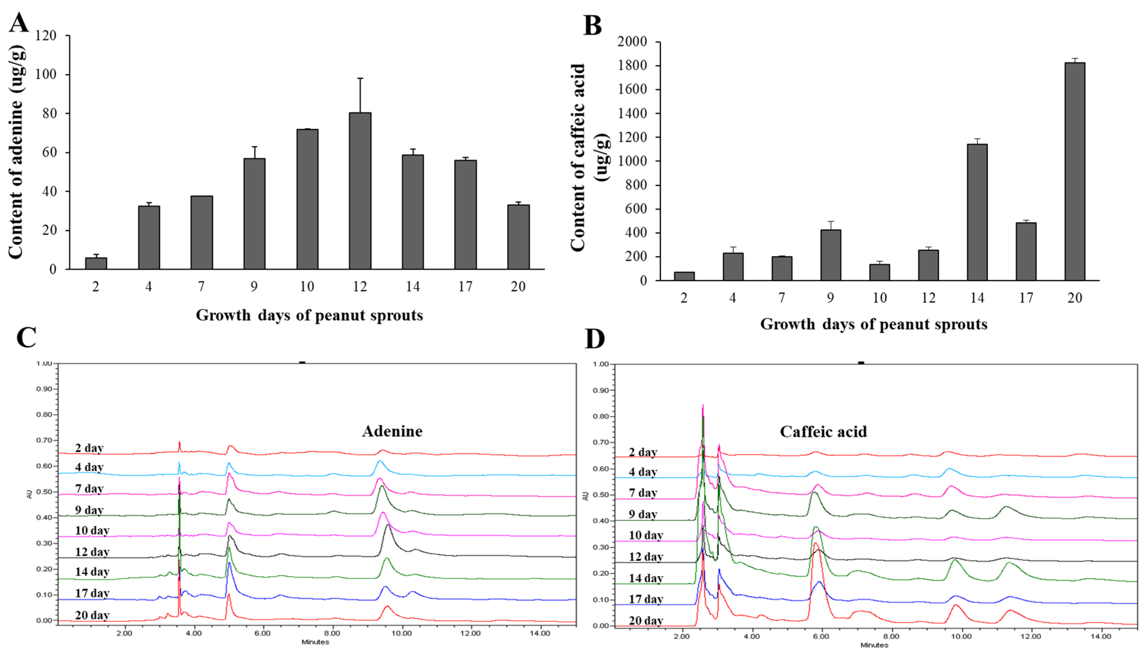
In order to determine the optimal growth days for peanut sprouts, peanut sprouts grown for 2–20 days were collected and extracted with 100% ethanol. The contents of adenine were between 5 and 80 μg/g. The highest content of adenine was 80.3 μg/g in the 12th-day extract (Figure 9A,C). In the case of caffeic acid, the contents of caffeic acid were between 100 and 2500 μg/g. The highest content of caffeic acid was 2549.6 μg/g in the 17th-day extract (Figure 9B,D).

3.4.4. Optimal Extraction Condition for PSE

Adenine and caffeic acid at 4 and 1 μg/mL, marker compounds of PSE, increased ALP activity by 40% and 30%, respectively, compared to the ODM group. The contents of adenine increased as the ethanol ratio decreased, whereas the contents of caffeic acid was the highest in 100% ethanol but similar in 60–100% ethanol extracts. In the case of growth days of peanut sprouts, the contents of adenine were relatively high in peanut sprouts grown for 9–17 days, and those of caffeic acid were high in peanut sprouts grown for 14–20 days. Taken together, the extraction of peanuts grown for 14–17 days with 60% ethanol was considered the optimal extraction method for nucleobases and phenolic acids of peanut sprouts in order to have better osteoblastic differentiation activity.

4. Discussion

Peanut sprouts are a relatively brand-new product derived from peanuts, one of the most popular foods in the world. Most reports regarding peanut sprouts have mainly focused on their antioxidant and anti-inflammatory properties, which are exerted by the presence of phenolic compounds, including resveratrol and caffeic acid, as active constituents [32,33,34,35]. In this study, we found that peanut sprouts efficiently promote the osteoblastic differentiation of MC3T3-E1 cells and nucleobases such as adenine, thymine, and uracil, and phenolic acids such as coumaric acid, ferulic acid, and caffeic acid, were identified as active compounds. To the best of our knowledge, this is the first report regarding nucleobases as inducers of osteoblastic differentiation.
PSEs have been known to alleviate benign prostatic hyperplasia [36] due to resveratrol, a main responsible compound [37]. In addition, the supplementation of PSE improved the indices of obesity, including waist circumference, systolic blood pressure, serum LDL and triglyceride, and serum aspartate transaminase and alanine transaminase [19]. It also reduced the lipid contents and mRNA expression of adipogenesis-related genes more efficiently than non-germinated peanuts [38]. A study on the nutrition difference between peanut sprouts and peanuts revealed that peanut sprouts have less carbohydrates, but more vitamin C, niacin, calcium, and proteins [39]. Furthermore, the germination of peanuts increased the amount of caffeic acid and this enhancement was effective in protecting erythrocytes from oxidative stress [34]. In addition, Kim et al. [40] reported the enhancement of osteogenic differentiation by peanut sprout water extract and soyasaponin Bb as active constituents. However, our study indicated that the ethyl acetate fraction of PSE is the active fraction, and neucleobases such as adenine, thymine, and uracil, and phenolic acids such as coumaric acid, ferulic acid, and caffeic acid, are the active constituents in PSE.
Nucleobases in nucleic acids are divided into two groups: purine type (adenine and guanine) and pyrimidine type (thymine, uracil, and cytosine). Nucleobases are not only associated with DNA and RNA synthesis but also involved in various physiological functions [23]. Thus, nucleobases are considered important templates for the development of new drugs with potent efficacy as anti-cancer, anti-viral, and anti-bacterial agents [41]. In particular, adenine has been reported to have pharmacological potential against allergies and inflammation. Adenine suppresses the allergic reaction in mast cells [42]. In addition, adenine exerted anti-inflammatory activity in LPS-stimulated macrophage cells, mast cells, and peritoneal cells in mice [43]. The anti-inflammatory effect of adenine against TNF-alpha-induced inflammation in MG-63 cells (human osteoblast-like cells) enhanced osteoblastic differentiation by increasing collagen and ALP enzyme activity [44]. This may be explained by the fact that adenine attenuates inflammation, which is one main factor among multiple pathogenic factors of osteoporosis [44]. These reports suggest that nucleobases, including adenine, could exert osteoblastic differentiation by reducing inflammatory responses.
The term “phenolic acid” generally describes phenolic compounds with one carboxylic acid group. Phenolic acids are found in a variety of plants, with the highest concentrations in seeds, fruit peels, and leaves. Phenolic acids are divided into two groups: hydroxybenzoic and hydroxycinnamic acid [45]. Phenolic acids have significantly higher in vitro antioxidant activity than vitamins [46]. It is also known that phenolic acids upregulate osteoblastogenesis and downregulate osteoclastogenesis via antioxidant activity by scavenging reactive oxygen species [47]. Furthermore, phenolic acids promote osteoblastic differentiation by increasing levels of Runx2, osteocalcin, and Wnt and by suppressing the expression of RANKL, TRAP, and MMPs [48]. In particular, caffeic acid has been reported to have pharmacological potential as an antioxidant [49]. In addition, caffeic acid attenuated neuroinflammation in LPS-stimulated mice, and inflammatory response in synovial tissues obtained from rheumatoid arthritis patients [50,51]. Caffeic acid also reduces osteoclastogenesis and bone resorption through its antioxidant potential and increases expression of osteoblast markers [52]. In addition, tannic acid also increases osteoblastic cell proliferation and mineralization [53]. In the case of 3(3-hydroxyphenyl) propionic acid, the treatment increased the production of ALPL in bone marrow-derived mouse stromal (ST2) cells and suppressed osteoclastogenesis through the RANKL-RANK independent pathway in Raw 264.7 cells [54,55]. These reports suggest that phenolic acids help protect bones by promoting osteoblastogenesis and reducing osteoclast formation and bone resorption.

5. Conclusions

In conclusion, peanut sprouts significantly upregulate ALP activity, indicating increased osteoblastic differentiation of pre-osteoblastic MC3T3-E1 cells. In addition, three nucleobases (adenine, thymine, and uracil) and three phenolic acids (caffeic acid, ferulic acid, and coumaric acid) were isolated as their active constituents. Furthermore, all nucleobases and phenolic acids significantly increased ALP activity. These results suggest that peanut sprouts and their constituents enhance osteogenic differentiation. In addition, the extraction of peanut sprouts grown for 14–17 days with 60% ethanol was determined as an optimal extraction condition for the isolation of marker compounds. Taken together, peanut sprouts, nucleobases, and phenolic acids have the potential to be developed as functional foods beneficial for osteoporosis. However, further in vivo animal studies are needed to confirm the beneficial effects of peanut sprouts, nucleobases, and phenolic acids in osteoporosis treatment.

Supplementary Materials

The following supporting information can be downloaded at: https://www.mdpi.com/article/10.3390/separations10080435/s1, Figure S1: Peanut sprouts with different growth days (2–20 days); Figure S2: Scheme for the isolation of active compounds from peanut sprouts based on activity guided isolation method.

Author Contributions

Conceptualization, S.-Y.P. and K.W.H.; methodology, E.-S.C. and C.H.L.; software, M.S.K.; validation, E.-S.C., C.H.L. and M.S.K.; formal analysis, E.-S.C. and C.H.L.; investigation, E.-S.C. and C.H.L.; resources, S.-Y.P.; data curation, M.S.K.; writing—original draft preparation, S.-Y.P. and K.W.H.; writing—review and editing, J.Y.C. and K.W.H.; visualization, M.S.K.; supervision, S.-Y.P.; project administration, K.W.H.; funding acquisition, S.-Y.P. and K.W.H. All authors have read and agreed to the published version of the manuscript.

Funding

This research was funded by the Ministry of Trade, Industry & Energy (MOTIE), Korea Institute for Advancement of Technology (KIAT), through the Encouragement Program for The Industries of Economic Cooperation Region (P0008626). This research was also supported by the Chung-Ang University research grant in 2021.

Data Availability Statement

Not applicable.

Acknowledgments

The authors thank the Center for Bio-Medical Engineering Core Facility at Dankook University for NMR experiments.

Conflicts of Interest

The authors declare no conflict of interest.

References

  1. Burge, R.; Dawson-Hughes, B.; Solomon, D.H.; Wong, J.B.; King, A.; Tosteson, A. Incidence and Economic Burden of Osteoporosis-Related Fractures in the United States. J. Bone Miner. Res. 2007, 22, 465–475. [Google Scholar] [CrossRef]
  2. KIM, T.H.; Jung, J.W.; Ha, B.G.; Hong, J.M.; Park, E.K.; Kim, H.J.; Kim, S.Y. The Effects of Luteolin on Osteoclast Differentiation, Function in Vitro and Ovariectomy-Induced Bone Loss. J. Nutr. Biochem. 2011, 22, 8–15. [Google Scholar] [CrossRef]
  3. Shiraishi, A.; Muyabe, S.; Nakano, T.; Umakoshi, Y.; Ito, M.; Mihara, M. The Combination Therapy with Alfacalcidol and Risedronate Improves the Mechanical Property in Lumbar Spine by Affecting the Material Properties in an Ovariectomized Rat model of Osteoporosis. BMC Musculoskelet. Disord. 2009, 10, 66. [Google Scholar] [CrossRef] [Green Version]
  4. Pavone, V.; Testa, G.; Giardina, S.M.C.; Vescio, A.; Restivo, D.A.; Sessa, G. Pharmacological Therapy of Osteoporosis: A systematic Current Review of Literature. Front. Pharmacol. 2017, 8, 803. [Google Scholar]
  5. Riggs, B.L.; Khosla, S.; Melton, L.J., 3rd. Sex Steroids and the Construction and Conservation of the Adult Skeleton. Endocr. Rev. 2002, 23, 279–302. [Google Scholar] [CrossRef]
  6. Park, M.H.; Kim, S.Y.; Cheon, J.H.; Lee, J.Y.; Kim, B.K.; Lee, S.H.; Kong, C.S.; Kim, Y.Y.; Kim, M.H. Effects of Scytosiphon lomentaria on Osteoblastic Proliferation and Differentiation of MC3T3-E1 cells. Nutr. Res. Pract. 2016, 10, 148–153. [Google Scholar] [PubMed] [Green Version]
  7. Lane, N.E.; Kelman, A. A Review of Anabolic Therapies for Osteoporosis. Arthritis Res. Ther. 2003, 5, 214–222. [Google Scholar] [PubMed] [Green Version]
  8. Teitelbaum, S.L. Bone Resorption by Osteoclasts. Science 2000, 289, 1504–1508. [Google Scholar] [CrossRef] [PubMed]
  9. Yoon, H.J.; Seo, C.R.; Kim, M.A.; Kim, Y.J.; Song, N.J.; Jang, W.S.; Kim, B.J.; Lee, J.H.; Hong, J.W.; Nho, C.W.; et al. Dichloromethane Extracts of Sophora japonica L. Stimulate Osteoblast Differentiation In mesenchymal Stem Cells. Nutr. Res. 2013, 33, 1053–1062. [Google Scholar]
  10. Golub, E.E.; Boesze-Battaglia, K. The Role of Alkaline Phosphatase in Mineralization. Curr. Opin. Orthop. 2007, 18, 444–448. [Google Scholar] [CrossRef]
  11. Gupta, T.; Das, N.; Imran, S. The Prevention and Therapy of Osteoporosis. A Review on Emerging Trends from Hormonal Therapy to Synthetic Drugs to Plant-Based Bioactives. J. Diet. Suppl. 2019, 16, 699–713. [Google Scholar] [CrossRef]
  12. Putnam, S.E.; Scutt, A.M.; Bicknell, K.; Priestley, C.M.; Williamson, E.M. Natural Products as Alternative Treatments for Metabolic Bone Disorders and for Maintenance of Bone Health. Phytother. Res. 2007, 21, 99–112. [Google Scholar] [CrossRef] [PubMed]
  13. Yen, P.H.; Kiem, P.V.; Nhiem, N.X.; Tung, N.H.; Quang, T.H.; Minh, C.V.; Kim, J.W.; Choi, E.M.; Kim, Y.H. A New Monoterpene Glycoside from the Roots of Paeonia lactiflora Increases the Differentiation of Osteoblastic MC3T3-E1 cells. Arch. Pharm. Res. 2007, 30, 1179–1185. [Google Scholar] [CrossRef]
  14. Yun, H.M.; Park, K.R.; Quang, T.H.; Oh, H.; Hong, J.T.; Kim, Y.C.; Kim, E.C. 2,4,5-Trimethoxyldalbergiquinol Promotes Osteoblastic Differentiation and Mineralization Via the BMP and Wnt/beta-catenin Pathway. Cell Death. Dis. 2015, 6, e1819. [Google Scholar] [CrossRef] [PubMed] [Green Version]
  15. Son, H.E.; Kim, T.H.; Jang, W.G. Curculactones A and B Induced the Differentiation of C3H10T1/2 and MC3T3-E1 Cells to Osteoblasts. Bioorg. Med. Chem. Lett. 2017, 27, 1301–1303. [Google Scholar] [CrossRef]
  16. Thu, H.E.; Hussain, Z.; Mohamed, I.N.; Shuid, A.N. Eurycoma Longifolia, a Promising Suppressor of RANKL-Induced Differentiation and Activation of Osteoclasts: An in Vitro Mechanistic Evaluation. J. Ayurveda Integr. Med. 2019, 10, 102–110. [Google Scholar] [CrossRef]
  17. Li, W.D.; Yan, C.P.; Wu, Y.; Weng, Z.B.; Yin, F.Z.; Yang, G.M.; Cai, B.C.; Chen, Z.P. Osteoblasts Proliferation and Differentiation Stimulating Activities of the Main Components of Fructus Psoraleae corylifoliae. Phytomedicine 2014, 21, 400–405. [Google Scholar] [CrossRef] [PubMed]
  18. Chai, L.; Zhou, K.; Wang, S.; Zhang, H.; Fan, N.; Li, J.; Tan, X.; Hu, L.; Fan, X. Psoralen and Bakuchiol Ameliorate M-CSF Plus RANKL-Induced Osteoclast Differentiation and Bone Resorption Via Inhibition of AKT and AP-1 Pathways in Vitro. Cell. Physiol. Biochem. 2018, 48, 2123–2133. [Google Scholar] [CrossRef]
  19. Ha, A.W.; Kim, W.K.; Kim, J.H.; Kang, N.E. The Supplementation Effects of Peanut Sprout on Reduction of Abdominal Fat and Health Indices in Overweight and Obese Women. Nutr. Res. Pract. 2015, 9, 249–255. [Google Scholar] [CrossRef] [Green Version]
  20. Ha, A.W.; Kang, N.E.; Kim, W.K. Ethanol Extract of Peanut Sprout Lowers Blood Triglyceride Levels, Possibly Through a Pathway Involving SREBP-1c in Rats Fed a High-Fat Diet. J. Med. Food 2015, 18, 850–855. [Google Scholar] [CrossRef]
  21. Limmongkon, A.; Nopprang, P.; Chaikeandee, P.; Somboon, T.; Wongshaya, P.; Pilaisangsuree, V. LC-MS/MS Profiles and Interrelationships Between the Anti-Inflammatory Activity, Total Phenolic Content and Antioxidant Potential of Kalasin 2 cultivar Peanut Sprout Crude Extract. Food Chem. 2018, 239, 569–578. [Google Scholar] [CrossRef]
  22. Lee, Y.M.; Son, E.J.; Kim, D.S. Treatment with Peanut Sprout Root Extract Alleviates Inflammation in a Lipopolysaccharide-Stimulated Mouse Macrophage Cell Line by Inhibiting the MAPK Signaling Pathway. Int. J. Mol. Sci. 2019, 20, 5907. [Google Scholar] [CrossRef] [PubMed] [Green Version]
  23. Mediero, A.; Cronstein, B.N. Adenosine and Bone Metabolism. Trends Endocrinol. Metab. 2013, 24, 290–300. [Google Scholar] [CrossRef] [Green Version]
  24. Fredholm, B.B.; Ijerman, A.P.; Jacobson, K.A.; Linden, J.; Muller, C.E. International Union of Basic and Clinical Pharmacology. LXXXI. Nomenclature and Classification of Adenosine Receptors-an Update. Pharmacol. Rev. 2011, 63, 1–34. [Google Scholar] [CrossRef] [Green Version]
  25. Strazzulla, L.C.; Cronstein, B.N. Regulation of Bone and Cartilage by Adenosine Signaling. Purinergic Signal. 2016, 12, 583–593. [Google Scholar] [CrossRef] [Green Version]
  26. Jacobsen, N.E. NMR Data Interpretation Explained: Understanding 1D and 2D NMR Spectra of Organic Compounds and Natural Products; John Wiley & Sons: Hoboken, NJ, USA, 2016; pp. 348–349. [Google Scholar]
  27. Mohamed, T.A.; Shabaan, I.A.; Zoghaib, W.M.; Husband, J.; Farag, R.S.; Alajhaz, A.M.A. Tautomerism, Normal Coordinate Analysis, Vibrational Assignments, Calculated IR, Raman and NMR Spectra of Adenine. J. Mol. Struct. 2009, 938, 263–276. [Google Scholar] [CrossRef]
  28. Benoit, R.L.; Frechette, M. 1H and 13C nuclear Magnetic Resonance and Ultraviolet Studies of the Protonation of Cytosine, Uracil, Thymine, and Related Compounds. Can. J. Chem. 1986, 64, 2348–2352. [Google Scholar] [CrossRef]
  29. Exarchou, V.; Troganis, A.; Gerothanassis, I.P.; Tsimidou, M.; Boskou, D. Identification and Quantification of Caffeic and Rosmarinic Acid in Complex Plant Extracts by the Use of Variable-Temperature Two-Dimensional Nuclear Magnetic Resonance Spectroscopy. J. Agric. Food Chem. 2001, 49, 2–8. [Google Scholar] [CrossRef] [PubMed]
  30. Sajjadi, S.E.; Shokoohinia, Y.; Moayedi, N.S. Isolation and Identification of Ferulic Acid from Aerial Parts of Kelussia odoratissima Mozaff. Jundishapur J. Nat. Pharm. 2012, 7, 159–162. [Google Scholar] [CrossRef] [Green Version]
  31. Shimomura, H.; Sashida, Y.; Mimaki, Y.; Kudo, Y.; Maeda, K. New Phenylpropanoid Glycerol Glucosides from the Bulbs of Lilium Species. Chem. Pharm. Bull. 1988, 36, 4841–4848. [Google Scholar] [CrossRef] [Green Version]
  32. Wang, K.H.; Lai, Y.H.; Chang, J.C.; Ko, T.F.; Shyu, S.L.; Chiou, R.Y. Germination of Peanut Kernels to Enhance Resveratrol Biosynthesis and Prepare Sprouts as a Functional Vegetable. J. Agric. Food Chem. 2005, 53, 242–246. [Google Scholar] [CrossRef] [PubMed]
  33. Kang, H.I.; Kim, J.Y.; Kwon, S.J.; Park, K.W.; Kang, J.S.; Seo, K.I. Antioxidative Effects of Peanut Sprout Extracts. J. Korean Soc. Food Sci. Nutr. 2010, 39, 941–946. [Google Scholar] [CrossRef]
  34. Wang, G.; Lei, Z.; Zhong, Q.; Wu, W.; Zhang, H.; Min, T.; Wu, H.; Lai, F. Enrichment of Caffeic acid in Peanut Sprouts and Evaluation of its In vitro Effectiveness Against Oxidative Stress-Induced Erythrocyte Hemolysis. Food Chem. 2017, 217, 332–341. [Google Scholar] [CrossRef]
  35. Choi, D.I.; Choi, J.Y.; Kim, Y.J.; Lee, J.B.; Kim, S.O.; Shin, H.T.; Lee, S.C. Ethanol Extract of Peanut Sprout Exhibits a Potent Anti-Inflammatory Activity in Both an Oxazolone-Induced Contact Dermatitis Mouse Model and Compound 48/80-Treated HaCaT Cells. Ann. Dermatol. 2015, 27, 142–151. [Google Scholar] [CrossRef]
  36. Song, J.H.; Hwang, B.D.; Chung, H.J.; Moon, B.K.; Kim, J.W.; Ko, K.S.; Kim, B.W.; Kim, W.R.; Kim, W.J.; Myung, S.C.; et al. Peanut Sprout Extracts Cultivated with Fermented Sawdust Medium Inhibits Benign Prostatic Hyperplasia In vitro and In vivo. World J. Men’s Health 2020, 38, 385–396. [Google Scholar] [CrossRef] [Green Version]
  37. Pyo, K.H.; Lee, Y.W.; Lee, S.H.; Xin, C.F.; Shin, J.H.; Shin, E.H. Preventive Effects of Resveratrol Enriched Extract of Peanut Sprout on Bacteria and Estradiol Induced Prostatitis in Mice. Nat. Prod. Commun. 2017, 12, 73–78. [Google Scholar] [CrossRef]
  38. Boonsong, T.; Pakwan, S.; Chawnawa, W. Anti-Adipogenesis and Antioxidative Defense Status of a Crude Extract of ‘Kalasin 2’ Peanut Sprouts in 3T3-L1 Mouse Adipocytes. Biochem. Cell Biol. 2019, 97, 740–749. [Google Scholar] [CrossRef] [PubMed]
  39. Bonku, R.; Yu, J. Health Aspects of Meanuts as an Outcome of its Chemical Composition. Food Sci. Hum. Wellness 2020, 9, 21–30. [Google Scholar] [CrossRef]
  40. Kim, S.H.; Yuk, H.J.; Ryu, H.W.; Oh, S.R.; Song, D.Y.; Lee, K.S.; Park, K.I.; Choi, S.W.; Seo, W.D. Biofunctional Soyasaponin Bb in Peanut (Arachis hypogaea L.) Sprouts Enhances Bone Morphogenetic Protein-2-Dependent Osteogenic Differentiation via Activation of Runt-Related Transcription Factor 2 in C2C12 Cells. Phytother. Res. 2019, 33, 1490–1500. [Google Scholar] [CrossRef] [Green Version]
  41. Wong, X.K.; Yeong, K.Y. From Nucleic Acids to Drug Discovery. Nucleobases as Emerging Templates for Drug Candidates. Curr. Med. Chem. 2021, 28, 7076–7121. [Google Scholar] [CrossRef]
  42. Hosoi, T.; Ino, S.; Ohnishi, F.; Todoroki, K.; Yoshii, M.; Kakimoto, M.; Muller, C.E.; Ozawa, K. Mechanisms of the Action of Adenine on Anti-Allergic Effect in Mast Cells. Immun. Inflamm. Dis. 2018, 6, 97–105. [Google Scholar] [CrossRef] [PubMed]
  43. Silwal, P.; Lim, K.; Heo, J.Y.; Park, J.I.; Namgung, U.; Park, S.K. Adenine Attenuates Lipopolysaccharide Induced Inflammatory Reactions. Korean J. Physiol. Pharmacol. 2018, 22, 379–389. [Google Scholar] [CrossRef] [Green Version]
  44. Chen, Y.P.; Chu, Y.L.; Tsuang, Y.H.; Wu, Y.; Kuo, C.Y.; Kuo, Y.J. Anti-Inflammatory Effects of Adenine Enhance Osteogenesis on the Osteoblast Like MG-63 Cells. Life 2020, 10, 116. [Google Scholar] [CrossRef] [PubMed]
  45. Clifford, M.N. Chlorogenic Acids and Other Cinnamates—Nature, Occurrence and Dietary Burden. J. Sci. Food Agric. 1999, 79, 3. [Google Scholar] [CrossRef]
  46. Tsao, R.; Deng, Z. Separation Procedures for Naturally Occurring Antioxidant Phytochemicals. J. Chromatogr. B Anal. Technol. Biomed. Life Sci. 2004, 812, 85–99. [Google Scholar] [CrossRef]
  47. Oh, T.W.; Park, K.-I.; Do, H.J.; Kim, K.; Yang, H.J.; Cho, W.K.; Ma, J.Y. Acer tegmentosum Maxim Prevents Bone Loss by Inhibiting Osteoclastogenesis and Promoting Osteoblast Mineralization in Ovariectomized Mice. Nat. Prod. Sci. 2020, 26, 83–89. [Google Scholar] [CrossRef]
  48. Zhou, Y.; Wu, Y.; Jiang, X.; Zhang, X.; Xia, L.; Lin, K.; Xu, Y. The Effect of Quercetin on the Osteogenesic Differentiation and Angiogenic Factor Expression of Bone Marrow-Derived Mesenchymal Stem Cells. PLoS ONE 2015, 10, e0129605. [Google Scholar] [CrossRef] [PubMed] [Green Version]
  49. Chen, J.H.; Ho, C.T. Antioxidant Activities of Caffeic Acid and Its Related Hydroxycinnamic Acid Compounds. J. Agric. Food Chem. 1997, 45, 2374–2378. [Google Scholar] [CrossRef]
  50. Mallik, S.B.; Mudgal, J.; Nampoothiri, M.; Hall, S.; Anoopkumar-Dukie, S.; Grant, G.; Rao, C.M.; Arora, D. Caffeic Acid Attenuates Lipopolysaccharide-Induced Sickness Behaviour and Neuroinflammation in Mice. Neurosci. Lett. 2016, 6, 218–223. [Google Scholar] [CrossRef]
  51. Wang, W.; Sun, W.; Jin, L. Caffeic Acid Alleviates Inflammatory Response in Rheumatoid Arthritis Fibroblast-Like Synoviocytes by Inhibiting Phosphorylation of IκB Kinase α/β and IκBα. Int. Immunopharmacol. 2017, 48, 61–66. [Google Scholar] [CrossRef]
  52. Ekeuku, S.O.; Pang, K.L.; Chin, K.Y. Effects of Caffeic Acid and Its Derivatives on Bone: A Systematic Review. Drug Des. Dev. Ther. 2021, 15, 259–275. [Google Scholar] [CrossRef] [PubMed]
  53. Hapidin, H.; Romli, N.A.A.R.; Abdullah, H. Proliferation Study and Microscopy Evaluation on the Effects of Tannic Acid in Human Fetal Osteoblast Cell Line (hFOB 1.19). Microsc. Res. Tech. 2019, 82, 1928–1940. [Google Scholar] [CrossRef] [PubMed]
  54. Chen, J.R.; Wankhade, U.D.; Alund, A.W.; Blackburn, M.L.; Shankar, K.; Lazarenko, O.P. 3-(3-Hydroxyphenyl)-Propionic Acid (PPA) Suppresses Osteoblastic Cell Senescence to Promote Bone Accretion in Mice. JBMR Plus 2019, 3, e10201. [Google Scholar] [CrossRef] [PubMed] [Green Version]
  55. Zhao, H.; Lazarenko, O.P.; Chen, J.R. Hippuric Acid and 3-(3-Hydroxyphenyl) Propionic Acid Inhibit Murine Osteoclastogenesis through RANKL-RANK Independent Pathway. J. Cell. Physiol. 2020, 235, 599–610. [Google Scholar] [CrossRef] [Green Version]
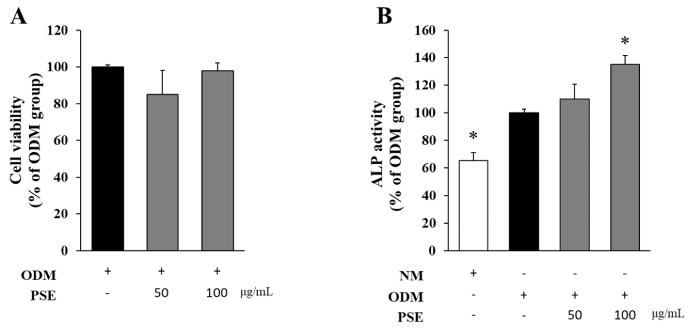
Figure 1. The effect of peanut sprout extract (PSE) on osteoblastic differentiation. The changes in cell viability (A) and ALP activity (B) with the treatment of PSE (100 μg/mL) were determined using the MTT and ALP assay, respectively. The experiments were repeated at least three times, and data were expressed as mean ± SD. * p < 0.05 in comparison to the ODM group. (ODM−osteoblastic differentiation media; NM−normal media).
Separations 10 00435 g001
Figure 2. The effect of solvent-partitioned layers of peanut sprout extract (PSE) on osteoblastic differentiation. The changes in cell viability (A) and ALP activity (B) were determined using the MTT and ALP assay, respectively. The experiments were repeated at least three times, and data were expressed as mean ± SD. * p < 0.05 in comparison to the ODM group. (ODM−osteoblastic differentiation media; NM−normal media).
Separations 10 00435 g002
Figure 3. Structures of nucleobases and phenolic acids isolated from PSE.
Separations 10 00435 g003
Figure 4. The effects of nucleobases and phenolic acids isolated from peanut sprout extract (PSE) on osteoblastic differentiation. The cell viability (A,B) and ALP activity (C,D) were determined using the MTT and ALP assay, respectively. The experiments were repeated at least three times, and data were expressed as mean ± SD. * p < 0.05 in comparison to the ODM group. (ODM−osteoblastic differentiation media; NM−normal media).
Separations 10 00435 g004
Figure 5. The effect of five nucleobases on osteoblastic differentiation. MC3T3-E1 cells were treated with five nucleobases (4 μg/mL) for 5 days in the presence of ODM (A), and ALP activity (B) was determined using the ALP assay. The experiments were repeated at least three times, and data were expressed as mean ± SD. * p < 0.05 in comparison to the ODM group. (ODM−osteoblastic differentiation media; NM−normal media).
Separations 10 00435 g005
Figure 6. HPLC analysis of peanut sprout extract (PSE). (A) Chromatographic analysis of nucleobases was performed using a C18 column with a gradient solvent system composed of 1% NAH2PO4 in water and acetonitrile in a gradient manner. The injection volume of the EA layers of PSE (3 mg/mL) was 10 μL. The detection wavelength was 260 nm. 1: Standard mixture (uracil, adenine, and thymine). 2: EA layers of PSE (3 mg/mL). (B) Chromatographic analysis of phenolic acids was performed using a C18 column with a gradient solvent system composed of 0.5% trifluoroacetic acid in water and acetonitrile in a gradient manner. 3: Standard mixture (caffeic acid, coumaric acid, and ferulic acid). 4: 100% Ethanol extract of peanut sprouts (20 mg/mL).
Separations 10 00435 g006
Figure 7. The yields of peanut sprout extracts (PSEs). (A) The yields of PSEs depending on the extraction solvents with different % of ethanol. (B) The yields of PSEs depending on peanut sprouts with different growth days with 100% ethanol.
Separations 10 00435 g007
Figure 8. The contents of marker compounds in PSEs and their chromatogram profiles. (A,B) The contents of marker compounds adenine and caffeic acid in PSE extracted with different % of ethanol. (C) HPLC chromatograms of PSEs for adenine. (D) HPLC chromatograms of PSEs for caffeic acid.
Separations 10 00435 g008
Figure 9. The contents of marker compounds in PSE according to the growth days of peanut sprouts and their chromatogram profiles. Peanut sprouts at different growth days, such as 2, 4, 7, 9, 10, 12, 14, 17, and 20 days, were extracted with 100% ethanol, and the extracts were analyzed using HPLC. (A,C) The content of adenine in PSEs and their HPLC chromatograms. (B,D) The content of caffeic acid in PSEs and their HPLC chromatograms.
Separations 10 00435 g009
Disclaimer/Publisher’s Note: The statements, opinions and data contained in all publications are solely those of the individual author(s) and contributor(s) and not of MDPI and/or the editor(s). MDPI and/or the editor(s) disclaim responsibility for any injury to people or property resulting from any ideas, methods, instructions or products referred to in the content.