Abstract
Capillary zone electrophoresis (CZE) is an important technique for the analysis of monoclonal antibodies (mAbs). A recently released light-emitting diode (LED)-induced fluorescence (LEDIF) detector equipped with a 275 nm LED for the detection of proteins through their native fluorescence was used in this study and compared to results obtained using the predominant detection mode, the measurement of the absorption of ultraviolet light (UV detection). This was accomplished using an established CZE method for the analysis of three mAbs: NISTmAb, matuzumab, and Intact Mass Check Standard (Waters). For this purpose, the detector’s settings were first optimized using a design of experiments approach. Three factors, rise time, photomultiplier high voltage supply, and acquisition frequency, were optimized by means of a D-optimal design. The optimal settings were then used for the investigation of signal-to-noise ratios (S/Ns), linearity, and precision. LEDIF detection offered a similar separation quality, up to 12 times higher S/Ns, and lower limits of detection compared to UV detection. Repeatability was excellent, with relative standard deviations (RSDs) of approximately 1% for percentage areas. For intermediate precision, RSDs of <2% (n = 3 × 10) were typically achieved. Overall, LEDIF detection was found to be an excellent and easily optimizable alternative to UV detection.
1. Introduction
Capillary electrophoresis (CE) combined with laser-induced fluorescence (LIF) for detection has been used since the 1980s [1]. It offers a higher selectivity and better sensitivity than detection through the absorption of ultraviolet light (UV detection). CE-LIF has been the topic of numerous excellent reviews [2,3,4,5,6] and may be applied to many analytes, e.g., glycans [7], DNA [8], RNA [9], amino acids [10], peptides, and proteins [6,11]. For the LIF detection of proteins, either the protein’s native fluorescence is used or the protein is labelled with a fluorescent dye. The latter usually offers better limits of detection (LODs) [4]; however, the protein’s physicochemical properties are altered in the process [12]. This is not the case if the native fluorescence of the protein is used. Although some proteins possess a specific fluorophore, e.g., green fluorescent protein, which can be used for detection [13,14], this is rather uncommon. Fortunately, the native or intrinsic fluorescence of the aromatic amino acids, phenylalanine, tyrosine, and especially tryptophan, may be utilized for most proteins. The required excitation wavelength is typically around 280 nm [15]. For CE applications, lasers were the predominant excitation source [4]. In recent years, light-emitting diodes (LEDs) have gained popularity as alternatives to lasers [16,17]. The performance of three selected LEDs was investigated by Rodat-Boutonet et al. for the capillary electrophoresis sodium dodecyl sulfate (CE-SDS) analysis of monoclonal antibodies (mAbs). They concluded that LEDs require less energy, are less expensive, and offer a better baseline than comparable lasers [18].
Recently, an LED-induced fluorescence (LEDIF) detector equipped with a suitable LED (275 nm) for the detection of proteins through their native fluorescence has become commercially available. It uses the established collinear ball lens arrangement [17,19,20], offers ease of use, and its control can be integrated into the CE system’s software. In the present case, it was used for the analysis of mAbs in combination with capillary zone electrophoresis (CZE). CZE is, among CE-SDS, capillary isoelectric focusing (CIEF), and imaged capillary isoelectric focusing (iCIEF), one of the routinely used electrophoretic techniques for the analysis of biopharmaceuticals in general and mAbs in particular [21,22,23]. While the detection of mAbs through induced fluorescence is described in combination with CE-SDS [18,24,25] and iCIEF [26,27], its combination with CZE has not been explored to date and is now being investigated for the first time. Since the labelling of mAbs with fluorescent dyes would change their charge-to-size ratio, native fluorescence should be favored so as to not impair the separation. The combination of CZE with native LEDIF detection could offer two advantages over UV detection. First, lower limits of quantification (LOQs) are expected. This might be beneficial for the detection of low abundant charge variants, e.g., in stability studies. Second, the better selectivity would allow for a wider selection of possible background electrolyte (BGE) constituents. Currently, the commonly employed detection at 214 nm [28] prevents the use of substances which strongly absorb in this region, effectively excluding many substances containing a conjugated electron system. Higher UV detection wavelengths (e.g., 280 nm) offer a reduced sensitivity [26] due to the lower molar absorption coefficient at this wavelength.
Within this study, an established CZE method set for the analysis of mAbs was adapted. It had been previously used in combination with UV detection during an interlaboratory study [29] and provided a good starting point. First, the LEDIF detector’s settings needed to be optimized. Given that several different detector settings are available, some with opposing effects, the optimization was performed using a design of experiments (DoE) approach. According to Fukuda et al., DoE can be defined as “…a structured and organized method for determining the relationships between input factors (xi—independent variables) affecting one or more output responses (y—dependent variables), through the establishment of mathematical models (y = f(xi)).” [30]. DoE approaches are important pieces in (analytical) quality-by-design concepts and frequently used in (analytical) method development [30,31]. Examples of DoE-guided CE-based separation method developments include labelling reactions in CE-LIF [32], CZE for adenovirus particles [33], chiral separations [34,35], as well as CE-SDS [36] and CZE [37] methods for mAb analysis. Within this manuscript, the separation method remained unmodified; the focus was on the LEDIF detector optimization. The presented approach should be easily transferable to similar tasks and could provide a template for future detector optimizations.
In the next step, the optimized detector settings were used for the evaluation of the signal-to-noise ratio (S/N), linearity, and precision. Determined S/Ns were used to estimate LODs and LOQs [38,39]. Additionally, the results obtained with LEDIF detection were compared to results obtained with UV detection.
In total, three different mAbs were used. The first mAb was the NIST monoclonal antibody reference material 8671 (NISTmAb). It is a humanized immunoglobulin G (IgG)1κ mAb, which has been excellently characterized and is intended as a reference material for method evaluation and comparison [40,41]. The second mAb was the Intact mAb Mass Check Standard (Waters mAb). It is a murine mAb distributed by the Waters Corporation and intended as an LC/MS standard for intact mass analysis [42]. The third and last mAb was matuzumab, a humanized IgG1 mAb that targets the extracellular domain of the epidermal growth factor receptor [43]. Matuzumab had been tested therapeutically in clinical trials [44,45], but its development has been discontinued [46].
The aim of this work was to demonstrate a general and systematic approach to LEDIF detector optimization and to evaluate the detector’s performance in comparison to UV detection.
2. Materials and Methods
2.1. Materials
Ultrapure water with an electrical conductivity of 0.055 µS/cm was provided by an arium pro VF system from Sartorius AG, Goettingen, Germany. Triethylenetetramine (TETA) (≥97%, catalog number (cat.) 90460), hydrochloric acid (HCl) (≥37%, p.a./reag. Ph.Eur., cat. 30721-M), and glacial acetic acid (≥99.8%, p.a./reag. Ph.Eur., cat. 33209-M) were purchased from Sigma-Aldrich, Steinheim, Germany. Hydroxypropylmethylcellulose (HPMC) with a viscosity of 2600–5600 cP (2.6–5.6 Pa × s) (2% in water at 20 °C, cat. H7509) was obtained from Sigma-Aldrich, St. Louis, MO, USA. ε-Aminocaproic acid (εACA) (99%, cat. A14719) was acquired from ThermoFisher (Kandel) GmbH, Kandel, Germany. Phosphoric acid (85%) (extra pure, cat. 29570) was supplied by Acros Organics, Geel, Belgium. Matuzumab (10 mg/mL) was received as a gift from Merck KGaA, Darmstadt, Germany. NIST monoclonal antibody reference material 8671 (NISTmAb), lot 14HB-D-002, was obtained from the National Institute of Standards and Technologies, Gaithersburg, MD, USA. Intact mAb Mass Check Standard (cat. 186006552, Waters mAb), lot W07012117, was sourced from Waters Corporation, Milford, MA, USA. NISTmAb, Waters mAb, and HPMC were kindly sponsored by AB SCIEX, Framingham, MA, USA, as part of a different project [29] and the surplus was used herein after its completion.
For this project, the mAbs were prepared according to the following procedure: NISTmAb was diluted to 1 mg/mL in water. For this, two NISTmAb vials (10 mg/mL) were thawed at room temperature, vortexed (3 × 5 s), and centrifuged. The vials were combined, vortexed (3 × 5 s) and centrifuged. Next, 1400 µL was diluted with 12,600 µL water, vortexed (3 × 5 s) and centrifuged. The diluted solution was divided into aliquots, and the aliquots were frozen at −32 °C. After approximately 14 h at −32 °C, the aliquots were stored at −80 °C until use. The Waters mAb lyophilisate was reconstituted with 400 µL water, leading to a final concentration of 2.5 mg/mL; it was then stored in aliquots at −20 °C. For use, the aliquoted mAb solutions were thawed at room temperature, vortexed (3 × 5 s) and centrifuged (1 min/10,000× g). The mAb solutions were thawed once and were subjected to ambient temperature for about 24 h, before they were stored again at −20 °C for 26 weeks. Then, they were used for the repeatability experiments described herein.
Bare-fused silica capillaries with a 50 µm internal diameter and a 365 µm external diameter were a gift from Polymicro Technologies, Phoenix, AZ, USA. They were cut to a total length of either 61 cm (LEDIF detection) or 48.5 cm (UV detection), resulting in an effective length of 40 cm in both cases. The polyimide coating at the detection window and at the capillary tips was removed with a gas torch.
2.2. Preparation of Reagent Solutions
A 1% (w/v) HPMC stock solution was prepared by slowly adding 300 mg of HPMC to 30.0 mL water under careful agitation. The dispersion was stirred overnight, resulting in a viscous solution. The solution was stored at 4 °C. A 420 mM εACA, 2.1 mM TETA, pH 5.7 stock solution was prepared by dissolving 13.8 g of εACA and 76.8 µL of TETA in approximately 200 mL water. The pH was adjusted to 5.70 ± 0.05 with glacial acetic acid using a FiveEasy™ FE20 pH-meter with an LE438 pH electrode from Mettler Toledo, Gießen, Germany. Water was added to a final volume of 250.0 mL and the solution was also stored at 4 °C. The BGE was prepared by mixing 1/20 (v/v) of the 1% (w/v) HPMC stock solution and 19/20 (v/v) of the 420 mM εACA, 2.1 mM TETA pH 5.7 stock solution. After mixing, the solution was left to stand for at least one hour. The BGE consisted of 400 mM εACA, 2.0 mM TETA and 0.05% (w/v) HPMC with pH 5.7.
The 0.1 M HCl was obtained by diluting 420 µL HCl (37%) to 50.0 mL with water. For the 0.01 M phosphoric acid solution, 17.5 µL of phosphoric acid (85%) was diluted to 25.0 mL with water. Pipetting of all viscous solutions was carried out with positive displacement pipettes.
2.3. Capillary Electrophoresis System
An Agilent 7100 CE system from Agilent Technologies, Waldbronn, Germany was used. A Zetalif LED-induced fluorescence detector from Adelis SAS, Grabels, France, was coupled to the CE system. OpenLab CDS ChemStation Edition Rev. C.01.10 [201] (Agilent Technologies) in combination with the integrated driver LEDIF Detector 1.7.3 (Adelis SAS) was used for instrument control and data analysis. The LEDIF detector used an excitation wavelength of 275 nm and recorded the emission between 300 nm to 450 nm. The optimal LEDIF detector settings derived from the DoE were used for all non-DoE experiments, namely 0.6 s rise time (RiT), 470 V photomultiplier high voltage supply (PMHV), and a frequency of 100 Hz.
The CE system’s built-in diode array detector was used for UV detection; the UV absorption at 214 nm was recorded with a bandwidth of 4 nm and a frequency of 10 Hz.
2.4. Capillary Electrophoresis Methods
In total, four CE methods for each detection mode were used. The cassette temperature was set to 25 °C in all cases. The conditioning method for new capillaries started with 0.1 M HCl for 5 min at 3.5 bar. Next, BGE was flushed through the capillary for 10 min at 3.5 bar. Then, a mAb sample was injected for 13 s at 34 mbar; afterwards, the inlet electrode was dipped in a water vial. Finally, 24.4 kV were applied for 35 min, using a voltage ramp from 0 to 24.4 kV over 0.17 min.
Used capillaries were conditioned by flushing with 0.1 M HCl for 3 min at 3.5 bar, followed by BGE for 5 min at 3.5 bar. Next, a mAb sample was injected for 13 s at 34 mbar, and the inlet electrode was dipped in a water vial. At last, 24.4 kV were applied for 35 min with a voltage ramp from 0 kV at 0 min to 24.4 kV at 0.17 min.
For separation, the capillaries were first flushed with 0.1 M HCl for 3 min at 3.5 bar and BGE for 5 min at 3.5 bar. Next, the sample was injected at 34 mbar for 13 s. After the injection, the inlet electrode was dipped in a water vial. The separation was carried out at 24.4 kV (field strength 400 V/cm) over 35 min (NISTmAb, matuzumab) or 37 min (Waters mAb), with a voltage ramp from 0 kV at 0 min to 24.4 kV at 0.17 min.
The shutdown method consisted of a flush step of 0.01 M phosphoric acid at 3.5 bar over 5 min. Afterwards, inlet and outlet vials were replaced with water vials. Capillaries were stored with their ends submerged in water.
In the case of UV detection, the following parameters differed. Injections were performed over 10 s at 35 mbar instead of 13 s at 34 mbar. In both cases, 12.4 nL was injected (calculated using zeecalc (v1.0b) [47]). The voltage ramp was from 0 kV at 0 min to 19.4 kV at 0.17 min, resulting in the same field strength of 400 V/cm. Moreover, the UV detector was zeroed at a run time of 2 min.
2.5. Design of Experiments
The DoE was designed and evaluated using Cornerstone version 7.2.0.3, camLine GmbH, Petershausen, Germany. The total design space included a full quadratic design with the following three factors: frequency (categorical factor, 20 Hz or 100 Hz), RiT (0.6 s to 1.0 s), and PMHV (470 V to 570 V). The experiments were selected by a D-optimal algorithm, which resulted in a set of 18 runs in randomized order (D-optimal design). The factors and their levels are summarized in columns 2–4 in Table S3. NISTmAb at a concentration of 1 mg/mL was used as analyte. Each NISTmAb run was preceded by a blank run, allowing for the determination of the noise. The BGE was replaced after six runs. Four responses were determined. The S/N was determined for the main peak (S/N Main) and the first basic peak (S/N Basic). The peak-to-valley ratio was calculated for the last basic peak (P/V Basic) and the first acidic peak (P/V Acidic); see Figure 1 for a graphical representation.
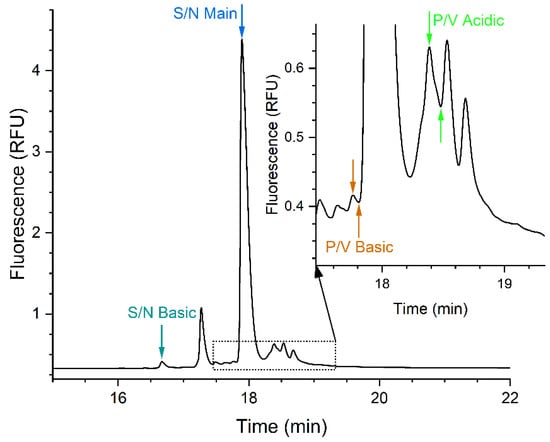
Figure 1.
Exemplary electropherogram of NISTmAb obtained with photomultiplier high voltage supply (PMHV) of 520 V, rise time (RiT) 0.8 s, and frequency of 20 Hz. The peaks and valleys used for the calculation of the responses’ signal-to-noise ratio of the main peak (S/N Main), the signal-to-noise ratio of the first basic peak (S/N Basic), the peak-to-valley ratio of the last basic peak (P/V Basic), and the peak-to-valley ratio of the first acidic peak (P/V Acidic) are indicated. The noise was determined by means of a blank run with water [38].
2.6. Linearity and Signal-to-Noise Ratio
Linearity and S/N were evaluated using different concentrations of matuzumab and NISTmAb. Matuzumab (10 mg/mL) was diluted to 1, 0.5, 0.25, 0.1, 0.05, 0.04, 0.03, 0.02, and 0.01 mg/mL with water. The detailed dilution scheme is provided in Table S1. NISTmAb (1 mg/mL) was diluted to 0.5, 0.2, 0.1, 0.05, 0.02, 0.01, 0.005, 0.004, 0.003, 0.002, and 0.001 mg/mL with water. The detailed dilution scheme is described in Table S2. Each dilution was injected three times, starting with a blank run (water) followed by the mAb samples with concentrations in ascending order. Experiments using either LEDIF or UV detection were conducted on consecutive measurement days with the same samples and BGE. The BGE was replaced after six runs.
2.7. Precision
Precision was assessed using Waters mAb (2.5 mg/mL) and NISTmAb (1 mg/mL). Each mAb was injected ten times on three measurement days. The first two measurement days were consecutive days in week 1, and the third measurement day was in week 2, precisely six (LEDIF) or seven (UV) days after the first measurement day. For each measurement day, the BGE was prepared freshly from the stock solutions. One set of BGE vials was used for up to ten injections, whereby one set each was used for conditioning, blank runs, Waters mAb and NISTmAb. The two measurement series (UV/LEDIF detection) used the same samples, which were stored between them at −20 °C. For use, they were thawed at room temperature, vortexed (3 × 5 s) and centrifuged (1 min/10,000× g).
2.8. Evaluation
ChemStation was used for the calculation of S/Ns and peak-to-valley ratios (P/Vs), according to the European Pharmacopoeia [38], and the determination of areas and migration times. For all non-DoE runs, exported data were summarized in Microsoft® Excel® 2019, version 1809 (build 10827.20138) from Microsoft Corporation, Redmond, WA, USA, and time corrected areas (TCAs) were calculated. Further data analysis was carried out in OriginPro 2022b, version 9.9.5.167, OriginLab Corporation, Northampton, MA, USA.
3. Results
3.1. Design of Experiments
For each run, the four responses (Figure 1) were calculated. Aggregated data for all 18 runs are provided in combination with the associated factor settings in Table S3. Based on this dataset, a quadratic regression model was constructed. For a predicted response ŷ, the regression function may be represented with constant, linear, interaction, and quadratic terms in the following way:
For this purpose, the responses S/N Main and S/N Basic were transformed in such a way that they were closer to normality. The best transformation found was their reciprocal square root. All terms with a significance level >0.1, as determined by Cornerstone, were regarded as insignificant and removed successively. Linear terms were not removed if they were part of a significant higher order term. Orthogonal scaling (−1 to +1) was used for the responses. An overview of the included terms is provided in Figure S1.
The estimated effect of the individual terms on the predicted responses can be seen in the Pareto charts in Figure 2. The charts show the relative effect of each included term on the indicated response. Each bar indicates the maximum size of the effect going from the lowest to the highest term value. Based on this, the most influential terms and underlying factors can be identified. For example, for the S/N basic, the RiT is the most influential term. The factor RiT is also part of the interaction term PMHV*RiT. Since the reciprocal square root is used for S/N Basic and S/N Main, the illustrated negative impact of the RiT on the transformed responses is a positive impact on the untransformed response. Or, simply put, if the RiT increases, S/N Basic will also increase.


Figure 2.
Pareto chart of the four responses: S/N Basic (a); P/V Basic (b); S/N Main (c); P/V Acidic (d). Please note that in case of the S/Ns, the transformed response (1/square root) is considered. The frequency is abbreviated to “Fr” in this figure.
The influence of factors on the responses is visualized in the surface response plots in Figure 3. They show the dependence of the predicted response (z-axis) on the two factors of PMHV and RiT. Since this type of plot allows only two dependent variables, the third dependent variable, frequency, was set to be constant, with a value of 100 Hz. For each combination of x- and y-value (factors), the corresponding predicted response value is shown (z-value). The calculated z-values result in a plane, which is colored accordingly to the z-values (high: dark red, low: dark blue). Curvatures of the plane are the result of interaction terms.
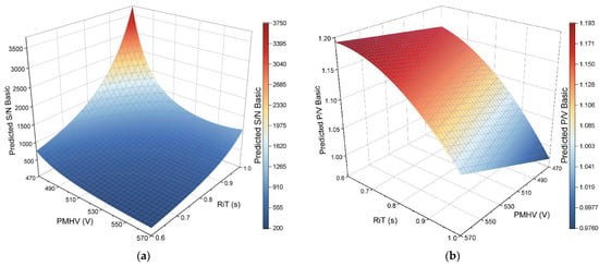
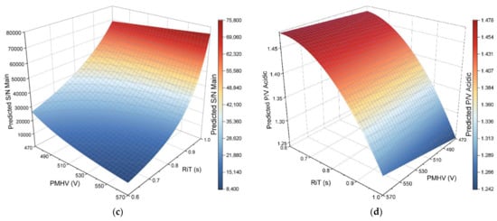
Figure 3.
Surface response plots of the responses: S/N Basic (a); P/V Basic (b); S/N Main (c); P/V Acidic (d). The factors PMHV and RiT are on the x- and y-axis. The factor “frequency” was set to 100 Hz. The predicted response is shown on the z-axis. Additionally, the color of the plane corresponds to the predicted z-value (high: dark red—mid: white—low: dark blue), as indicated in the respective color scale.
The software was then used for the determination of optimal factor settings. Its optimization algorithm should maximize the S/N Main and S/N Basic, while for the P/V Acidic and P/V Basic, target values of 1.38 and 1.16, respectively, were set. The target values were experimentally determined mean values from previous experiments with UV detection (n = 3 × 10). All responses were treated with equal weight. The optimal predicted values for the factors were 0.6 s RiT, 470 V PMHV, and a frequency of 100 Hz.
Verification of the Predicted Optimal Factor Settings
Five consecutive injections of NISTmAb were carried out to verify the optimal predicted parameters of the DoE, and the experimental values of the responses were determined. The results are summarized in Table 1. The mean value, standard deviation (SD), 95% confidence interval (CI), relative standard deviation (RSD), and the absolute and relative deviation from the predicted value were calculated. While all experimental values are slightly lower than the predicted values, they show good agreement with the prediction.

Table 1.
Overview of the results from five consecutive runs with optimized parameters with mean, standard deviation (SD), 95% confidence interval (CI), and relative standard deviation (RSD), as well as the absolute and relative deviation of the experimentally determined value from the predicted value.
The values for the response S/N Basic were calculated with n = 4. One experimental value was identified as a significant outlier using Dixon’s Q test (significance level 0.05) and removed for calculations. The remaining data, as well as the data in the other four data sets, were tested for normality using the Shapiro–Wilk test. The null hypothesis H0 that the data came from a normally distributed population could not be rejected on a significance level of 0.05. Experimental values and calculated values for S/N Basic with n = 5 are provided in Table S4 and Table S5, respectively.
3.2. Signal-to-Noise Ratio
The S/N was evaluated using the main peak of each mAb. Blank electropherograms (water injection) were used for the calculation of the noise [38]. The obtained values are summarized in Table 2. Exemplary electropherograms of matuzumab at all concentration levels and obtained with both detection modes are provided in Figures S2–S5. Likewise, exemplary electropherograms of NISTmAb are provided in Figures S6–S9. Based on the S/N, LODs and LOQs were estimated, whereby the limits of S/N ≥ 3 for the LOD and S/N ≥ 10 for the LOQ were derived from the Ph. Eur. 2.2.47 and ICH Q2(R1) guideline [38,39]. For the estimation, the S/Ns at 0.01 and 0.02 mg/mL (both mAbs and detection modes) were used, and linear regression was performed. The obtained linear equation was further used, values of 3 or 10 were inserted for the S/N, and the equation was solved for the concentration. The results were estimated LODs of 6.924 µg/mL and 5.662 µg/mL for matuzumab with UV detection and LEDIF detection, respectively. The LOQs were estimated at 10.73 µg/mL (UV detection) and 6.203 µg/mL (LEDIF detection). For NISTmAb, the estimated LODs were 10.27 µg/mL (UV detection) and 5.699 µg/mL (LEDIF detection). Likewise, the estimated LOQs were 16.23 µg/mL (UV detection) and 6.919 µg/mL (LEDIF detection).

Table 2.
Determined S/N for the matuzumab main peak and NISTmAb main peak at different concentrations with both detection modes. * n = 2.
3.3. Linearity
The same runs used for the determination of the S/Ns were used (Table 2). The total time corrected area (TCA) of the mAb peak was determined and all runs with an S/N ≥ 10 (main peak) were used for the calculations. Linear regression was performed and the compiled results and calibration curves are presented in Figure 4.
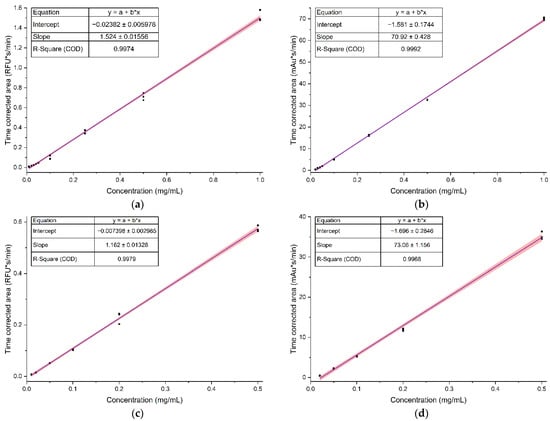
Figure 4.
Results from linear regression obtained with: (a) matuzumab and LEDIF detection; (b) matuzumab and UV detection; (c) NISTmAb and LEDIF detection; (d) NISTmAb and UV detection. Black dots are the individual data points (n = 3 for each concentration level), the blue line is the calibration curve and the red band is the 95% confidence band.
The ascertained linear ranges for matuzumab were 0.01 mg/mL to 1 mg/mL and 0.02 mg/mL to 1 mg/mL with LEDIF and UV detection, respectively. For NISTmAb, the linear ranges were from 0.01 mg/mL to 0.50 mg/mL using LEDIF detection, and from 0.02 mg/mL to 0.50 mg/mL using UV detection. The calibration curves for UV or LEDIF detection of both mAbs have a similar slope, indicating a similar sensitivity. This is in line with expectations due to the structural similarity of the two analytes. With both detection modes and mAbs, high correlations between concentration and TCA were observed (adjusted R2 > 0.99).
Additionally, linear regression using two forms of weighting was performed. The first weighting procedure used a weighting factor wi of
as recommended by Baumann and Wätzig [48]. For this, the built-in weighting procedure of Origin (“Instrumental”) was used. The resulting calibration curves are shown in Figure S10. In contrast to the calibration curves presented in Figure 2, the mean values were used for the regression in combination with the aforementioned weighting.
The second weighting procedure used a weighting factor wi of
as described in annex D of DIN 38402-51 [49]. The resulting calibration curves are also provided in Figure S10.
Both weighting procedures lead to intercepts closer to zero and slightly different slopes. The resulting residuals are closer to zero for low concentration levels. At higher concentration levels, the residuals from the (unweighted) regular regression function were closer to zero (Figure S11). In all cases, the residuals increased with increasing concentration, indicating that the error variance increases with the concentration.
3.4. Precision
The precision was assessed for each of the three measurement days, in the following termed “repeatability”, and for all three measurement days combined (week 1 and 2), which is referred to as “intermediate precision” in the following [39]. Thus, repeatability mainly covers the variation introduced through the instrument, method, and integration. Intermediate precision additionally reflects the variation introduced through preparation of the BGE from stock solutions, possible time-related capillary changes, and variation in the aforementioned elements over a longer period of time.
Exemplary electropherograms are provided in Figure 5a,b (Waters mAb), and Figure 5c,d (NISTmAb). The obtained mAb profiles (Figure 5) showed a very good agreement, and all peaks observed with UV detection were also seen in the electropherograms obtained with LEDIF detection. As indicated in Figure 5a,b, the Waters mAb peak profile was divided into five groups: basic, main 1, mid, main 2, and acidic. The division of the peak profile into five groups, with two peaks labelled “main”, was adopted from previous experiments with this mAb [29]. The migration times of the highest peak (“main 2”) and the P/Vs of the indicated acidic peak were determined. Furthermore, the TCAs and from these, the percentage of the TCA from the total TCA (%area), were calculated for all aforementioned peak groups. Four electropherograms of Waters mAb obtained with LEDIF detection displayed a baseline shift, namely runs 1, 4, 5, and 26 (Figure S12). Therefore, they were excluded from the evaluation presented in Table 3. However, the evaluation with these four values included is provided in Table S6. For each measurement day, as well as for all three measurement days combined, the mean values, 95% CIs, SDs, and RSDs were calculated and are provided for both detection modes in Table 3. The individual measurements over the course of the measurement series are depicted in Figure S13, and the corresponding data are summarized in Table S7.
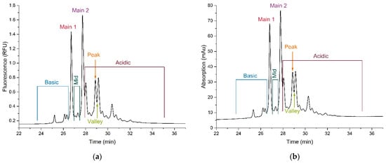
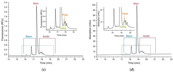
Figure 5.
Exemplary electropherograms of (a) Waters mAb with LEDIF detection; (b) Waters mAb with UV detection; (c) NISTmAb with LEDIF detection; (d) NISTmAb with UV detection. In each electropherogram, the partition of the peak profile into the respective groups and the peak and valley used for the calculation of the peak-to-valley ratio (P/V) are indicated.

Table 3.
Obtained values for Waters mAb over different time intervals of the measurement series with both detection modes. Values denoted with an asterisk are affected by the removal of the runs 1, 4, 5, and 26.
The NISTmAb runs were evaluated in a similar way. The peak profile was divided into three groups: basic, main, and acidic. For each group, the TCAs and from them, the %areas, were calculated. Further determined parameters were the migration time of the main peak and the P/V of the first acidic peak. They are also indicated in the exemplary electropherograms in Figure 5. Likewise, the corresponding mean values, 95% CIs, SDs, and RSDs are provided in Table 4, for each measurement day and for all three measurement days combined. The individual values over the course of the measurement series are depicted in Figure S14, and the corresponding data are provided in Table S8.

Table 4.
Obtained values for NISTmAb over different time intervals of the measurement series with both detection modes.
The graphical representation of the course of the measured values over time (Figures S13 and S14) indicated a trend in some cases. This was investigated in more detail through a trend test using linear regression [50,51]. For this, the injection number was plotted against the respective value. Two cases were considered; trends over the whole measurement series (interday) were investigated by assigning numbers from 1 to 30 to the injections in the order in which they were performed. Trends during a measurement day (intraday) were analyzed by assigning them injection numbers from 1 to 10, according to the order in which they were measured during the individual measurement day. If the slope of the resulting calibration function was significantly different from zero (p < 0.05), a trend was assumed.
For NISTmAb analyzed with LEDIF detection, an interday upwards trend was found for the migration time and P/V, and a downwards trend for the %area main. An intraday upwards trend was determined for the %area acidic.
The data from NISTmAb obtained with UV detection showed an interday downwards trend for the %area basic and an interday upwards trend for the %area main, %area acidic, migration time, and P/V. Intraday, an upwards trend was observed for the %area acidic.
In case of the data obtained for Waters mAb with LEDIF detection, an interday downwards trend was found for the %area basic and %area main 2, and an interday upwards trend was found for the %area main 1 and %area acidic. Intraday, the migration time exhibited an upwards trend, and the P/V a downwards trend.
For the data obtained for Waters mAb with UV detection, an interday downwards trend was observed for the %area main 1, and an interday upwards trend for the %area acidic, migration time, and P/V. For the P/V and the %area mid, an intraday upwards trend was found.
Equivalence of the data obtained with LEDIF and UV detection was further investigated through equivalence testing. This was achieved through a two one-sided t-test (TOST) using an Excel sheet provided by the “Wasserchemische Gesellschaft—Fachgruppe in der Gesellschaft Deutscher Chemiker e.V. (GDCh)” [52]. Further details about the TOST can be found elsewhere [53,54]. The data from all 30 runs (week 1 and 2) were considered, except for the data from Waters mAb obtained with LEDIF detection, which consisted of 26 runs, as previously described. The acceptance criterion θ was determined based on the tolerated deviation aa from the combined arithmetic mean value. The tolerated deviation depended on the investigated parameter, and was either 2% (migration time) or 5% (%areas, P/Vs). With θ, a [−θ, θ] interval around the parameter δ, “the absolute value of the true difference between the groups’ mean values” [53], which was set to zero, was constructed. The null hypothesis was that the mean values are not equal. If the 90% CI of the combined mean value was completely contained within the [−θ, θ] interval, the null hypothesis was rejected and the values were accepted as equivalent. The results are summarized in Table 5.

Table 5.
Results obtained by TOST with a defined tolerated deviation from the combined mean value of either 2% or 5%.
4. Discussion
4.1. Design of Experiments
4.1.1. Selection of Factors and Responses
The software for detector control offers four adjustable parameters. The first is the offset, which reduces the signal by the specified value. However, the software does not support the output of negative values. Instead, all (theoretically) negative values are reported as zero. Since the electropherograms showed only very low background fluorescence, the offset was disregarded as a factor and set to zero.
The second parameter is the (acquisition) frequency. It has three possible settings, 10 Hz, 20 Hz, and 100 Hz. Due to the fact that frequencies in between these settings cannot be chosen, the factor was set as categorial. Preliminary experiments indicated that a higher frequency was beneficial. Therefore, only 20 Hz and 100 Hz were chosen as possible levels, and 10 Hz was omitted to reduce the complexity of the design.
The third parameter is the RiT. According to the manual, it acts as a smoothing and filtering function. Thus, it may reduce noise and increase signals. At the same time, there is the risk of losing narrow peaks if set too high. For CE applications, an RiT between 0.5 s and 1 s is recommended by the manual. In preliminary experiments, RiTs between 0 s and 0.8 s were tested. The results indicated that a higher RiT, 0.8 s, was beneficial. A symmetrical interval around 0.8 s with a maximum of 1 s (corresponding to the recommendation from the manual) and a minimum of 0.6 s was chosen for the design.
The fourth parameter is the photomultiplier high-voltage supply (PMHV). The instrument’s manual recommends the setting used during installation qualification as starting point. In this case, this was 570 V; however, this value may differ from instrument to instrument. Increasing its value increases both, noise and signal. Preliminary experiments included PMHV values between 460 V and 620 V. The results from the preliminary experiments indicated that a lower value offered better responses. At 460 V and an RiT of 1 s, the last basic peak had a P/V of 1, indicating that a distinction between peak and valley was no longer possible (Figure S15). The P/V Basic for lower PMHV values in combination with a high RiT would always have been 1. Since this would have negatively influenced the regression, lower PMHV values were therefore regarded as unfavorable. With a small safety margin in mind, the factor range was set from 470 V to 570 V.
LEDIF detection was expected to provide lower LOQs and LODS than UV detection. The S/N is an established metric to determine the LOQ and LOD of an analytical method [38,39]. Furthermore, it can be easily calculated using ChemStation. The S/Ns of two peaks were evaluated. The first choice was the NISTmAb main peak (S/N Main). It can be easily located and is the peak with the highest concentration. Since there are several known concentration-dependent effects which may lead to a lower signal than theoretically expected [2,15,55], and the desired detector settings should be well-suited for all peaks, an additional peak was considered. The choice was one of the smaller peaks, which can also be easily identified: the first basic peak (S/N Basic).
At the same time, the separation quality should remain at least at an equal level as with UV detection. Usually, mAbs display a distinct charge profile, which consists of numerous closely related charge variants without baseline separation. Thus, the P/V is better suited to monitoring changes in separation quality than resolution [56]. The P/V of the first acidic peak (P/V Acidic) had been determined in a previous investigation using UV detection [29]. Hence, it seemed reasonable to collect the same data again for good comparability. Additionally, this already existing data served as target value for the optimization. However, changes in separation quality due to RiT changes were first expected to be seen with the narrowest peaks. The last basic peak before the main peak was one of the narrowest peaks, and at the same time, it could be easily located, making it an ideal choice for assessing the optimal RiT. Thus, its P/V was selected as the response (P/V Basic).
4.1.2. Results from the Design of Experiments
DoE was successfully utilized to predict settings which maximize the S/N while keeping the separation quality in terms of P/V constant. The presented results indicate that the frequency is the least important factor. It is only included in terms of the response S/N Basic (Figure 2). If one wished to further reduce the complexity and exclude a categorical factor, its removal from the design could be an option. The highest S/N Basic was predicted for low PMHV and high RiT values, and S/N Main had near maximal predicted values for this combination, too. Likewise, both S/Ns were minimal for high PMHV and low RiT values. Interestingly, there are some differences in the prediction of the S/N. Due to the similar nature of both responses, a very similar prediction was expected. Contrary to the expectation, the S/N Main is predicted to be maximal for high PMHV and RiT values, while the S/N Basic is predicted to be rather low (Figure 3). The specific reason is unknown and might be the subject of further research. Furthermore, this observation would not have been possible if only one S/N had been selected as a response, supporting this choice for further experiments. In contrast, the predictions for both investigated P/Vs were very similar (Figure 3). For the aforementioned combination of low PMHV and high RiT values, minimal P/Vs are predicted, and at low RiT and high PMHV values, both responses are predicted to be maximal. Thus, it can be seen that a balance between the factors is required to reach the desired optimization.
The factors influenced the response in an expected way. It is noteworthy that although a low PMHV value lowered both signal and noise, the noise was reduced to a higher degree than the signal, as indicated by the increasing S/N. As aforementioned, even lower PMHV values would not have been beneficial, since the response P/V basic would have been 1 for many combinations with high RiT values.
The predicted values are situated on the edge of the design space. Hence, the question arises as to whether there is an even better set of factors outside of this space. This question could only be answered by conducting another DoE. However, the predicted factor settings provided excellent results. Thus, we decided to use them for all further experiments.
4.2. Signal-to-Noise Ratio
According to the presented results (Table 2), using LEDIF detection instead of UV detection improved the S/N of the matuzumab main peak by a factor of about 6 (0.03 mg/mL) to 12 (0.25 mg/mL). Similarly, the S/N of the NISTmAb main peak improved by 2 (0.5 mg/mL) to 10 (0.01 mg/mL) times. The concentrations of 0.03 mg/mL and 0.04 mg/mL matuzumab have the same mean S/N using UV detection. Since the determined signal height increases as expected, the noise is the determinant factor. The peaks at 0.04 mg/mL are slightly wider than the peaks at 0.03 mg/mL, and consequently, a wider range of the blank electropherogram is used for the noise calculation [38]. In this specific case, this leads to an apparent increase in the noise and stagnant S/Ns.
Another interesting observation was the behavior of the S/Ns at low concentrations. For example, at 0.01 mg/mL, an S/N of 28 was determined for NISTmAb using LEDIF detection. At half the concentration, 0.005 mg/mL, the expected S/N would be half the previous S/N. However, at 0.005 mg/mL, an S/N of 2 was experimentally determined, which is 1/14 of the previous value. Additionally, for the concentrations 0.003 mg/mL and 0.004 mg/mL, similar S/Ns of about 2 were determined. There is also a difference in the peak shape, which becomes more refined with higher concentrations (Figures S6–S9). One possible explanation for this observation is that a small portion of the injected mAb is adsorbed during the separation process. Adsorption is a well-known and often encountered phenomenon during CE-based separations of proteins [57]. Although the method set employs several principles to reduce adsorption [58], it will most likely never be possible to suppress it completely. If a certain amount of protein is lost during separation due to adsorption, it is only logical that this affects the lowest concentrations the most.
Another contributing factor could be sample-induced transient isotachophoresis, which could lead to sharpened zones and better S/Ns [59,60,61]. During sample preparation, the mAbs in their storage buffer were diluted with water. The storage buffer of, e.g., NISTmAb, is composed of 12.5 mmol/L histidine and 12.5 mmol/L histidine hydrochloride, pH 6.0 [40]. NISTmAb itself also carries a positive net charge, and is present in different forms with slightly different mobilities. Additional ions, namely TETA, εACA, and acetate are contained in the BGE. If either one of those small ions or a NISTmAb form would be able to act as the leading and another as the terminating electrolyte, isotachophoresis could transiently take place. The in-depth investigation of such effects is outside the scope of this work and offers room for further research.
Since the mAb samples were diluted with water, the samples with the lowest concentration should have the lowest conductivity. There is a risk that this leads to excessive heat generation, which could denature proteins and thus have a negative effect on the separation and the peak profile obtained. If such samples must be routinely analyzed, additional investigations should be performed [62].
The estimated LODs and LOQs were not experimentally verified due to limited resources. However, they are derived from experimental data with actual concentrations close to the estimated concentrations, and should therefore be considered relatively good estimates. He et al. reported an LOQ of 20 µg/mL for their method using UV detection [58]. The estimates for the LOQ within this article using UV detection are slightly lower and within the same order of magnitude as their value.
The estimated values are good approximations of obtainable LODs and LOQs for other mAbs, but differences between them are to be expected. First, the LODs and LOQs are also dependent on the content of the aforementioned aromatic amino acids [15]. The NISTmAb primary structure contains 24 tryptophans, 52 tyrosines, and 50 phenylalanines [40,63], and the matuzumab primary structure includes 32 tryptophans, 64 tyrosines, and 44 phenylalanines [43]. Second, the separation itself is also a contributing factor. The better the individual forms are separated from each other, the lower the individual peak heights, and as a consequence thereof, the lower the S/Ns may be.
All the electropherograms obtained with UV detection showed a slight baseline drift. This baseline drift had been observed in previous experiments with this method, as well as during the repeatability runs. Its root cause has not been investigated so far. Since integration was possible at all times without difficulties, it was not seen as an issue. However, it was noted that LEDIF detection did not show such a baseline drift.
This study shows that substantially lower LODs and LOQs are obtainable through the simple switch from UV to LEDIF detection. Further improvements may be obtainable through additional measures and could be explored in further studies.
The same principal detector design in combination with lasers instead of an LED was used by Rodat et al. in combination with a bubble cell capillary. For an IgG2 antibody, the LOD was reduced by more than three times. A small negative effect of the bubble cell on the resolution of a separation of five small molecules was observed [25].
The described optimization approach balances the desire for maximal S/Ns with the requirement for adequate P/Vs. If maximal S/Ns were the only optimization target, different optimal factor settings would have been predicted. For example, looking at Figure 3a, S/N Basic is predicted to be maximal for low PMHV (470 V) and high RiT (1.0 s) values, a factor combination which is predicted to provide the lowest P/Vs (Figure 3b,d).
The studied BGE was initially used in CZE-UV and then used in CZE-LEDIF without modification. None of the BGE’s constituents were found to be a known quencher of protein’s native fluorescence. Morani et al. investigated so-called “inorganic-species-free BGEs” for C(Z)E-LIF and found them better suited than “conventional BGEs” for the analysis of model proteins [5]. The BGE used herein fulfills their requirements for an “inorganic-species-free BGE”, so their suggested ideas are already implemented in this case.
Currently, mAbs in their formulation buffer are diluted with water before injection. Furthermore, the BGE contains a high amount (400 mM) of εACA. Thus, it is assumed that some field-enhanced sample stacking takes place. Optimization of sample stacking conditions, e.g., the length of the sample zone, the composition of the sample, etc., could further enhance the S/N. Such optimizations should also consider possible sample-induced transient isotachophoresis, which may provide a synergistic effect [59,61].
Galievsky et al. outlined many considerations for the improvement of the sensitivity of CE-LIF [2]. Most concepts should also be applicable to CE-LEDIF. However, a lot of the suggested ideas pertain to the design of the detector, which is the intellectual property of its manufacturer and typically cannot be changed by the analyst.
4.3. Linearity
The investigated linear range spans several orders of magnitude for both detection modes. Except for the case of matuzumab with LEDIF detection, the lower limit was set due to the S/N of the preceding concentration level. The upper limit resulted from the experimental design. In this case, it would be worthwhile to investigate even higher concentrations in future experiments.
With increasing concentrations, increasing variances were observed, indicating heteroscedasticity. This is in accordance with previous investigations, and one of the weighted linear regression procedures suggested therein was applied to the presented data [48]. A weighting of 1/ var(yi) was chosen due to its good practicability and comprehensibility. It should be noted that the variance was estimated from three replicates. For an improved estimation, a higher number of replicates and repeats is recommended.
Linearity and S/N were evaluated in parallel using the same runs due to limited resources. This led to a suboptimal distribution of the concentration levels for a calibration curve with increasing distances between the individual data points. Another weighting procedure was derived from annex D of DIN 38402-51, which suggests a weighting factor of 1/xi for non-equidistant calibrations over a wide calibration interval with a cumulation of calibration points in the lower calibration range [49]. This procedure is also described in DIN ISO 11095, where the weighting is recommended if “the residual standard deviation increases with the accepted values of the RMs [reference materials]” [64]. This is the same use case as described by Baumann and Wätzig [48].
As a consequence, both weighting factors have a similar effect; the lower concentration levels receive a higher weight, either through their lower variance or through their lower concentration. It is therefore not surprising that the absolute value of the residuals of the lower concentration levels was reduced when using a weighting factor, while the absolute values of the residuals of the higher concentration levels were increased.
The goal of the linearity experiments was to provide an initial assessment and comparison of the linearity. For this, all three functions are well suited. If the method should be used for quantification, validation is mandatory. In the process, the outlined limitations of these experiments should be overcome. Depending on the covered range and resulting data, either weighted or unweighted regression could be employed. However, weighted regression is recommended, since “… results can never get worse using WLS [weighted least squares]” [48].
4.4. Precision
As aforementioned, four Waters mAb runs with LEDIF detection were excluded from the evaluation presented in Table 3 due to their irregular baseline (Figure S12). The baseline behavior was not observed in other runs before or after and could not be replicated. Other recorded parameters, e.g., current, voltage, and cassette temperature, were within the same range as in all other experiments. Hence, the root cause of this phenomenon could not be investigated and is unknown.
Equivalence testing was performed to allow for a more objective assessment of the obtained values. The tolerated deviations were conservatively chosen. A level of 2% has been used in the past as a rather strict criterion [65]. Migration times should not depend on the employed detector, so only a small deviation was allowed. The other parameters, %areas and P/Vs, showed higher variability. In some cases, e.g., Waters mAb %area basic, the obtained 90% CI would have been larger than the [−θ, θ] interval with a 2% level. Hence, the test would have failed simply due to the imprecision, which is inherent to the method and not due to the detector. Thus, a larger interval was selected.
It should be noted that the set-up for the precision experiments was chosen in this way due to previous experiments with this method, and equivalence testing was performed post hoc. Ideally, the sample size, acceptance limits, and experimental setup would have been determined a priori [53,65]. However, the sample size was sufficiently large, and equivalence testing provided an objective criterion for the comparison.
The obtained migration times were found to be equal for Waters mAb, but not for NISTmAb. Although different capillaries had to be used for each detection mode, they were cut from the same capillary coil and should not influence the results to such an extent. The most likely cause is the fact that different, fresh stock solutions were used for the BGE preparation during the precision runs with UV detection, which was necessary for technical reasons. It is presumed that even better values might be obtained if the same BGE is used for all runs. Additionally, NISTmAb seems to be more susceptible to such changes than Waters mAb.
Excellent RSD values were obtained for repeatability, with values between 0.26% and 0.57% for Waters mAb, and 0.12% and 0.58% for NISTmAb. The values for the intermediate precision are slightly higher, but still very acceptable, with RSD values of up to 1.25% (Waters mAb) or 1.19% (NISTmAb). The increase is related to differences between the measurement days, which can be seen for both mAbs (Figures S13b,e and S14b,e). Moreover, the individual values obtained during a measurement day show an upward trend for Waters mAb, while no clear trend is observed for NISTmAb, again highlighting the slightly different behavior of both mAbs.
The %areas of Waters mAb were found to be equal except for the %area basic and % area mid. The %area basic has the highest RSDs (repeatability and intermediate precision) of all investigated %areas. Since this is true for both detection modes, it is most likely related to the CZE method. Furthermore, there is a noticeable difference in measured values and resulting mean values with LEDIF detection between the first measurement day (mean 7.4%) and the other two measurement days (mean 6.5% and 6.2%), which contributes to the observed variability. Concomitant with the decrease in the %area basic, the %area main 1 increases, leading to the assumption that the separation between the peak groups has slightly changed here.
A similar observation can be made for the %area basic and %area main of the NISTmAb series with UV detection, with a decrease in the %area basic mean value and an increase in the %area main mean value in week 2. This is also the reason for the worse intermediate precision of the %area basic (RSD: 5.8%) in comparison to the excellent repeatability (RSDs < 0.82%). With LEDIF detection, the repeatability of the %area basic is slightly higher but still very acceptable. The %area acidic was not found to be equivalent and is slightly higher for LEDIF detection. The other values are at a consistent and good level. In many cases, the intraday RSDs (repeatability) were <1% for the %areas, and their interday RSDs (intermediate precision) were typically below 2%.
The P/Vs of the investigated Waters mAb peak obtained with LEDIF detection are about 17% higher than the P/Vs obtained with UV detection. The detector optimization was conducted with NISTmAb, so a slight difference for another analyte can be expected. If other peaks were affected in a similar way, this could be a reason for the slightly different %areas. With both detection modes, a downwards trend during a measurement day is observed, and at the beginning of a new measurement day, a higher value is obtained. This indicates a small but noticeable change in the separation quality during a measurement day, and might be due to changes in the BGE or capillary. Changing the BGE more frequently or implementing additional capillary flushing steps could provide further insight into the underlying cause of this trend. Since the overall separation was not impaired, no additional measures were taken during this study.
For NISTmAb, this difference is only about 3.6%, and the values are in good agreement with the values in Table 1. The first value of the repeatability series obtained with UV detection (injection 1, P/V 1.185; Table S8) is noticeably lower than all other P/V values. According to Grubb’s test (significance level 0.05), the value 1.185 is an outlier. With this value removed, the remaining data were significantly drawn from a normally distributed population (Shapiro–Wilk test, significance level 0.05), and the calculated mean value (n = 29) was 1.371, reducing the difference between the means to 3.2%. Additionally, the RSD improved from 2.672% (n = 30) to 0.9935% (n = 29) and was again equal (TOST, as described earlier) to the value obtained with LEDIF detection (n = 30, RSD 0.7138%).
Overall, the achieved results are satisfactory, with close mean values for both detection modes and excellent precision.
5. Conclusions and Outlook
For the first time, CZE-LEDIF has been successfully applied for the charge-based analysis of three mAbs. The DoE-based detector optimization led to detector settings which balanced the desire for maximal S/Ns with the requirement for adequate separation quality in terms of P/Vs. The demonstrated systematic optimization approach can be easily adapted for similar tasks, and the commercially available detector has good technical accessibility for all interested researchers. Using LEDIF detection, the S/Ns were increased by two- to twelve-fold compared to UV detection. Consequently, the LOQs and LODs were significantly lower, and the linear ranges extended. The precision experiments provided very similar and often equal results to those of UV detection. Overall, LEDIF detection demonstrated the ability to act as a sound alternative to UV detection for the CZE analysis of mAbs. This opens up opportunities for different BGE constituents and the detection of low-abundance charge variants in, e.g., stability studies. Future investigations may also include validation and robustness studies. Furthermore, leveraging the advantages of the LEDIF detector in combination with other CZE as well as CIEF methods may provide additional research opportunities.
Supplementary Materials
The following supporting information can be downloaded at: https://www.mdpi.com/article/10.3390/separations10050320/s1, Table S1: matuzumab dilution scheme; Table S2: NISTmAb dilution scheme; Table S3: factor settings and obtained response values of all 18 runs of the experimental design; Figure S1: overview over the terms and corresponding responses of the DoE; Table S4: individual results of the runs summarized in Table 1; Table S5: calculation, with outlier included (n = 5), for the response S/N Basic; Figure S2: exemplary electropherograms of matuzumab at different concentration levels (0 mg/mL to 0.02 mg/mL); Figure S3: exemplary electropherograms of matuzumab at different concentration levels (0.03 mg/mL to 0.05 mg/mL); Figure S4: exemplary electropherograms of matuzumab at different concentration levels (0.10 mg/mL to 0.50 mg/mL); Figure S5: exemplary electropherograms of matuzumab at 1 mg/mL; Figure S6: exemplary electropherograms of NISTmAb at different concentration levels (0 mg/mL to 0.002 mg/mL); Figure S7: exemplary electropherograms of NISTmAb at different concentration levels (0.003 mg/mL to 0.005 mg/mL); Figure S8: exemplary electropherograms of NISTmAb at different concentration levels (0.01 mg/mL to 0.05 mg/mL); Figure S9: exemplary electropherograms of NISTmAb at different concentration levels (0.10 mg/mL to 0.50 mg/mL); Figure S10: obtained calibration curves using linear regression without weighting, with 1/xi as weight (red) and with 1/var(yi) as weight; Figure S11: residuals determined for different regression functions; Figure S12: electropherograms of the four Waters mAb runs which were excluded for the evaluation; Table S6: obtained values for Waters mAb over different time intervals of the measurement series with both detection modes and all 30 runs included; Figure S13: course of the individual measured values over the injection series for Waters mAb; Table S7: individual determined values for each injection of Waters mAb with both detection modes over the course of the measurement series; Figure S14: Course of the individual measured values over the injection series for NISTmAb; Table S8: Individual determined values for each injection of NISTmAb with both detection modes over the course of the measurement series; Figure S15: Electropherogram of NISTmAb obtained with 460 V PMHV and 1 s RiT, frequency 20 Hz.
Author Contributions
Conceptualization, H.Z.; methodology, H.Z.; validation, H.Z.; formal analysis, H.Z.; investigation, H.Z., S.H. and D.-M.M., A.W.; resources, H.W.; data curation, H.Z.; writing—original draft preparation, H.Z.; writing—review and editing, H.Z., S.H. and H.W.; visualization, H.Z.; supervision, H.W.; project administration, H.Z. and S.H.; funding acquisition, H.W. All authors have read and agreed to the published version of the manuscript.
Funding
This research received no external funding.
Data Availability Statement
Most of the data presented in this study are available in the article and Supporting Information. Further data are available upon reasonable request from the corresponding author.
Acknowledgments
AB SCIEX kindly sponsored Waters mAb, NISTmAb, and HPMC as part of a different research project, whose logistics were also supported by Kantisto BV and Bristol Myers Squibb. Polymicro Technologies kindly provided the capillaries used. Merck KGaA is gratefully acknowledged for the matuzumab gift. Matthias Stein is gratefully acknowledged for revising the manuscript.
Conflicts of Interest
The authors declare no conflict of interest.
References
- Gassmann, E.; Kuo, J.E.; Zare, R.N. Electrokinetic separation of chiral compounds. Science 1985, 230, 813–814. [Google Scholar] [CrossRef] [PubMed]
- Galievsky, V.A.; Stasheuski, A.S.; Krylov, S.N. “Getting the best sensitivity from on-capillary fluorescence detection in capillary electrophoresis”—A tutorial. Anal. Chim. Acta 2016, 935, 58–81. [Google Scholar] [CrossRef]
- Ban, E.; Song, E.J. Recent developments and applications of capillary electrophoresis with laser-induced fluorescence detection in biological samples. J. Chromatogr. B Analyt. Technol. Biomed. Life Sci. 2013, 929, 180–186. [Google Scholar] [CrossRef]
- Couderc, F.; Ong-Meang, V.; Poinsot, V. Capillary electrophoresis hyphenated with UV-native-laser induced fluorescence detection (CE/UV-native-LIF). Electrophoresis 2017, 38, 135–149. [Google Scholar] [CrossRef] [PubMed]
- Morani, M.; Taverna, M.; Mai, T.D. A fresh look into background electrolyte selection for capillary electrophoresis-laser induced fluorescence of peptides and proteins. Electrophoresis 2019, 40, 2618–2624. [Google Scholar] [CrossRef] [PubMed]
- García-Campaña, A.M.; Taverna, M.; Fabre, H. LIF detection of peptides and proteins in CE. Electrophoresis 2007, 28, 208–232. [Google Scholar] [CrossRef]
- Szekrényes, Á.; Park, S.S.; Santos, M.; Lew, C.; Jones, A.; Haxo, T.; Kimzey, M.; Pourkaveh, S.; Szabó, Z.; Sosic, Z.; et al. Multi-Site N-glycan mapping study 1: Capillary electrophoresis—Laser induced fluorescence. MAbs 2016, 8, 56–64. [Google Scholar] [CrossRef]
- Skeidsvoll, J.; Ueland, P.M. Analysis of double-stranded DNA by capillary electrophoresis with laser-induced fluorescence detection using the monomeric dye SYBR green I. Anal. Biochem. 1995, 231, 359–365. [Google Scholar] [CrossRef]
- Ban, E.; Kwon, H.; Song, E.J. A rapid and reliable CE-LIF method for the quantitative analysis of miRNA-497 in plasma and organs and its application to a pharmacokinetic and biodistribution study. RSC Adv. 2020, 10, 18648–18654. [Google Scholar] [CrossRef]
- Ta, H.Y.; Collin, F.; Perquis, L.; Poinsot, V.; Ong-Meang, V.; Couderc, F. Twenty years of amino acid determination using capillary electrophoresis: A review. Anal. Chim. Acta 2021, 1174, 338233. [Google Scholar] [CrossRef]
- Le Potier, I.; Boutonnet, A.; Ecochard, V.; Couderc, F. Chemical and Instrumental Approaches for Capillary Electrophoresis (CE)-Fluorescence Analysis of Proteins. Methods Mol. Biol. 2016, 1466, 1–10. [Google Scholar] [CrossRef]
- Toseland, C.P. Fluorescent labeling and modification of proteins. J. Chem. Biol. 2013, 6, 85–95. [Google Scholar] [CrossRef] [PubMed]
- Malek, A.; Khaledi, M.G. Expression and analysis of green fluorescent proteins in human embryonic kidney cells by capillary electrophoresis. Anal. Biochem. 1999, 268, 262–269. [Google Scholar] [CrossRef]
- Jin, Y.; Chen, C.; Meng, L.; Chen, J.; Li, M.; Zhu, Z.; Lin, J. A CE-LIF method to monitor autophagy by directly detecting LC3 proteins in HeLa cells. Analyst 2012, 137, 5571–5575. [Google Scholar] [CrossRef] [PubMed]
- Lakowicz, J.R. Principles of Fluorescence Spectroscopy, 3rd ed.; Springer Science+Business Media LLC: Boston, MA, USA, 2006; ISBN 978-0-387-46312-4. [Google Scholar]
- Mukunda, D.C.; Joshi, V.K.; Mahato, K.K. Light emitting diodes (LEDs) in fluorescence-based analytical applications: A review. Appl. Spectrosc. Rev. 2022, 57, 1–38. [Google Scholar] [CrossRef]
- de Kort, B.J.; de Jong, G.J.; Somsen, G.W. Native fluorescence detection of biomolecular and pharmaceutical compounds in capillary electrophoresis: Detector designs, performance and applications: A review. Anal. Chim. Acta 2013, 766, 13–33. [Google Scholar] [CrossRef]
- Rodat-Boutonnet, A.; Naccache, P.; Morin, A.; Fabre, J.; Feurer, B.; Couderc, F. A comparative study of LED-induced fluorescence and laser-induced fluorescence in SDS-CGE: Application to the analysis of antibodies. Electrophoresis 2012, 33, 1709–1714. [Google Scholar] [CrossRef]
- Couderc, F.; Nertz, M.; Nouadje, G. Laser-Induced Fluorescence Detector and Method for the Implementation of Said Device. WO1999FR00800, 7 April 1999. [Google Scholar]
- Bayle, C.; Siri, N.; Poinsot, V.; Treilhou, M.; Caussé, E.; Couderc, F. Analysis of tryptophan and tyrosine in cerebrospinal fluid by capillary electrophoresis and “ball lens” UV-pulsed laser-induced fluorescence detection. J. Chromatogr. A 2003, 1013, 123–130. [Google Scholar] [CrossRef]
- Kumar, R.; Guttman, A.; Rathore, A.S. Applications of capillary electrophoresis for biopharmaceutical product characterization. Electrophoresis 2022, 43, 143–166. [Google Scholar] [CrossRef]
- Gilardoni, E.; Regazzoni, L. Liquid phase separation techniques for the characterization of monoclonal antibodies and bioconjugates. J. Chromatogr. Open 2022, 2, 100034. [Google Scholar] [CrossRef]
- Dadouch, M.; Ladner, Y.; Perrin, C. Analysis of Monoclonal Antibodies by Capillary Electrophoresis: Sample Preparation, Separation, and Detection. Separations 2021, 8, 4. [Google Scholar] [CrossRef]
- Salas-Solano, O.; Tomlinson, B.; Du, S.; Parker, M.; Strahan, A.; Ma, S. Optimization and validation of a quantitative capillary electrophoresis sodium dodecyl sulfate method for quality control and stability monitoring of monoclonal antibodies. Anal. Chem. 2006, 78, 6583–6594. [Google Scholar] [CrossRef] [PubMed]
- Rodat, A.; Gavard, P.; Couderc, F. Improving detection in capillary electrophoresis with laser induced fluorescence via a bubble cell capillary and laser power adjustment. Biomed. Chromatogr. 2009, 23, 42–47. [Google Scholar] [CrossRef]
- Kahle, J.; Zagst, H.; Wiesner, R.; Wätzig, H. Comparative charge-based separation study with various capillary electrophoresis (CE) modes and cation exchange chromatography (CEX) for the analysis of monoclonal antibodies. J. Pharm. Biomed. Anal. 2019, 174, 460–470. [Google Scholar] [CrossRef]
- Candreva, J.; Esterman, A.L.; Ge, D.; Patel, P.; Flagg, S.C.; Das, T.K.; Li, X. Dual-detection approach for a charge variant analysis of monoclonal antibody combination products using imaged capillary isoelectric focusing. Electrophoresis 2022, 43, 1701–1709. [Google Scholar] [CrossRef]
- Kahle, J.; Wätzig, H. Determination of protein charge variants with (imaged) capillary isoelectric focusing and capillary zone electrophoresis. Electrophoresis 2018, 39, 2492–2511. [Google Scholar] [CrossRef]
- Wiesner, R.; Zagst, H.; Lan, W.; Bigelow, S.; Holper, P.; Hübner, G.; Josefsson, L.; Lancaster, C.; Lo, L.; Lößner, C.; et al. An interlaboratory capillary zone electrophoresis-UV study of various monoclonal antibodies, instruments, and ε-aminocaproic acid lots. Electrophoresis 2023. accepted manuscript. [Google Scholar] [CrossRef] [PubMed]
- Fukuda, I.M.; Pinto, C.F.F.; Moreira, C.d.S.; Saviano, A.M.; Lourenço, F.R. Design of Experiments (DoE) applied to Pharmaceutical and Analytical Quality by Design (QbD). Braz. J. Pharm. Sci. 2018, 54. [Google Scholar] [CrossRef]
- Silva Araújo, A.; Fernandes Andrade, D.; Babos, D.V.; Castro, J.P.; Garcia, J.A.; Sperança, M.A.; Gamela, R.R.; Cardoso Machado, R.; Câmara Costa, V.; Nascimento Guedes, W.; et al. Key information related to quality by design (QbD) applications in analytical methods development. Braz. J. Anal. Chem. 2020, 8, 14–28. [Google Scholar] [CrossRef]
- Emonts, P.; Avohou, H.T.; Hubert, P.; Ziemons, E.; Fillet, M.; Dispas, A. Optimization of a robust and reliable FITC labeling process for CE-LIF analysis of pharmaceutical compounds using design of experiments strategy. J. Pharm. Biomed. Anal. 2021, 205, 114304. [Google Scholar] [CrossRef] [PubMed]
- van Tricht, E.; Geurink, L.; Backus, H.; Germano, M.; Somsen, G.W.; Sänger-van de Griend, C.E. One single, fast and robust capillary electrophoresis method for the direct quantification of intact adenovirus particles in upstream and downstream processing samples. Talanta 2017, 166, 8–14. [Google Scholar] [CrossRef] [PubMed]
- Hancu, G.; Orlandini, S.; Papp, L.A.; Modroiu, A.; Gotti, R.; Furlanetto, S. Application of Experimental Design Methodologies in the Enantioseparation of Pharmaceuticals by Capillary Electrophoresis: A Review. Molecules 2021, 26, 4681. [Google Scholar] [CrossRef]
- Krait, S.; Heuermann, M.; Scriba, G.K.E. Development of a capillary electrophoresis method for the determination of the chiral purity of dextromethorphan by a dual selector system using quality by design methodology. J. Sep. Sci. 2018, 41, 1405–1413. [Google Scholar] [CrossRef] [PubMed]
- Michels, D.A.; Parker, M.; Salas-Solano, O. Quantitative impurity analysis of monoclonal antibody size heterogeneity by CE-LIF: Example of development and validation through a quality-by-design framework. Electrophoresis 2012, 33, 815–826. [Google Scholar] [CrossRef]
- Moritz, B.; Locatelli, V.; Niess, M.; Bathke, A.; Kiessig, S.; Entler, B.; Finkler, C.; Wegele, H.; Stracke, J. Optimization of capillary zone electrophoresis for charge heterogeneity testing of biopharmaceuticals using enhanced method development principles. Electrophoresis 2017, 38, 3136–3146. [Google Scholar] [CrossRef]
- EDQM Council of Europe. 2.2.47 Kapillarelektrophorese. In Europäisches Arzneibuch 10.5: Ph. Eur. 10.5—Grundwerk 2020 inkl. 5. Nachtrag (Online Version), 10th ed.; Deutscher Apotheker Verlag Dr. Roland Schmiedel GmbH & Co. KG: Stuttgart, Germany, 2020; pp. 119–126. [Google Scholar]
- International Council for Harmonisation of Technical Requirements for Pharmaceuticals for Human Use. Validation of Analytical Procedures: Text and Methodology Q2(R1): ICH Q2(R1), Step 4. International Council for Harmonisation of Technical Requirements for Pharmaceuticals for Human Use: 2005; (Q2(R1)). Available online: https://database.ich.org/sites/default/files/Q2%28R1%29%20Guideline.pdf (accessed on 17 January 2023).
- National Institute of Standards and Technology. Reference Material Information Sheet, Reference Material 8671 NISTmAb, Humanized IgG1κ Monoclonal Antibody Lot 14HB-D-002; 2022. Available online: https://tsapps.nist.gov/srmext/certificates/8671.pdf (accessed on 12 January 2023).
- Schiel, J.E.; Davis, D.L.; Borisov, O. (Eds.) Biopharmaceutical Characterization: The NISTmAb Case Study; American Chemical Society: Washington, DC, USA; Distributed in Print by Oxford University Press: Oxford, UK, 2015; ISBN 9780841230293. [Google Scholar]
- Waters Corporation. Intact mAb Mass Check Standard Care and Use Manual. 720004420EN. 2013. Available online: https://www.waters.com/webassets/cms/support/docs/720004420en.pdf (accessed on 12 January 2023).
- Knoechel, T.; Schmiedel, J.; Ferguson, K.M. Crystalline Egfr—Matuzumab Complex and Matuzumab Mimetics Obtained Thereof. WO2008EP07889, 19 September 2008. [Google Scholar]
- Seiden, M.V.; Burris, H.A.; Matulonis, U.; Hall, J.B.; Armstrong, D.K.; Speyer, J.; Weber, J.D.A.; Muggia, F. A phase II trial of EMD72000 (matuzumab), a humanized anti-EGFR monoclonal antibody, in patients with platinum-resistant ovarian and primary peritoneal malignancies. Gynecol. Oncol. 2007, 104, 727–731. [Google Scholar] [CrossRef]
- Rao, S.; Starling, N.; Cunningham, D.; Sumpter, K.; Gilligan, D.; Ruhstaller, T.; Valladares-Ayerbes, M.; Wilke, H.; Archer, C.; Kurek, R.; et al. Matuzumab plus epirubicin, cisplatin and capecitabine (ECX) compared with epirubicin, cisplatin and capecitabine alone as first-line treatment in patients with advanced oesophago-gastric cancer: A randomised, multicentre open-label phase II study. Ann. Oncol. 2010, 21, 2213–2219. [Google Scholar] [CrossRef]
- Taiga Uranaka. UPDATE 1-Merck, Takeda Cancel Development of Cancer Drug. Thomson Reuters. 2008. Available online: https://www.reuters.com/article/takeda-idUST35282120080218 (accessed on 12 January 2023).
- González-Ruiz, V.; Drouin, N.; Reginato, E.; Rudaz, S.; Schappler, J. zeecalc: 1.0b. Available online: https://ispso.unige.ch/labs/fanal/zeecalc:en (accessed on 31 August 2018).
- Baumann, K.; Wätzig, H. Appropriate calibration functions for capillary electrophoresis II. Heteroscedasticity and its consequences. J. Chromatogr. A 1995, 700, 9–20. [Google Scholar] [CrossRef]
- DIN Deutsches Institut für Normung e. V. Deutsche Einheitsverfahren zur Wasser-, Abwasser- und Schlammuntersuchung—Allgemeine Angaben (Gruppe A)—Teil 51: Kalibrierung von Analysenverfahren—Lineare Kalibrierfunktion (A 51): (German Standard Methods for the Examination of Water, Waste Water and Sludge—General Information (Group A)—Part 51: Calibration of Analytical Methods—Linear Calibration (A 51)), 2017-05; Beuth Verlag GmbH: Berlin, Germany, 2017; 13.060.50 (DIN 38402-51). [Google Scholar]
- Köppel, H.; Wätzig, H. Trends in der statistischen QC Teil 2: Verteilungsabhägige Test. PZ Prisma 2009, 16, 251–256. [Google Scholar]
- Köppel, H.; Cianciulli, C.; Wätzig, H. Trendtests für die statistische Qualitätskontrolle Teil 3: Anwendung und Leistungsbewertung. PZ Prisma 2010, 17, 229–243. [Google Scholar]
- Platen, H.; Jähnichen, S. Prüfung der Gleichwertigkeit Zweier Analysenverfahren Mittels Two One-Sided t-Test (TOST) nach DIN 38402-71:2020-10. Available online: https://www.wasserchemische-gesellschaft.de/dev/validierungsdokumente?download=204:a71-gleichwertigkeitspruefung-tost-verfahren&lang=de (accessed on 24 January 2023).
- Limentani, G.B.; Ringo, M.C.; Ye, F.; Berquist, M.L.; McSorley, E.O. Beyond the t-test: Statistical equivalence testing. Anal. Chem. 2005, 77, 221A–226A. [Google Scholar] [CrossRef]
- DIN Deutsches Institut für Normung e. V. Deutsche Einheitsverfahren zur Wasser-, Abwasser- und Schlammuntersuchung—Allgemeine Angaben (Gruppe A)—Teil 71: Gleichwertigkeit von Zwei Analysenverfahren Aufgrund des Vergleichs von Analysenergebnissen (A 71): (German Standard Methods for the Examination of Water, Waste Water and Sludge—General Information (Group A)—Part 71: Equivalence of Two Analysis Methods Based on the Comparison of Analysis Results (A 71)), 2020-10; Beuth Verlag GmbH: Berlin, Germany, 2020; 13.060.45 (DIN 38402-71). [Google Scholar]
- Valeur, B.; Berberan-Santos, M.N. Molecular Fluorescence: Principles and Applications, 2nd ed.; Wiley-VCH: Weinheim, Germany, 2012; ISBN 9783527650002. [Google Scholar]
- Christophe, A.B. Valley to peak ratio as a measure for the separation of two chromatographic peaks. Chromatographia 1971, 4, 455–458. [Google Scholar] [CrossRef]
- Stutz, H. Protein attachment onto silica surfaces—A survey of molecular fundamentals, resulting effects and novel preventive strategies in CE. Electrophoresis 2009, 30, 2032–2061. [Google Scholar] [CrossRef] [PubMed]
- He, Y.; Isele, C.; Hou, W.; Ruesch, M. Rapid analysis of charge variants of monoclonal antibodies with capillary zone electrophoresis in dynamically coated fused-silica capillary. J. Sep. Sci. 2011, 34, 548–555. [Google Scholar] [CrossRef]
- Foret, F.; Szoko, E.; Karger, B.L. On-column transient and coupled column isotachophoretic preconcentration of protein samples in capillary zone electrophoresis. J. Chromatogr. A 1992, 608, 3–12. [Google Scholar] [CrossRef]
- Malá, Z.; Gebauer, P. Analytical isotachophoresis 1967–2022: From standard analytical technique to universal on-line concentration tool. TrAC Trends Anal. Chem. 2023, 158, 116837. [Google Scholar] [CrossRef]
- Křivánková, L.; Boček, P. Synergism of capillary isotachophoresis and capillary zone electrophoresis. J. Chromatogr. B Biomed. Sci. Appl. 1997, 689, 13–34. [Google Scholar] [CrossRef]
- Vinther, A.; Soeberg, H.; Nielsen, L.; Pedersen, J.; Biedermann, K. Thermal degradation of a thermolabile Serratia marcescens nuclease using capillary electrophoresis with stacking conditions. Anal. Chem. 1992, 64, 187–191. [Google Scholar] [CrossRef]
- Formolo, T.; Ly, M.; Levy, M.; Kilpatrick, L.; Lute, S.; Phinney, K.; Marzilli, L.; Brorson, K.; Boyne, M.; Davis, D.; et al. Determination of the NISTmAb Primary Structure. In Biopharmaceutical Characterization: The NISTmAb Case Study; Schiel, J.E., Davis, D.L., Borisov, O., Eds.; American Chemical Society: Washington, DC, USA; Distributed in Print by Oxford University Press: Oxford, UK, 2015; pp. 1–62. ISBN 9780841230293. [Google Scholar]
- DIN Deutsches Institut für Normung e. V. Lineare Kalibrierung unter Verwendung von Referenzmaterialien (ISO 11095:1996); Text Deutsch und Englisch: (Linear Calibration Using Reference Materials (ISO 11095:1996); Text German and English), 2008-04; Beuth Verlag GmbH: Berlin, Germany, 2008; 17.020 (DIN ISO 11095). [Google Scholar]
- Kaminski, L.; Schepers, U.; Wätzig, H. Analytical method transfer using equivalence tests with reasonable acceptance criteria and appropriate effort: Extension of the ISPE concept. J. Pharm. Biomed. Anal. 2010, 53, 1124–1129. [Google Scholar] [CrossRef]
Disclaimer/Publisher’s Note: The statements, opinions and data contained in all publications are solely those of the individual author(s) and contributor(s) and not of MDPI and/or the editor(s). MDPI and/or the editor(s) disclaim responsibility for any injury to people or property resulting from any ideas, methods, instructions or products referred to in the content. |
© 2023 by the authors. Licensee MDPI, Basel, Switzerland. This article is an open access article distributed under the terms and conditions of the Creative Commons Attribution (CC BY) license (https://creativecommons.org/licenses/by/4.0/).