Article

An UPLC–ESI–MS/MS Bioanalytical Methodology for the Quantification of Gilteritinib in Human Liver Microsomes: Application to In Vitro and In Silico Metabolic Stability Estimation

Department of Pharmaceutical Chemistry, College of Pharmacy, King Saud University, Riyadh 11451, Saudi Arabia
*
Author to whom correspondence should be addressed.
Separations 2023, 10(5), 278; https://doi.org/10.3390/separations10050278
Submission received: 19 March 2023 / Revised: 16 April 2023 / Accepted: 23 April 2023 / Published: 25 April 2023
(This article belongs to the Section Analysis of Natural Products and Pharmaceuticals)

Abstract

:
Gilteritinib (Xospata®) is a tyrosine kinase inhibitor (TKI) that works by inhibiting numerous receptor tyrosine kinases, involving AXL and FMS-like tyrosine kinase 3 (FLT3). Gilteritinib (GTB) was approved (28 November 2018) by the USFDA for the treatment of refractory or relapsed (R/R) acute myeloid leukemia (AML) with a FLT3 mutation. In the current study, a fast, highly sensitive, and specific ultra-performance liquid chromatography tandem mass spectrometry (UPLC–MS/MS) analytical methodology was created for GTB determination in human liver microsomes (HLMs) utilizing an electrospray ionization (ESI) source. The developed methodology (UPLC–ESI–MS/MS) was utilized in the assessment of GTB metabolic stability. The UPLC–ESI–MS/MS methodology was validated following the rules of the FDA that include selectivity, linearity, accuracy, precision, matrix effect, stability, and extraction recovery. The generated data of the optimized validation parameters of the current UPLC–ESI–MS/MS methodology were acceptable as reported in the FDA guidelines. GTB parent ions were generated in the ESI source (positive mode) and GTB daughter ions (two) were quantified in the mass analyzer utilizing multiple reaction monitoring (MRM) modes. The plotted GTB calibration curve showed a wide range of linearity from 1 ng/mL to 3000 ng/mL in HLMs matrix (y = 1.7298x + 3.62941 and r2 = 0.9949). The intraday and interday precision and accuracy outcomes of the current UPLC–ESI–MS/MS methodology were 0.35–11.39% and 0.27–4.32%, respectively. GTB and encorafenib (EFB) (internal standard; IS) were resoluted utilizing a reversed stationary phase (ZORBAX Eclipse plus C18 column; 1.8 μm PS, 2.1 mm ID, and 50 mm L) at 22 ± 2 °C. The calculated lower limit of quantification (LLOQ) was 0.94 ng/mL, revealing the UPLC–ESI–MS/MS methodology sensitivity. The two metabolic stability factors including in vitro half-life (t1/2) and intrinsic clearance (Clint) of GTB were 14.32 min and 56.64 mL/min/kg, respectively, predicting the moderate extraction ratio and good bioavailability of GTB. The current UPLC–ESI–MS/MS methodology is fast, sensitive and exhibits a wider range of linearity (1 to 3000 ng/mL) compared to other reported methods and is considered the first validated methodology for the determination of GTB metabolic stability.

1. Introduction

Cancer is thought to be the chief reason of death worldwide and is caused by the unmanageable splitting of cells in specific organs of the human body; furthermore, it can be translocated to different organs of the body owing to damaged genes that are accountable for managing various cellular functions which, if mismanaged, can permit the formation of malignant cells [1]. In the last twenty years, more than fifty tyrosine kinase inhibitors (TKIs) have been approved for the treatment of different solid tumors and hematological malignancies [2]. Molecular targeting approaches have been utilized to control cancer through the tumor oncogenes and suppressor genes contributing to human cancer development [3].
Gilteritinib (GTB; Figure 1) is a TKIs that works by inhibiting various receptor tyrosine kinases (RTK), involving AXL and FMS-like tyrosine kinase 3 (FLT3) [4]. On 28 November 2018, the USFDA approved GTB (Xospata®) for the treatment of refractory or relapsed (R/R) acute myeloid leukemia (AML) with a mutation in the FLT3 that was revealed by FDA-approved test [4,5,6]. In addition, GTB is being tested for the possibility of antiviral activity for COVID-19 treatment [7].
Although there are important survival advantages in patients with AML that were treated with GTB if compared to standard chemotherapy, GTB can result in a variety of side effects, including febrile neutropenia, anemia, diarrhea, and thrombocytopenia, that can require the undesirable reduction of the dose or even treatment stoppage by physicians [8,9]. Then, the final conc. of GTB at the target gene can influence survival [10,11]. The metabolic stability assessment in HLMs is important for GTB to recognize the rate of excretion and metabolism. Here, the GTB metabolic stability factors involving Clint and in vitro t1/2 in HLMs were calculated utilizing the well-stirred mode (in vitro t1/2 approach) [12,13] as it is the commonly used model for in vitro metabolic experiments for metabolic stability evaluation because of its simplicity. In vitro t1/2 and Clint could be utilized for computing other physiological factors (e.g., clearance capability of the liver and in vivo t1/2). The estimation of the drug’s bioavailability is a good strategy for proposing its in vivo metabolic pathways. If the analyte reveals a rapid metabolic rate, it will exhibit a reduced action duration and decreased in vivo bioavailability [14,15].
The current work involved the validation and development of an analytical methodology for estimating GTB in HLMs with the application for metabolic stability evaluation. Upon reviewing the literature, three LC-MS/MS analytical methodologies were published for determining GTB in human plasma or mouse plasma with the application for GTB pharmacokinetic study in patients, rats or mice [16,17,18]. In the three cited analytical methodologies, plotted calibration curves were performed from 10 to 1000 ng/mL or 5 to 500 ng/mL or 1 to 500 ng/mL utilizing a gradient mobile phase elution system with no chromatographic separation using the selected stationary phase between analytes. Additionally, the published articles utilized one mass transition (parent ion to one daughter ion; 553 m/z → 436 m/z) that is less sensitive and selective compared to using two mass transitions (parent ion to two daughter ions; 553 m/z → 453 m/z and 553 m/z → 436 m/z) in the current UPLC-ESI-MS/MS that was approved practically; this resulted in generating more sensitivity and a wider range (1 ng/mL to 3000 ng/mL) compared to other published methodologies [17,18]. The present UPLC–ESI–MS/MS methodology has another advantage of using an isocratic mobile phase system in a faster elution time (2 min) and less flow rate (0.2 mL) that is more accurate and consumes a smaller percent of ACN as organic solvent (green chemistry) if compared with the other published methodologies. In the current experiment, we developed a UPLC–ESI–MS/MS method for estimating GTB in HLMs that was applied for GTB metabolic stability determination. The present UPLC–ESI–MS/MS methodology is considered the first study for GTB metabolic stability determination.
In silico prediction of the GTB metabolic lability was performed utilizing StarDrop’s software package that contains the P450 metabolism model before starting the in vitro metabolic incubation study to exhibit the worth of the current UPLC–ESI–MS/MS methodology and to spare time and resources [19]. After validation of the established UPLC–ESI–MS/MS method, the application was done in a practical assessment of the two metabolic stability factors [the in vitro half-life (t1/2) and intrinsic clearance (Clint)] of GTB [20]. In silico software (P450 metabolic model) and in vitro HLMs metabolic incubation with HLMs were utilized for the determination of GTB metabolic stability to reveal more information about the rate of GTB metabolism and to permit the determination of in vivo bioavailability.

2. Results and Discussions

2.1. In Silico GTB Metabolic Stability

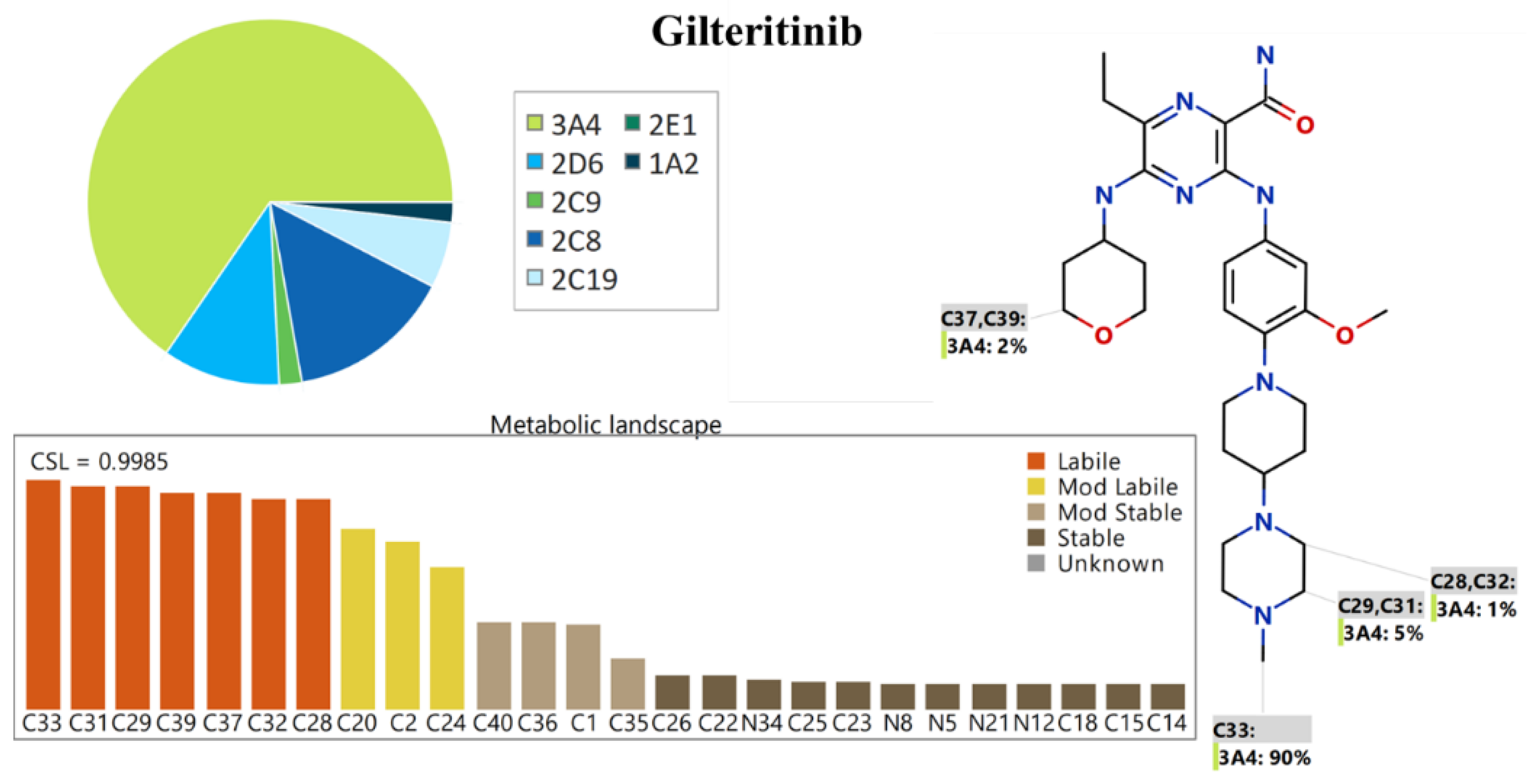
The metabolic lability of the various active sites of GTB towards CYP3A4 enzyme metabolism is outlined in the pie chart [21,22,23]. The CSL (0.9985) proposed high GTB metabolic lability; therefore, the current LC–ESI–MS/MS method was applied for GTB metabolic stability evaluation as GTB is proposed to be a high metabolic rate drug (Figure 2). The generated data revealed that C28, C29, C31, C32, and C33 of the N-methyl piperazine group, and C37 and C39 of the 2H-pyran ring are labile to metabolism while C20 of the methoxy group, C24 of piperidine, C2 of ethyl group are moderately labile to metabolism. The in silico P450 outcomes approved that the N-methyl piperazine group (C28, C29, C31, C32, and C33) is the main reason for GTB metabolic instability (0.9985), showing a high lability to metabolism that agreed with the in vitro HLMs metabolic incubations (mentioned in Section 2.5).

2.2. In Silico GTB Utilizing DEREK Software

In silico toxicity prediction of GTB was performed utilizing the DEREK model of StarDrop software package (Figure 3). GTB was proposed to have skin sensitization (PLAUSIBLE) owing to structural alert (Amino- or hydroxy-aniline) and HERG channel inhibition (PLAUSIBLE) owing to HERG Pharmacophore I as a reported structural alert. The N-methyl piperazine group is predicted to be blamed for the high metabolic lability of GTB in addition to the toxic side effects that were proposed by the DEREK software. Utilizing these results may help develop new drugs with improved metabolic stability and safety.

2.3. UPLC–ESI–MS/MS Methodology Development

Encorafenib (EFB) was selected as the internal standard (IS) in the established UPLC–ESI–MS/MS methodology for the determination of GTB concentration in the HLMs incubation matrix due to three causes: First: both GTB and EFB were extracted from the HLMs matrix utilizing the extraction methodology (protein precipitation) with a good recovery percent for GTB (101.43 ± 3.37) and EFB (104.34 ± 2.82%). Second: the analytes peaks of EFB (1.16 min) and GTB (0.55 min) appeared with perfect separation and a short running time (2 min) which is a fast analytical methodology. Third: there is no written prescription for the simultaneous administration of GTB and EFB for the same case to the same patient as anticancer drugs. The current UPLC–ESI–MS/MS methodology could be utilized for pharmacokinetics studies or therapeutic drug monitoring (TDM) of GTB. No carry-over influence was seen for GTB in the HLMs negative control (Figure 4A) and positive control containing EFB (1000 ng/mL) (Figure 4C). Figure 4D explains the overlaid GTB calibration levels (1: 3000 ng/mL). LLQC of GTB is shown in Figure 4C revealing the sensitivity of the established UPLC-ESI-MS/MS method.
In MRM mode for detection of GTB, two mass transitions (parent ion to two daughter ions; 553 m/z → 453 m/z and 553 m/z → 436 m/z) were selected in the current UPLC-ESI-MS/MS, revealing that the two mass transition showing higher peak area compared to one mass transition (Figure 5) and resulted in generating more sensitivity and a wider range (1 ng/mL to 3000 ng/mL).
Fragmentation of EFB parent ion generated many daughter ions but the two most intense daughter ions at 359 m/z and 116 m/z were seen in the product ion mass spectrum (Figure 6A). These two daughter ions were selected for MRM mass transitions (Figure 6B). Fragmentation of GTB parent ion generated two main daughter ions (453 m/z and 436 m/z) as seen in the product ion mass spectrum (Figure 6C). These two daughter ions were selected for MRM mass transitions (Figure 6D). The MRM mass transitions for GTB and FTB and the corresponding optimized parameters were listed in Table 1.

2.4. Validation Steps of the LC–ESI–MS/MS Methodology

2.4.1. Specificity

The specificity of the LC–ESI–MS/MS methodology was confirmed by the optimum separation of GTB and EFB peaks as seen in Figure 4. There was no influence from the HLMs matrix components with the eluted peaks of GTB and EFB (Figure 4A). No carry-over apparent influence of GTB was noticed in the positive control (blank HLMs + EFB) MRM chromatograms (Figure 4B).

2.4.2. Sensitivity and Linearity of the UPLC-SEI-MS/MS Method

The linearity of the UPLC–ESI–MS/MS analytical methodology was confirmed statistically over a wide range from 1 ng/mL to 3000 ng/mL (y = 2.137x + 0.963 and R2: 0.9964) by injecting seven GTB calibration levels and three QCs in HLMs matrix then back calculating as unknowns. Due to the wide range (1–3000 ng/mL) of the plotted calibration curve, weighting (1/x) was applied during the calculation of the linearity and the regression equation. The RSD values for the six replicates (seven calibration standards and three QCs) were <4.32% (Table 2). The limit of detection (0.31 ng/mL) and LOQ (0.95 ng/mL) revealed the developed UPLC-ESI-MS/MD method.

2.4.3. Precision and Accuracy of the UPLC-SEI-MS/MS Method

The interday and intraday precision and accuracy of the UPLC–ESI–MS/MS methodology were confirmed by the analysis of six repeats (three QCs and LLOQ) in three days and 12 repeats (three QCs and LLOQ) in the same day. The results were in the allowed reported range following the stated FDA guidelines. The intraday and interday precision and accuracy results of the UPLC–ESI–MS/MS method were 0.35–11.39% and 0.27–4.32%, respectively (Table 3).

2.4.4. Matrix Effects of HLMs and GTB Extraction Recovery of the UPLC-SEI-MS/MS Method

The effectiveness of the chosen extraction methodology (protein precipitation utilizing ACN) of GTB and EFB was confirmed by the analysis of six repeats (three QCs and LLOQ) in the HLMs matrix compared to the three QCs and LLOQ that were made in the mobile phase. The results confirmed the efficiency of the chosen extraction method by the high extraction recovery rate of GTB (101.43 ± 3.37 and RSD < 3.6%) and EFB (104.34 ± 2.82%). The HLMs matrix has no influence on the degree of ionization of GTB or EFB parent ions which was verified by preparing two groups of samples. The HLMs incubation matrix (Sample Group 1) was spiked with the GTB LQC (3 ng/mL) and EFB (1000 ng/mL), while Samples Group 2 was done utilizing the mobile phase instead of the HLMs incubation matrix. The HLMs had GTB and EFB exhibited a matrix effect (ME) of 101.92 ± 3.68% and 103.27 ± 3.43%, respectively. The IS normalized ME was 0.987, which was good according to the FDA guidelines. These outcomes revealed that the HLMs incubation matrix exhibited no effect on the EFB or GTB ionization.

2.4.5. Stability of GTB in DMSO and HLMs incubation Matrix

The stability of GTB in DMSO and in the HLM metabolic incubation matrix was assessed. GTB exhibited good stability in the stock solution (DMSO; 1 mg/mL) after kept for 28 days at −80 °C. The stability results ranged from 98.73 to 101.24% for GTB in the HLM incubation matrix (Table 4). There was no noticeable degradation of GTB after short-term storage (4 h at Room T), auto-sampler storage (24 h at 15 °C), long-term storage (−80 °C for 28 d), and three freeze–thaw cycles (3 Cycles, −80 °C). The outcomes show that good stability of GTB was attained.

2.5. The In Vitro Metabolic Incubation for the Determination of GTB Metabolic Stability

In in vitro metabolic incubation with HLMs matrix for GTB metabolic stability determination, the conc. of GTB should be 1 µmole/mL to be less than the Michaelis–Menten constant to establish a statistically linear relationship among the selected time points for metabolic incubation and GTB metabolic rate [24]. HLMs protein conc. should be 1 mg/mL to escape the influence of the protein binding on the concentration of GTB. The GTB concentration in unknown samples was computed utilizing a concurrent injected seven GTB calibration levels. The first plotted GTB metabolic stability curve was made by plotting the selected quenching time points where the metabolic pathways were stopped (x-axis) from 0 to 50 min. against the % of GTB concentration remaining (y-axis) (Figure 7A). The linear part (0–20 min) was nominated to plot the natural logarithmic curve of incubation (y = −0.04842x + 4.659 with R2 = 0.985) (Figure 7B) and the slope (0.04842) represented the GTB metabolic rate constant that was used for computing GTB in vitro t1/2 (Table 5). The in vitro t1/2 (ln2/slope) was 14.32 min. GTB Clint was computed using the in vitro t1/2 methodology, so the Clint of GTB was 56.64 mL/min/kg.
C l i n t , = 0.693 14.32 × 1 1 × 45 1 × 26 1
Following the scoring reported by McNaney et al. [25], GTB is an intermediate clearance ratio drug. By utilizing other in silico software (the Cloe PK software and simulation), these data could also be utilized to propose the GTB in vivo pharmacokinetics [26].

3. Methodologies and Material

3.1. Instruments and Materials

All solvents that were used in the current work were HPLC grade. Milli-Q plus filtration instrument was purchased from the Millipore Company (Billerica, MA, USA) and was utilized for filtration and purification of water at the HPLC grade level. Gilteritinib, encorafenib and solid chemicals are at analytical grade. Formic acid, ammonium formate, acetonitrile, and HLMs (M0567) were procured from Sigma-Aldrich company (St. Louis, MO, USA). After HLMs (20 mg/mL) receiving, it was kept at −70 °C until the time of performing the metabolic incubation. HLMs are composed of a mixture of HLMs pooled from various human donors. The HLMs protein content was labeled as 20 mg/mL of 250 mM sucrose solution. Encorafenib (Synonyms: LGX818; at purity: 99.63%) and GTB (Synonyms: ASP2215; at purity: 98.47%) were procured from MedChem Express Company (Princeton, NJ, USA). The UPLC–ESI–MS/MS system consisted of mass analyzer (Acquity TQD MS; QBB1203) and a liquid chromatography system (Acquity UPLC; H10UPH)] was utilized for separation, mass characterization and quantification of GTB and EFB chromatographic peaks after being extracted from the HLMs incubation matrix. MassLynx 4.1 software (Version 4.1, SCN 805) managed the UPLC–ESI–MS/MS instrument. Processing and interpretation of the generated results were done utilizing the QuanLynx application manager. Optimization of GTB and EFB spectrometric parameters were optimized utilizing a smart tool of IntelliStart® software. The required Nitrogen gas was obtained from a generator that was purchased from Peak Scientific company (Renfrewshire, Scotland, UK) and was used as a drying gas of the mobile phase in the ESI source. The required vacuum inside the mass analyzer was generated from a vacuum pump that was purchased from Sogevac® company (Murrysville, PA, USA). Argon gas (99.999%) was used as a collision gas.

3.2. In Silico Evaluation of GTB Metabolic Stability

In silico GTB metabolic lability assessment was made using the P450 metabolism model (StarDrop software package from Optibrium Ltd., Cambridge, MA, USA). The outcomes are shown as composite site lability (CSL) values reflecting the GTB metabolic lability [24,25,26]. CSL is a crucial factor in proposing the GTB metabolism rate prior to initiating the practical steps to reveal the value of the metabolic incubation work. The GTB SMILES format was uploaded to the metabolic software for CSL computation. To evaluate the GTB metabolic stability, the atom’s labilities can be collected to calculate the CSL indicating the total GTB metabolic lability [27,28] according to Equation (1):
C S L = k t o t a l ( k t o t a l + k w )
as kw is the rate constant for water generation.

3.3. In Silico Proposal of GTB DEREK Software

In silico proposal of GTB toxicity utilizing the DEREK model of StarDrop software that was used for screening the structural alerts. The chemical structure of GTB SMILES format was uploaded to the in silico program software (DEREK software) for the toxicity alerts prediction.

3.4. LC–ESI–MS/MS

3.4.1. LC Chromatographic Tuned Parameters

LC parameters contributing to the chromatographic resolution of GTB and EFB, such as the stationary phase type, and the pH and composition of the mobile phase, were adjusted to get the best chromatographic separation and the increased intensity of analytes peaks. The mobile phase contained an organic part (50%; ACN) and an aqueous part (50%: 0.1% formic acid in H2O; pH: 3.2). Elevating the % of ACN revealed overlapping and poorly resoluted analytes peaks, while the lower percent of ACN generated long running time. The aqueous part was 50% of the mobile phase. Elevating the pH value more than 3.2 reasoned long elution time and analytical peak tailing. Second: the chosen stationary phase was the Eclipse plus-C18 column (50 mm length, 1.8 μm particle size and 2.1 mm internal diameter). Various stationary phase columns were tested, such as normal phase (polar HILIC columns). Neither GTB nor EFB was retained or separated, and the best outcomes were attained utilizing the chosen ZORBAX Eclipse plus-C18 column. GTB and EFB were separated in a short elytion time (2 min) at a flow rate of 0.2 mL/min. after injecting 5 µL into the LC–ESI–MS/MS instrument.

3.4.2. MS/MS Tuned Parameters

MS/MS factors contributing to the mass spectrometric detection and analysis of GTB and EFB were adjusted to attain the optimum sensitivity and ionization of the eluted peaks from the LC instrument followed by spraying and evaporating inside the ESI source. Positive mode for ESI source was utilized for GTB and EFB due to their basic nature (containing basic nitrogens). The optimization of the mass spectrometric parameters (MS/MS) of GTB (Molecular formulae: C29H44N8O3) and EFB (Molecular formulae: C22H27ClFN7O4S) were done utilizing the IntelliStart® software through direct infusion into the mobile phase from the GTB and EFB (10 µg/mL). The cone gas flow rate was optimized at 100 L/H. Nitrogen gas (650 L/H at 350 °C) was supplied by a nitrogen generator. MRM mode for ions detection (parent ion to two product ions) was utilized for estimating GTB and EFB, so as to elevate the sensitivity and selectivity of the developed UPLC–ESI–MS/MS methodology. Argon was utilized as a collision gas at flow rate (0.14 mL/min) for dissociating the parent ion into fragment ions. The cone voltages for GTB and EFB were adjusted at 38 (V) and 56 (V), respectively. The extractor voltage and RF lens were set at 3.0 (V) and 0.1 (V), respectively. The dwell time for GTB and EFB mass transitions (parent to fragment ions) was 0.025 s.

3.5. GTB Working Solutions

GTB and EFB have a reasonable solubility in DMSO at 2 mg/mL (3.62 mM) and 50 mg/mL (92.59 mM), respectively. The prepared stock solutions (1 mg/mL) of GTB and EFB were made in DMSO due to the good stability and solubility features in DMSO. Consecutive dilutions of the GTB (1 mg/mL) utilizing mobile phase were done to make the working solutions (WK) at 100 µg/mL (GTB WK1), 10 µg/mL (GTB WK2) and 1 µg/mL (GTB WK3). Consecutive dilution of the EFB (1 mg/mL) was done to make the EFB WK3 (10 µg/mL).

3.6. Calibration Curve of GTB

Deactivation of HLMs was performed before spiking with GTB and EFB to discard the metabolic influence in the concentration of GTB and EFB during the analytical methodology validation steps. HLMs were deactivated utilizing DMSO (2%) as it stopped metabolic pathways [29] with slight warming (5 min at 50 °C) as heat stopped HLMs enzymatic activity [30,31,32]. The HLMs matrix for the validation steps was made at a conc. of 1 mg protein/mL by diluting 30 µL of the quenched HLMs to 1 mL with the buffer (0.1 M sodium phosphate buffer: pH 7.4) inclosing the metabolism cofactor (1 mM NADPH) to mimic the in vitro metabolic incubation steps that could be applied to metabolic stability determination. GTB calibration points were made by consecutive dilution of GTB WK2 (10 µg/mL) and GTB WK3 (1 µg/mL) with the quenched HLMs matrix making seven standards: 1, 15, 150, 300, 500, 1500, and 3000 ng/mL making the volume of the HLMs matrix (90%) so as to decrease the effect of dilution if compared to real metabolic incubations. These GTB calibration levels were utilized for plotting a calibration curve. Three quality controls (QCs) were prepared as LQC (3 ng/mL), MQC (900 ng/mL) and HQC (2400 ng/mL). A lower limit of quantification (LLOQ; 1 ng/mL) was involved in the validation procedure. QCs were treated as unknowns and the concentration were estimated utilizing the regression equation of a freshly constructed GTB calibration curve. EFB WK1 (100 µL; 1000 ng/mL) was added to all seven calibration standards and the three QCs as IS.

3.7. Extraction Methodology of GTB and EFB

The protein precipitation methodology utilizing ACN was used for GTB and EFB extraction from the HLMs matrix as described in the next steps. Two mL. of the precipitating solvent (ACN) was added to the seven GTB calibration levels, the three QCs or unknown metabolic incubation samples. Then, good shaking of all incubates were performed for 5 min, and then centrifugation was done at 14,000 rpm for 12 min (4 °C). Filtration of all supernatants utilizing syringe filters (0.22 µm pore size) was done into HPLC vials to verify the purity that is required for samples to be loaded into the UPLC–ESI–MS/MS instrument. Positive controls (HLMs matrix + IS) and negative control (HLMs matrix) were made utilizing the identical procedure previously detailed to confirm the lack of any interference chromatographic peaks at the retention time of analytes (GTB and EFB) and carry over effect of GTB, respectively. A GTB calibration curve was made by plotting the peak area ratio of GTB to EFB (y-axis) against GTB standards concentration (x-axis) that generated a linear regression equation (y = ax + b; r2) that confirmed the range of linearity.

3.8. Validation of the UPLC–ESI–MS/MS Methodology

The validation parameters of the LC–esi–MS/MS methodology were done utilizing specificity, sensitivity, linearity, precision, accuracy, stability, matrix effect and extraction recovery following the USFDA regulations for bioanalytical methodology validation [33].

3.8.1. Specificity of the UPLC–ESI–MS/MS Methodology

The specificity of the LC–ESI–MS/MS methodology was tested by loading six blank HLMs incubation matrices after doing the selected protein precipitation method for purification. Then, 5 µL of these purified extracts were loaded into LC–ESI–MS/MS and verified for any interfering endogenous peaks at GTB or EFB retention times, and comparing the generated MRM total ion chromatogram of spiked HLMs matrix incubation samples with GTB and EFB. MRM mass analyzer mode was used to decrease the carryover effects of GTB and EFB in the mass detector as verified by the data generated from the negative control HLMs without any GTB and EFB chromatographic peaks.

3.8.2. Linearity and Sensitivity of the UPLC–ESI–MS/MS Methodology

The linearity and sensitivity of the current UPLC–ESI–MS/MS methodology were revealed by preparing 12 repeats calibration standards (seven standards and three QCs) of GTB in HLMs matrix in one day then back computing of peak area ratio of the calibration points and QCs as unknowns by substituting in the linear regression equation. The LOQ and LOD were calculated as stated in the Pharmacopeia utilizing the SD of the intercept and the slope of the plotted calibration levels following Equations (2) and (3), respectively:
L O Q = 10 × S D   o f   t h e   i n t e r c e p t S l o p e
L O D = 3.3 × S D   o f   t h e   i n t e r c e p t S l o p e
The linearity of the UPLC–ESI–MS/MS method was confirmed by utilizing the least squared statistical methodology (y = ax + b) and the coefficient of variation (R2).

3.8.3. Accuracy and Precision of the UPLC–ESI–MS/MS Methodology

Precision and accuracy of the current LC–ESI–MS/MS methodology were computed as % relative standard deviation (% RSD) and % error (% E), respectively as computed in Equations (4) and (5), respectively.
%   R S D = S D A v e r a g e
%   E = ( a v e r a g e   c o m u t e d   c o n c . s u p p o s e d   c o n c . ) s u p p o s e d   c o n c .
The intraday and interday accuracy and precision of the current UPLC–ESI–MS/MS methodology were verified by loading six repeats of GTB QCs in 3 days and 12 repeats of GTB QCs in one day, respectively following the USFDA guidelines.

3.8.4. Extraction Recovery and Matrix Effect

The influence of the HLMs constituents on the GTB ionization and the percent of extraction recovery of GTB from the incubation HLMs matrix were determined by loading the three GTB QCs samples and the GTB LLOQ into the UPLC-ESI-MS/MS instrument. The efficacy of the selected extraction methodology (protein precipitation utilizing ACN) for GTB and EFB was verified by loading six repeats of GTB QCs and LLOQ in HLMs matrix (B) and comparing it with the three QCs and LLQC that were prepared in the mobile phase (A). The % recovery of the GTB and EFB was calculated as the ratio of B/A × 100. The effect of HLMs matrix constituents on the degree of ionization of GTB or EFB was revealed by loading two sets of samples. HLMs matrix (set 1) was spiked with the GTB LQC (3 ng/mL) and EFB (IS; 1000 ng/mL), while set 2 was made by using the mobile phase. The matrix effects (ME) for GTB and EFB were computed following Equation (6). The IS normalized ME was calculated following Equation (7).
M E   o f   G T B   o r   E F B = a v e r a g e   p e a k   a r e a   r a t i o   S e t   1 S e t   2 × 100
I S   n o r m a l i z e d   M E = M E   o f   G T B M E   o f   E F B   ( I S )

3.8.5. Stability

GTB stability in DMSO and in HLMs samples (in vitro metabolic incubations) were evaluated utilizing laboratory parameters that an analyte may be exposed to before the injection to LC–ESI–MS/MS instrument, involving after auto-sampler storage, short-term storage, cycles long-term storage, and three freeze–thaw.

3.9. In Vitro Assessment of GTB Metabolic Stability

The parameters of GTB metabolic stability (in vitro t1/2 and the CLint) were computed by estimating the remaining % GTB conc. after in vitro metabolic incubation with HLMs matrix (active) that involves NADPH as a metabolic reaction cofactor that is responsible for the initialization of the in vitro metabolic pathways. The metabolic incubation procedure for GTB and HLMs matrix was done in four steps. The first step was the pre-incubation (conditioning) of 1 µmole/mL of GTB with the prepared HLMs matrix (without the co-factor: NADPH) at 37 °C for 10 min to obtain the perfect factors for initiating metabolic incubation. The second step included beginning the metabolic pathway (incubation) by adding 1 mM of NADPH as a co-factor. In the third step, the metabolic reaction (quenching) was stopped by adding 2 mL of ACN. In the fourth step, the addition of 100 µL of EFB WK3 (IS addition) was performed before the ACN addition to remove the metabolic influence on the EFB conc. Quenching of the incubation metabolic reaction was made at chosen time points: 0, 2.5, 5, 7.5, 15, 20, 25, 30, 40, and 50 min. The same methodology for extraction of GTB from HLMs matrix incubations was applied as described before.
The GTB conc. in HLMs incubation matrix was estimated utilizing simultaneous injected GTB calibration seven levels for regression equation generation. GTB metabolic stability curve was made by plotting the stopping time points (x-axis) from 0 to 50 min. against the of % of the GTB conc. remaining (y-axis). Choosing the part of the established curve that exhibited (0–20 min) was done to construct another curve by plotting the natural logarithm (Ln) of the % GTB remaining against the time of metabolic incubation points (linear range) and the slope of the constructed curve revealed the rate constant for the metabolic reaction that was used to determine the in vitro t1/2 (ln2/slope). Then, the CLint (µL/min/mg) of GTB was calculated [25], utilizing a value of 26 g for liver tissue per Kg of body weight and 45 mg of microsomal protein (HLMs matrix) per gram of liver tissue [34] (Equation (8)).
C L i n t , = 0.693 i n   v i t r o   t ½ × m g   H L M s g   l i v e r × m L   i n c u b a t i o n m g   m i c r o s o m e s × g   l i v e r K g   b . w .

4. Conclusions

A validated UPLC-ESI-MS/MS method was established for estimating GTB in metabolic incubation matrix (HLMs) that was applied for the in vitro determination of GTB metabolic stability. The UPLC-ESI-MS/MS methodology revealed good sensitivity and selectivity. The current UPLC-ESI-MS/MS methodology also revealed high recovery utilizing protein precipitation extraction methodology of EFB and GTB from HLMs matrix. The utilization of less ACN in the mobile phase with short elution time and flow rate make the UPLC-ESI-MS/MS methodology eco-friendly. The in silico metabolism P450 model was utilized to predict the GTB metabolic stability that matched the data of the in vitro HLMs incubations. GTB metabolic stability parameters [moderate Clint (56.64 mL/min/kg) and in vitro t1/2 values (14.32 min)] showed the moderate clearance feature of GTB and proposed good in vivo bioavailability. The N-methyl piperazine group is predicted to be blamed for the high metabolic lability of GTB in addition to toxic side effects that were proposed by DEREK software. Future work may be done utilizing in vitro and in silico practical experiments as a part of designing new drugs with good metabolic stability behavior.

Author Contributions

M.W.A., A.S.A., H.A. and A.A.K. establish the experimental steps. M.W.A., H.A. and A.M.A. made the in vitro metabolic incubations, running the in silico software and wrote the first draft of the manuscript. A.S.A., H.A. and A.A.K. helped software applications and design the methodology. All authors have read and agreed to the published version of the manuscript.

Funding

This study was funded by the Deputyship for Research & Innovation, “Ministry of Education” in Saudi Arabia, through project no. (IFKSURG-2-1023).

Institutional Review Board Statement

Ethical review and approval were discharged for the current experiment owing to the use of commercially available human liver microsomes in the in vitro metabolic incubation experiments that were purchased from Sigma company.

Data Availability Statement

All data are available within the manuscript.

Acknowledgments

The authors extend their appreciation to the Deputyship for Research & Innovation, Ministry of Education in Saudi Arabia for funding this research work through project no. (IFKSURG-2-1023).

Conflicts of Interest

The authors state no conflict of interest.

Abbreviations

AUCarea under the curve
Clintintrinsic clearance
DMSOdimethyl sulfoxide
EFBencorafenib
ESIelectrospray ionization
EGFRepidermal growth factor receptor
GTBgilteritinib
HLMshuman liver microsomes
ISinternal standard
LLOQlower limit of quantification
LC-MS/MSliquid chromatography tandem mass spectrometry
MRMmultiple reaction monitoring
NSCLCsNon-small cell lung cancers
QCquality control
RSDrelative standard deviation
RErelative error
S/Nsignal to noise ratio
SDstandard deviation
TKIstyrosine kinase inhibitors
t1/2half-life

References

  1. Mattiuzzi, C.; Lippi, G. Current Cancer Epidemiology. J. Epidemiol. Glob. Health 2019, 9, 217–222. [Google Scholar] [CrossRef] [PubMed]
  2. Roskoski, R., Jr. Properties of FDA-approved small molecule protein kinase inhibitors: A 2020 update. Pharmacol. Res. 2020, 152, 104609. [Google Scholar] [CrossRef] [PubMed]
  3. Barinaga, M. From Bench Top to Bedside. Science 1997, 278, 1036–1039. [Google Scholar] [CrossRef] [PubMed]
  4. Kawase, T.; Nakazawa, T.; Eguchi, T.; Tsuzuki, H.; Ueno, Y.; Amano, Y.; Suzuki, T.; Mori, M.; Yoshida, T. Effect of Fms-like tyrosine kinase 3 (FLT3) ligand (FL) on antitumor activity of gilteritinib, a FLT3 inhibitor, in mice xenografted with FL-overexpressing cells. Oncotarget 2019, 10, 6111–6123. [Google Scholar] [CrossRef] [PubMed]
  5. Pulte, E.D.; Norsworthy, K.J.; Wang, Y.; Xu, Q.; Qosa, H.; Gudi, R.; Przepiorka, D.; Fu, W.; Okusanya, O.O.; Goldberg, K.B.; et al. FDA Approval Summary: Gilteritinib for Relapsed or Refractory Acute Myeloid Leukemia with a FLT3 Mutation. Clin. Cancer Res. 2021, 27, 3515–3521. [Google Scholar] [CrossRef]
  6. Dhillon, S. Gilteritinib: First Global Approval. Drugs 2019, 79, 331–339. [Google Scholar] [CrossRef]
  7. Bouhaddou, M.; Memon, D.; Meyer, B.; White, K.M.; Rezelj, V.V.; Correa Marrero, M.; Polacco, B.J.; Melnyk, J.E.; Ulferts, S.; Kaake, R.M.; et al. The Global Phosphorylation Landscape of SARS-CoV-2 Infection. Cell 2020, 182, 685–712.e19. [Google Scholar] [CrossRef]
  8. Usuki, K.; Sakura, T.; Kobayashi, Y.; Miyamoto, T.; Iida, H.; Morita, S.; Bahceci, E.; Kaneko, M.; Kusano, M.; Yamada, S.; et al. Clinical profile of gilteritinib in Japanese patients with relapsed/refractory acute myeloid leukemia: An open-label phase 1 study. Cancer Sci. 2018, 109, 3235–3244. [Google Scholar] [CrossRef]
  9. Perl, A.E.; Altman, J.K.; Cortes, J.; Smith, C.; Litzow, M.; Baer, M.R.; Claxton, D.; Erba, H.P.; Gill, S.; Goldberg, S.; et al. Selective inhibition of FLT3 by gilteritinib in relapsed or refractory acute myeloid leukaemia: A multicentre, first-in-human, open-label, phase 1-2 study. Lancet Oncol. 2017, 18, 1061–1075. [Google Scholar] [CrossRef]
  10. Cardoso, E.; Csajka, C.; Schneider, M.P.; Widmer, N. Effect of Adherence on Pharmacokinetic/Pharmacodynamic Relationships of Oral Targeted Anticancer Drugs. Clin. Pharmacokinet. 2018, 57, 1–6. [Google Scholar] [CrossRef]
  11. Bryant, A.L.; LeBlanc, T.W.; Albrecht, T.; Chan, Y.N.; Richardson, J.; Foster, M.; Dang, M.; Dudley, W.; Owenby, S.; Wujcik, D. Oral adherence in adults with acute myeloid leukemia (AML): Results of a mixed methods study. Support Care Cancer 2020, 28, 5157–5164. [Google Scholar] [CrossRef]
  12. Houston, J.B. Utility of in vitro drug metabolism data in predicting in vivo metabolic clearance. Biochem. Pharmacol. 1994, 47, 1469–1479. [Google Scholar] [CrossRef]
  13. Obach, R.S.; Baxter, J.G.; Liston, T.E.; Silber, B.M.; Jones, B.C.; MacIntyre, F.; Rance, D.J.; Wastall, P. The prediction of human pharmacokinetic parameters from preclinical and in vitro metabolism data. J. Pharmacol. Exp. Ther. 1997, 283, 46–58. [Google Scholar]
  14. Attwa, M.W.; Kadi, A.A.; Darwish, H.W.; Amer, S.M.; Alrabiah, H. A reliable and stable method for the determination of foretinib in human plasma by LC-MS/MS: Application to metabolic stability investigation and excretion rate. Eur. J. Mass Spectrom. 2018, 24, 344–351. [Google Scholar] [CrossRef]
  15. Amer, S.M.; Kadi, A.A.; Darwish, H.W.; Attwa, M.W. LC–MS/MS method for the quantification of masitinib in RLMs matrix and rat urine: Application to metabolic stability and excretion rate. Chem. Cent. J. 2017, 11, 136. [Google Scholar] [CrossRef]
  16. Zhang, M.; Tajima, S.; Suetsugu, K.; Hirota, T.; Tsuchiya, Y.; Yamauchi, T.; Yoshimoto, G.; Miyamoto, T.; Egashira, N.; Akashi, K.; et al. Development and Validation of an LC-MS/MS Method to Quantify Gilteritinib and Its Clinical Application in Patients With FLT3 Mutation-Positive Acute Myelogenous Leukemia. Ther. Drug Monit. 2022, 44, 592–596. [Google Scholar] [CrossRef]
  17. Garrison, D.A.; Jin, Y.; Uddin, M.E.; Sparreboom, A.; Baker, S.D. Development, validation, and application of an LC-MS/MS method for the determination of the AXL/FLT3 inhibitor gilteritinib in mouse plasma. J. Chromatogr. B Analyt. Technol. Biomed. Life Sci. 2021, 1179, 122882. [Google Scholar] [CrossRef]
  18. Wang, Q.; Chen, Z.; Chen, D.; Ye, X.Y. An LC-MS/MS Bioanalytical Assay for the Determination of Gilteritinib in Rat Plasma and Application to a Drug-Drug Interaction Study. Drug Des. Devel Ther. 2020, 14, 2061–2067. [Google Scholar] [CrossRef]
  19. Tyzack, J.D.; Kirchmair, J. Computational methods and tools to predict cytochrome P450 metabolism for drug discovery. Chem. Biol. Drug Des. 2019, 93, 377–386. [Google Scholar] [CrossRef]
  20. Marothu Vamsi, K.; Kantamaneni, P.; Gorrepati, M. In vitro Metabolic Stability of Drugs and Applications of LC-MS in Metabolite Profiling. In Drug Metabolism; Katherine, D., Ed.; IntechOpen: Rijeka, Croatia, 2021; p. 77. [Google Scholar]
  21. Kazmi, S.R.; Jun, R.; Yu, M.S.; Jung, C.; Na, D. In silico approaches and tools for the prediction of drug metabolism and fate: A review. Comput. Biol. Med. 2019, 106, 54–64. [Google Scholar] [CrossRef]
  22. Hunt, P.A.; Segall, M.D.; Tyzack, J.D. WhichP450: A multi-class categorical model to predict the major metabolising CYP450 isoform for a compound. J. Comput. Aided Mol. Des. 2018, 32, 537–546. [Google Scholar] [CrossRef] [PubMed]
  23. Shin, Y.G.; Le, H.; Khojasteh, C.; Hop, C.E.C.A. Comparison of metabolic soft spot predictions of CYP3A4, CYP2C9 and CYP2D6 substrates using MetaSite and StarDrop. Comb. Chem. High Throughput Screen. 2011, 14, 811–823. [Google Scholar] [CrossRef]
  24. Attwa, M.W.; Darwish, H.W.; Al-Shakliah, N.S.; Kadi, A.A. A validated lc–ms/ms assay for the simultaneous quantification of the fda-approved anticancer mixture (Encorafenib and binimetinib): Metabolic stability estimation. Molecules 2021, 26, 2717. [Google Scholar] [CrossRef] [PubMed]
  25. McNaney, C.A.; Drexler, D.M.; Hnatyshyn, S.Y.; Zvyaga, T.A.; Knipe, J.O.; Belcastro, J.V.; Sanders, M. An automated liquid chromatography-mass spectrometry process to determine metabolic stability half-life and intrinsic clearance of drug candidates by substrate depletion. Assay Drug Dev. Technol. 2008, 6, 121–129. [Google Scholar] [CrossRef]
  26. Leahy, D.E. Integrating invitro ADMET data through generic physiologically based pharmacokinetic models. Expert Opin. Drug Metab. Toxicol. 2006, 2, 619–628. [Google Scholar] [CrossRef]
  27. Alrabiah, H.; Kadi, A.A.; Attwa, M.W.; Abdelhameed, A.S. A simple liquid chromatography-tandem mass spectrometry method to accurately determine the novel third-generation EGFR-TKI naquotinib with its applicability to metabolic stability assessment. RSC Adv. 2019, 9, 4862–4869. [Google Scholar] [CrossRef] [PubMed]
  28. Kadi, A.A.; Darwish, H.W.; Abuelizz, H.A.; Alsubi, T.A.; Attwa, M.W. Identification of reactive intermediate formation and bioactivation pathways in Abemaciclib metabolism by LC-MS/MS: In vitro metabolic investigation. R. Soc. Open Sci. 2019, 6, 181714. [Google Scholar] [CrossRef]
  29. Busby, W.F., Jr.; Ackermann, J.M.; Crespi, C.L. Effect of methanol, ethanol, dimethyl sulfoxide, and acetonitrile on in vitro activities of cDNA-expressed human cytochromes P-450. Drug Metab. Dispos. 1999, 27, 246–249. [Google Scholar]
  30. Störmer, E.; Roots, I.; Brockmöller, J. Benzydamine N-oxidation as an index reaction reflecting FMO activity in human liver microsomes and impact of FMO3 polymorphisms on enzyme activity. Br. J. Clin. Pharmacol. 2000, 50, 553–561. [Google Scholar] [CrossRef]
  31. Fouin-Fortunet, H.; Tinel, M.; Descatoire, V.; Letteron, P.; Larrey, D.; Geneve, J.; Pessayre, D. Inactivation of cytochrome P-450 by the drug methoxsalen. J. Pharmacol. Exp. Ther. 1986, 236, 237–247. [Google Scholar]
  32. Attwa, M.W.; AlRabiah, H.; Mostafa, G.A.E.; Kadi, A.A. Development of an LC-MS/MS Method for Quantification of Sapitinib in Human Liver Microsomes: In Silico and In Vitro Metabolic Stability Evaluation. Molecules 2023, 28, 2322. [Google Scholar] [CrossRef]
  33. United State of America–Food and Drug Administration. Bioanalytical Method Validation Guidance for Industry. 2018. Available online: https://www.fda.gov/ucm/groups/fdagov-public/@fdagov-drugsgen/documents/document/ucm070107.pdf (accessed on 10 February 2022).
  34. Słoczyńska, K.; Gunia-Krzyżak, A.; Koczurkiewicz, P.; Wójcik-Pszczoła, K.; Żelaszczyk, D.; Popiół, J.; Pękala, E. Metabolic stability and its role in the discovery of new chemical entities. Acta Pharm. 2019, 69, 345–361. [Google Scholar] [CrossRef]
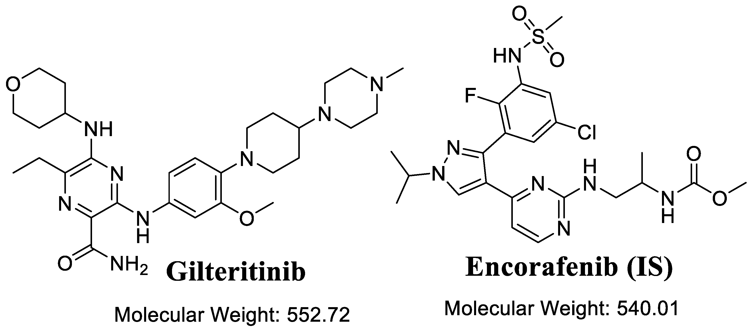
Figure 1. Chemical structures of gilteritinib and encorafenib (internal standard; IS).
Separations 10 00278 g001
Figure 2. Composite site lability (CSL: 0.9985) exhibiting the high metabolic instability of GTB. These results were done utilizing the StarDrop software package (P450 metabolic model).
Separations 10 00278 g002
Figure 3. Proposed toxicity alerts of GTB utilizing the DEREK model of StarDrop software.
Separations 10 00278 g003
Figure 4. Blank control (HLMs matrix) exhibiting no chromatographic peaks at the elution time of GTB and EFB (A), LLOQ of GTB revealing the sensitivity of the method (B). Blank HLMs plus EFB (C) chromatogram, and overlaid GTB calibration levels exhibiting the GTB peak (0.55 min) and EFB peak (1.16 min) (D).
Separations 10 00278 g004
Figure 5. Mass transitions of GTB revealing that the two mass transition showing higher peak area compared to one mass transition.
Separations 10 00278 g005
Figure 6. Product ion mass spectrum of EFB (A). MRM mass spectrum of EFB (B). Product ion mass spectrum of GTB (C). MRM mass spectrum of GTB (D). The proposed fragmentation patterns were exhibited.
Separations 10 00278 g006
Figure 7. The plotted metabolic stability curve of GTB after in vitro metabolic incubation with HLMs matrix (A). The selected linear range of the metabolic stability curve (ln curve) exhibiting the regression equation (B). The GTB MRM chromatogram for in vitro metabolic incubation at 0 time (100%) (C).
Separations 10 00278 g007
Table 1. MRM mass transitions of GTB and EFB showing tuned parameters.
AnalyteESI RtParent
(m/z)
Daughter Ion (1)Daughter Ion (2)CE, eVCone Voltage
(V)
GTB+ve0.56553.043645330/2438
EFB (IS)+ve1.16540.011635932/3656
Table 2. Back-calculation data of six replicates (seven calibration levels and three QCs) of GTB.
GTB (ng/mL)AverageSDRSD (%)Accuracy (%)Recovery
1 (LLOQ)1.100.011.3310.10110.10
3 (LQC)3.060.134.322.10102.10
15 14.810.201.38−1.2898.72
150 146.812.291.56−2.1397.87
300305.012.620.861.67101.67
500508.114.690.921.62101.62
900 (MQC)905.534.610.510.61100.61
15001485.845.670.38−0.9499.06
2400 (HQC)2430.9314.010.581.29101.29
30003039.4420.480.671.31101.31
% Recovery 101.43 ± 3.37
Table 3. Accuracy and precision (interday and intraday) of the UPLC-SEI-MS/MS method.
GTB in HLMs Matrix (ng/mL)Intra-Day Assay
(Twelve Replicates in One Day)
Inter-Day Assay
(Six Replicates in Three Days)
1
(LLOQ)
3
(LQC)
900
(MQC)
2400 (HQC)1
(LLOQ)
3
(LQC)
900
(MQC)
2400 (HQC)
Mean1.103.06905.532430.931.113.11910.302408.49
SD0.010.134.6114.010.020.104.046.52
Precision (% RSD)1.334.320.510.582.023.240.440.27
% Accuracy10.102.100.611.2911.393.691.140.35
Recovery (%)110.10102.10100.61101.29111.39103.69101.14100.35
Table 4. Stability data of GTB.
GTB Concentration (ng/mL)Short-Term StabilityFreeze–Thaw StabilityAutosampler StabilityLong-Term Stability
LQC (3)99.7 ± 3.2199.24 ± 3.4699.52 ± 3.0898.73 ± 4.18
HQC (2400)101.24 ± 3.62100.82 ± 3.48100.68 ± 2.8299.85 ± 3.28
Table 5. GTB in vitro metabolic stability.
Time of Quenching (min)Average a (ng/mL)X bLN XThe Linearity
0.00672.00100.004.61Regression equation:
y = −0.04842x + 4.659
2.50626.5793.244.54
5.00561.5983.574.43R2 = 0.985
7.50512.4076.254.33
15.00365.0354.323.99Slope: −0.04842
20.00254.3537.853.63
25.00197.9029.453.38t1/2: 14.32 min and
30.00187.1527.853.33Clint: 56.64 mL/min/kg
40.00166.3224.753.21
50.00146.1621.753.08
Notes: a Mean of three repeats, b X: Average of the % remaining GTB of the three replicates.
Disclaimer/Publisher’s Note: The statements, opinions and data contained in all publications are solely those of the individual author(s) and contributor(s) and not of MDPI and/or the editor(s). MDPI and/or the editor(s) disclaim responsibility for any injury to people or property resulting from any ideas, methods, instructions or products referred to in the content.