Article

Phytochemical Characterization and Antifungal Efficacy of Camphor (Cinnamomum camphora L.) Extract against Phytopathogenic Fungi

1
Plant Protection and Biomolecular Diagnosis Department, ALCRI—Arid Lands Cultivation Research Institute, City of Scientific Research and Technological Applications, New Borg El Arab City, Alexandria 21934, Egypt
2
Department of Botany and Microbiology, College of Science, King Saud University, P.O. Box 2455, Riyadh 11451, Saudi Arabia
3
Department of Botany and Microbiology, Faculty of Science, Al-Azhar University, Assiut 71524, Egypt
4
Department of Agricultural Botany, Faculty of Agriculture, Kafrelsheikh University, Kafrelsheikh 33516, Egypt
5
School of Molecular Sciences, The University of Western Australia, Perth, WA 6009, Australia
6
Agricultural Botany Department, Faculty of Agriculture (Saba Basha), Alexandria University, Alexandria 21531, Egypt
*
Authors to whom correspondence should be addressed.
Separations 2023, 10(3), 189; https://doi.org/10.3390/separations10030189
Submission received: 8 February 2023 / Revised: 2 March 2023 / Accepted: 7 March 2023 / Published: 9 March 2023

Abstract

:
Cinnamomum camphora methanolic extract was tested for antifungal activity against three common, isolated, widespread phytopathogens: Alternaria alternata, Fusarium solani, and Fusarium oxysporum, which were molecularly identified and assigned accession numbers ON795987, ON795988, and ON795989, respectively. At 4000 µg/mL, the highest concentration of C. camphora methanolic extract inhibits the fungal mycelia weight of F. oxysporum, A. alternata, and F. solani by 60, 49, and 24%, respectively. The presence of several bioactive metabolites in the C. camphora extract could explain its antifungal activity. The presence of numerous phenolic and flavonoid compounds in the extract was revealed by HPLC analysis, including catechin and gallic acid, which had the highest concentrations of 6.21 and 6.98 µg/mL, respectively. Furthermore, osmoprotectants, total amino acids, and glycine betaine were abundant. Furthermore, total antioxidant activities, as measured by PMA and DPPH, were significant. The most abundant compound in the extract, according to GC-MS analysis, was mono(2-ethylhexyl) ester of 1,2-benzene dicarboxylic acid. Based on its in vitro efficacy in inhibiting mycelial growth weight, the tested extract could be recommended as a safe fungicide instead of a chemical treatment.

1. Introduction

Global crop yields suffer from many pathogens that affect their productivity and quality. The major pathogens causing these diseases comprise fungi, viruses, nematodes, and bacteria [1]. The intensity of underground fungi is significantly higher than that of aboveground fungi. These soil-borne fungi can endure for extended periods within the soil due to the production of structures that help them survive, such as sclerotia, melanized hyphae, oospores, and chlamydospores [2]. A prevalent soil-borne fungus, the Fusarium species, is responsible for a variety of plant diseases, including Fusarium head blight, Fusarium root rot, Fusarium crown rot, and others. This fungus can impact a diverse range of crops, including small grain cereals, maize, various vegetables, bananas, lilies, trees, and many more, each with its own unique disease pattern, leading to substantial reductions in yield [3]. In addition to Fusarium, Alternaria sp. is responsible for severe pre- and post-harvest crop losses. Alternaria alternata has previously been reported to cause post-harvest losses at high frequency in tomatoes [4] and produce leaf blight symptoms. Moreover, Alternaria invades not only vegetables, but is also responsible for the late blight symptom of nuts, which affects both foliage and fruit and is characterized by the development of large necrotic spots or lesions that eventually coalesce and consume the entire leaf and defoliate the tree [5].
There are many ways to control and deal with the rise of plant fungal diseases around the world. The most common and useful method in agriculture for getting rid of pests and diseases is to use pesticides [6]. Unfortunately, the widespread use of pesticides in agriculture to deal with problems before and after harvest has led to dangerous outbreaks and fungal infections that are resistant to fungicides [7]. In addition, once the pesticide has been applied, the chemical residue makes its way into groundwater, lakes, and marine water through various environmental processes, negatively impacting the species living in those bodies of water [8,9]. Therefore, research is now focused on finding new ways to reduce the pesticide levels left on the soil. Among these, natural extracts are safer and cheaper alternatives to replace the widespread use of synthetic chemical antifungal substances to create a chemical-free environment.
Plant extracts are characterized by diverse active compounds, minerals, secondary metabolites, and antioxidants, which give the extracts their efficacy [10,11]. Natural compounds extracted from various plants have been shown to have broad biological applications as antifungal, antibacterial, and antiviral agents [11,12,13,14]. Cinnamomum camphora (L.) is a huge tree commonly planted for landscaping and forestation. This tree synthesizes plenty of compounds known as terpenoids, which prevent herbivore attacks and repel fungal infections [15]. Due to its antioxidant constituents and antimicrobial activities, C. camphora has been widely used in the cosmetic and medical industries [16]. Moreover, it is a rich source of alcohols, ketones, terpenoids, and esters, which may increase the algaecidal functions. The C. camphora leaf extract has antifungal activity against Choanephora cucurbitarum [8] and Aspergillus niger [17], antibacterial activity against Pasturella multocida [18], and insecticidal properties against Drosophila melanogaster, Chaoborus plumicornis, and Pieris rapae [17]. Therefore, our research endeavors to assess the antifungal properties of a methanolic extract of C. camphora against three molecularly identified soil-borne phytopathogenic fungi (F. oxysporum, A. alternata, and F. solani) isolated from tomato plants. Additionally, total flavonoid, phenolic, ascorbic acid, amino acids, saponin, tannins, proline, glycine betaine content, total antioxidant, and DPPH scavenging activities were estimated. Additionally, the chemical composition of the extract was determined through HPLC and GC-MS analysis.

2. Materials and Methods

2.1. Plant Sampling and Extract Preparation

The leaves of Cinnamomum camphora plants grown in Tanta, Egypt, were picked for experimental research. The selected leaves were healthy and did not suffer from any morphological aberrations. Initially, the leaves underwent thorough washing with tap water to remove any surface dust or contaminants and were left to dry at room temperature for 10 days or until completely dry. The dried leaves were then ground into a fine powder. Subsequently, 50 g of the powder was combined with 500 mL of 80% methanol in an Erlenmeyer flask and agitated on a rotary shaker (100 rpm) at room temperature for a period of one night. The resulting methanolic leaf extract was filtered through Whatman No. 1 filter paper. To remove any remaining methanol in the extract, vacuum-assisted drying was employed. By applying a temperature range of 25–30 °C, the residual solvent can be evaporated. The dried extract was stored in the fridge at 4 °C. To use the extract in the antifungal activity test, it was dissolved in 10% dimethyl sulfoxide (DMSO). We weighed 0.1, 0.2, 0.3, and 0.4 g of the extract and dissolved each in 1 mL of 10% DMSO. We then used these stock solutions to prepare the final concentrations in 100 mL of broth media in a flask as 1000, 2000, 3000, and 4000 µg/mL of media. The control flask received 1 mL of 10% DMSO (free of extract).

2.2. Fungal Isolates, Culture Media, and Growth Conditions

Three phytopathogenic fungi were obtained from the rhizosphere of squash plants. The purity of these fungi was verified by growing them on potato dextrose agar (PDA) medium in 9-cm Petri dishes at 27 °C for a week in an incubator. Afterward, the three fungi were preserved on slants in the refrigerator at 4 °C for further investigation.

2.3. Morphological and Molecular Characterization

The three fungal isolates were re-grown on PDA media at 27 °C for 7 days, and then the mycelia and spores were morphologically examined under the light microscope according to the identification protocols [19,20]. Afterward, the mycelia were harvested for DNA extraction using the CTAB method described by Wang et al. [21]. The DNA concentration was measured using a Nanodrop 2000 spectrophotometry device (Thermo Fisher Scientific, Wilmington, DE, USA) and stored at −20 °C in a dilution of 100 ng/μL for use in PCR amplification. The complete rDNA-ITS (ITS) region was amplified using specific PCR primers, such as ITS1/ITS4 [22], from all fungal isolates. The PCR amplification conditions were initial denaturing at 94 °C for 5 min; 35 cycles of denaturing (94 °C for 30 s each cycle); annealing at 55 °C for 30 s; extension at 72 °C for 1 min; and a final extension at 72 °C for 10 min. The PCR amplicons were visualized, purified, and sent to the Macrogen company for Sanger sequencing after being electrophoresed on a 1% agarose gel.

2.4. Antifungal Activity

The mycelial weight of the fungal mat was used to evaluate the antifungal activity of C. camphora extract. The final concentrations of C. camphora extract (1000, 2000, 3000, and 4000 µg/mL) in potato broth media were made as illustrated in Section 2.1. Each flask of broth medium was inoculated with 0.5 cm of the fungal disc and shaken for a week on an orbital shaker (100 rpm) at 27 ± 2 °C before being filtered. The fungal mats were weighed and compared to the control to find out the reduction in growth, thereby estimating the most effective concentration for decreasing fungal growth. The inhibition of fungal growth in the experiment was calculated as the percentage of inhibition of mycelial growth compared to the control. All the tests were repeated three times.
Mycelial growth inhibition (MGI %) = [(D0 − Dt)/D0] × 100
where D0 and Dt are the control and treatment fungi growth weights, respectively.

2.5. Phytochemical Analysis of Camphor Extracts

The number of active compounds and antioxidants in camphor methanolic extract was determined. Flavonoids, phenolics, and ascorbate were assayed as previously described [23,24]. Saponin was estimated quantitatively by the method described by Hiai et al. [25]. Tannin content was determined by Broadhurst et al. [26]. Proline, glycine betaine, and total amino acids were also investigated [27,28,29]. The camphor methanolic extract’s antioxidant capacity was determined using the 2,2-diphenyl-1-picrylhydrazyl (DPPH) and phosphomolybdate assay (PMA) methods described previously [30,31].

2.6. HPLC Conditions for Phenolic and Flavonoid Compounds

The presence of phenolic and flavonoid compounds in the methanolic extract of C. camphora was determined by using a set of standard compounds, including gallic acid, catechol, vanillic acid, p-hydroxybenzoic acid, syringic acid, vanillin, p-coumaric acid, ellagic acid, ferulic acid, caffeic acid, benzoic acid, salicylic acid, cinnamic acid, o-coumaric acid, rutin, quercetin, myricetin, apigenin, naringenin, and kaempferol. To identify the phenolic compounds, an Agilent 1260 Infinity HPLC Series was utilized, which included a Quaternary pump and a Zorbax Eclipse Plus C18 column (100 mm × 4.6 mm i.d.). The extract was injected into a volume of 25 µL, and the separation was performed at a temperature of 30 °C with a gradient elution consisting of (A) HPLC-grade water containing 0.2% H3PO4 (v/v), (B) methanol, and (C) acetonitrile at a flow rate of 1 mL/min. A variable-wavelength detector was used to detect the compounds at 284 nm.
To quantify flavonoid compounds, a Knauer HPLC Smart Line equipped with a binary pump and a Zorbax Eclipse Plus C18 column (150 mm × 4.6 mm i.d.) was used. The separation process involved an eluent containing methanol and water with 0.5% H3PO4 in a 50:50 ratio and a flow rate of 0.7 mL/min. The extract was injected into a volume of 25 µL, and a UV detector was set at 273 nm. Data integration was carried out using ClarityChrom® Version 7.2.0 (Knauer Wissenschaftliche Geräte GmbH, Berlin, Germany) [32,33].

2.7. Gas Chromatography–Mass Spectrometry (GC-MS) Analysis

The chemical composition of the methanolic extract of C. camphora was studied using a Trace GC Ultra ISQ mass spectrometer from Thermo Scientific in Waltham, Massachusetts, USA. The analysis was performed using a direct capillary column, TraceGOLD TG–5MS, with specifications of 30 m × 0.25 mm × 0.25 μm film thickness. The extract was dissolved in high-quality methanol suitable for spectroscopy. The temperature of the column oven was initially set to 50 °C and increased at a rate of 5 °C/min to reach 230 °C, where it was maintained for 2 min before being increased to the final temperature of 290 °C and held for another 2 min. The injector and MS transfer line temperatures were kept at 250 and 260 °C, respectively. Using helium as a carrier gas, a 1 µL sample was injected at 250 °C, split at a 1:30 ratio. The mass spectrometer was operated in the electron ionization (EI) mode at 200 °C and 70 eV, with a scan range of 40–1000 m/z [34]. The components were identified by comparing the mass spectra and retention times to the data in the Wiley and NIST MS library databases [35,36].

2.8. Statistical Tests

A randomized design was used in the experiments, and the results were analyzed using analysis of variance with the aid of “CoSTAT” software. The results are presented as mean ± SD and are considered statistically significant if p ≤ 0.05.

3. Results

3.1. Isolation Trails and Identification

From the rhizosphere samples, the isolation trails led to three isolates of fungi, which were found to be Alternaria alternata, Fusarium solani, and F. oxysporum. Their sequences were deposited into the GenBank database under the accession numbers (ON795987, ON795988, and ON795989), respectively.

3.2. Effect of Plant Extracts on the Fungal Mat Weight

The obtained results in Table 1 revealed a gradual decrease in the fungal mat weight by increasing the concentration of methanolic camphor extract; the most effective concentration giving the least fungal mat weight was (4000 µg/mL), where the mycelia growth inhibition (MGI) reached 24, 49, and 60% in F. solani, A. alternata, and F. oxysporum, respectively, compared to the untreated control.

3.3. Phytochemical Screening of C. camphora Methanolic Extract

The results presented in Table 2 revealed that C. camphora methanolic extract comprises high levels of antioxidant compounds such as ascorbate and flavonoids. In addition to its antioxidant content, C. camphora is considered a rich source of saponin and tannins as secondary metabolites. Moreover, osmoprotectants, total amino acids, and glycine betaine were present in a noteworthy amount. Additionally, total antioxidant activities, estimated on the basis of PMA and DPPH, were also present to a considerable extent.

3.4. HPLC Analysis

The HPLC fingerprints of detected polyphenolic (phenolic and flavonoid) compounds in C. camphora are presented in Table 3 and Figure 1. The identified phenolic compounds were syringic acid, p-coumaric acid, caffeic acid, ferulic acid, gallic acid, benzoic acid, ellagic acid, iso-ferulic acid, and catechol (Table 3). On the other hand, the detected flavonoid compounds were naringin, quercetin, hesperidin, catechin, 7-OH flavone, and apigenin (Table 3). Among the fifteen polyphenolic compounds, ferulic acid was the main compound detected at a retention time (RT) of 7 min with a concentration of 7.22 µg/mL. The gallic acid and catechin compounds showed relatively high concentrations of 6.98 and 6.21 µg/mL, respectively (Table 3). The other compounds, such as 7-OH flavone, apigenin, naringin, and quercetin, were detected with moderated concentrations of 4.36, 3.98, 3.78, and 2.46, respectively. The three compounds (ellagic acid, catechol, and benzoic acid) were identified at low concentrations (2.11, 1.65, and 1.55 µg/mL, respectively), while the two compounds (p-coumaric acid and iso-ferulic acid) showed the lowest concentrations (0.98 and 0.87 µg/mL, respectively).

3.5. GC-MS Analysis of the C. camphora Methanolic Extract

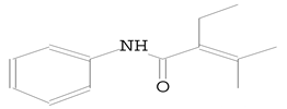
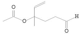
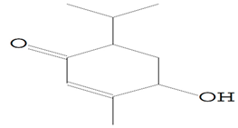
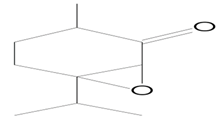
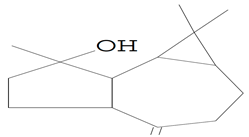
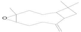
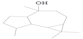
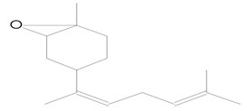
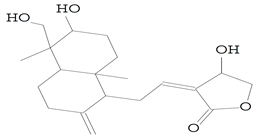
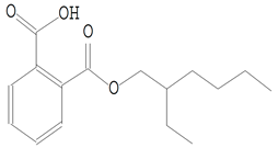
A GC-MS analysis was performed to identify the main secondary metabolites in the extract. Table 4 lists the 14 chemical components that have been found with their retention time (RT), molecular formula, and chemical structure. The most abundant compound detected at an RT of 23.267 min was 1,2-benzenedicarboxylic acid, mono(2-ethylhexyl) ester, with the molecular formula of C16H22O4. The second highest compound was 2-Butenamide, 2-ethyl-3-methyl-N-phenyl, detected at a RT of 10.595 min with a molecular weight of C13H17NO. The third abundant compound was 1H-Cycloprop[e]azulen-7-ol, decahydro-1,1,7-trimethyl-4-methylene- have a molecular formula of C15H24O and was observed at a RT of 12.971 min. On the other hand, 9,19-Cyclolanostan-3-ol, acetate, (3.beta.)-; Bicyclo(3.1.1)heptane-2,3-diol, 2,6,6-trimethyl, and 7-Methyl-Z-tetradecen-1-ol acetate were detected at RT of 14.115, 10.547, and 12.211 min, respectively, with a very similar moderate ratio (Table 4). The other compounds, such as 2-Cyclohexen-1-one,4-hydroxy-3-methyl-6-(1-methylethyl)-;7-Oxabicyclo[4.1.0]heptan-2-one,3-methyl-6-(1-methylethyl); trans-Z-alpha-Bisaboleneepoxide;2 Oxabicyclo[2.2.2]octan-6-ol, 1,3,3-trimethyl-; 5-Hexenal, 4-(acetyloxy)-4-methyl-; Ledol and Caryophyllene oxide, were detected at different RTs with low-level ratios (Table 4). The andrographolide compound, detected at a RT of 14.859 min with a molecular formula of C20H30O5, showed the lowest ratio among the identified compounds (Table 4).

4. Discussion

Cinnamomum camphora (L.) is an evergreen species widely distributed and cultivated in many tropical and subtropical areas, including Southeast and East Asia. It plays a crucial role in protecting the environment and has a volatile essential oil, camphor, which has been reported to have antibacterial properties [37,38]. The extracts from C. camphora have been traditionally used for herbal medicine and furniture in China. They are more commonly used as essential oils, obtained from the trunk, leaves, and twigs by steam distillation or various solvents [39]. The C. camphora extracts have been shown to kill pathogenic bacteria and can be used as a flavoring or preservative for food. It can also stop bacteria from growing and kill insects [40,41]. C. camphora extracts contain several active ingredients, including camphor, α-terpineol, linalool, eucalyptol, and safrole [42]. Additionally, the C. camphora tree’s essential oils derived from its different parts have been reported by several authors to be effective against pathogenic fungi, bacteria, algae, and insects [17,43,44]. Our work concluded that the methanolic extract of C. camphora showed potential bio-fungicide against the growth of F. solani, A. alternata, and F. oxysporum, which could be considered a prospective antifungal extract with a good number of antioxidants and secondary metabolites. It is believed that several types of compounds, including terpenoids, alkaloids, flavones, glycosides, saponins, quinines, coumarins, stilbenes, esters, phenols, aldehydes, alcohols, steroids, and organic acids, contribute to antimicrobial activity.
HPLC analysis results revealed the presence of antimicrobial compounds—naringenin, catechin, 7-OH flavone, apigenin, ferulic, benzoic acid, gallic acid, and catechol. Several researchers noticed that C. camphora extracts contained several phenolics that exhibited antimicrobial, antifungal, anticandidal, and antioxidant activities, such as gallic acid, naringenin, and its derivatives, and ferulic acid, offering protection against oxidative stress [14,45,46,47,48,49]. Research has shown that phenolic compounds play a role in the durability of natural wood, and resin acids inhibit fungi growth [50]. In the same regard, the results obtained in this work are in accord with those of Al-Huqail et al. [14], who found several active compounds in acacia extract, such as benzoic acid, o-coumaric acid, naringenin, quercetin, and kaempferol that showed significant inhibition of P. chrysogenum mycelial growth. Additionally, our results align with those in previous work by Yakefu et al. [51], who affirmed the potential values for C. camphora developing as an algaecide. Additionally, Tomazoni et al. [52] discovered that treatment with the extract of Eucalyptus staigeriana, E. globulus, and C. camphora resulted in inhibition of Alternaria mycelial growth and spore germination in vitro and a decrease in early blight symptoms in plants in vivo. Furthermore, our study aligns with that of Manilal and Idhayadhulla [53], who found natural plant extracts like Prosopis juliflora, Ricinus communis, and Carica papaya, respectively, and Polyalthia longifolia is effective in controlling Alternaria solani [54].
The potent antimicrobial activity of C. camphora extract is believed to be related to its secondary metabolites and phytochemicals, which can impact the membrane integrity, hyphae initiation, and cell cycle of microbes causing structural and functional alterations, swelling, and increased permeability [55,56].
GC-MS analysis of the methanolic leaf extract of C. camphora identified various compounds. The most abundant compound found in the extract was 1,2-benzenedicarboxylic acid, mono (2-ethylhexyl) ester, belonging to the class of phthalate esters, has been previously reported for its biological properties, including cytotoxicity, antioxidant, anti-inflammatory, antimicrobial, and antiviral activities [57,58,59]. The same compound was noticed to be the major component (58.05%) in a study conducted by Ali et al. [60], who noticed that it may potentially contribute to the antifungal activity of the methanolic root extract of Chenopodium album. On the other hand, different compounds detected in our GC-MS analysis had potential activities, such as the detected compound 1H-Cycloprop[e]azulen-7-ol, decahydro-1,1,7-trimethyl-4-methylene-, which is called spathulenol and is used as a scent in foods. According to Sousa et al. [61], the primary component of Eugenia calycina essential leaf oil, spathulenol (21.36%), has demonstrated antimicrobial activity against the anaerobic bacteria Prevotella nigrescens and Porphyromonas gingivalis, with a minimum inhibitory concentration (MIC) of 100 μg/mL. Similarly, Tan et al. [62] reported that spathulenol (23.8%) and caryophyllene (14.9%), the primary components of Salvia cilicica essential oil, exhibited antimicrobial activity against Mycobacterium tuberculosis, Microsporum gypseum, Trichophyton mentagrophytes, and Candida spp.
In our study, the detected compound, 7-methyl-Z-tetradecen-1-ol acetate, has previously been reported in GC-MS analysis of Mentha viridis and Urospermum picroides methanolic leaf extracts, and proved to possess anticancer, anti-inflammatory, and hepatoprotective activities [63,64]. According to Arora and Kumar [65], a compound known as 9.19-cyclolanostan-3-ol,24-methylene-(3.beta) can be used as an anti-HIV agent for the prevention of HIV. Additionally, the GC-MS results confirmed the existence of the compound 2-Oxabicyclo[2.2.2]octan-6-ol, 1,3,3-trimethyl-, also known as eucalyptol or 1,8-cineole. Many researchers have shown that 1,8-cineole has strong antimicrobial properties against various pathogens and spoilage organisms, as 1,8-cineole exhibited high inhibitory activity against C. albicans and Proetus vulgaris, with MIC values of 31.3 and 62.5 µg/mL, respectively [66,67,68]. The synergistic effect of 1,8-cineole combined with other molecules in essential oils, such as limonene, α-pinene, P-cymene, and terpineol-4-ol, could reveal strong antimicrobial activities [69,70].
Beta-caryophyllene (BCP) is a natural plant compound that belongs to the bicyclic sesquiterpene family. In its natural state, BCP is primarily found as a trans-caryophyllene ((E)-BCP), along with small quantities of its isomers such as iso-caryophyllene (Z)-β-caryophyllene, α-humulene (α-caryophyllene), and its oxidation derivative known as β-caryophyllene oxide (BCPO). Our results showed that the extract possessed BCPO, which has been studied in recent years because it has potent biological activities. BCPO, whether used in its pure form or as a component of plant essential oils, has been shown to have anti-inflammatory, antioxidant, antiviral, anticarcinogenic, and analgesic properties [71,72,73,74]. While various plant species’ antibacterial and antifungal properties have been widely studied, the specific mechanisms behind these properties have not been thoroughly explored. This study highlights the need for more trials to support the in vitro research studies of the camphor extract. Further research is required to determine the extract’s chemical nature and mechanisms of action, which will provide accurate and reproducible discoveries that could be used as eco-friendly alternative molecules to treat plant diseases caused by microbial pathogens.

5. Conclusions

Cinnamomum camphora methanolic extract was investigated for antifungal efficacy against three common, isolated, extensive phytopathogens: Alternaria alternata, Fusarium solani, and Fusarium oxysporum. C. camphora methanolic extract suppresses F. oxysporum, A. alternata, and F. solani fungal mycelia weight by 60, 49, and 24% at 4000 μg/mL. C. camphora extract may be antifungal due to its bioactive metabolites. HPLC examination indicated several phenolic and flavonoid components in the extract, including catechin and gallic acid, which had the highest amounts of 6.21 and 6.98 μg/mL, respectively. Glycine betaine, total amino acids, and osmoprotectants were plentiful. PMA and DPPH showed considerable total antioxidant activity. Mono(2-ethylhexyl) ester of 1,2-benzene dicarboxylic acid was the most abundant chemical in the extract’s GC-MS study. The tested extract may be a safe alternative to chemical fungicides because it inhibits mycelial growth weight in vitro.

Author Contributions

Conceptualization, S.S., A.A., A.A.A.-A., E.K.B., M.M.E., A.A.A. and S.I.B.; methodology, S.S., A.A., A.A.A.-A., E.K.B., M.M.E., A.A.A. and S.I.B.; software, S.I.B. and A.A.; validation, S.S., A.A., A.A.A.-A., E.K.B., M.M.E., A.A.A. and S.I.B.; formal analysis, S.S., A.A., A.A.A.-A., E.K.B., M.M.E., A.A.A. and S.I.B.; investigation, S.S. and A.A.; resources, S.S., A.A., A.A.A.-A., E.K.B., M.M.E., A.A.A. and S.I.B.; data curation, M.M.E.; writing—original draft preparation, S.S., A.A., A.A.A.-A., E.K.B., M.M.E., A.A.A. and S.I.B.; writing—review and editing, S.S., A.A., A.A.A.-A., E.K.B., M.M.E., A.A.A. and S.I.B.; visualization, A.A.A.; supervision, A.A.A.-A. and A.A.; project administration, A.A.A. and A.A.A.-A.; funding acquisition, A.A.A.-A. All authors have read and agreed to the published version of the manuscript.

Funding

This research was financially supported by the Researchers Supporting Project number (RSP2023R505), King Saud University, Riyadh, Saudi Arabia.

Data Availability Statement

Not applicable.

Acknowledgments

The authors would like to extend their appreciation to the Researchers Supporting Project number (RSP2023R505), King Saud University, Riyadh, Saudi Arabia.

Conflicts of Interest

The authors declare no conflict of interest.

References

  1. Abdelkhalek, A.; Behiry, S.I.; Al-Askar, A.A. Bacillus velezensis PEA1 Inhibits Fusarium oxysporum Growth and Induces Systemic Resistance to Cucumber Mosaic Virus. Agronomy 2020, 10, 1312. [Google Scholar] [CrossRef]
  2. Baysal-Gurel, F.; Gardener, B.M.; Miller, S.A. Soil Borne Disease Management in Organic Vegetable Production. 2012. Available online: https://eorganic.org/node/7581 (accessed on 5 February 2023).
  3. Hibar, K.; Daami-Remadi, M.; Ayed, F.; El-Mahjoub, M. Fusarium crown and root rot of tomato and its chemical control. Int. J. Agric. Res. 2007, 2, 687–695. [Google Scholar] [CrossRef] [Green Version]
  4. Akhtar, K.P.; Saleem, M.Y.; Asghar, M.; Haq, M.A. New report of Alternaria alternata causing leaf blight of tomato in Pakistan. Plant Pathol. 2004, 53, 816. [Google Scholar] [CrossRef]
  5. Chao, C.-C.T.; Parfitt, D.E.; Michailides, T.J. Alternaria late blight (Alternaria alternata) resistance in pistachio (Pistacia vera) and selection of resistant genotypes. J. Am. Soc. Hortic. Sci. 2001, 126, 481–485. [Google Scholar] [CrossRef] [Green Version]
  6. Abdelkhalek, A.; Hafez, E. Plant Viral Diseases in Egypt and Their Control. In Cottage Industry of Biocontrol Agents and Their Applications; Springer: Berlin/Heidelberg, Germany, 2020; pp. 403–421. [Google Scholar]
  7. Heflish, A.A.; Abdelkhalek, A.; Al-Askar, A.A.; Behiry, S.I. Protective and Curative Effects of Trichoderma asperelloides Ta41 on Tomato Root Rot Caused by Rhizoctonia solani Rs33. Agronomy 2021, 11, 1162. [Google Scholar] [CrossRef]
  8. Pragadheesh, V.S.; Saroj, A.; Yadav, A.; Chanotiya, C.S.; Alam, M.; Samad, A. Chemical characterization and antifungal activity of Cinnamomum camphora essential oil. Ind. Crops Prod. 2013, 49, 628–633. [Google Scholar] [CrossRef]
  9. El-Gendi, H.; Al-Askar, A.A.; Király, L.; Samy, M.A.; Moawad, H.; Abdelkhalek, A. Foliar Applications of Bacillus subtilis HA1 Culture Filtrate Enhance Tomato Growth and Induce Systemic Resistance against Tobacco mosaic virus Infection. Horticulturae 2022, 8, 301. [Google Scholar] [CrossRef]
  10. Behiry, S.I.; Soliman, S.A.; Al-Askar, A.A.; Alotibi, F.O.; Basile, A.; Abdelkhalek, A.; Elsharkawy, M.M.; Salem, M.Z.M.; Hafez, E.E.; Heflish, A.A. Plantago lagopus extract as a green fungicide induces systemic resistance against Rhizoctonia root rot disease in tomato plants. Front. Plant Sci. 2022, 13, 2818. [Google Scholar] [CrossRef]
  11. Abdelkhalek, A.; Al-Askar, A.A.; Alsubaie, M.M.; Behiry, S.I. First Report of Protective Activity of Paronychia argentea Extract against Tobacco Mosaic Virus Infection. Plants 2021, 10, 2435. [Google Scholar] [CrossRef]
  12. Youssef, N.H.; Qari, S.H.; Behiry, S.I.; Dessoky, E.S.; El-Hallous, E.I.; Elshaer, M.M.; Kordy, A.; Maresca, V.; Abdelkhalek, A.; Heflish, A.A. Antimycotoxigenic Activity of Beetroot Extracts against Alternaria alternata Mycotoxins on Potato Crop. Appl. Sci. 2021, 11, 4239. [Google Scholar] [CrossRef]
  13. Ismail, I.A.; Qari, S.H.; Shawer, R.; Elshaer, M.M.; Dessoky, E.S.; Youssef, N.H.; Hamad, N.A.; Abdelkhalek, A.; Elsamra, I.A.; Behiry, S.I. The Application of Pomegranate, Sugar Apple, and Eggplant Peel Extracts Suppresses Aspergillus flavus Growth and Aflatoxin B1 Biosynthesis Pathway. Horticulturae 2021, 7, 558. [Google Scholar] [CrossRef]
  14. Al-Huqail, A.A.; Behiry, S.I.; Salem, M.Z.M.; Ali, H.M.; Siddiqui, M.H.; Salem, A.Z.M. Antifungal, antibacterial, and antioxidant activities of Acacia saligna (Labill.) HL Wendl. flower extract: HPLC analysis of phenolic and flavonoid compounds. Molecules 2019, 24, 700. [Google Scholar] [CrossRef] [Green Version]
  15. Chen, S.; Zheng, T.; Ye, C.; Huannixi, W.; Yakefu, Z.; Meng, Y.; Peng, X.; Tian, Z.; Wang, J.; Ma, Y. Algicidal properties of extracts from Cinnamomum camphora fresh leaves and their main compounds. Ecotoxicol. Environ. Saf. 2018, 163, 594–603. [Google Scholar] [CrossRef] [PubMed]
  16. Ghabraie, M.; Vu, K.D.; Tata, L.; Salmieri, S.; Lacroix, M. Antimicrobial effect of essential oils in combinations against five bacteria and their effect on sensorial quality of ground meat. LWT Food Sci. Technol. 2016, 66, 332–339. [Google Scholar] [CrossRef] [Green Version]
  17. Satyal, P.; Paudel, P.; Poudel, A.; Dosoky, N.S.; Pokharel, K.K.; Setzer, W.N. Bioactivities and compositional analyses of Cinnamomum essential oils from Nepal: C. camphora, C. tamala, and C. glaucescens. Nat. Prod. Commun. 2013, 8, 1934578X1300801232. [Google Scholar] [CrossRef] [Green Version]
  18. Agarwal, R.; Pant, A.K.; Prakash, O. Chemical composition and biological activities of essential oils of Cinnamomum tamala, Cinnamomum zeylenicum and Cinnamomum camphora growing in Uttarakhand. In Chemistry of Phytopotentials: Health, Energy and Environmental Perspectives; Springer: Berlin/Heidelberg, Germany, 2012; pp. 87–92. [Google Scholar]
  19. Simmons, E.G. Alternaria: An Identification Manual: Fully Illustrated and with Catalogue Raisonné 1796–2007; CBS Fungal Biodiversity Centre: Utrecht, The Netherlands, 2007; ISBN 9789070351687. [Google Scholar]
  20. Leslie, J.F.; Summerell, B.A. The Fusarium Laboratory Manual; John Wiley & Sons: New York, NY, USA, 2007; ISBN 0813819199. [Google Scholar]
  21. Wang, Y.-B.; Yu, H.; Dai, Y.-D.; Wu, C.-K.; Zeng, W.-B.; Yuan, F.; Liang, Z.-Q. Polycephalomyces agaricus, a new hyperparasite of Ophiocordyceps sp. infecting melolonthid larvae in southwestern China. Mycol. Prog. 2015, 14, 70. [Google Scholar] [CrossRef]
  22. White, T.J.; Bruns, T.; Lee, S.; Taylor, J. Amplification and direct sequencing of fungal ribosomal RNA genes for phylogenetics. PCR Protoc. Guid. Methods Appl. 1990, 18, 315–322. [Google Scholar]
  23. Jindal, K.K.; Singh, R.N. Phenolic content in male and female Carica papaya: A possible physiological marker for sex identification of vegetative seedlings. Physiol. Plant. 1975, 33, 104–107. [Google Scholar] [CrossRef]
  24. Oser, B.L.; Hawk, P.B. Hawk’s Physiological Chemistry; McGraw-Hill: New York, NY, USA, 1965. [Google Scholar]
  25. Hiai, S.; Oura, H.; Hamanaka, H.; Odaka, Y. A color reaction of panaxadiol with vanillin and sulfuric acid. Planta Med. 1975, 28, 131–138. [Google Scholar] [CrossRef]
  26. Broadhurst, R.B.; Jones, W.T. Analysis of condensed tannins using acidified vanillin. J. Sci. Food Agric. 1978, 29, 788–794. [Google Scholar] [CrossRef]
  27. Lee, Y.P.; Takahashi, T. An improved colorimetric determination of amino acids with the use of ninhydrin. Anal. Biochem. 1966, 14, 71–77. [Google Scholar] [CrossRef]
  28. Bates, L.S.; Waldren, R.P.; Teare, I.D. Rapid determination of free proline for water-stress studies. Plant Soil 1973, 39, 205–207. [Google Scholar] [CrossRef]
  29. Grieve, C.M.; Grattan, S.R. Rapid assay for determination of water soluble quaternary ammonium compounds. Plant Soil 1983, 70, 303–307. [Google Scholar] [CrossRef]
  30. Bondet, V.; Brand-Williams, W.; Berset, C. Kinetics and mechanisms of antioxidant activity using the DPPH·free radical method. LWT Food Sci. Technol. 1997, 30, 609–615. [Google Scholar] [CrossRef]
  31. Jayaprakasha, G.K.; Rao, L.J.; Sakariah, K.K. Antioxidant activities of curcumin, demethoxycurcumin and bisdemethoxycurcumin. Food Chem. 2006, 98, 720–724. [Google Scholar] [CrossRef]
  32. Abdelkhalek, A.; Al-Askar, A.A.; Behiry, S.I. Bacillus licheniformis strain POT1 mediated polyphenol biosynthetic pathways genes activation and systemic resistance in potato plants against Alfalfa mosaic virus. Sci. Rep. 2020, 10, 16120. [Google Scholar] [CrossRef]
  33. Abdelkhalek, A.; El-Gendi, H.; Al-Askar, A.A.; Maresca, V.; Moawad, H.; Elsharkawy, M.M.; Younes, H.A.; Behiry, S.I. Enhancing systemic resistance in faba bean (Vicia faba L.) to Bean yellow mosaic virus via soil application and foliar spray of nitrogen-fixing Rhizobium leguminosarum bv. viciae strain 33504-Alex1. Front. Plant Sci. 2022, 13, 933498. [Google Scholar] [CrossRef]
  34. Okla, M.K.; Alamri, S.A.; Salem, M.Z.M.; Ali, H.M.; Behiry, S.I.; Nasser, R.A.; Alaraidh, I.A.; Al-Ghtani, S.M.; Soufan, W. Yield, phytochemical constituents, and antibacterial activity of essential oils from the leaves/twigs, branches, branch wood, and branch bark of Sour Orange (Citrus aurantium L.). Processes 2019, 7, 363. [Google Scholar] [CrossRef] [Green Version]
  35. Abdelkhalek, A.; Al-Askar, A.A.; Elbeaino, T.; Moawad, H.; El-Gendi, H. Protective and Curative Activities of Paenibacillus polymyxa against Zucchini yellow mosaic virus Infestation in Squash Plants. Biology 2022, 11, 1150. [Google Scholar] [CrossRef]
  36. Youssef, N.H.; Qari, S.H.; Matar, S.; Hamad, N.A.; Dessoky, E.S.; Elshaer, M.M.; Sobhy, S.; Abdelkhalek, A.; Zakaria, H.M.; Heflish, A.A. Licorice, Doum, and Banana Peel Extracts Inhibit Aspergillus flavus Growth and Suppress Metabolic Pathway of Aflatoxin B1 Production. Agronomy 2021, 11, 1587. [Google Scholar] [CrossRef]
  37. Kim, D.-I.; Park, J.-D.; Kim, S.-G.; Kuk, H.; Jang, M.-S.; Kim, S.-S. Screening of some crude plant extracts for their acaricidal and insecticidal efficacies. J. Asia-Pac. Entomol. 2005, 8, 93–100. [Google Scholar] [CrossRef]
  38. Chuanmao, W.; Zhouhe, W.; Yesong, W. Study on antibacterial activity of ethanol extract of Cinamomum camphora. Amino Acids Biot. Resour. 2000, 22, 41–42. [Google Scholar]
  39. Chua, M.-T.; Tung, Y.-T.; Chang, S.-T. Antioxidant activities of ethanolic extracts from the twigs of Cinnamomum osmophloeum. Bioresour. Technol. 2008, 99, 1918–1925. [Google Scholar] [CrossRef] [PubMed]
  40. Liu, C.H.; Mishra, A.K.; Tan, R.X.; Tang, C.; Yang, H.; Shen, Y.F. Repellent and insecticidal activities of essential oils from Artemisia princeps and Cinnamomum camphora and their effect on seed germination of wheat and broad bean. Bioresour. Technol. 2006, 97, 1969–1973. [Google Scholar] [CrossRef]
  41. Walch, S.G.; Kuballa, T.; Stühlinger, W.; Lachenmeier, D.W. Determination of the biologically active flavour substances thujone and camphor in foods and medicines containing sage (Salvia officinalis L.). Chem. Cent. J. 2011, 5, 44. [Google Scholar] [CrossRef] [Green Version]
  42. Pandey, A.K.; Bora, H.R.; Deka, S.C.; Rastogi, R.C.; Baruah, A.K.S. Composition of the essential oil of the bark of Cinnamomum camphora. J. Med. Aromat. Plant Sci. 1997, 19, 408–409. [Google Scholar]
  43. Poudel, D.K.; Rokaya, A.; Ojha, P.K.; Timsina, S.; Satyal, R.; Dosoky, N.S.; Satyal, P.; Setzer, W.N. The chemical profiling of essential oils from different tissues of Cinnamomum camphora L. and their antimicrobial activities. Molecules 2021, 26, 5132. [Google Scholar] [CrossRef]
  44. Lee, S.-H.; Kim, D.-S.; Park, S.-H.; Park, H. Phytochemistry and applications of Cinnamomum camphora essential oils. Molecules 2022, 27, 2695. [Google Scholar] [CrossRef] [PubMed]
  45. Duda-Madej, A.; Stecko, J.; Sobieraj, J.; Szymańska, N.; Kozłowska, J. Naringenin and Its Derivatives—Health-Promoting Phytobiotic against Resistant Bacteria and Fungi in Humans. Antibiotics 2022, 11, 1628. [Google Scholar] [CrossRef]
  46. Teodoro, G.R.; Ellepola, K.; Seneviratne, C.J.; Koga-Ito, C.Y. Potential use of phenolic acids as anti-Candida agents: A review. Front. Microbiol. 2015, 6, 1420. [Google Scholar] [CrossRef] [Green Version]
  47. Borges, A.; Ferreira, C.; Saavedra, M.J.; Simões, M. Antibacterial activity and mode of action of ferulic and gallic acids against pathogenic bacteria. Microb. Drug Resist. 2013, 19, 256–265. [Google Scholar] [CrossRef]
  48. Alves, C.T.; Ferreira, I.C.F.R.; Barros, L.; Silva, S.; Azeredo, J.; Henriques, M. Antifungal activity of phenolic compounds identified in flowers from North Eastern Portugal against Candida species. Future Microbiol. 2014, 9, 139–146. [Google Scholar] [CrossRef] [Green Version]
  49. Badhani, B.; Sharma, N.; Kakkar, R. Gallic acid: A versatile antioxidant with promising therapeutic and industrial applications. RSC Adv. 2015, 5, 27540–27557. [Google Scholar] [CrossRef]
  50. Ekeberg, D.; Flæte, P.-O.; Eikenes, M.; Fongen, M.; Naess-Andresen, C.F. Qualitative and quantitative determination of extractives in heartwood of Scots pine (Pinus sylvestris L.) by gas chromatography. J. Chromatogr. A 2006, 1109, 267–272. [Google Scholar] [CrossRef]
  51. Yakefu, Z.; Huannixi, W.; Ye, C.; Zheng, T.; Chen, S.; Peng, X.; Tian, Z.; Wang, J.; Yang, Y.; Ma, Z. Inhibitory effects of extracts from Cinnamomum camphora fallen leaves on algae. Water Sci. Technol. 2018, 77, 2545–2554. [Google Scholar] [CrossRef] [PubMed]
  52. Tomazoni, E.Z.; Pauletti, G.F.; da Silva Ribeiro, R.T.; Moura, S.; Schwambach, J. In vitro and in vivo activity of essential oils extracted from Eucalyptus staigeriana, Eucalyptus globulus and Cinnamomum camphora against Alternaria solani Sorauer causing early blight in tomato. Sci. Hortic. 2017, 223, 72–77. [Google Scholar] [CrossRef]
  53. Manilal, A.; Idhayadhulla, A. Potential in vitro antimicrobial efficacy of Holigarna arnottiana (Hook F). Asian Pac. J. Trop. Biomed. 2014, 4, 25–29. [Google Scholar] [CrossRef]
  54. Kalpashree, M.M.; Raveesha, K.A. Efficacy of aqueous plant extracts against Alternaria solani, the causative agent of early blight of tomato. Int. J. Herb. Med 2016, 4, 184–188. [Google Scholar]
  55. Pinto, E.; Gonçalves, M.J.; Oliveira, P.; Coelho, J.; Cavaleiro, C.; Salgueiro, L. Activity of Thymus caespititius essential oil and α-terpineol against yeasts and filamentous fungi. Ind. Crops Prod. 2014, 62, 107–112. [Google Scholar] [CrossRef]
  56. Mishra, A.K.; Dwivedi, S.K.; Kishore, N.; Dubey, N.K. Fungistatic properties of essential oil of Cinnamomum camphora. Int. J. Pharmacogn. 1991, 29, 259–262. [Google Scholar] [CrossRef]
  57. Govindappa, M.; Prathap, S.; Vinay, V.; Channabasava, R. Chemical composition of methanol extract of endophytic fungi, Alternaria sp. of Tebebuia argentea and their antimicrobial and antioxidant activity. Int. J. Biol. Pharm. Res. 2014, 5, 861–869. [Google Scholar]
  58. Krishnan, K.; Mani, A.; Jasmine, S. Cytotoxic Activity of Bioactive Compound 1, 2-Benzene Dicarboxylic Acid, Mono 2-Ethylhexyl Ester Extracted from a Marine Derived Streptomyces sp. VITSJK8. Int. J. Mol. Cell. Med. 2014, 3, 246–254. [Google Scholar] [PubMed]
  59. Mukund, S.; Jegan, G.; Palanisamy, M.; Muthukumaran, M.; Sivasubramanian, V. GC-MS and FT-IR analysis of some bioactive constituents from Oscillatoria terebriformis. Indian J. Pharm. Sci. Res. 2014, 4, 102–107. [Google Scholar]
  60. Ali, A.; Javaid, A.; Shoaib, A. GC-MS analysis and antifungal activity of methanolic root extract of Chenopodium album against Sclerotium rolfsii. Planta Daninha 2017, 35, e017164713. [Google Scholar] [CrossRef] [Green Version]
  61. Sousa, R.M.F.; de Morais, S.A.L.; Vieira, R.B.K.; Napolitano, D.R.; Guzman, V.B.; Moraes, T.S.; Cunha, L.C.S.; Martins, C.H.G.; Chang, R.; de Aquino, F.J.T. Chemical composition, cytotoxic, and antibacterial activity of the essential oil from Eugenia calycina Cambess. leaves against oral bacteria. Ind. Crops Prod. 2015, 65, 71–78. [Google Scholar] [CrossRef]
  62. Tan, N.; Satana, D.; Sen, B.; Tan, E.; Altan, H.B.; Demirci, B.; Uzun, M. Antimycobacterial and Antifungal Activities of Selected Four Salvia Species; ACG Publicaitons: Kocaeli, Türkiye, 2016. [Google Scholar]
  63. Hameed, I.H.; Hussein, H.J.; Kareem, M.A.; Hamad, N.S. Identification of five newly described bioactive chemical compounds in methanolic extract of Mentha viridis by using gas chromatography-mass spectrometry (GC-MS). J. Pharmacogn. Phyther. 2015, 7, 107–125. [Google Scholar]
  64. Al-Mawla, A.H.; Abu-Serag, N.A. Determination of bioactive compounds in leaves of Urospermum picroides (L.) Desf. Plant Arch. 2019, 19, 1299–1308. [Google Scholar]
  65. Arora, S.; Kumar, G. Gas Chromatography-Mass Spectrometry (GC-MS) determination of bioactive constituents from the methanolic and ethyl acetate extract of Cenchrus setigerus Vahl (Poaceae). Pharma Innov. J. 2017, 6, 635–640. [Google Scholar]
  66. Rosato, A.; Vitali, C.; De Laurentis, N.; Armenise, D.; Milillo, M.A. Antibacterial effect of some essential oils administered alone or in combination with Norfloxacin. Phytomedicine 2007, 14, 727–732. [Google Scholar] [CrossRef]
  67. Sonboli, A.; Babakhani, B.; Mehrabian, A.R. Antimicrobial activity of six constituents of essential oil from Salvia. Zeitschrift für Naturforschung C 2006, 61, 160–164. [Google Scholar] [CrossRef]
  68. Safaei-Ghomi, J.; Ahd, A.A. Antimicrobial and antifungal properties of the essential oil and methanol extracts of Eucalyptus largiflorens and Eucalyptus intertexta. Pharmacogn. Mag. 2010, 6, 172–175. [Google Scholar] [CrossRef] [PubMed] [Green Version]
  69. Dorman, H.; Deans, S.G. Antimicrobial agents from plants: Antibacterial activity of plant volatile oils. J. Appl. Microbiol. 2000, 88, 308–316. [Google Scholar] [CrossRef]
  70. Bakkali, F.; Averbeck, S.; Averbeck, D.; Idaomar, M. Biological effects of essential oils—A review. Food Chem. Toxicol. 2008, 46, 446–475. [Google Scholar] [CrossRef] [PubMed]
  71. Tung, Y.-T.; Chua, M.-T.; Wang, S.-Y.; Chang, S.-T. Anti-inflammation activities of essential oil and its constituents from indigenous cinnamon (Cinnamomum osmophloeum) twigs. Bioresour. Technol. 2008, 99, 3908–3913. [Google Scholar] [CrossRef]
  72. Hammami, S.; Jmii, H.; El Mokni, R.; Khmiri, A.; Faidi, K.; Dhaouadi, H.; El Aouni, M.H.; Aouni, M.; Joshi, R.K. Essential oil composition, antioxidant, cytotoxic and antiviral activities of Teucrium pseudochamaepitys growing spontaneously in Tunisia. Molecules 2015, 20, 20426–20433. [Google Scholar] [CrossRef] [PubMed]
  73. Zheng, G.-Q.; Kenney, P.M.; Lam, L.K.T. Sesquiterpenes from clove (Eugenia caryophyllata) as potential anticarcinogenic agents. J. Nat. Prod. 1992, 55, 999–1003. [Google Scholar] [CrossRef] [PubMed]
  74. Singh, T.P.; Singh, R.K.; Malik, P. Analgesic and anti-inflammatory activities of Annona squamosa Linn bark. J. Sci. Innov. Res 2014, 3, 60–64. [Google Scholar] [CrossRef]
Figure 1. HPLC chromatograms of polyphenolic compounds identified in Cinnamomum camphora extract at (a) λ = 284 nm, the peaks represent the following phenolic compounds: 1 = syringic acid, 2 = p-coumaric acid, 3 = caffeic acid, 4 = ferulic acid, 5 = gallic acid, 6 = benzoic acid, 7 = ellagic acid, 8 = iso-ferulic acid, and 9 = catechol acid; (b) λ = 273 nm, the peaks represent the following flavonoid compounds: 1 = naringenin, 2 = quercetin, 3 = hesperidin, 4 = catechin, 5 = 7-OH flavone and 6 = apigenin.
Separations 10 00189 g001
Table 1. The effect of different concentrations of Cinnamomum camphora methanolic extract on fungal growth weight in broth media.
Concentration (µg/mL)Weight of Fungal Mat (g)
F. solaniInhibition %A. alternataInhibition %F. oxysporumInhibition %
Control (10% DMSO)3.38 ± 0.2 a0.004.12 ± 0.12 a0.001.37 ± 0.1 a0.00
10003.27 ± 0.1 ab3.253.65 ± 0.14 b11.411.20 ± 0.1 a12.41
20003.22 ± 0.2 ab4.732.95 ± 0.3 c28.400.81 ± 0.1 b40.88
30003.05 ± 0.2 b9.762.33 ± 0.1 d43.450.66 ± 0.1 bc51.82
40002.56 ± 0.1 c24.262.09 ± 0.2 d49.270.55 ± 0.1 c59.85
LSD 0.050.24 ** 0.33 ** 0.18 **
** The values with the same letters in each column are not significantly different from each other.
Table 2. Screening of some active components of methanolic Cinnamomum camphora extract.
ClassificationTested ParametersConcentration (mg/g DM)
AntioxidantsFlavonoids48.0 ± 2.3
Phenols22.7 ± 1.1
Ascorbic acid46.6 ± 1.9
Secondary metabolitesSaponin12.0 ± 0.9
Tannins16.5 ± 0.6
Osmo-regulatory moleculesProline2.6 ± 0.4
Glycine betaine (GB)16.0 ± 1.2
Total amino acids37.4 ± 3.4
Antioxidant activityTotal antioxidant activity (PMA)22.0 ± 2.1
DPPH scavenging activity %13.7 ± 1.6
Table 3. The level of phenolic and flavonoid compounds (concentration, µg/mL) present in the methanolic extract of Cinnamomum camphora was determined using high-performance liquid chromatography (HPLC).
Phenolic CompoundsFlavonoid Compounds
Compound* RTµg/mLCompoundRTµg/mL
Syringic acid2.81.08Naringin3.83.78
p-coumaric acid3.50.98Quercetin5.02.46
Caffeic acid4.81.11Hesperidin7.01.25
Ferulic acid7.07.22Catechin7.86.21
Gallic acid8.96.987-OH flavone8.94.36
Benzoic acid10.01.55Apigenin10.03.98
Ellagic acid11.02.11
Iso-Ferulic acid12.00.87
Catechol13.01.65
* RT = retention time (minutes).
Table 4. Phytochemical components identified in the Cinnamomum camphora extract by GC-MS.
Retention Timem/zAreaHeightNameMolecular FormulaClassChemical Structure
10.47543.0018,66083512-Oxabicyclo[2.2.2]octan-6-ol, 1,3,3-trimethyl- (1,8-Cineole)C10H18O2monoterpeneSeparations 10 00189 i001
10.54743.0040,62022,112Bicyclo(3.1.1)heptane-2,3-diol, 2,6,6-trimethyl- (Pinanediol)C10H18O2terpeneSeparations 10 00189 i002
10.595111.00113,13034,8182-Butenamide, 2-ethyl-3-methyl-N-phenyl (Crotamiton)C13H17NOanilidesSeparations 10 00189 i003
11.25343.0016,39776655-Hexenal, 4-(acetyloxy)-4-methyl-,C9H14O3acetate esterSeparations 10 00189 i004
11.46098.0030,56614,0372-Cyclohexen-1-one, 4-hydroxy-3-methyl-6-(1-methylethyl)-,
(Barosma camphor)
C10H16O2cyclic monoterpene ketoneSeparations 10 00189 i005
11.71241.0022,15012,3077-Oxabicyclo[4.1.0]heptan-2-one, 3-methyl-6-(1-methylethyl)C10H16O2ketoneSeparations 10 00189 i006
12.21143.0040,59422,9727-Methyl-Z-tetradecen-1-ol acetateC17H32O2fatty acid estersSeparations 10 00189 i007
12.97143.0087,24755,0411H-Cycloprop[e]azulen-7-ol, decahydro-1,1,7-trimethyl-4-methylene- (Spathulenol)C15H24Otricyclic sesquiterpeneSeparations 10 00189 i008
14.11543.0045,45132,0869,19-Cyclolanostan-3-ol, acetate, (3.beta)-C32H54O2triterpeneSeparations 10 00189 i009
14.15043.0014,8249135Caryophyllene oxideC15H24Ocyclic sesquiterpeneSeparations 10 00189 i010
14.25843.0015,66610,273LedolC15H26OsesquiterpeneSeparations 10 00189 i011
14.31743.0020,59512,396trans-Z-alpha.-Bisabolene epoxideC32H54O2sesquiterpeneSeparations 10 00189 i012
14.85941.0093637179AndrographolideC20H30O5diterpenoidsSeparations 10 00189 i013
23.267149.00437,98483,3891,2-Benzenedicarboxylic acid, mono(2-ethylhexyl) esterC16H22O4phthalate estersSeparations 10 00189 i014
Disclaimer/Publisher’s Note: The statements, opinions and data contained in all publications are solely those of the individual author(s) and contributor(s) and not of MDPI and/or the editor(s). MDPI and/or the editor(s) disclaim responsibility for any injury to people or property resulting from any ideas, methods, instructions or products referred to in the content.