Introduction
Functional neurological disorder (FND) is a fascinating entity defined by neurological symptoms such as weakness, abnormal movements including psychogenic non-epileptic seizures (PNES), and sensory, swallowing or speech abnormalities that are incompatible with lesions in the central or peripheral nervous systems or recognised medical illness [1]. FND is frequently encountered in neurological clinics [2, 3], yet patients often remain in the interdisciplinary no-man’s-land between neurology and psychiatry, being labeled as “difficult to help” [4].
Despite historical interest from the founding fathers of modern neurology [5] and psychiatry [6], functional disorders have long been conceptualised as an exclusion diagnosis. Slater [7] concluded that long-term follow-up of “hysteria” patients showed an organic disorder in 75% of the cases. As a consequence, medical doctors were reluctant to diagnose functional disorders unless an in-depth neurological work-up had been completed to appropriately exclude all possible organic causes. The rate of misdiagnosis in conversion disorder as such (around 4%) [8] is much lower than in other organic movement disorders such as Parkinson’s disease (up to 47% when performed in the primary care setting) [9]. Nonetheless, patients with FND often undergo a number of sometimes unnecessary and expensive medical examinations in order to exclude as many organic disorders as possible.
Here, we discuss the case of a patient with a rare presentation of FND involving eye movements (functional convergence insufficiency), where this systematic exclusion-oriented approach was ended by a mosaic of interdisciplinary anamnestic and clinical elements that finally led to the diagnosis of FND.
We use this case to recall the importance of a detailed knowledge about functional neurological disorders, and functional eye movement disorders (FEMDs) in particular, a largely under-investigated subsection of FND. In addition, we emphasise the importance of close collaboration between neurologists, ophthalmologists and psychiatrists.
Case report
A 37-year-old manager of his own cleaning company was brought to the emergency department of another Swiss University Hospital following a sudden fall and loss of consciousness while standing alone in his mother’s kitchen, after breakfast. There were no apparent contributing factors and no prodromal sensation. He thinks he remained on the floor for 5 minutes and then was unable to get up alone, and felt vertigo, nausea and severe pain in both arms. During the hours at emergency department, he developed double vision. The consulting neurologist found binocular horizontal diplopia, which led him to suspect bilateral internuclear ophthalmoplegia. computed tomography angiography was normal, and the patient was discharged for ambulatory brain magnetic resonance imaging (MRI), on which “many white matter lesions” where described, and because of which he was referred to our unit for neurological work-up.
On admission, the patient complained about horizontal diplopia, especially when looking at near objects. In addition, he described episodes of undirected vertigo, of frequently loosing the thread of discussion and of forgetting the past. No further falls had occurred. Past medical history revealed a benign familial pemphigus with a particular sensitivity to skin irritation, as well as weight excess with a reduction of his body mass index from 40 to 30 kg/m2 in the past year.
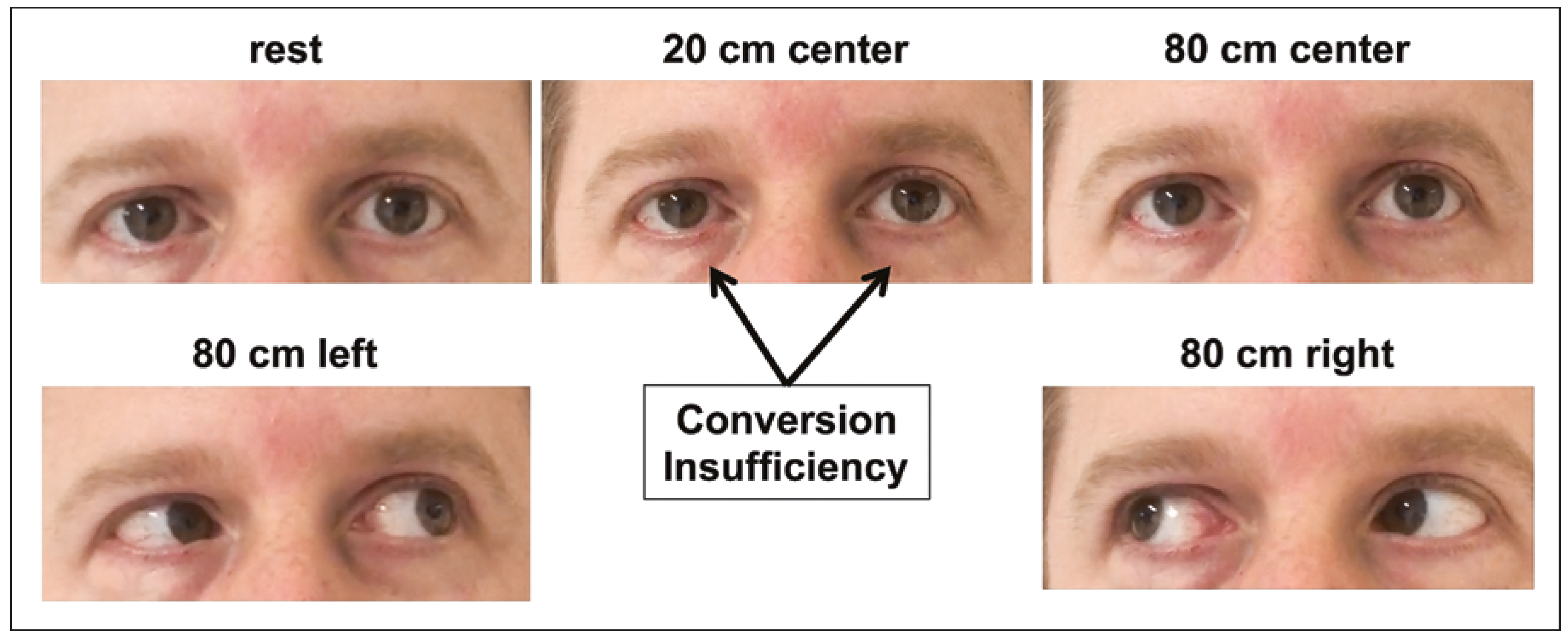
Neurological examination was normal except for the examination of the eye position, with a cover test revealing the impossibility of any convergence movement with near sight, together with a lack of pupillary constriction for accommodation in both eyes. With distant sight, there was no visible deviation. There was no nystagmus, and motility of both eyes was within the normal range. Nevertheless, convergence could not be elicited when looking at a near small object. The reaction of both pupils to light was normal and symmetrical (
Figure 1).
Figure 1.
Convergence insufficiency. The patient complained of horizontal diplopia in any gaze position. No convergence movement and no pupillary constriction were elicited. The photograph is published with the informed consent of the patient.
Figure 1.
Convergence insufficiency. The patient complained of horizontal diplopia in any gaze position. No convergence movement and no pupillary constriction were elicited. The photograph is published with the informed consent of the patient.
In response to the suspicion of his referring doctor, our patient had a complete work-up for an unexplained fall with loss of consciousness (looking for syncope, stroke, hypoglycaemia, or seizure) and for horizontal diplopia (looking for a brain lesion in the midbrain or pons, as well as signs of inflammation of the brain or cranial nerves). A second brain MRI scan did not find any visible lesion on the midbrain or pons on the diffusion, T2 or SWI sequences; the numerous unspecific white matter signal abnormalities were related to cerebrovascular risk factors. Neurological ultrasound showed chronic hypoplasia of the right vertebral artery, as well as flow acceleration in the left middle cerebral artery, suggesting moderate intracranial stenosis (70%); however, this did not explain the symptoms in question. An infectious and inflammatory origin was excluded by a normal lumbar puncture without oligoclonal bands, and negative serology for human immunodeficiency virus, Lyme disease, hepatitis and syphilis, as well as a negative immunological work-up motivated by his pemphigus history. EEG, ECG and a transthoracic heart ultrasound were also normal. Because the tilt table test was unavailable, the patient underwent a stress myocardial perfusion scan, during which (at 175 W) he complained of nonspecific vertigo, although the test was clinically and electrically negative.
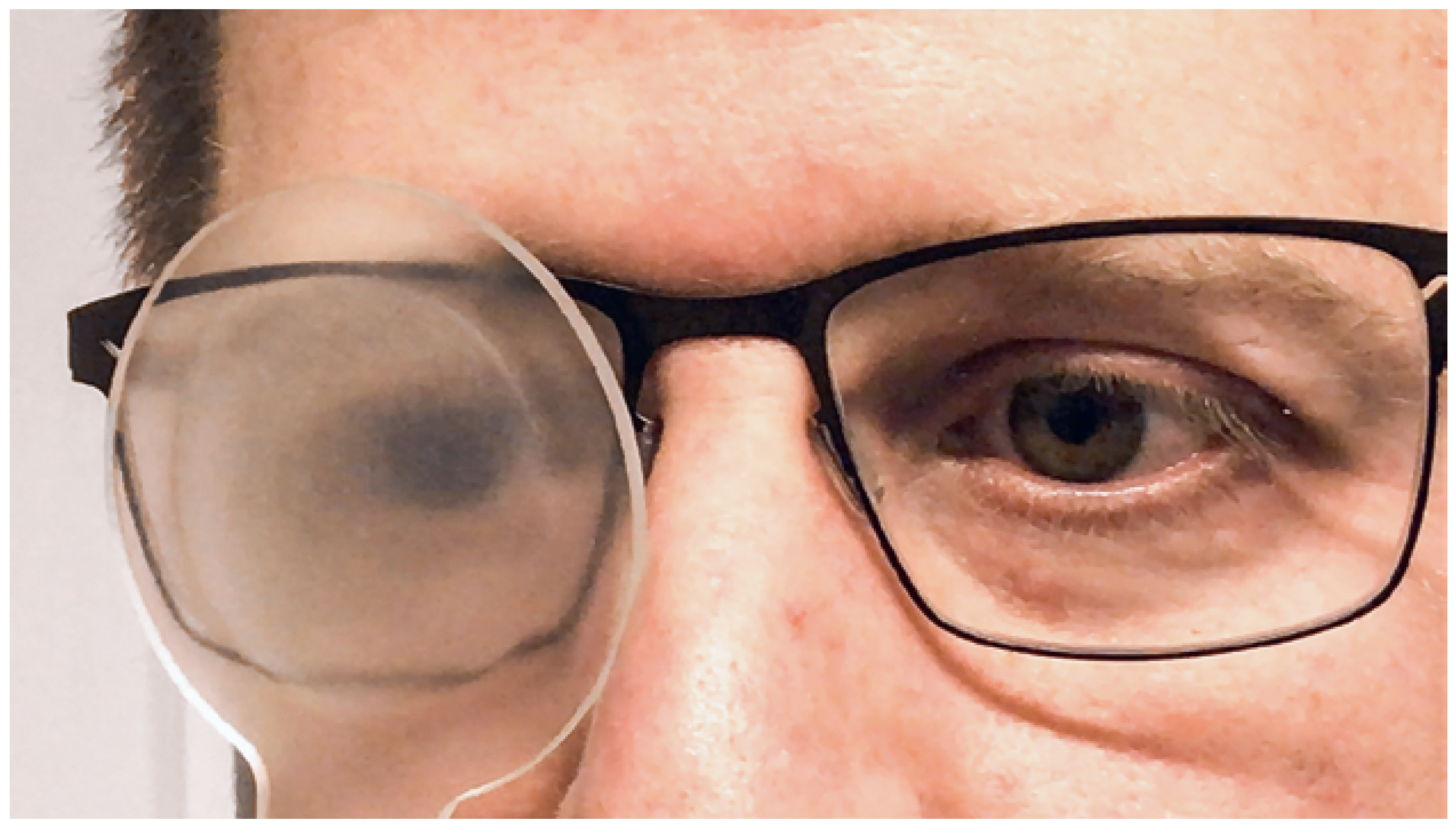
The neuro-ophthalmological examination confirmed complete absence of convergence when the patient was looking near. He reported, in addition, that he could hardly recognise the near optotype on the testing object presented to elicit convergence (the “E” on the Lang’s Würfel). No eye movement and no pupillary constriction could be detected during his “accommodative effort”. Accommodation was repeatedly measured under monocular viewing conditions for each eye with an accommodometer and ranged between 4 and 5 dpt (20 cm) under monocular viewing conditions. In addition, the presence of accommodation was assessed with a −3.0 dpt lens in front of both eyes during visual acuity testing with both eyes open. When he looked through −3.0 dpt lenses with both eyes open, visual acuity was 10/10 (the same as without the minus lens, there was no anisometropia). In order to test whether a vergence movement was linked to accommodation, a −3.0 dpt lens was then placed in front of one eye during monocular visual acuity testing at distance, and a semitranslucent occluder was held in front of the fellow eye. The eye behind the semitranslucent occluder clearly made a convergence movement during this distraction testing (
Figure 2). This asymmetrical convergence gradually increased up to 15 dpt when we repeated this test on near vision by progressively moving the optotype closer to the patient (corresponding to a near point of around 8 cm without the −3 dpt lens). In addition, pupillary constriction now occurred while the patient was performing this task. This asymmetrical convergence was therefore normal for both eyes under the semi-transparent occluder, confirming a normal ratio between accommodative convergence and accommodation (AC/A ratio, normal about 4:1). The rest of the neuroopthalmological evaluation was normal, including visual fields and colour vision. Hence, the patient could be diagnosed with an inconsistent convergence insufficiency and lack of accommodation effort.
Figure 2.
During the distraction testing, a −3 dpt lens was placed in front of his fixating left eye, while the right eye was covered with a semitranslucent occulder. The non-fixating semi-occluded right eye made a clear convergence movement. The test was symmetric for both eyes, showing preserved convergence ability. The photograph is published with the informed consent of the patient.
Figure 2.
During the distraction testing, a −3 dpt lens was placed in front of his fixating left eye, while the right eye was covered with a semitranslucent occulder. The non-fixating semi-occluded right eye made a clear convergence movement. The test was symmetric for both eyes, showing preserved convergence ability. The photograph is published with the informed consent of the patient.
During a psychiatric assessment, the patient reported chronic fatigue, a feeling of worthlessness and irritability. He complained about attention difficulties and fluctuating memory deficits. He also mentioned sleep disturbances with frequent nocturnal awakenings for over 6 months and difficulties with getting going in the morning. However, two scales for anxiety and depression did not show any significant result and, overall, the patient met no criterion for current mood disorders. During the evaluation, the patient reported being very worried about his health and having frequent consultations with his family doctor for occipital headaches (as if “the skull would inflate and open from behind”), dizziness, and back and muscle pain. In this context, a diagnosis of somatic symptom disorder (SSD, F45.1) was made (SSD is defined as “somatic symptoms that are either very distressing or result in significant disruption of functioning, as well as excessive and dispropor tionate thoughts, feelings and behaviors regarding those symptoms” (DSM-5). The patient must be persistently (at least for 6 months) symptomatic). He related episodes of childhood trauma with physical and emotional abuse. Moreover, 5 months before, he had experienced psychological distress when his company went bankrupt. Probing for psychiatric antecedents revealed a diagnosis of PNES in 2001 in addition to panic disorder (F41.0) in 2003.
The functional nature of his deficit was explained to the patient by both the neurologist and the psychiatrist. During the following 12 months, he was regularely followed up in the ambulatory setting by his neurologist and in weekly sessions of psychotherapy. He clearly accepted the diagnosis of FND and was slowly improving. However, after the ocular symptoms had resolved, he still suffered from concentration difficulties. To date, he has still not resumed professional activities.
Discussion
The case of our patient who, out of the blue, suffered a sudden unwitnessed fall from standing with loss of consciousness but without any sequelae, and who then developed isolated horizontal diplopia, could only be solved with a multidisciplinary approach: the initial presentation suggested a neurological origin, meticulous ophthalmological testing unveiled the hidden inconsistency, and psychiatric history taking collected numerous elements that completed the mosaic in favour of a functional condition.
Neurological point of view
From the initial neurological point of view there were two elements: the unwitnessed sudden fall with loss of consciousness, and the later appearance of the convergence insufficiency. A sudden fall with no prodromi during a quiet moment of standing in a previously normal patient evokes above all syncope or a stroke, as well as hypoglycaemia, vitamin B1 deficiency or, rather atypically, a seizure. However, complete recovery within minutes limits it to a transitory or functional problem (syncope, transient ischaemic attack). Organic convergence paralysis originates from a lesion of the dorsal midbrain that interrupts the pathways of the eye vergence control. These fibres descend from the visual occipital cortex, decussate in the thalamotectal area and control premotor vergence neurones. These so-called near-response cells lie in the midbrain in the medial nucleus reticularis tegmenti pontis, dorsolateral to the oculomotor nucleus [10]. As a result of the narrow space in the midbrain, a lesion in this area is usually accompanied by other signs of midbrain damage including impaired vertical gaze, upbeat or downbeat nystagmus, convergence-retraction nystagmus and eyelid retraction. Such accompanying symptoms were not found in our patient. Organic convergence paralysis has been described in patients with trauma, subdural haematoma, midbrain tumours, haemorrhage or infarction, multiple sclerosis or encephalitis, as a result of metabolic causes or drug-related, as well as with Parkinson’s disease and progressive supranuclear palsy [11, 12]. Convergence paralysis can also be caused by strategic supranuclear lesions, including bilateral paramedian thalamic infarction [13], thalamotectal haemorrhage [14] or bilateral superior colliculus lesion [15]. Convergence insufficiency, as opposed to convergence paralysis, is common among children and teenagers (appears often with an increased visual work load). It can also be seen after mild head trauma, and with acquired cerebral lesions (especially in the nondominant parietal lobe) in the elderly [16]. In organic convergence paralysis, convergence movements are always absent, whereas in functional convergence paralysis or insufficiency, as in our patient, convergence movements may be observed under distraction during examination of an alternated near task. The lack of pupillary constriction in our patient was another clue to the functional origin of his incapacity to elicit convergence movements of the eyes when looking at a near object, since in organic convergence insufficiency the effort to converge is always associated with preserved pupillary constriction. The accommodative and vergence systems are controlled together by an interactive negative feedback loop, and clinically also the two systems are not commonly separated [17].
Psychiatric point of view
From the psychiatric point of view, our patient’s detailed history unveiled several clue elements such as previous trauma, mood and anxiety comorbidities, as well as multiple functional symptoms such as back pain, PNES, tremor, posterior headaches, dizziness, and back and muscle pain, on the basis of which a diagnosis of somatic symptom disorder was made (
Table 1). Patients with FND frequently have comorbid mood and anxiety disorders [3]. Moreover, they often present with multiple functional symptoms that co-occur, like in our patient. In one study, pain disorder was a comorbidity in 84% of patients with FND, 67% of whom suffered from more than one functional neurological symptom [18]. PNES patients suffer from more severe forms of migraine compared with epilepsy patients without PNES [19], and the presence of “chronic pain” or “fibromyalgia” independently predicts a diagnosis of psychogenic seizures (predictive value 75%) [20]. In keeping with this, it has recently been suggested that the presence of illnesses such as chronic pain, severe and chronic migraine, fibromyalgia, asthma, and gastro-oesophageal reflux disease may provide valuable clinical clues to the suspicion of PNES [21].
Table 1.
Definition of functional neurological symptom disorder in DSM–5.
Table 1.
Definition of functional neurological symptom disorder in DSM–5.
Ophthalmological perspective
From the ophthalmological perspective, two elements uncovered the functional origin of this patients problem: the absent convergence effort, as seen in thelack of accommodative effort, and the reappearing convergence movement in an artificial anisometropic condition created by putting a −3 dpt lens in front of only one eye while the non-fixating eye was covered with a semitranslucent occluder. In this artificial condition, accommodation was elicited. Since accommodation is always bilateral, the associated convergence could be observed in the non-fixating eye. The fixating eye was stationary on the target and fixation prevented the eye from converging. By putting a −3 lens in front of both eyes while the patient was fixating on a target bilaterally, we tested two properties: first, the degree of accommodation (as under monocular viewing conditions) and second, the fusional ability of the patient, which counterbalanced the convergence impulse provoked by the minus lenses. Our patient had a visual acuity of 10/10 and no diplopia under binocular viewing conditions (there was no manifest tropia in the unilateral cover test). These investigations clearly showed that the anatomical requirements of convergence, accommodation and fusion maintenance were normal in our patient. Based on these results, a diagnosis of a functional convergence insufficiency was made.
Functional eye movement disorders
Functional eye movement disorders (FEMDs) include (1) functional convergence spasm disorder (defined as transient ocular convergence, miosis and accommodation) [22], (2) functional eye oscillations (“voluntary nystagmus”, episodes lasting for 3–20 seconds), (3) gaze limitation (limited capacity to direct gaze in different directions), (4) tonic gaze deviation, and (5) convergence paralysis (inability to fixate proximally and difficulties with reading) [23]. They present generally with an abrupt onset (96.7%) [24] and a nonprogressive course (85.2%), fluctuate or disappear with distraction (89.6%), worsen with emotional anxiety or increased attention (86%), and improve under suggestion or placebo treatment (89.5%) [25]. In addition, FEMDs have a female preponderance (a 9:1 female-to-male ratio) and almost one in two patients (49.2%) present with other somatisations (for a review see [24]).
Looking back, many of the neurological examinations in this patient were probably not necessary. Post hoc we always know better, and it is clear that the unequal, immediate danger of, for example, a TIA versus a diagnosis of FND will always oblige physicians to take at least into account the dangerous condition. However, the less precise our clinical knowledge is, the likelier we are to exclude other conditions by any test at hand. New DSM-5 criteria have recently emphasized that the diagnosis of FND should be based on positive signs of discrepancy between clinical findings and neuropathology [26–28]. Because of the high incidence of FND, the neurologist needs to routinely evaluate clues to the diagnosis, such as inconsistency, sudden onset and variability over time, in addition to distractibility and suggestibility [29]. However, it is important to note that most validated positive clinical signs of FND, such as the Hoover’s sign, give-away weakness, pronator drift, etc. (for review see [27]), have been developed for volitional motor systems. Eye movements are semi-volitional with reflex components that are highly complex and fine-tuned. To date, no valid, specific and sensitive positive clinical signs exist for FEMD, thus often leaving neurologists without proper tools to validly examine functional eye movement disorders.
Finally, to complicate the situation even more, it is important to remember that having an organic disease does not preclude a comorbid functional neurological disorder. Indeed, some classic examples show that well-established neurological conditions can coexist with their FND mimics and chameleons: for example, epilepsy and psychogenic non-epileptic seizures (in up to 10% of patients [30]), or Parkinson’s disease and functional movement disorders (in up to 34% of patients [31, 32]). Moreover, functional variations of symptoms and non-congruence with anatomical laws do not automatically equate with “psychogenic” disorder. The differential diagnoses of FND include two other entities: factitious disorder and malingering. In our patient’s case, there was no evidence of any immediate extrinsic reward, and we therefore excluded malingering. Factitious disorder [33] manifests with frequent demands for hospitalisations and examinations in relation to a voluntary intoxication or a self-afflicted lesion in order to implicitly benefit from the sick role, rather than a specific isolated absence of a symptom (e.g., the vergence movement) as in our patient’s case.
Overall, this case report illustrates the importance of knowledge about FEMD in the neurological clinic and the importance of close interdisciplinary collaboration in order to advance straightforwardly the diagnostic work-up and prevent invasive investigations and unnecessary treatments [2]. Future clinical research efforts should be dedicated to developing positive signs for functional eye movement control disorders. This will allow for better knowledge and, hopefully, more useful instruments for clinical assessment thus limiting exclusion-based diagnostic approaches of looking for a needle (the FND) in a haystack (all possible disorders to be excluded).