Introduction
Many studies have shown that verbal memory deficits, abnormalities in the Stroop-paradigm and psycho-motor slowing are common in the early course of epileptic syndromes in adults and that depression is a substantial risk factor for a first unprovoked seizure [1]. In 2011 it was proposed that the neuropsychological status of persons with new-onset epilepsy should be a focus of future clinical research in the neuro-psychology of epilepsy [2]. Additionally it was stressed that a baseline neuropsychological assessment should be performed at an early stage of epilepsy [3]. In a group of 247 untreated patients screened for cognitive defi cits within an average of 92.4 days after their first unprovoked seizure [4], verbal memory deficits were seen in 47.8% of the patients. In the baseline survey for this study we found verbal memory deficits in 60% of the patients [5]. Twenty-one percent of the patients delivered a self-rating that was suggestive of at least a minor depression in the week before the seizure. In one study [6], reduced verbal memory performance in patients with newly diagnosed epilepsy was identified as a prognostic risk factor for a refractory course. Elevated depression scores in the first month after an unprovoked epileptic seizure predicted another seizure in the next 2 months [7]. The aim of this study was to investigate whether cognitive deficits after a first unprovoked seizure or depressive symptoms in the week before indicate an increased risk of a second seizure during the following 3 years. The method of data acquisition and the baseline data of 53 patients including the 37 patients who could be followed up have been published previously [5].
Patients and methods
In this prospective study we tested all adults aged 18 to 70 years who presented with a first unprovoked seizure in our emergency room from November 2009 until November 2012 for cognitive deficits and symp toms of depression. The study was approved by the institutional ethics board in Rostock under the regis tration number HV-2009-0009. Informed consent was obtained from all individual participants included in the study. Only patients with a single unprovoked seizure were included. Patients with more than one seizure or status epilepticus were excluded. Patients with aphasia, dementia or severe psychiatric illness like acute psychosis were excluded as well. As a part of the routine procedure all patients had at least one electroencephalogram (EEG). With the exception of one patient with a pacemaker, who was investigated via computed tomography (CT) only, all patients had a magnetic resonance imaging (MRI) scan. For evalua tion, the CT results of the patient with the pacemaker were combined with the MRI results of the other patients. Initially, abnormalities on MRI scans were classified as either significant or non-significant based on an a priori system of classification used in a previous study on the association of MRI findings and neuro psychological functioning after the first recognised seizure in children [8]. Using this classificationsystem, abnormalities that fell under the classification of “significant” included leucomalacia, encephalomala-cia, any gray matter lesion, mass lesion, haemorrhage, vascular lesion, hippocampal abnormality, ventricular enlargement >1.5 cm, or prominence of extra-axial fluid spaces >1 cm. In a post-hoc analysis, abnormalities on MRI scans were classified as being suggestive of either a primary brain tumour or brain metastasis, or of neither. After informed consent was obtained, the patients were investigated with three neuropsycho logical tests and the Self-rating Depression Scale (SDS; Zung depression scale) [9]. The patients were instructed to answer the questions for the week before the sei zure. The Verbal Learning Memory Test (VLMT) [10], which is a German version of the Rey Auditory Verbal Learning Test [11], was performed to detect verbal memory deficits. Results were regarded as pathological when they were more than one standard deviation below the published normal limits. The Diagnosticum für Cerebralschädigung (DCS) [12], which is a visual learning and memory test, was performed in the modification of Helmstaedter et al. [13]. In this version the test is especially sensitive for visual memory deficits in patients with right temporal lobe epilepsy. The cut-off for pathological results was taken from the same publication [13]. For the assessment of mental speed and selective attention, the Farbe-Wort-Inter-ferenztest (FWIT) [14], which follows the Stroop-para-digm [15], was used. Results were regarded as patho logical when they were more than one standard deviation below the published normal limits. By this procedure the baseline data of 53 patients were ob tained and published previously [5]. Three years later the patients were asked by letter if another seizure had occurred. The data of patients who showed up with a second seizure in our department earlier were in cluded as well. One patient had to be excluded because we found out that he had several unrecognised seizures before the first presentation at our department. Fifteen patients were lost to follow up. Therefore the data of 37 patients are available for this analysis. Asso ciations between the occurrence of a second seizure and psychometric abnormalities according to the published norms, significant MRI lesions or abnormal ities in the EEG were tested for significance with the χ2-test using the Yates correction for small sample sizes. We performed a subgroup analysis of 22 patients without a significant lesion in the MRI. This is an exploratory study. Therefore we performed no Bon-ferroni-Holmes procedure to correct for multiple statistical testing.
Results
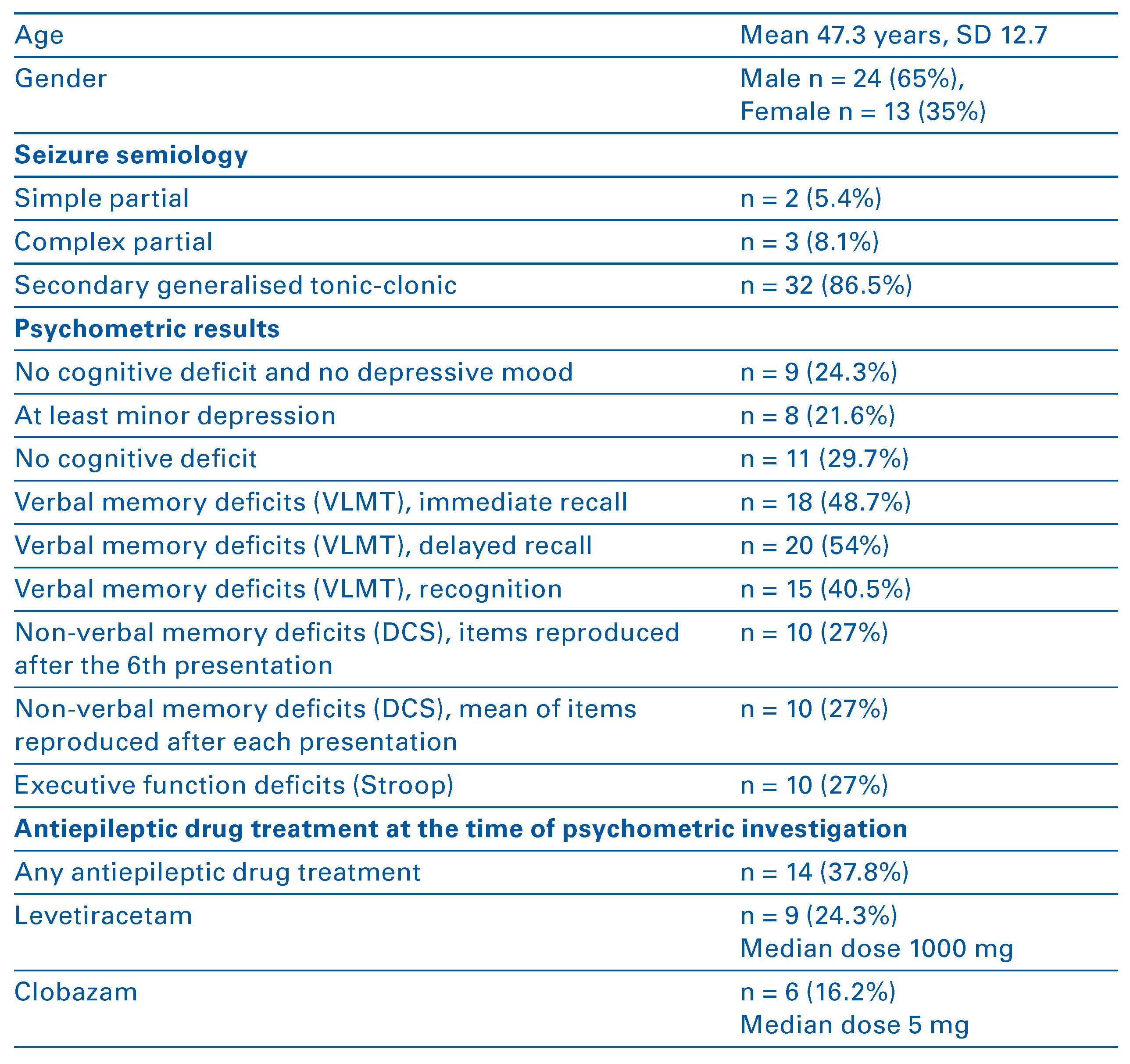
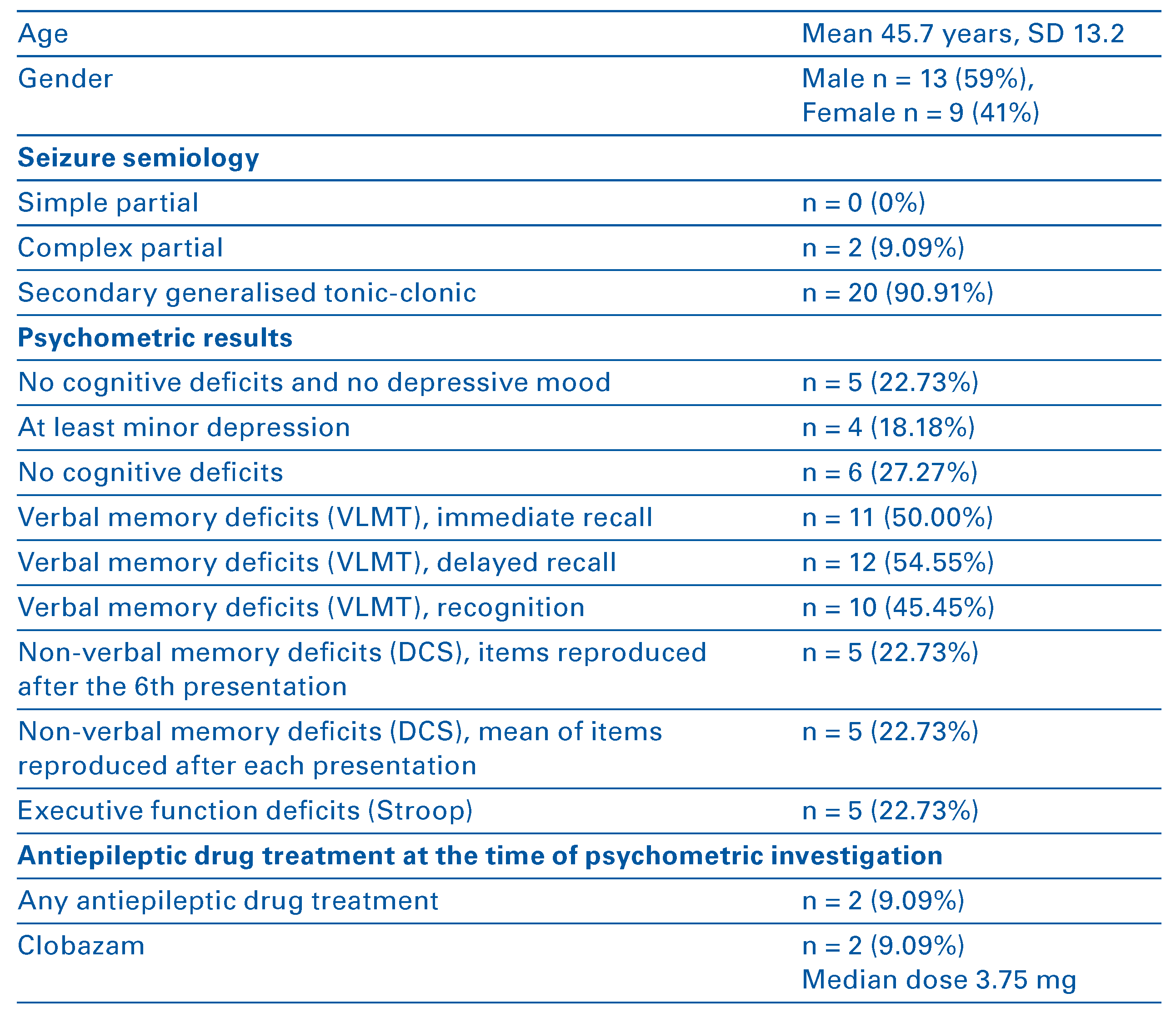
The data of 37 patients are available for this analysis. For patients characteristics and psychometric results see
Table 1. Only 24.3% of the patients showed no psychometric deficit in the whole psychometric inves tigation. The mean score in the SDS was 40.7 (standard deviation [SD] 11.1); 21.6% of the patients had an SDS score indicating at least minor depression (score of 50 or more) according to the published cut-offs. But only two patients (5.4%) had a score suggestive of major de pression (score of 60 or more). At least one pathological result in the neuropsychological battery was found in 69.7% of cases. This was mainly due to pathological results in the VLMT. Twenty-two patients (59.5% of the whole group) had at least one pathological result in the VLMT. The most common deficit in the VLMT was in the delayed recall task (n = 20, 54% of the whole group). The pathological results in the FWIT were mainly in the initial reading task (n = 8, 21.6% of the whole group). Therefore the deficits were mainly in the realm of mental speed and not in the field of selective attention. Five patients (13.5%) had pathological results in all three neuropsychological tests, three patients (8.1%) had pathological results in both memory tests only and another three patients had pathological results in one of the memory tests and the FWIT. For the lesions found with MRI see
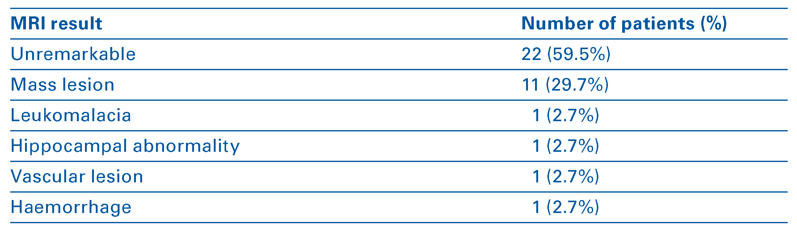
Table 2. Of the mass lesions, two were in the left temporal lobe, three in the right temporal lobe, four the left frontal lobe and the remaining two in the right frontal lobe. The majority of patients did not have a kind of lesion that is usually related to seizures. Among the pathological MRI scans, those suggestive of an astrocytoma had the highest fre quency (astrocytoma WHO II° n = 3, astrocytoma WHO III° n = 2). Other MRI scans with mass lesions were suggestive of cerebral metastases (n = 1) or showed lesions after brain surgery (n = 2). Especially patients with pathological MRI findings were treated with an antiepileptic drug (AED) immediately after the MRI (for details see
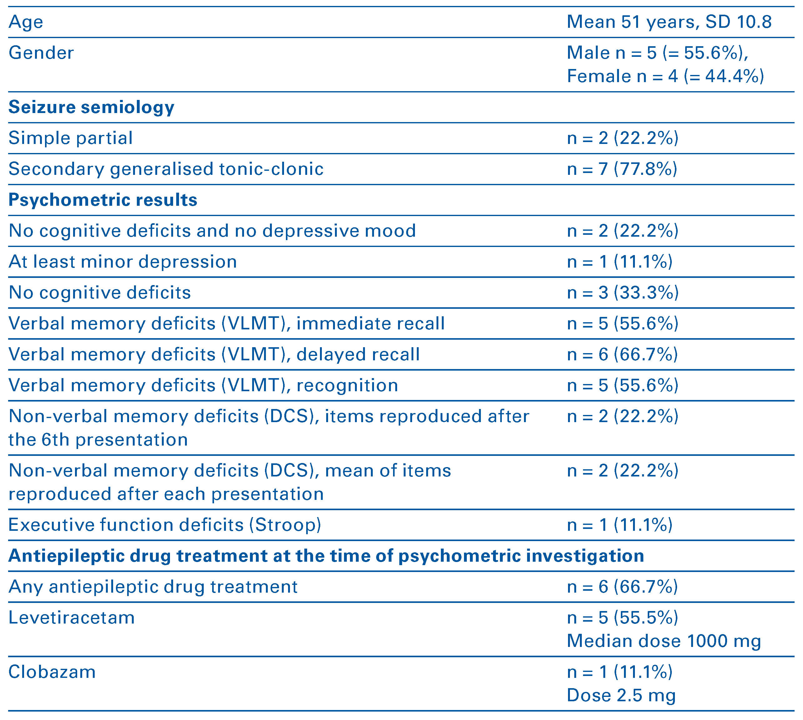
Table 1). In some cases clobazam 5 mg was used as an emergency treatment to prevent furfocal EEG abnormalities mainly in the leh temporal leads (n = 8). With the exception of three patients, all those with focal EEG abnormalities had focal epileptic patterns in their EEG. Nine patients (24%) experienced a second seizure. The time between the first and the second seizure was 293 days on average (for the charac teristics of the patients who experienced a second seizure see
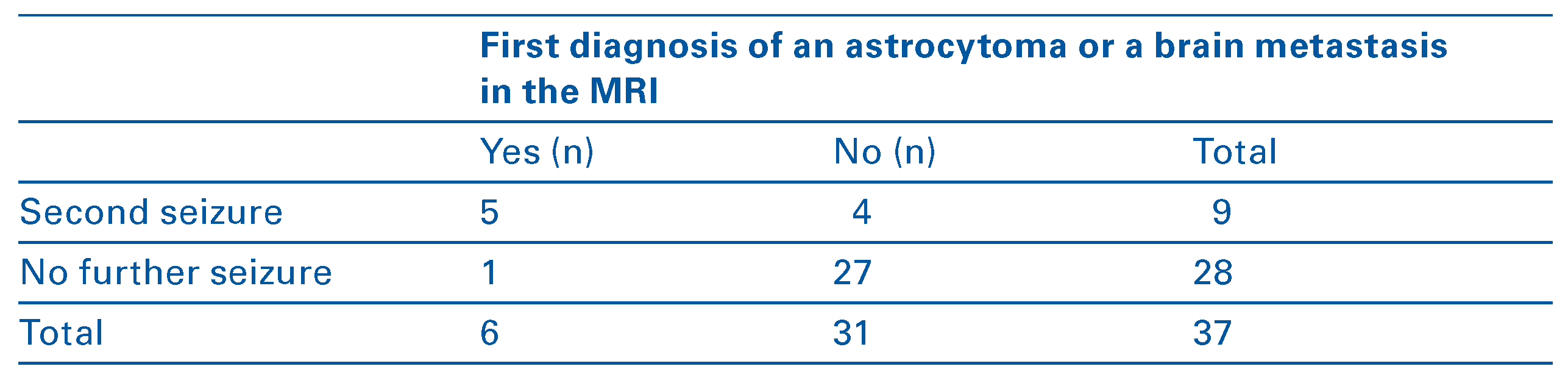
Table 3). We found no significant association between the baseline data mentioned above and the occurrence of a second seizure during the follow-up period, with the exception of the first diagnosis of an astrocytoma or a brain metastasis in the MRI (p <0.0016; odds-ratio 6.4, 95% confidence interval 2.1–19.5) (for sample size and cell size see
Table 4). All patients in this subgroup were discharged on leve tiracetam. One patient discontinued levetiracetam after 22 months of seizure freedom and experienced his second seizure 9 months later. At this time his astrocytoma had relapsed. It has to be acknowl edged that in three other patients with a second seizure, biopsy revealed a glioblastoma multiforme instead of an astrocytoma as suspected from MRI. Two of them had their second seizure within 2 weeks of the first one; the other patient experienced his second seizure after 6 months, when his glioblastoma relapsed.
Table 1.
Patients’characteristics (whole group).
Table 1.
Patients’characteristics (whole group).
Table 2.
Frequency of lesions detected by MRI according to the system of Byars et al. [8].
Table 2.
Frequency of lesions detected by MRI according to the system of Byars et al. [8].
Table 3.
Baseline characteristics of patients who experienced a second seizure.
Table 3.
Baseline characteristics of patients who experienced a second seizure.
Table 4.
Association of a second seizure with the first diagnosis of an astrocytoma or a brain metastasis in the MRI.
Table 4.
Association of a second seizure with the first diagnosis of an astrocytoma or a brain metastasis in the MRI.
We performed a subgroup analysis excluding all patients with a significant lesion according to the system of Byars et al. [8]. The data of this subgroup of patients are given in
Table 5. The amount of pathologi cal psychometric results was roughly in the same range as in the entire group. Here we found a trend for an association between pathological immediate recall in the VLMT after Yates correction for small sample size (p = 0.097). No patient without a significant lesion according to the system of Byars et al. [8] and normal results for immediate or delayed recall in the VLMT had a second seizure in the following 3 years. Therefore no odd ratios can be calculated. All patients without a significant lesion according to the system of Byars et al. [8] who experienced a second seizure had a patho logical result for immediate recall in the VLMT.
Table 5.
Characteristics of patients without a significant lesion according to the system of Byars et al. [8].
Table 5.
Characteristics of patients without a significant lesion according to the system of Byars et al. [8].
Discussion
The main limitation of this study is the high dropout rate of 30.2%. Since the baseline data of the remaining sample are not much different from the baseline data of the entire group published before [5], the main problem is the resulting small sample size. Therefore the data presented here are very preliminary and have to be interpreted very cautiously in both directions. Another limitation is the small number of tests per formed. Short and working memory, as well as some aspects of attentional function, were not assessed. Setting the cut-off for pathological results to one standard deviation below the published normal limit implies that 16% of population will have a pathological result. So just four patients more than expected had a pathological result in the FWIT. Nevertheless, our data show that at least mild cognitive deficits, mainly in the realms of verbal memory, are a frequent finding after a first unprovoked seizure. But the occurrence of further seizures seems to depend mainly on the brain pathology shown by MRI. Additionally, deficits in verbal learning may indicate an increased risk for a second seizure in the next 3 years. This assumption is in line with the finding that reduced verbal memory performance in patients with newly diagnosed epi lepsy is a prognostic risk factor for a refractory course [6]. In a sample of persons with refractory epilepsy, we found a negative correlation between age at the onset of epilepsy and performance in a vocabulary test [16]. This result also points to a high prevalence of deficits in verbal learning at the onset of epilepsy. Unfortunately, due to the high drop-out rate resulting in a small sample size, we cannot confirm these as sumptions with significant data. Probably because of the small sample size, there was no hint of prognostic significance of depressive symptoms in the week be fore the first seizure. Nevertheless, to the best of our knowledge here we present the results of the first prospective study designed to elucidate the prognostic significance of cognitive deficits after a first unpro voked seizure in adulthood. Further studies in this field should take into account that a high drop-out rate has to be expected at follow up.
Conclusion
After a first unprovoked seizure the occurrence of further seizures seems to depend mainly on the brain pathology shown by MRI. Deficits in verbal learning additionally may indicate an increased risk for a sec ond seizure in the next 3 years.