Introduction
The changes in electrical activity associated with epileptic seizures had been documented in the early 1900’s [
1], and with Berger began an era of research and diagnosis of cerebral diseases especially epilepsy. EEG machines became commercially available in the 1930’s, the technical development which made this possible was the development of the electronic valve amplifier, and with this discovery began the era of EEG-orientated epileptology. Epileptologists were among the first to implement this new tool for the diagnosis and therapy control of patients with epilepsy. Since this time the progress made in clinical neurophysiology has been to a large degree dependent on technological development. The importance of electroencephalography was soon recognised and epilepsy centres all over the world began with the installation of laboratories.
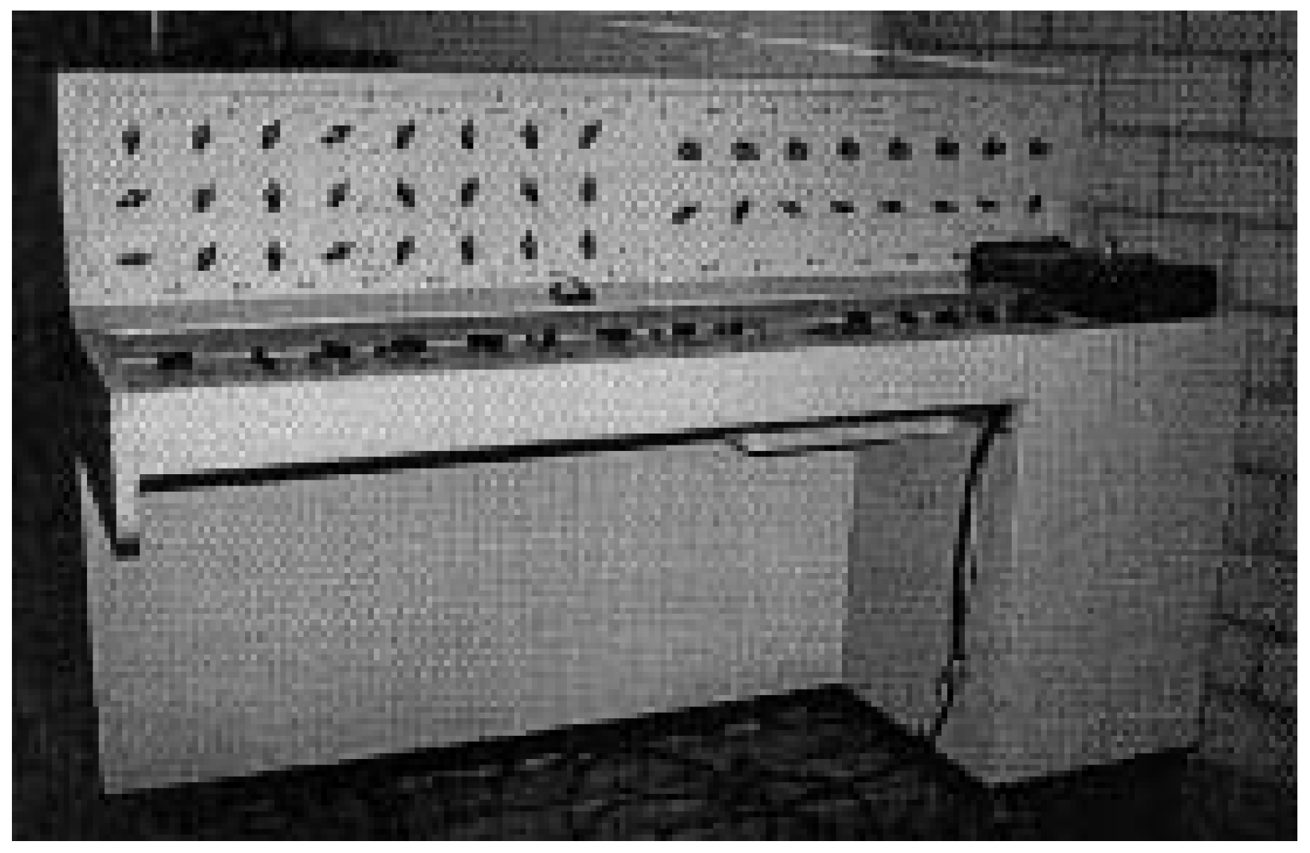
Figure 1.
The first EEG machine at the Swiss Epilepsy Centre, built by Kaiser from the company Schou in Denmark put into clinical use in the spring of 1948.
Figure 1.
The first EEG machine at the Swiss Epilepsy Centre, built by Kaiser from the company Schou in Denmark put into clinical use in the spring of 1948.
The Swiss Epilepsy Centre was also aware of the importance and necessity of recording EEGs, as can be seen in the annual report of the centre from 1942 [
2] where the medical director wrote: “We tried even before the second world war to obtain a machine which would allow us to record and measure the electrical activity of the brain, the so-called electroencephalogram.” It was not, however, possible, due to the war itself and the price of such an installation, which was at the time too high, to realise this plan. Finally in 1948, however, the first machine (Kaiser from the company Schou in Denmark, Fig. 1) was delivered. It was put into clinical use in the spring of 1948, one of the first reports being from march 26 on patient H. J. In the case history of this patient the following notation was made: “An EEG was recorded yesterday with a very nice and regular alpha rhythm, the EEG is normal.”
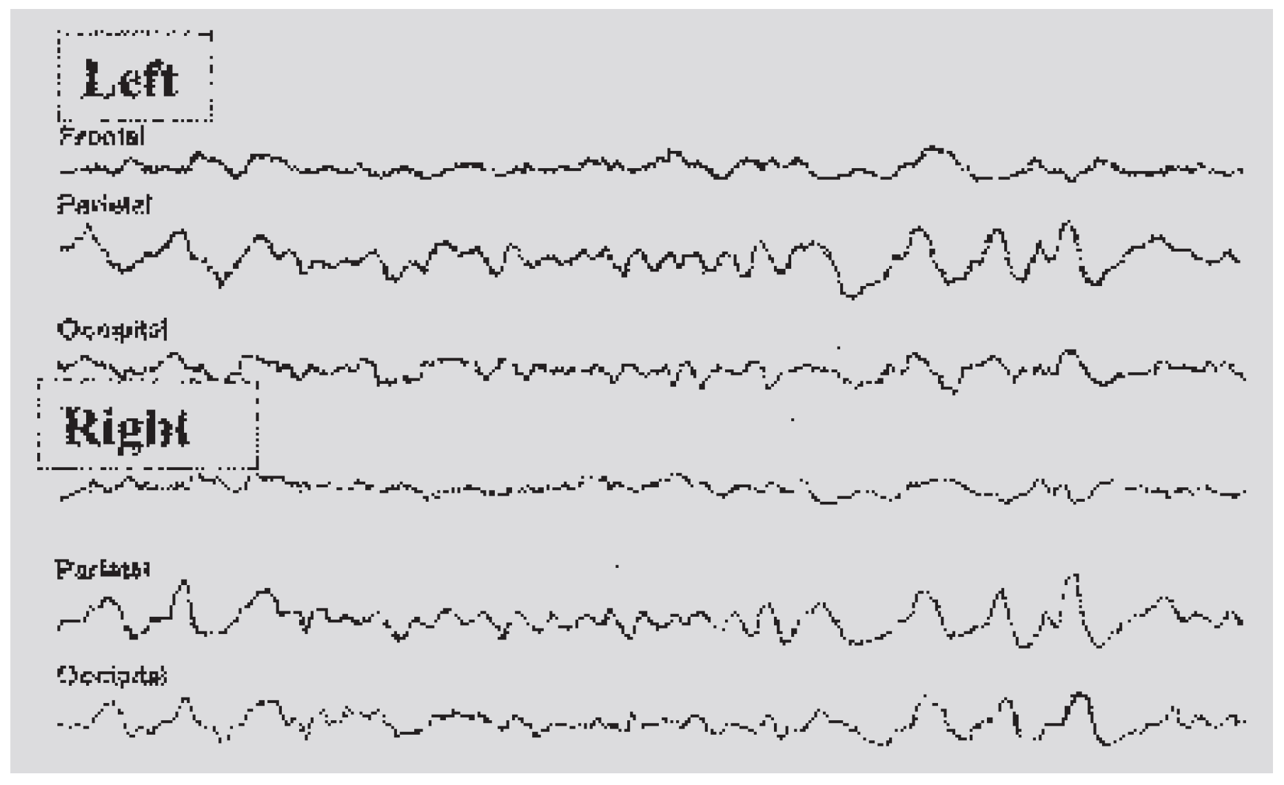
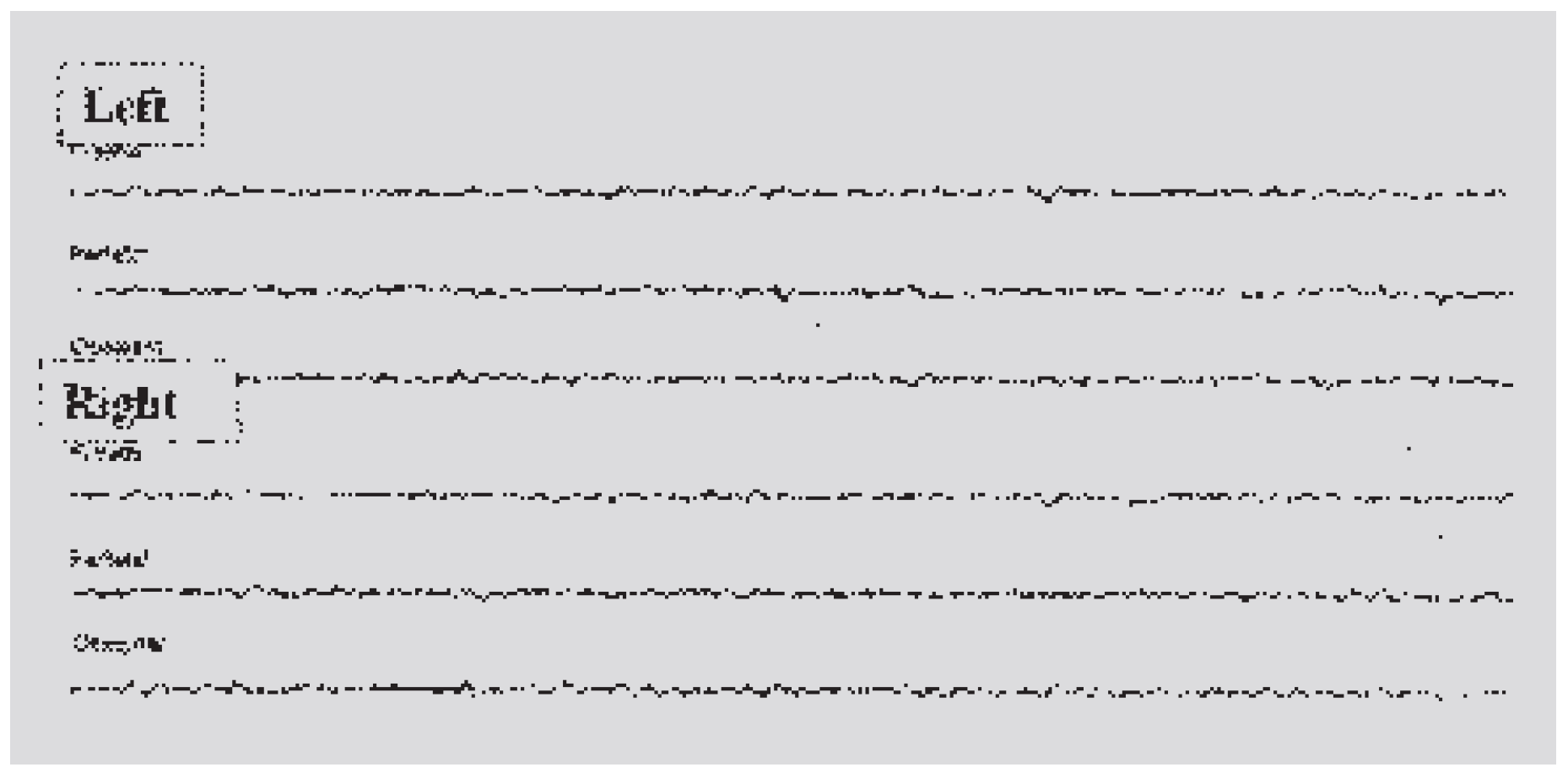
The following Figures (2 and 3) show some of the first 6-channel recordings, and illustrate how the EEG was used for both diagnosis and therapy control.
Figure 2.
EEG recorded in 1948 showing the pathological findings before therapy (notation: “Genuine epilepsy before successful therapy”).
Figure 2.
EEG recorded in 1948 showing the pathological findings before therapy (notation: “Genuine epilepsy before successful therapy”).
Figure 3.
EEG recorded in 1948 showing normal findings after therapy (notation: “Genuine epilepsy after successful therapy”).
Figure 3.
EEG recorded in 1948 showing normal findings after therapy (notation: “Genuine epilepsy after successful therapy”).
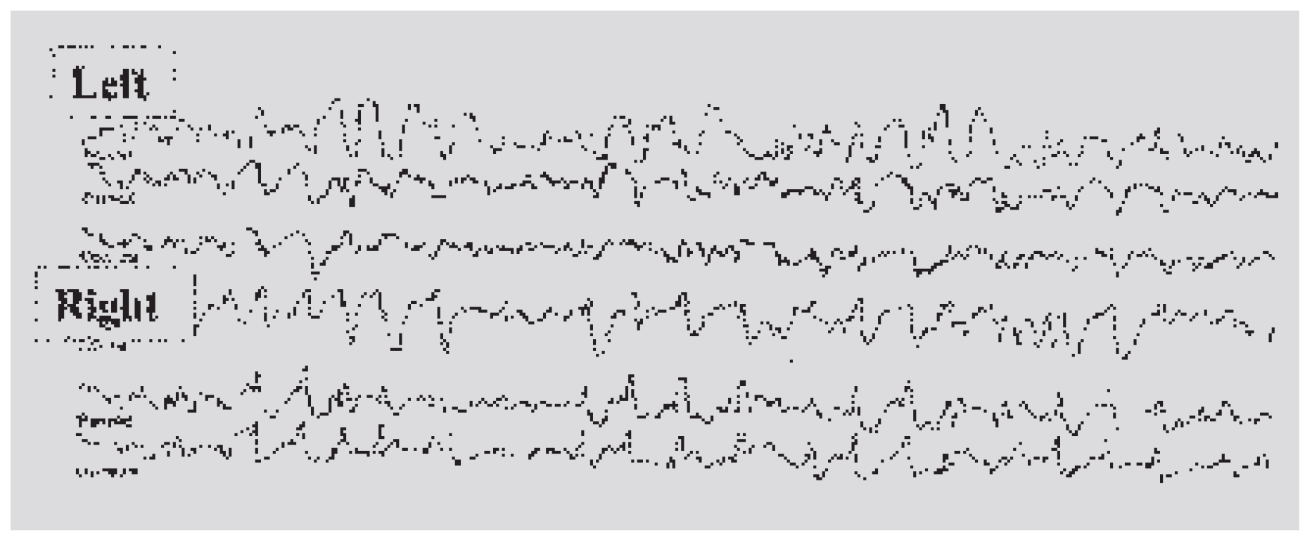
In the first year 440 EEGs were recorded and even at this time the questions asked from the EEG were not only whether the patient had epilepsy but also whether he was able to drive a motor vehicle (Fig. 4).
Figure 4.
EEG recorded in 1948 (notation: “Irregular spike-and-waves. Immediate decision for the motor vehicle authorities in an out-patient; question of epilepsy was confirmed; driving licence denied”).
Figure 4.
EEG recorded in 1948 (notation: “Irregular spike-and-waves. Immediate decision for the motor vehicle authorities in an out-patient; question of epilepsy was confirmed; driving licence denied”).
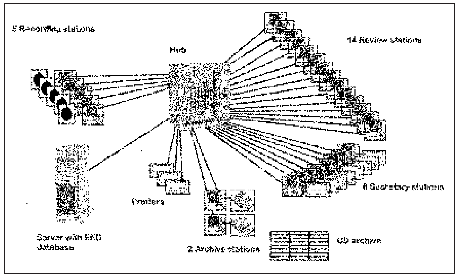
In the following fifty years there has been a continuos development of the EEG department utilising state of the art technology. The routine EEG machines were continuously updated from the original six channels to a modern network of digital (paperless) recording and review stations (Fig. 5).
Figure 5.
Schematic diagram of the digital (paperless) EEG network including recording, review, archive, and secretary stations.
Figure 5.
Schematic diagram of the digital (paperless) EEG network including recording, review, archive, and secretary stations.
Even with this continuous development in the field of routine EEG it soon became clear that for an optimal diagnosis of unclear seizure disorders the attacks themselves, either spontaneously occurring or being specifically provoked, should be recorded. In addition to the EEG other electrophysiological parameters, such as electrocardiogram, electromyogram, etc., in combination with documentation of the clinical manifestations allowed precise electro-clinical correlations.
The development has therefore largely concentrated on the field of “long-term monitoring” (LTM) for the documentation of the ictal EEG and seizure symptomatology [
3].
The following methods were developed and implemented:
- –
radio telemetry;
- –
ambulatory EEG.
Radio telemetry
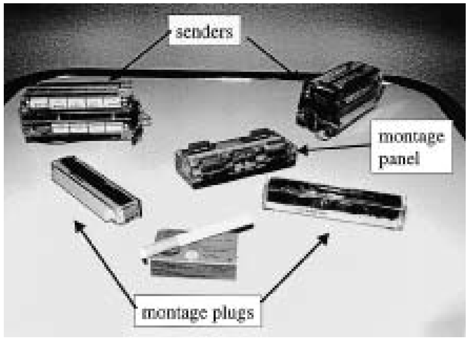
The first radio-telemetry system was put into use as early as 1975 using at this time a 16-channel sender (2 parallel 8-channel FM senders) (Fig. 6). Video technology although in its early days was also implemented at this time with
1/
2-inch reels. Split screen (video and EEG) recordings were obtained by using two cameras, one following the patient the other being fixed above the EEG paper write out. From these early pioneer days there has been a continuous development to 28-channel digital telemetry (1985) allowing polygraphic recordings, VAX computer for the analysis of the ictal EEG (1988) [
4], 32-channel digital sender (1998). One of the early prerequisites of our laboratory was that all clinically relevant video recordings should be archived in the highest image quality available, allowing multiple copies of tapes to be made with minimal or no loss of picture quality. This was achieved with broadcast image quality starting with
1/
2-inch reels (1975) developing to 1-inch broadcast recorders (C format 1979) and finally to digital video recording (D3 format 1993). This high technical standard allowed us to build up a comprehensive library of master tapes of seizures.
Figure 6.
First 16-channel radio-telemetry sender at the Swiss Epilepsy Centre. It was put into use in 1975 and consisted of two 8-channel sender modules, two montage plugs and one montage/electrode panel.
Figure 6.
First 16-channel radio-telemetry sender at the Swiss Epilepsy Centre. It was put into use in 1975 and consisted of two 8-channel sender modules, two montage plugs and one montage/electrode panel.
Ambulatory EEG
The possibility of recording outside the hospital environment over long periods of time became technically feasible in 1975 [
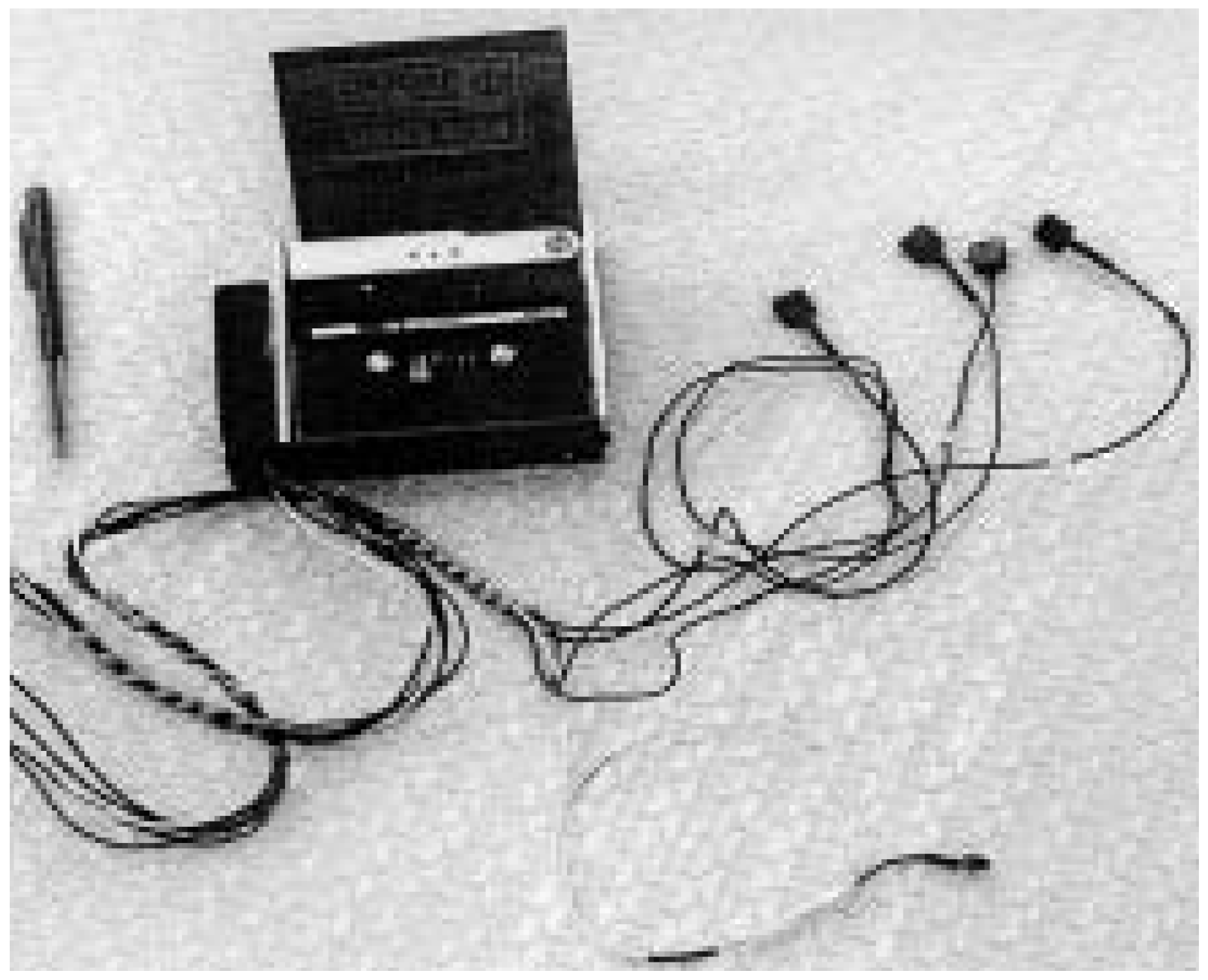
5]. In 1977, before such systems became commercially available, the first 4-channel system was put into use at our centre (Fig. 7). The tapes were initially replayed at high speed onto an EEG paper write out, which was followed by a page mode graphic display with computer analysis (1979), and the introduction of five 8-channel recorders in (1985) which are currently being replaced with 16–32-channel digital recorders.
Our long experience with these systems has shown that high technological standards combined with an individualised problem-solving approach, with flexible interchange from one form of examination to another, allow a cost-effective service with a high diagnostic yield.
Figure 7.
One of the first 4-channel recorders put into use at the Swiss Epilepsy Centre.
Figure 7.
One of the first 4-channel recorders put into use at the Swiss Epilepsy Centre.