Next Article in Journal
Lamotrigin bei schwierig zu behandelnden Epilepsien – eine offene Studie an 437 Patienten
Previous Article in Journal
Acute management of mild traumatic brain injury
 
 
Swiss Archives of Neurology, Psychiatry and Psychotherapy is published by MDPI from Volume 176 Issue 1 (2026). Previous articles were published by another publisher in Open Access under a CC-BY (or CC-BY-NC-ND) licence, and they are hosted by MDPI on mdpi.com as a courtesy and upon agreement with EMH Editores Medicorum Helveticorum.
Case Report

Neural basis of neuropsychological rehabilitation

Division de neuropsychologie, CHUV, CH-1011 Lausanne
Swiss Arch. Neurol. Psychiatry Psychother. 1998, 149(2), 73-78; https://doi.org/10.4414/sanp.1998.01027
Published: 1 January 1998

Simple Summary

Neuropsychological rehabilitation is often guided by pragmatic approaches. There is, however, a strong belief that models of recovery derived from cognitive and basic neurosciences may yield more efficient strategies. This paper summarises evidence from neuropsychological, activation and anatomical studies, which supports a model of parallel distributed processing of human cognitive functions. Parallel distributed processing models can account for recovery after brain damage and may prove useful for designing new rehabilitation strategies in future.

Introduction

Since Broca’s seminal paper [1], much of neuropsychological research has been devoted to clarify the anatomical substrates of deficits observed after cerebral lesions (e.g. [2,3]). This review paper addresses a complementary question, namely that of anatomical substrate underlying cognitive recovery following acute lesions. In particular, it highlights those organisational principles of the human brain which may play an important role in recovery and which should thus guide rehabilitation strategies.
Models of recovery from brain damage are certainly not a new venture. Based on his clinical observations,Kurt Goldstein (for historical review see [4]) introduced the distinction between restitutive and compensatory therapy.The former tries to restore the patient’s earlier capacities, while the latter concentrates on the development of substitutive strategies for lost or impaired functions. Goldstein did not believe in assigning individual functions to strictly delimited areas of the brain, but he assumed a co-operation between different brain structures for a given function. If only part of this structure was damaged, restitutive therapy would be successful, but if all parts were damaged, compensatory therapy should be used. During Goldstein’s active period, relatively little was known about the functional organisation of structures underlying cognitive functions. The cerebral cortex, e.g., was subdivided into primary areas and association cortex; although the latter was believed to contain different hierarchical levels of association, precise data and models were sparse (for review see e.g. [5]).
Current neuropsychological rehabilitation is often guided by pragmatic approaches. At the same time, there is a strong belief that efficient rehabilitation should be based on understanding the basic mechanisms of recovery; much hope is given today to cognitive models (see e.g. [6]) and to the combination of cognitive models and basic neuroscience [7].

Parallel distributed processing

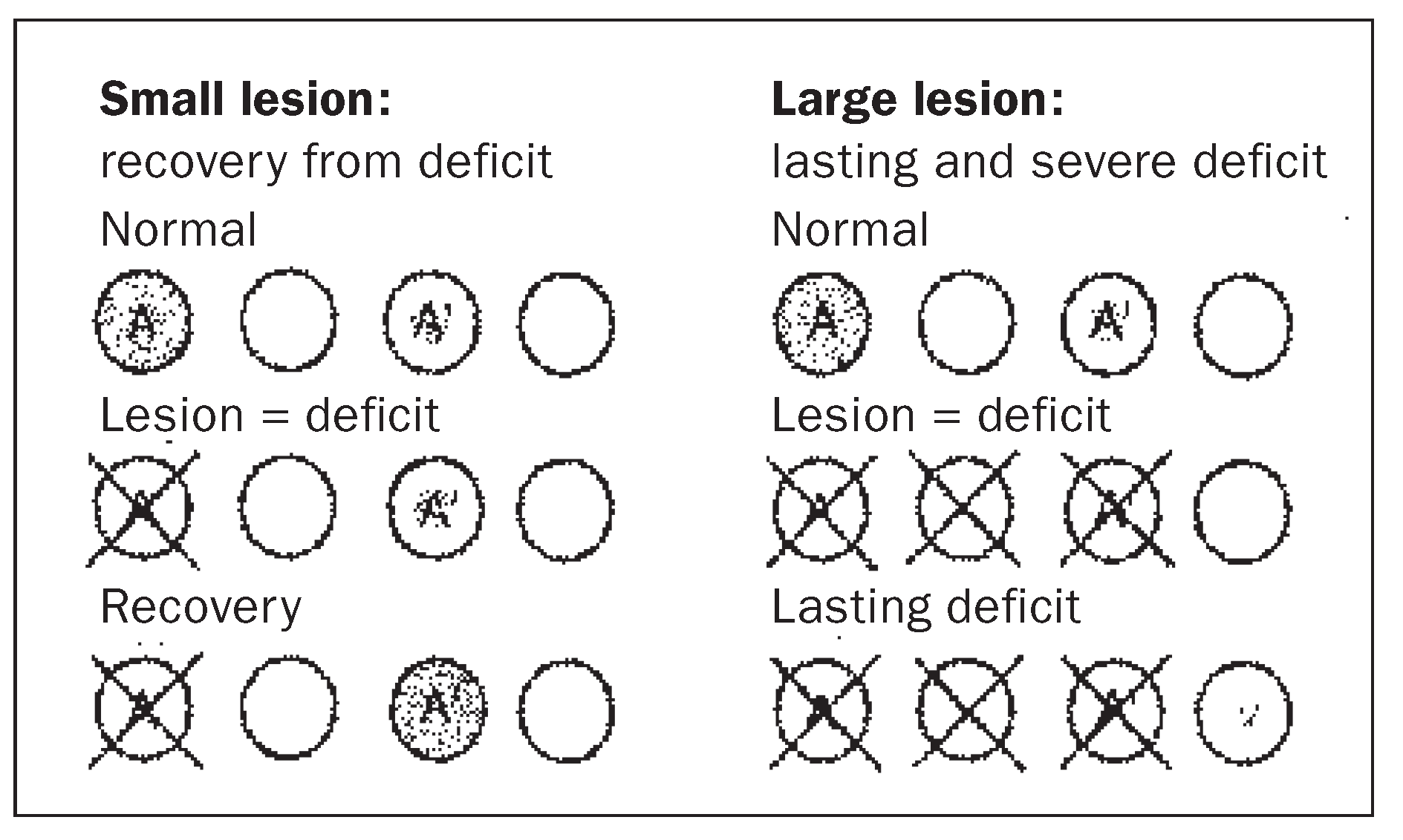
A simple model of what may happen during recovery is represented in Figure 1. Following a small lesion, recovery is accompanied by functional reorganisation. A given function, represented by A in Figure 1, is then supported by a different cerebral region than in normal subjects. Such a reorganisation was demonstrated in cases of recovery from hand paralysis and from aphasia; recovered functions activated foci which were not or only partially activated in normal subjects [8,9].
Several questions remain open in relation to the functional reorganisation underlying recovery. First, the nature of functional units is very poorly understood. Although language is known to depend critically on specific regions within the left hemisphere,the precise organisation of these areas is still unknown. Evidence summarised below indicates that speech areas are likely to consist of several specialised subareas. If so, new rehabilitation strategies could be designed for cases with specific lesions. Second, the flow of information between functional units is almost entirely unknown. Work on non-human primates showed repeatedly that parallel processing occurs at different levels of analysis.The same principle seems to apply to man and existence of parallel pathways was confirmed by human tracing studies (see below). Third, the degree of functional plasticity within the adult central nervous system is not determined. Recent evidence shows that a lesion limited to a specialised region is not accompanied with a lasting and severe deficit in the corresponding function; the latter occurs only in case of much larger lesions (see example of colour vision below).

Multiple visual areas in non-human primates

The last three decades of neurophysiological and neuroanatomical studies transformed what was previously believed to be a rather homogenous association cortex into a wealth of functionally and anatomically different cortical areas.The visuallyrelated cortex of macaque monkeys was shown to consist of over 30 functionally defined visual areas. Some of these areas contain a high proportion of functionally selective neurons, such as the colour specific neurons in V4 and the motion specific neurons in V5 (for review see [10]). Other, less precisely defined areas have been described more rostrally in the parietal and temporal cortices (for review see [11]). E.g., neurons selectively responsive for faces have been found in the inferotemporal cortex and the superior temporal sulcus.

Specialised visual areas in man

Case reports of selective loss following focal brain damage,and activation studies with positron emission tomography, have suggested the existence of extrastriate regions specialised in motion perception, colour vision or face recognition in man (for review see [10]). It has been proposed that these deficits result from lesions of human equivalents of V5, V4 or inferotemporal cortex [12,13]. Anatomical studies in man identified several extrastriate areas [14,15,16] and strengthened thus evidence from neuropsychological and activation studies.
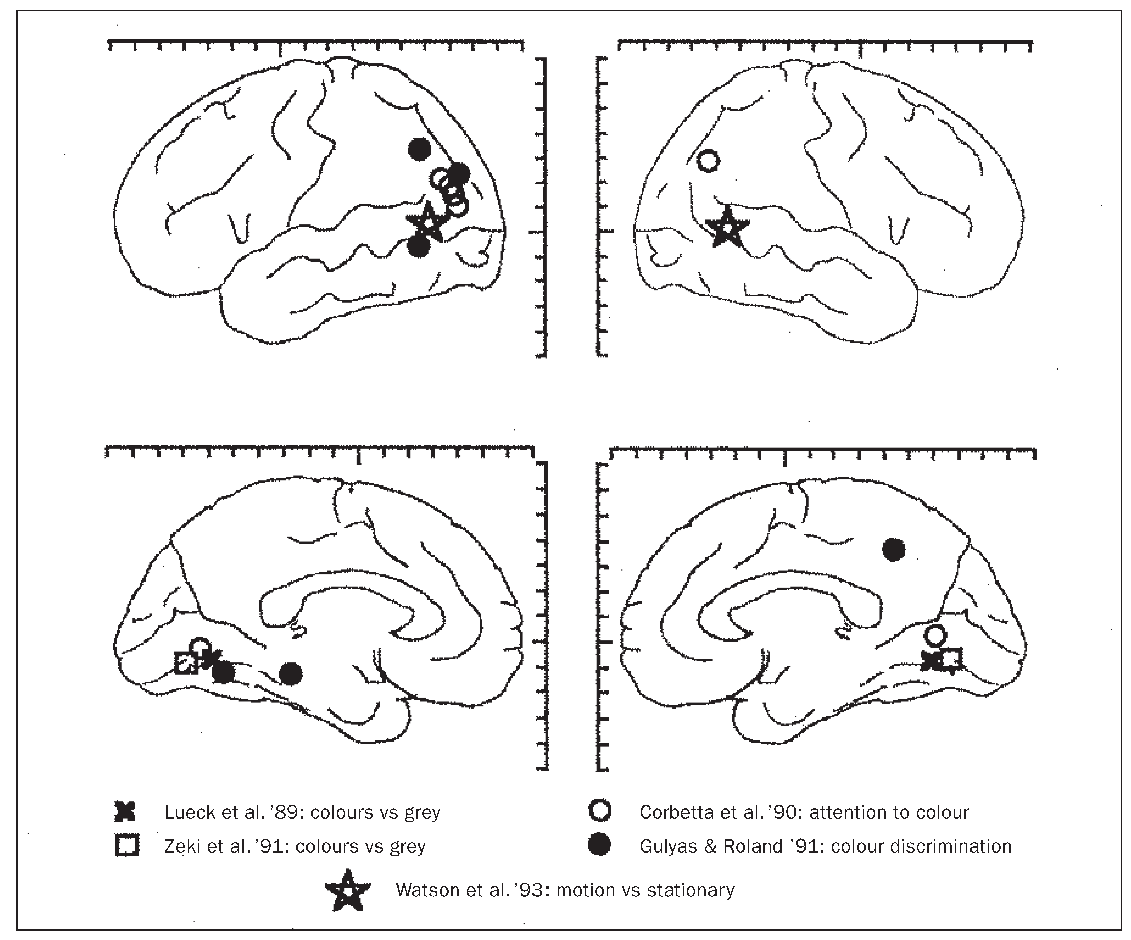
The relationship between specialised areas as observed in the normal human extrastriate cortex and the corresponding deficits observed after lesions is far from clear. One of the best understood examples is colour vision.A series of activation studies showed that colour tasks activate selectively specific regions (Figure 2; [17,18,19,20]).The most heavily activated region was at the inferior occipitotemporal junction, corresponding to putative human area V4, and additional foci were on the parieto-occipital convexity, predominantly on the left side. The foci activated by colour tasks were anatomically distinct from a region at the posterior part of the superior temporal sulcus that was shown to be selectively activated by visual motion stimuli [21]. Evidence from clinical reports predicted a separation of colour and motion processing: lasting achromatopsia was most often reported in association with relatively large inferior occipitotemporal lesions [12], whereas akinetopsia was reported in association with lesions of the occipital convexity [22].
We have investigated the role of the human extrastriate cortex in colour perception by testing a series of patients with circumscribed posterior lesions. In most cases reported in the literature, achromatopsia was associated with prosopagnosia following large occipitotemporal lesions. We examined two patients with severe and lasting prosopagnosia of whom one was achromatopsic and the other not [23]. Both patients had bilateral, almost symmetrical lesions within the posteroinfe-rior parts of the hemispheres. Comparison of the lesions by means of the Talairach and Tournoux coordinate system showed that the lesion associated with achromatopsia included the inferior occipitotemporal junction (site of putative V4), whereas the other one did not.This finding suggests that V4 lesion is necessary for achromatopsia to occur. In another series of experiments, we tested patients with relatively small circumscribed lesions of which some were limited to V4. In the chronic stage, patients with bilateral lesions limited to V4 were not at all or only very little impaired in colour discrimination and colour short-term memory tasks [24].This finding shows that V4 lesion is not sufficient for lasting and severe achromatopsia to occur.

Heterotopic interhemispheric connections

Many influential models of human cognitive functions have presumed that human interhemispheric connections link predominantly symmetrical parts of cortex (e.g. [25,26]). Our recent work shows that, in addition to homotopic connections, there is a wide array of heterotopic connections, some of which may be specific to man. In many instances, the heterotopic connections may represent alternative pathways and thus substrate for compensatory strategies developed following brain damage.
In a recent study, we traced interhemispheric connections originating in the medial part of the right occipital lobe using the Nauta method for anterogradely degenerating axons [27]. Serial coronal sections from the posterior third of the hemisphere contralateral to the lesion were stained and analysed for anterogradely degenerating axons, i.e., interhemispheric afferents from the damaged site. Callosal afferents were found within early stage visual areas,and also in the posterior parietal and temporal cortices. The latter regions received medium to high density of callosal afferents originating in the contralateral medio-occipital cortex. Among the callosal-recipient regions was the (left) angular gyrus.
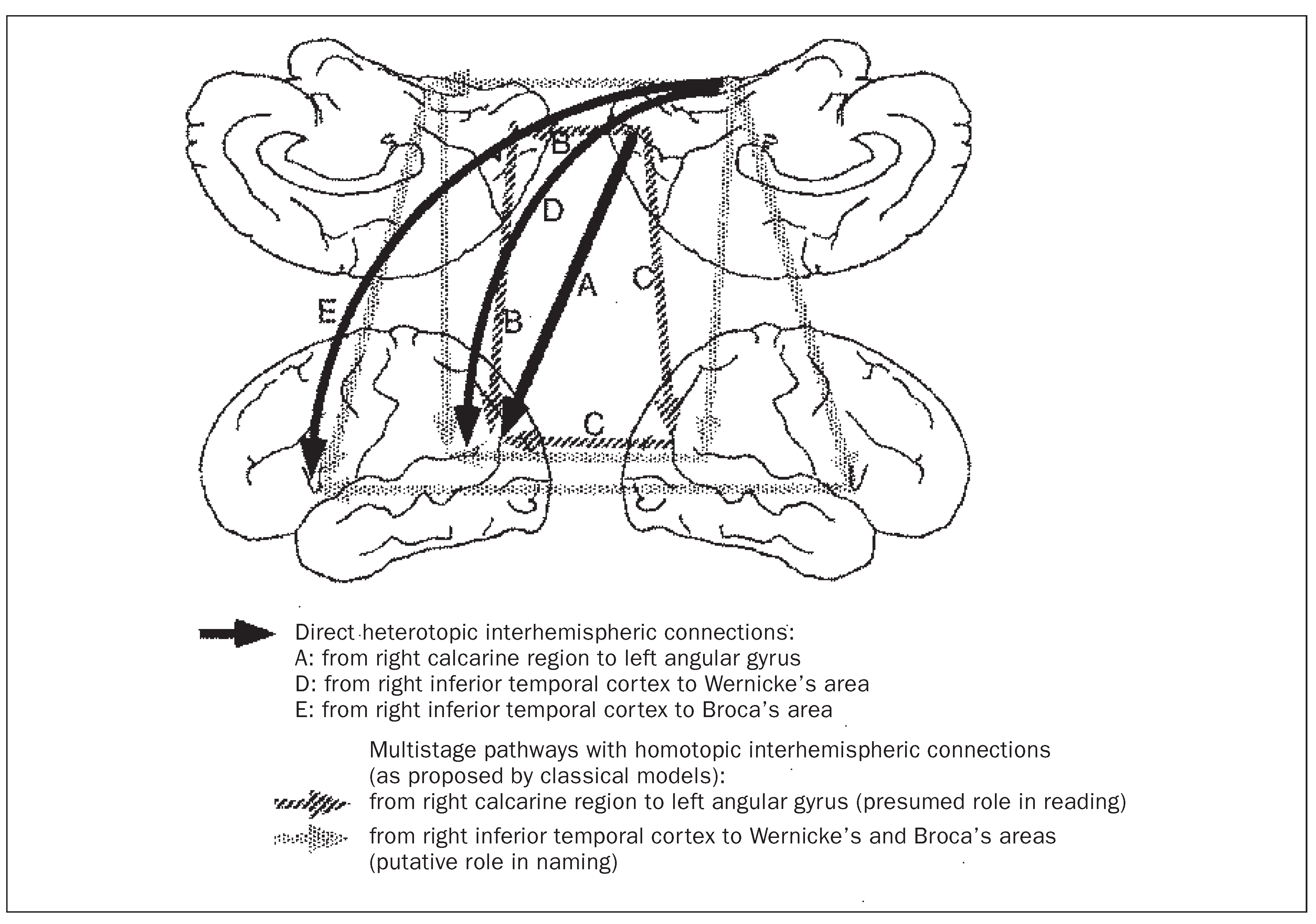
In cases of left calcarine and splenial lesions, pure alexia was attributed to disconnection of the right, non-injured calcarine region from the left angular gyrus, which was proposed to be involved in reading [28]. At least three routes link right calcarine region to left angular gyrus (Figure 3): (A) heterotopic callosal connection from right calcarine region directly to left angular gyrus; (B) combination of homotopic callosal connections from right calcarine region to the left one and then association connections from left calcarine region to left angular gyrus; (C) combination of association connections from right calcarine region to right angular gyrus and then a homotopic callosal connection from right angular gyrus to its homologue on the left side. Neuropsychological evidence showed that the ability to read does not rely critically on route B [29]. Route A, i.e. a monosynaptic pathway from right calcarine region to left angular gyrus was demonstrated in man [27], but its role in reading is presently unknown. Our current hypothesis is that it may underlie spared reading capacities that are present in some alexic patients.
The second example of heterotopic connections is that of monosynaptic interhemispheric input from right inferior temporal cortex to Wernicke’s and Broca’s areas [30]. The connections were traced in a brain with a right inferior temporal infarction by means of the Nauta method. The lesion from which the interhemispheric connections were traced was within a region known to be activated selectively in tasks of recognition of known faces (comparison of lesion site with activation foci reported by others [31]). Afferents were found both in Broca’s and Wernicke’s areas, with a higher density in the latter. These interhemispheric pathways are part of what is most likely a large network linking higher order visual and speech areas. When parts of this network are damaged, processing within the remaining parts determines spared capacities.It is conceivable that connections from right inferior temporal cortex to the speech areas may account, e.g., for successful phonemic cueing in otherwise anomic patients (see e.g. [32]): simultaneous activation of auditory and visual inputs in both Wernicke’s and Broca’s areas may be strong enough to initiate correct naming.

Multiple auditory areas in man

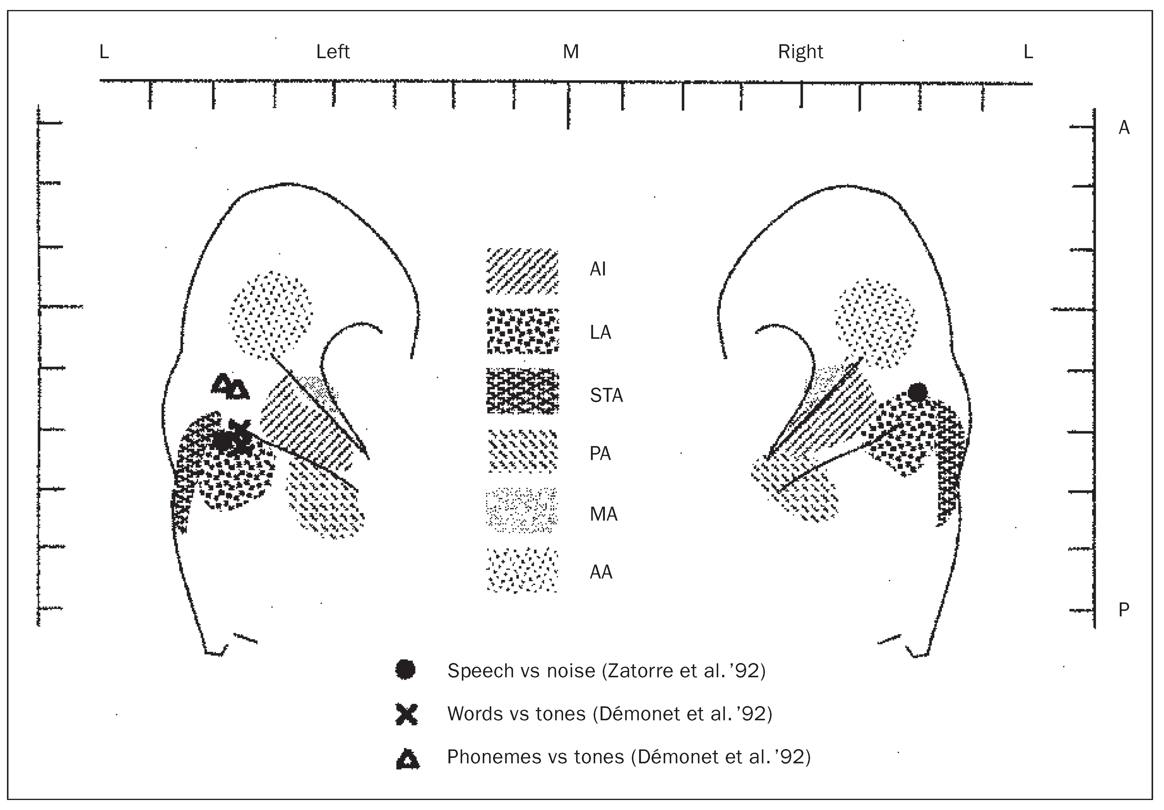
The human auditory cortex is the entry to the most powerful communication system and yet relatively little is known about its functional organisation. Most current beliefs are derived from work on nonhuman primates or extrapolated from work on the human visual cortex. It is often assumed that the auditory cortex outside the primary auditory cortex contains several, perhaps functionally specialised areas. Such a view is supported by recent anatomical and functional studies (Figure 4). We have studied the functional subdivision of the human supratemporal plane and insula using stains for cytochrome oxidase and acetylcholinesterase activity [33]. Seven distinct areas outside the primary auditory area were identified (for their relative positions see Figure 4). Comparison with activation and electrophysiological studies (by others) showed that all these areas respond to auditory stimuli.Some of these areas may belong to specialised processing streams, such as area LA (lateral to the primary auditory area) shown to be selectively activated by speech-related sounds [34,35], or anterior insula, by auditory motion [36]. Neuropsychological studies confirm further a putative functional specialisation, since different aspects of e.g. non-verbal auditory recognition can be disrupted independently [37].
Human primary auditory area contains anatomically distinct compartments that may be involved in processing of specific functions. The cytochrome oxidase staining revealed in the supragranular layers a pattern of dark and light stripes [38]. Comparison with tonotopic maps of human primary auditory area obtained by activation studies suggests that these cytochrome oxidase stripes are perpendicular to isofrequency lines.They may be related to particular binaural or ampliotopic domains, whose presence is suggested by evidence from animal experiments and from magnetoencephalographic studies in man, or else to specifically human functions, such as speech.
This work has been supported by the Swiss National Science Foundation grants 31-36297.92, 4038-044081 and 323141607.94 («Score A» fellowship) and the Human Capital and Mobility project CHRX CT930261.

References

  1. Broca PP. Remarques sur le siège de la faculté du langage articulé, suivies d’une observation d’aphémie. Bull Soc Anat 1861;36:330–57.
  2. Damasio A, Yamada T, Damasio H, Corbett J, McKee J. Central achromatopsia: behavioral, anatomic, and physiologic aspects. Neurology 1980;30:1064–71.
  3. Damasio AR, Damasio H, Van Hoesen GW. Prosopagnosia: anatomic basis and behavioral mechanisms. Neurology 1982;32:331–41.
  4. Poser U, Kohler JA, Schonle PW. A historical review of neuropsychological rehabilitation in Germany. Neuropsych Rehab 1996;6:257–78.
  5. Dow BM, Vautin RG. Horizontal segregation of color information in the middle layers of foveal striate cortex. J Neurophysiol 1987;57:712–39.
  6. Wilson BA, Robertson IH. Editorial. Neuropsych Rehab 1992;2:1–2.
  7. Kosslyn SM. On cognitive neuroscience. J Cogn Neurosci 1992;6:297–303.
  8. Frackowiak RS, Weiller C, Chollet F. The functional anatomy of recovery from brain injury, Ciba. Found Symp 1991;163:235–44.
  9. Weiller C, Isensee C, Rijntjes M, Huber W, Muller S, et al. Recovery from Wernicke’s aphasia: a positron emission tomographic study. Ann Neurol 1995;37:723–32.
  10. Zeki S. A century of cerebral achromatopsia [Review]. Brain 1990;113:1721–77.
  11. Knierim JJ, Van Essen DC. Visual cortex: cartography, connectivity, and concurrent processing. Curr Opinion Neurobiol 1992;2:150–55.
  12. Meadows JC. Disturbed perception of colours associated with localized cerebral lesions. Brain 1974;97:615–32.
  13. Zeki S. Cerebral akinetopsia (visual motion blindness). A review. [Review], Brain, 1991;114:811–24.
  14. Clarke S, Miklossy J. Occipital cortex in man: organization of callosal connections, related myelo- and cytoarchitecture, and putative boundaries of functional visual areas. J Comp Neurol 1990;298:188–214.
  15. Clarke S. Modular organisation of human extrastriate visual cortex: evidence from cytochrome oxidase pattern in normal and macular degen-eration cases. Eur J Neurosci 1994;6:725–36.
  16. Tootell RB, Taylor JB. Anatomical evidence for MT and additional cortical visual areas in humans. Cereb Cortex 1995;5:39–55.
  17. Lueck CJ, Zeki S, Friston KJ, Deiber MP, Cope P, Cunningham VJ, et al. The colour centre in the cerebral cortex of man. Nature 1989;430:386–9.
  18. Corbetta M, Miezin FM, Dobmeyer S, Shulman GL, Petersen SE. Attentional modulation of neuronal processing of shape, color, and velocity in humans. Science 1990;248:1556–9.
  19. Gulyas B, Roland PE. Cortical fields participating in form and colour discrimination in the human brain. Neuroreport 1991;2:585–8.
  20. Zeki S, Watson JDG, Lueck CJ, Friston KJ, Kennard C, Frackowiak RSJ. A direct demonstration of functional specialisation in human visual cortex. J Neurosci 1991;11:641–9.
  21. Watson JD, Myers R, Frackowiak RS, Hajnal JV, Woods RP, Mazziotta JC, et al. Area V5 of the human brain: evidence from a combined study using positron emission tomography and magnetic resonance imaging. Cereb Cortex 1993;3:79–94.
  22. Zihl J, Von Cramon D, Mai N. Selective disturbance of movement vision after bilateral brain damage. Brain 1983;106:313–40.
  23. Clarke S, Lindemann A, Maeder P, Borruat FX, Assal G. Face recognition and posterior hemispheric lesions. Neuropsychologia 1997;35:1555–63.
  24. Schoppig A,Clarke S, Walsh V, Assal G, Meuli R, Cowey A. Discrimination and short-term memory for colour following circumscribed posterior hemispheric lesions in man. J Neurol Neurosurg Psychiatry 1998 (submitted).
  25. Geschwind N. Disconnexion syndromes in animals and man. Brain 1965;88:237–94 and 585–644.
  26. Geschwind N. The apraxias: Neural mechanisms of disorders of learned movement. Am Sci 1975;63:188–95.
  27. Clarke S, Van Essen DC, Hadjikhani N, Drury H, Coogan TA. Understanding human area 19 and 37: contribution of two-dimensional maps of visual callosal afferents [Abstract]. Human Brain Mapping 1995;Suppl.1:33.
  28. Dejerine J, Vialet N. Contribution à l’étude de la localisation de la cécité verbale pure. Comptes Rendus Hebdomadaires des Scéances et Mémoires de la Société de Biologie 1893;5:790–3.
  29. Binder JR, Mohr JP. The topography of callosal reading pathways. A case-control analysis. Brain 1992;115:1807–26.
  30. Di Virgilio G, Clarke S. Direct interhemispheric visual input to human speech areas. Human Brain Mapping 1997;5:347–54.
  31. Sergent J, Ohta S, MacDonald B. Functional neuroanatomy of face and object processing. A positron emission tomography study. Brain 1992;115:15–36.
  32. Semenza C, Sgaramella TM. Production of proper names: a clinical case study of the effects of phenemic cueing. Memory 1993;1:249–63.
  33. Rivier F, Clarke S. Cytochrome oxidase, acetylcholinesterase, and NADPH-diaphorase staining in human supratemporal and insular cortex: evidence for multiple auditory areas. Neuroimage 1997;6:288–304.
  34. Zatorre RJ, Evans AC, Meyer E, Gjedde A. Lateralization of phonetic and pitch discrimination in speech processing. Science 1992;256:846–8.
  35. Demonet JF, Celsis P, Nespoulous JL, Viallard G, MarcVergnes JP, Rascol A. Cerebral blood flow correlates of word monitoring in sentences: influence of semantic incoherence. A spect study in normals. Neuropsychologia 1992;30:1–11.
  36. Griffiths TD, Bench CJ, Frackowiak, RSJ. Human cortical areas selectively activated by apparent sound movement. Curr Biol 1994:892–5.
  37. Clarke S, Bellmann A, de Ribaupierre F, Assal G. Nonverbal auditory recognition in normal subjects and braindamaged patients: evidence for parallel processing, Neuropsychologia 1996;34:587–603.
  38. Clarke S, Rivier F. Compartments within human primary auditoy cortex: evidence from cytochrome oxidase and ace-tylcholinesterase staining. Eur J Neurosci 1997;10:741–5.
  39. Talairach J, Tournoux P. Co-planar stereotaxic atlas of the human brain. New York: Thieme, 1998.
Figure 1. Recovery after small (on the left) or large lesions (on the right). A function, represented here by A, activates in the normal subject a distinct region, represented by the shaded circle, and marginally also A’. A lesion restricted to circle A is first accompanied with the corresponding deficit. Later function A recovers, activating a region, or a set of regions, normally only marginally (here A’) or not at all activated in this function. A much larger lesion is not accompanied with such a recovery.
Sanpp 149 00073 g001
Figure 2. Centres of foci selectively activated by colour or motion tasks, represented on the lateral and medial views of the hemispheres. Bars indicate Talairach and Tournoux [39] coordinates.
Sanpp 149 00073 g002
Figure 3. Heterotopic interhemispheric connections demonstrated in man (black arrows) and the alternative pathways that were proposed in classical models (shaded arrows). A: direct, monosynaptic connections were found to link the right calcarine region to the left angular gyrus [27]; it may be one of several pathways (B, C) involved in reading (see text). D, E: direct, monosynaptic connections were demonstrated from the inferior temporal cortex to Wernicke’s and Broca’s areas [30]; it is part of a most likely large network linking higher order visual and speech areas and sustaining visuo-verbal functions.
Sanpp 149 00073 g003
Figure 4. Human auditory areas and putative specialisation for speech processing. Anatomical studies [33] identified five distinct areas on the supratemporal plane outside the primary auditory cortex (AI) and two on the insula (not shown here). These areas may be involved in specific aspects of auditory processing, similarly to the functional specialisation of some extrastriate visual areas. Here we compare the sites of anatomically identified auditory areas (hatched; from Rivier and Clarke [33]) and activation foci to speech-related auditory tasks [34,35]. Areas lateral to AI, but not those anterior, medial and posterior to it, show specialisation for speech processing. Bars indicate Talairach and Tournoux [39] coordinates.
Sanpp 149 00073 g004